An unusual ‘heart attack’ – Takotsubo cardiomyopathy

Br J Cardiol 2009;16:197–8 Leave a comment
Click any image to enlarge
Authors:

The authors describe a case of Takotsubo-like syndrome in a 59-year-old Caucasian woman.

Case report

A 59-year-old woman was admitted with symptoms and signs suggesting acute coronary syndrome. A 12-lead electrocardiogram (ECG) demonstrated ST segment elevation in leads V2-V6, I, II and aVL consistent with ST segment elevation myocardial infarction. She underwent emergency coronary angiography, which demonstrated only minor irregularities in coronaries. Chest pain resolved completely after four hours.

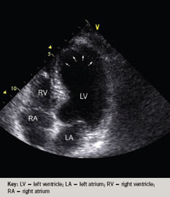
Figure 1b. Transthoracic echocardiography during the initial admission (apical four-chamber view, diastole)
Figure 1a. Transthoracic echocardiography during the initial admission (apical four-chamber view, systole) demonstrating ballooning of left ventricular apical function as indicated by white arrows
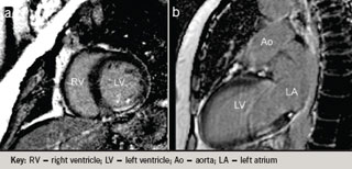
Figure 2. Cardiac magnetic resonance (CMR) TruFisp cine inflow/outflow in systole (a) with ‘ballooning’ of the left ventricular mid body and apex; (b) diastole
Figure 3. Delayed viability CMR scan (a) short axis and (b) two-chamber view

After 24 hours from admission, serial daily 12-lead ECGs demonstrated marked T-wave inversion in leads V2-V6. This pattern persisted throughout the patient’s hospital stay. Transthoracic echocardiography studies demonstrated significant left ventricular impairment due to apical dyskinesia with ballooning of the apex (figure 1). She also underwent cardiac magnetic resonance (CMR) studies, which demonstrated stunned but viable left ventricular (LV) myocardium (figures 2 and 3).

Follow-up echocardiography studies demonstrated that LV function had returned to normal (figures 4 and 5).

Clinical features and pattern of recovery was consistent with Takotsubo cardiomyopathy. This clinical entity, although rare, has been more prevalent in Japan.1 There are a few case reports describing Caucasian patients affected by this disorder.2,3 The aetiology of Takotsubo cardiomyopathy remains unclear. It is a diagnosis of exclusion. Most of the patients recover after one to two months without serious sequelae.
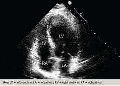
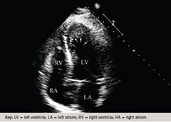
Figure 1b. Transthoracic echocardiography during the initial admission (apical four-chamber view, diastole)
Figure 4b. Transthoracic echocardiography three months after the initial admission (apical four-chamber view, diastole)
Figure 1a. Transthoracic echocardiography during the initial admission (apical four-chamber view, systole) demonstrating ballooning of left ventricular apical function as indicated by white arrows
Figure 4a. Transthoracic echocardiography after the initial admission (apical four-chamber view, systole) demonstrating full recovery of the apex as indicated by white arrows
Figure 5. 3D echocardiography studies three months after the initial admission demonstrating full recovery of left ventricular function

Conflict of interest

None declared.

References

  1. Tsuchihashi K, Ueshima K, Uchida T et al. Transient left ventricular apical ballooning without coronary artery stenosis: a novel heart syndrome mimicking acute myocardial infarction. Angina Pectoris-Myocardial Infarction Investigations in Japan. J Am Coll Cardiol 2001;38:11–18.
  2. Pillière R, Mansencal N, Digne F, Lacombe P, Joseph T, Dubourg O. Prevalence of tako-tsubo syndrome in a large urban agglomeration. Am J Cardiol 2006;98:662–5.
  3. Lisi M, Zacà V, Maffei S et al. Takotsubo cardiomyopathy in a Caucasian Italian woman: case report. Cardiovasc Ultrasound 2007;5:18.
THERE ARE CURRENTLY NO COMMENTS FOR THIS ARTICLE - LEAVE A COMMENT