Infective endocarditis: acne to zoonoses on the valve, an A to Z perspective

Br J Cardiol 2022;29:112–6doi:10.5837/bjc.2022.025 Leave a comment
Click any image to enlarge
Authors:
First published online 19th July 2022

Cutibacterium acnes (C. acnes), previously known as Propionibacterium acnes, is a rare cause of infective endocarditis (IE). We provide a review of the literature and describe two recent cases from a single centre to provide insight into the various clinical presentations, progression and management of patients with this infection.

The primary objective of our review is to highlight the difficulty in the initial assessment of these patients with an aim to improve the time and accuracy of diagnosis and expedite subsequent treatment. There are currently no guidelines in the literature specific to the management of IE caused by C. acnes. Our secondary objectives are to disseminate information about the indolent course of the disease and add to the growing body of evidence around this rare, yet complex, cause of IE.

Introduction

The incidence of Cutibacterium acnes as the causative organism for infective endocarditis (IE) is reported as 0.3%.1 C. acnes IE is associated with both native and prosthetic valves, but is much more commonly found on prosthetic valves. Studies show that middle-aged men are mostly affected, with serious infections increasingly reported in association with bioprosthetic material.1 C. acnes is part of the commensal flora of the skin, colonising pilous follicles and sebaceous glands, and may also be found in the mucosa of the mouth, nose, urogenital tract and large intestine, this difference might account for the gender-specific bacterial colonisation and subsequent male predominance seen in C. acnes infections.2 In a more recent study, the proportion of prosthetic valve endocarditis (PVE) cases due to C. acnes was nearly three times that previously reported from the International Collaboration on Endocarditis – Prospective Cohort Study (ICE-PCS) (23/606, 3.8% vs. 13/923, 1.4%).1,3

The increasing incidence of C. acnes endocarditis may, in part, be secondary to the increasing number of prosthetic valves being inserted, or be a product of increased diagnosis due to the use of valve sequencing via PCR (polymerase chain reaction).4 In addition, patients with C. acnes IE usually have advanced disease by the time they present, with a substantial proportion of patients having invasive disease and embolic complications.1 There typically is a delayed presentation, in one case series, the mean onset of infection was four years from surgery.5

The mortality rates due to C. acnes IE have been described in one recent study as 16%.5 This may be due to the prevalence of C. acnes associated PVE, late diagnosis and subsequent delay in targeted treatment of the disease.1 Herein, we present our experience of two patients affected by C. acnes associated IE.

Case presentations

Case 1: 29-year-old man with three re-do aortic valve replacements

A 29-year-old man, with a known congenital bicuspid aortic valve underwent minimal access aortic valve replacement (AVR), receiving a mechanical (25 mm On-X) prosthesis in November 2015.

A routine postoperative transthoracic echocardiogram (TTE) three months later showed two distinct jets of moderate aortic regurgitation (AR), one of which was paravalvular. A subsequent transoesophageal echocardiogram (TOE) showed good left ventricular (LV) systolic function with moderate-to-severe paravalvular AR, and he was referred once more for a surgical opinion. In the interim, the patient presented to the emergency department (six months following his original AVR) with a two-day history of rigors and a fever of 40.4 degrees Celsius. Two sets of blood cultures, taken 24 hours apart, were reported as negative in the initial 48-hour period. These cultures were observed for a further eight days as per trust policy in suspected IE with no identifiable growth. The patient was admitted under the medical team and, following satisfactory routine bloods, chest X-ray and electrocardiogram (ECG) tests, was discharged home, with advice to seek medical attention if he developed any further symptoms.

Boyle - Figure 1. Transoesophageal echocardiography (TOE) showing vegetation measuring over 3.3 cm<sup>2</sup> in the aortic valve with large aortic root abscess
Figure 1. Transoesophageal echocardiography (TOE) showing vegetation measuring over 3.3 cm2 in the aortic valve with large aortic root abscess

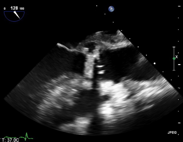
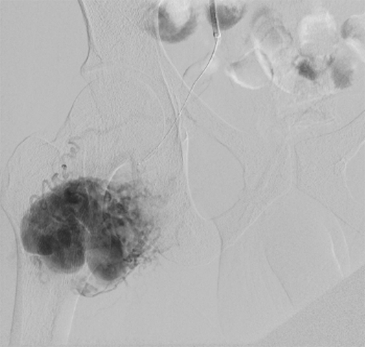
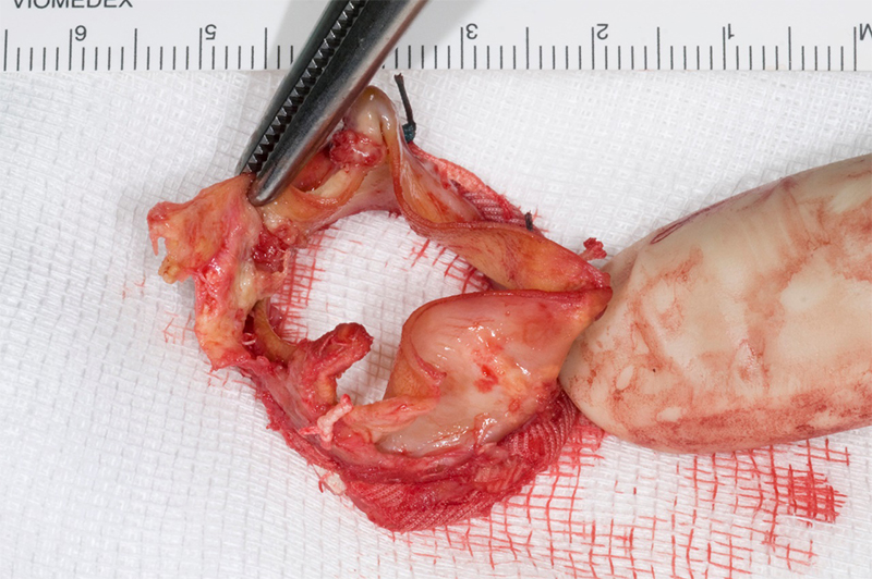
He had intermittent rigors for several weeks following discharge, however, when reviewed by his general practitioner (GP) all observations were recorded to be within normal parameters. The patient was readmitted to hospital due to progression of his symptoms of general malaise and intermittent rigors. A full blood count on this occasion showed a raised white cell count 14.9 × 10⁹/L (WCC) and C-reactive protein (CRP) 96 mg/L, blood cultures were taken. He had a known penicillin allergy, and so the patient was treated with empiric antibiotics as per trust guidelines (table 1). The anaerobic blood culture on this admission was positive for C. acnes. A repeat TOE showed a large vegetation measuring over 3.3 cm2 on the aortic valve causing mild-to-moderate obstruction of left ventricular outflow tract (LVOT) (figure 1). Furthermore, he had a large aortic root abscess and new ECG findings of a prolonged PR interval with right bundle branch block.

Table 1. Case 1 antibiotic regimen

Admission 1
Vancomycin 1 g IV BD 6 days Vancomycin 1.5 g IV BD 1 day
Rifampicin 600 mg PO BD 7 days
Gentamicin 80 mg IV BD 7 days
Re-do operation
Vancomycin 1.5 g IV BD 4 days Vancomycin 3 g IV continuous infusion 3 doses
Rifampicin 600 mg IV/PO BD 7 days
Gentamicin 80 mg IV BD 2 days
Cefuroxime 750 mg IV 3 doses
Discharged home
Daptomycin 500 mg IV OD 5 weeks
Admission 2
Vancomycin 1 g IV BD 3 days Vancomycin 1.5 g IV continuous infusion 5 days
Rifampicin 600 mg PO BD 8 days
Gentamicin 80 mg IV BD 9 days
Re-do operation
Ceftriaxone 2 g IV OD 6 weeks
Admission 3
Ceftriaxone 2 g IV OD 8 weeks
Key: BD = twice daily; IV = intravenous; OD = once daily; PO = per oral

The patient underwent an emergency re-do aortic valve replacement with a Sorin, Bicarbon 23 mm mechanical prosthesis. Intra-operatively, there was a large vegetation in the outflow tract with areas of abscess under the right and non-coronary cusps. There was a further cavity below the non-coronary cusp which was obliterated with 4-0 prolene.

The patient came off cardiopulmonary bypass and had an uneventful recovery. Intra-operative valve tissue sample was subjected to 16S rDNA real-time PCR and confirmed as C. acnes. The patient weighed 76.5 kg. Once discharged home the patient was continued on antibiotic therapy in the community (table 1).

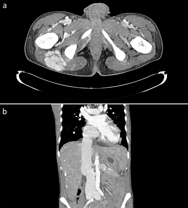
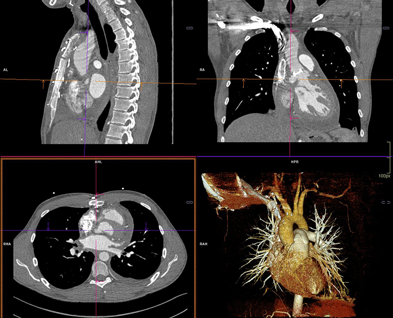
Three weeks following completion of intravenous (IV) antibiotic therapy he re-presented with chest pain, shortness of breath and general malaise. A full blood count showed a WCC within normal ranges but a raised CRP of 210 mg/L. Blood cultures grew C. acnes and he was recommenced on antibiotic therapy (table 1). Further cardiothoracic imaging was necessary and an ECG-gated computed tomography (CT) of the thorax showed an irregular and slightly dilated aortic root with a small focal outpouching, in keeping with a small aortic root abscess (figure 2). The patient had several episodes of supraventricular tachycardia managed with adenosine. Following a multi-disciplinary team discussion, the next operation deemed appropriate would involve a homograft replacement, and the patient was transferred to an appropriate surgical centre specialising in adult congenital surgery.

Boyle - Figure 2. Computed tomography (CT) imaging and 3D reconstruction of focal outpouchings in keeping with aortic root abscesses
Figure 2. Computed tomography (CT) imaging and 3D reconstruction of focal outpouchings in keeping with aortic root abscesses

The third operation was uneventful. During this re-do operation, the aortic valve showed evidence of dehiscence along the non-coronary cusp (NCC) with evidence of a chronic large abscess below the NCC onto the anterior mitral valve leaflet (AMVL), there was no evidence of pus intra-operatively. The abscess was debrided and cleaned, and a 27 mm Perimount Magna tissue aortic valve was inserted using interrupted 2/0 ethibond sutures.

Postoperatively, a gated contrast-enhanced thoracic CT was performed to examine the area adjacent to the NCC. It showed a blind ending 15 mm outpouching from the LVOT adjacent to the interatrial septum, with a thin extension posterior to the annulus. There was no visible communication to the ascending aorta or evidence of vegetation or valvular dehiscence. He made an uneventful recovery and was discharged home 15 days later to complete a prolonged antibiotic course for six weeks from date of operation (table 1).

Fourth readmission to hospital

Unfortunately, the patient re-presented to hospital for a fourth time two weeks after his third operation with symptoms of fever and night sweats. A repeat TOE showed no evidence of valvular compromise, no dehiscence and no obvious vegetations on aortic valve leaflets. Furthermore, a positron-emission tomography (PET) scan was performed and excluded an aortic root abscess and any active signs of active focal infection. The patient had a further course of antibiotics and completed a total of eight weeks’ treatment.

Follow-up

One year later, the patient reported feeling well in himself. Reassuringly, a repeat TOE showed no evidence of aortic regurgitation and he continues to be monitored closely in a clinical setting.

Case 2: 74-year-old man with heart failure and urosepsis

A 74-year-old man, with no known allergies to antibiotics, underwent a bioprosthetic aortic valve replacement for severe aortic stenosis in 2014. He had a trans-urethral resection of the prostate (TURP) one year later and required readmission eight weeks following this for treatment of urosepsis. This patient had multiple subsequent admissions for management of sepsis and exacerbations of his heart failure. It was noted that during this time C. acnes grew on a set of blood cultures, but was considered a skin contaminant.

Boyle - Figure 3. Transthoracic echocardiography (TTE) showing severe aortic regurgitation
Figure 3. Transthoracic echocardiography (TTE) showing severe aortic regurgitation

He was seen frequently in a heart failure clinic and optimised on diuretics, however, his symptoms continued to deteriorate. Following a TTE and TOE, he was referred as an outpatient to cardiothoracic surgeons with severe paravalvular aortic regurgitation and a dilated left ventricle with moderate-to-severe systolic impairment (figure 3).

An outpatient ECG-gated thoracic CT showed small outpouchings at the aortic root, possibly in keeping with micro abscesses. He was subsequently admitted from the outpatient clinic with evidence of acute heart failure with ongoing orthopnoea, and persistent episodes of paroxysmal nocturnal dyspnoea. He was transferred to the intensive care unit (ICU) for pre-operative optimisation with IV furosemide and dobutamine.

While on dobutamine he had a repeat TOE, which showed moderate ventricular function with mild tricuspid regurgitation (TR) and no evidence of mitral regurgitation (MR). There was also a marked reduction in the left ventricular end diastolic diameter (EDD). His previous TTE had suggested an EDD >7 cm with poor LV function and severe MR and TR. Due to this improvement with dobutamine, the decision was taken to proceed with surgery for severe aortic regurgitation.

Boyle - Figure 4. Excised bioprosthetic aortic valve replacement
Figure 4. Excised bioprosthetic aortic valve replacement

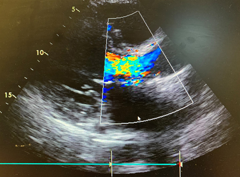
Intra-operatively, there was destruction of the strut at the NCC/right coronary cusp (RCC) junction with dehiscence of the leaflets from the valve frame, leading to the prolapse of the NCC leaflet (figure 4). A new size 23 mm trifecta biological prosthesis was inserted using interrupted plegeted Ethibond sutures × 11. The excised valve was sent for microbiology PCR examination, the 16S rDNA real-time PCR identified C. acnes. Blood cultures were negative.

The patient spent four days in the cardiac intensive therapy unit (CITU) and was gradually weaned off IV dobutamine and furosemide infusions. He developed postoperative delirium and an acute-on-chronic kidney injury requiring haemofiltration for two days. Two weeks following his valve replacement he was repatriated to his district general hospital for further rehabilitation. An antibiotic protocol was detailed on discharge and PICC (peripherally inserted central catheter) line inserted to facilitate a regimen of benzylpenicillin 2.4 g IV four times daily for two weeks, then changed to ceftriaxone 2 g IV once daily for four weeks.

Microbiotica

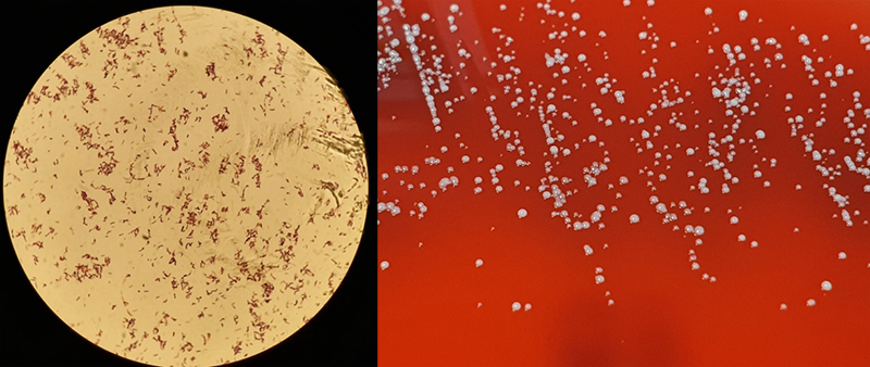
Cutibacteria (formerly propionibacteria) are part of normal flora of human skin and mucosal surfaces. They are slow growing, anaerobic yet aerotolerant, Gram-positive, non-spore forming, pleomorphic rods (figure 5) of relatively low virulence. Cutibacteria, due to their commensal presence on skin and low virulence, are commonly considered contaminants of blood cultures. Infrequently, they can cause significant infections of orthopaedic prosthesis, endovascular devices and cerebrospinal shunts.6

Boyle - Figure 5. Cutibacterium species anaerobic, Gram-positive bacilli
Figure 5. Cutibacterium species anaerobic, Gram-positive bacilli

Culture

Given the rarity of IE secondary to C. acnes, and that this bacterium is commonly grown as a commensal, it is pertinent to distinguish between simple culture contamination and a true bacteraemia. Multiple blood cultures must be positive with the same isolate to consider this the causative organism of infection.3 The time to detection of the bacteria in blood cultures is 6.4 days in anaerobic bottles and 6.1 days in aerobic bottles.3 However, the general consensus from numerous studies is, to reduce false negatives, cultures should be incubated for 10 to 14 days.3,7,8 Banzon et al. elucidated that C. acnes is identified in only 12.5% of routine blood cultures, greater success is achieved with extended incubation of blood cultures to 75%.9 However, the gold standard of identifying C. acnes is valve sequencing – PCR base diagnosis targeting 16S rDNA – achieving growth in 95–96% of cases.9,10 Notably, 46% of cases in Banzon et al.’s study, would have found no cause for IE without valve sequencing.9

Clinical course

Time from symptoms, including fever, chill, malaise, fatigue, myalgias and weight loss, to diagnosis averages at four weeks, however, this has been reported to be as long as 32 weeks in some studies. The bacterium can remain intracellular for weeks and months, explaining the long incubation period.11 Given the subtle nature of presentation, delayed incubation period and possibility of skin commensal contamination in blood cultures; diagnosis and subsequent early intervention can present a significant challenge to a clinician.3,4,12 A clear demographic has been shown: C. acnes IE is typically found in middle-aged men. The median age of diagnosis is 52 years, with various studies reporting between 90% and 98% of cases being male.5,7 However, the time from valve/device insertion to diagnosis of cutibacteria IE is less clear, with an average of four years and range between three weeks to 23 years.5 C. acnes has been associated with both native and prosthetic valves, however, the incidence is much higher in the latter.3,4 PVE or IE on an annuloplasty ring accounts for 96% of cases.9 The aortic valve is involved in the majority of cases (71%), mitral valve less so, with involvement in 24% of cases. Tricuspid valve involvement is uncommon (3%).5

Investigations and findings

In our cases, C. acnes has been found to be the causative organism for IE in both men, both of whom have prosthetic valves in situ, all with extensive valve or annulus destruction or abscess formation. Reinforced by the findings of Corvec, C. acnes causes extensive decalcification, abscess formation and valvular destruction.12 These cases displayed systemic symptoms; this is typical in 75% of cases. Case 1 presented primarily with a normal white blood cell count, it was not until his second presentation to hospital that he displayed leucocytosis and, indeed, leucocytosis is typical in only 57% of cases.5 Careful attention to the history, and consideration of potential sources of C. acnes seeding, should be considered. A recent study evaluated the rate of cardiac device-related endocarditis at 1.9 per 1,000 device-years.13 Typical complications of C. acnes include peripheral emboli in 16%, brain emboli in 10%, myocardial abscess in 36% and valvular insufficiency in 52%.5 Complications such as invasive disease (71%) and embolic complications (29%) are common.4,9

Management

Braun et al. strongly recommend a treatment combination of rifampicin with daptomycin or penicillin G.11 The combination of rifampicin and ciprofloxacin might be a reasonable alternative with excellent oral bioavailability for maintenance therapy in complicated IE.11 The European Committee on Antimicrobial Susceptibility Testing (EUCAST) has set breakpoints for benzylpenicillin, advocating the use of benzylpenicillin instead of vancomycin or ceftriaxone.8 The dose and addition of rifampicin in PVE remains to be elucidated.8

Conclusion

In summary, IE is a disease of high morbidity and mortality. Specifically, with C. acnes as a causative organism, it can be difficult to diagnose, and when established on a prosthetic valve it is very malignant to treat. The initial treating clinician’s awareness and willingness to consider the diagnosis should be encouraged. Once there is an index of suspicion, this should be preserved and the diagnosis pursued with combined modalities such as echo, CT and blood cultures. Due diligence from the multi-disciplinary team is essential in distinguishing between blood culture contamination and a true bacteraemia secondary to C. acnes. The awareness of this pathogen must be highlighted in the greater cardiothoracic and microbiology community in an effort to expedite appropriate treatment and improve patient outcomes.

Key messages

  • Infective endocarditis secondary to Cutibacterium acnes is an interesting clinical issue, as it is frequently grown in blood culture and discounted as a skin contaminant. In this clinical setting it should be considered significant and pursued aggressively, given the high rates of severe complication
  • Similarities in demographics, including age, gender and a prosthetic aortic valve in this cohort, should be noted and increase the physician level of suspicion when faced with this clinical presentation

Conflicts of interest

None declared.

Funding

None.

Study approval

In accordance with local guidelines of the ethics commission, no ethical application was needed. Institutional review board approval was not required.

Patient consent

Consent was gained from the patients.

References

1. Lalani T, Person AK, Hedayati SS et al. Propionibacterium endocarditis: a case series from the international collaboration on endocarditis merged database and prospective cohort study. Scand J Infect Dis 2007;39:840–8. https://doi.org/10.1080/00365540701367793

2. Patel A, Calfee RP, Plante M, Fischer SA, Green A. Propionibacterium acnes colonization of the human shoulder. J Shoulder Elbow Surg 2009;18:897–902. https://doi.org/10.1016/j.jse.2009.01.023

3. Achermann Y, Ellie J, Goldstein C, Coenye T, Shirtliff ME. Propionibacterium acnes: from commensal to opportunistic biofilm-associated implant pathogen. Am Soc Microbiol 2014;10:419–40. https://doi.org/10.1128/CMR.00092-13

4. Clayton JJ, Baig W, Reynolds GW, Sandoe JA. Endocarditis caused by Propionibacterium species: a report of three cases and a review of clinical features and diagnostic difficulties. J Med Microbiol 2006;55:981–7. https://doi.org/10.1099/jmm.0.46613-0

5. Sohail MR, Gray AL, Baddour LM, Tleyjeh IM, Virk A. Infective endocarditis due to Propionibacterium species. Clin Microbiol Infect 2009;15:387–94. https://doi.org/10.1111/j.1469-0691.2009.02703.x

6. Kanafani Z. Invasive Cutibacterium (formerly Propionibacterium) infections. Uptodate.com 2020. Available at: https://www.uptodate.com/contents/invasive-cutibacterium-formerly-propionibacterium-infections [accessed 18 June 2020].

7. Lindell F, Söderquist B, Sundman K, Olaison L, Källman J. Prosthetic valve endocarditis caused by Propionibacterium species: a national registry-based study of 51 Swedish cases. Eur J Clin Microbiol Infect Dis 2018;37:765–71. https://doi.org/10.1007/s10096-017-3172-8

8. Verkaik NJ, Schurink CAM, Melles DC. Letter to the editor, Antibiotic treatment of Propionibacterium acnes endocarditis. Clin Microbiol Infect 2018;24:209.  https://doi.org/10.1016/j.cmi.2017.07.024

9. Banzon JM, Rehm SJ, Gordon SM, Hussain ST, Pettersson GB, Shrestha NK. Propionibacterium acnes endocarditis: a case series. Clin Microbiol Infect 2017;23:396–9. https://doi.org/10.1016/j.cmi.2016.12.026

10. Yamamoto R, Miyagawa S, Hagiya H et al. Silent native-valve endocarditis caused by Propionibacterium acnes. Japanese Intern Med 2018;57:2417–20. https://doi.org/10.2169/internalmedicine.9833-17

11. Braun DL, Hasse BK, Stricker J, Fehr JS. Prosthetic valve endocarditis caused by Propionibacterium species successfully treated with coadministered rifampin: report of two cases. BMJ Case Rep 2013;2013:bcr2012007204. https://doi.org/10.1136/bcr-2012-007204

12. Corvec S. Clinical and biological features of Cutibacterium (formerly Propionibacterium) avidum, an underrecognized microorganism. Clin Microbiol Rev 2018;31:1–42. https://doi.org/10.1128/CMR.00064-17

13. Uslan DZ, Sohail MR, St Sauver JL et al. Permanent pacemaker and implantable cardioverter defibrillator infection: a population-based study. Arch Intern Med 2007;167:669–75. https://doi.org/10.1001/archinte.167.7.669

High-output heart failure due to arteriovenous malformation treated by endovascular embolisation

Br J Cardiol 2022;29:117–8doi:10.5837/bjc.2022.026 Leave a comment
Click any image to enlarge
Authors:
First published online 19th July 2022

High-output heart failure (HF) is an uncommon condition. This occurs when HF syndrome patients have a cardiac output higher than eight litres per minute. Shunts, such as fistulas and arteriovenous malformations are an important reversible cause. We present the case of a 30-year-old man who presented to the emergency department due to decompensated HF. Echocardiogram showed dilated myocardiopathy with high cardiac output (19.5 L/min calculated on long-axis view). He was diagnosed with arteriovenous malformation by computed tomography (CT) and subsequent angiography, and a multi-disciplinary team decided to perform endovascular embolisation with ethylene vinyl alcohol/dimethyl sulfoxide at different times. The transthoracic echocardiogram showed a significant decrease in cardiac output (9.8 L/min) and his general condition improved significantly.

Introduction

High-output heart failure (HF) is an uncommon condition with varying aetiologies, often left uninvestigated. This type of HF is characterised by an elevated cardiac output, usually with high stroke volume, leading to biventricular dilation. According to a Mayo Clinic series involving 120 patients with high-output HF diagnosed between 2000 and 2014, the most important causes included obesity, liver cirrhosis, chronic obstructive pulmonary disease, and shunting, such as fistulas or arteriovenous malformations.1

Case report

A 30-year-old man presented to the emergency department for dyspnoea, abdominal distention, and oedema lasting 20 days. He had a history of thyroid medullar carcinoma, diagnosed 11 years ago, and treated with surgery and oral chemotherapy with sorafenib without subsequent follow-up.

Liberman - Figure 1. Abdomen and pelvis computed tomography (CT) scan showing arteriovenous malformation (AVM) in axial section (a) and severe inferior vena cava dilatation in coronal section (b)
Figure 1. Abdomen and pelvis computed tomography (CT) scan showing arteriovenous malformation (AVM) in axial section (a) and severe inferior vena cava dilatation in coronal section (b)

On admission, he had a blood pressure of 155/95 mmHg, a heart rate of 107 bpm, and a respiratory rate of 30 breaths per minute. The patient was afebrile with an oxygen saturation of 96%. Cardiovascular examination revealed a 3/6 systolic murmur in the mesocardium. He presented with jugular vein distention that was not collapsible, bipedal oedema reaching the upper thigh, and marked hepatomegaly palpable up to the right iliac fossa. The chest X-ray showed cardiomegaly. His N-terminal pro-B-type natriuretic peptide (NT-proBNP) measured 950 pg/ml, while his calcitonin level was higher than 2,000 pg/ml, thyroid stimulating hormone (TSH) of 9.4 mU/ml with free T4 of 0.88 ng/dL. The bedside echocardiogram showed dilated myocardiopathy with preserved biventricular systolic function (61% by biplane method) with left ventricular end diastolic diameter of 6.4 cm, severe tricuspid regurgitation and cardiac output calculated on long-axis view of 19.5 L/min. A computed tomography (CT) scan ruled out pulmonary thromboembolism, and identified bilateral ectasia of the pulmonary artery branches, severely dilated inferior vena cava, multiple lithic metastatic-type lesions in the cervical and dorsal vertebral bodies, and a large right pelvic arteriovenous malformation (AVM) (figure 1). In order to rule out other possible aetiologies of dilated cardiomyopathy, a cardiac magnetic resonance imaging (MRI) perfusion scan was performed without signs of ischaemia and absence of late gadolinium enhancement.

Liberman - Figure 2. Diagnostic arteriography showing right pelvic AVM
Figure 2. Diagnostic arteriography showing right pelvic AVM

Bone biopsy revealed poorly differentiated neoplasia infiltration. The immunohistochemistry confirmed the metastatic origin of the thyroid medullar carcinoma. Aortic–iliac–femoral Doppler ultrasound demonstrated a flow compatible with AVM. A bibliographic review was carried out and the case was discussed by a multi-disciplinary team, before deciding to make a diagnostic arteriography. Right pelvic AVM tributary branches (hypogastric, upper gluteal, and superficial femoral) and a smaller contralateral AVM were identified (figure 2).

A right AVM embolisation through the left femoral artery (7 French access) was performed with a micro-catheter for the selective injection of ethylene vinyl alcohol/dimethyl sulfoxide. The control angiography at the end showed the material correctly distributed without complications.

Subsequently, the patient was admitted to the coronary unit due to decompensated HF. We initiated treatment with endovenous diuretics, but the patient responded poorly. He developed oliguria and dyspnoea, so right heart catheterisation was performed. It revealed a cardiac output of 18 L/min with a pulmonary wedge pressure of 24 mmHg and central venous pressure of 20 mmHg. Triple diuretic treatment was prescribed, eliciting an improved response. A new arteriography ruled out complications related to the procedure and showed the persistence of tributary branches with residual AVM.

Four endovascular procedures were successfully carried out. The transthoracic echocardiogram (seven days after the last procedure) showed a significant decrease in cardiac output (9.8 L/min). The patient’s decompensated HF symptoms disappeared, and his general condition improved significantly. Chemotherapy for his underlying disease was then planned.

Discussion

HF is characterised by a normal or low cardiac output, associated with increased
left-ventricle filling pressures.2 However, some HF syndrome patients present with a high output. This includes HF patients with a cardiac output higher than eight litres or 3.0 L/min/m2.3

High-output HF may be classified under two main categories:

  • Rise in body oxygen consumption due to increased metabolic demand
  • Low systemic vascular resistance due to peripheral vasodilation or arteriovenous shunting.

In the latter, the arteries and veins are tangled and form direct connections, bypassing normal tissues. In the second group, the shunt may be caused by arteriovenous fistulas in patients undergoing haemodialysis, or isolated AVMs, which may be congenital, post-traumatic, iatrogenic, or syndromic, particularly hereditary haemorrhagic telangiectasia.4

As seen in this case, the diagnosis is not straightforward. In most instances, various imaging tools are necessary to diagnose high-output HF and the aetiologic cause. Specifically, in this case, an echocardiogram, CT and cardiac MRI were performed in order to arrive at the diagnosis and to the aetiologic cause. About the AVM, the patient had no history of interventions or any other history to consider an iatrogenic or traumatic cause for AVM. He was not on haemodialysis, nor did he meet the clinical criteria for diagnosing hereditary haemorrhagic telangiectasia or other apparent cause, therefore, it should be considered congenital. Since high-output HF is not frequent, nor is it usual to consider it initially, diagnosis is often late. For this reason, thinking about the possibility of it is essential.

To conclude, patients with high-output HF secondary to extended AVM have been treated with different techniques, including embolisation through arterial catheterisation, stent placement with sclerotherapy, and surgical closure in selected cases.5 No treatment was reportedly better than the others. The treatment is individualised and decided by the multi-disciplinary team.

Conflicts of interest

None declared.

Funding

Research Committee, Allende Sanatorium.

Consent

The patient, in his full capacity, gave informed consent for the report of this case for educational and scientific purposes.

References

1. Reddy YNV, Melenovsky V, Redfield MM et al. High-output heart failure: a 15-year experience. J Am Coll Cardiol 2016;68:473–82. https://doi.org/10.1016/j.jacc.2016.05.043

2. Murphy SP, Ibrahim NE, Januzzi JL. Heart failure with reduced ejection fraction: a review. JAMA 2020;324:488–504. https://doi.org/10.1001/jama.2020.10262

3. Mehta PA, Dubrey SW. High output heart failure. QJM 2009;102:235–41. https://doi.org/10.1093/qjmed/hcn147

4. Koyalakonda SP, Pyatt J. High output heart failure caused by a large pelvic arteriovenous malformation. JRSM Short Rep 2011;2:66. https://doi.org/10.1258/shorts.2011.011057

5. Okada M, Kato M, Uchida K et al. Transcatheter and percutaneous procedures for huge pelvic arteriovenous malformations causing high-output heart failure. J Cardiol Cases 2015;12:162–5. https://doi.org/10.1016/j.jccase.2015.07.001

BJC accepted onto PubMed Central®

Leave a comment
Click any image to enlarge
Authors:

We are delighted to announce that the British Journal of Cardiology (BJC) has been accepted onto the prestigious international digital archive PubMed Central® after meeting the rigorous scientific and technical standards it requires.

Pub Med Central

PubMed Central® is a full-text archive of biomedical and life sciences journal literature at the U.S. National Institutes of Health’s National Library of Medicine. BJC joins thousands of journals from all over the world depositing content, with articles published in the BJC since January 2020 being added to the digital archive.

The entry to PubMed Central® comes as BJC marks almost 30 years in peer-reviewed cardiovascular medicine linking primary and secondary care. The journal publishes high-quality reviews, state of the art clinical research and original papers in cardiovascular medicine, news, meeting reports, as well as comprehensive e-learning programmes for CPD through BJC Learning.

What’s new in heart failure guidance – a user’s guide: Introduction

Br J Cardiol 2022;29(suppl 2):S2 Leave a comment
Click any image to enlarge
Authors:
Sponsorship Statement: AstraZeneca has provided a sponsorship grant towards this independent Programme.

Heart failure is such a common problem that all healthcare professionals should be familiar with the contemporary management of these patients. When there are delays in making the diagnosis or treatment is not optimised, patients are at risk of premature death, hospitalisation and many suffer impaired quality of life. To improve outcomes for patients with heart failure, it is imperative that the diagnosis is suspected and made as early as possible. Whilst heart failure specialists are integral to the delivery of optimal patient-centred care, every opportunity should be taken to optimise treatment. We can all help make a real difference for patients.

This supplement provides a user’s guide to what’s new in the guidelines for the diagnosis and treatment of heart failure. This primarily relates to recommendations provided in the updated (2021) European Society of Cardiology (ESC) guidelines for the diagnosis and treatment of acute and chronic heart failure.

The articles summarise the contemporary guidance with respect to the diagnosis and investigation of patients presenting with heart failure; drug therapy (including the early implementation of the ‘four pillars’ of drug treatment for heart failure with reduced ejection fraction); and recommendations on lifestyle, rehabilitation, remote monitoring and device use.

The authors commonly refer to the ESC classes of recommendation and level of evidence and this should be considered as one interprets the data and makes clinical decisions. In summary these are:

Classes of recommendation:

  • Class I – is recommended or indicated
  • Class IIa – should be considered
  • Class IIb – may be considered
  • Class III – is not recommended

Levels of evidence:

  • A – data derived from multiple randomised clinical trials or meta-analysis
  • B – data derived from a single randomised clinical trial or large non-randomised studies
  • C – expert consensus of opinion and/or small studies, retrospective studies or registries.

Please also note that these articles are the authors’ personal interpretation of the most recently available evidence and how this affects practice. It should not replace consulting the original sources and guidance.

Paul Kalra
Guest Editor

Articles in this supplement

New developments in the investigations and diagnosis of heart failure
Drug therapy in heart failure – an update from the 2021 ESC heart failure guideline
Guidance on lifestyle, rehabilitation and devices in heart failure patients

Disclaimer:

Medical knowledge is constantly changing. As new information becomes available, changes in treatment, procedures, equipment and the use of drugs become necessary. The editors/authors/contributors and the publishers Medinews (Cardiology) Ltd have taken care to ensure that the information given in this text is accurate and up to date at the time of publication.

Readers are strongly advised to confirm that the information, especially with regard to drug usage, complies with the latest legislation and standards of practice. Medinews (Cardiology) Limited advises healthcare professionals to consult up-to-date Prescribing Information and the full Summary of Product Characteristics available from the manufacturers before prescribing any product. Medinews (Cardiology) Limited cannot accept responsibility for any errors in prescribing which may occur.

The opinions, data and statements that appear are those of the contributors. The publishers, editors, and members of the editorial board do not necessarily share the views expressed herein. Although every effort is made to ensure accuracy and avoid mistakes, no liability on the part of the publisher, editors, the editorial board or their agents or employees is accepted for the consequences of any inaccurate or misleading information.

© Medinews (Cardiology) Ltd 2022. All rights reserved. No part of this publication may be reproduced, stored in a retrieval system, or transmitted in any form or by any means, electronic, mechanical, photocopying, recording or otherwise, without the prior written permission of the publishers, Medinews (Cardiology) Ltd. It shall not, by way of trade or otherwise, be lent, re-sold, hired or otherwise circulated without the publisher’s prior consent.

New developments in the investigations and diagnosis of heart failure

Br J Cardiol 2022;29(suppl 2):S3–S6doi:10.5837/bjc.2022.s06 Leave a comment
Click any image to enlarge
Authors:
Sponsorship Statement: AstraZeneca has provided a sponsorship grant towards this independent Programme.

This article reviews some of the new concepts, new recommendations, along with changes to recommendations, in the diagnosis and investigation of heart failure (HF) in the European Society of Cardiology (ESC) 2021 Guidelines for the diagnosis and treatment of acute and chronic heart failure, and contrasts these with the 2016 version of the guidelines.

Introduction

The heart failure (HF) community has seen huge advances in the care of HF, and we see a turning point in the narrative of doom and gloom, which has traditionally been associated with HF – we see cause for optimism. We recognise the urgency of putting these advances to prompt use, as demonstrated by the 2021 ESC Guidelines for the diagnosis and treatment of acute and chronic HF.1 The 2021 updated guidelines make it clear that we have the means to diagnose HF early, to classify it more accurately, the tools to change the HF trajectory, and the duty and ability to intervene – and to do so early.

Diagnosis

While the ESC diagnostic algorithm for HF is not new per se, it is worth reiterating and emphasising, that checking N-terminal pro-B-type natriuretic peptide (NTproBNP) levels and acting on them is the path to timely diagnosis. It allows for the early initiation of therapies to positively alter patient outcomes. The recommended cut-offs for NTproBNP are significantly lower in the ESC guidelines1 than in the National Institute for Health and Care Excellence (NICE) guidelines,2 as outlined in figure 1. They reflect that levels of NTproBNP below 125 pg/ml have excellent negative predictive value (0.94–0.98).

BJC 2022 Heart failure supplement - Figure 1. Simplified detail from the European Society of Cardiology (ESC) guidance and the National Institute for Health and Care Excellence (NICE) guidance to compare NTproBNP cut-offs. Always refer to the original guidance for full detail
Figure 1. Simplified detail from the European Society of Cardiology (ESC) guidance and the National Institute for Health and Care Excellence (NICE) guidance to compare NTproBNP cut-offs. Always refer to the original guidance for full detail

New concepts

These include:

  • A change of the term ‘heart failure with mid-range ejection fraction’ to ‘heart failure with mildly reduced ejection fraction’ (HFmrEF).
  • The nomenclature for HF with left ventricular ejection fraction (LVEF) of 41–49% has been revised to HF with mildly reduced EF (HFmEF).

HF with LVEF ≤40% remains HF with reduced EF (HFrEF), and HF with LVEF ≥50% remains HF with preserved EF (HFpEF).

For the diagnosis of HFmrEF, elevated natriuretic peptides plus other evidence of structural heart disease make the diagnosis more likely. But these are not mandatory if there is certainty on measurement of LVEF. The next article by Hardy and Kalra (pages S7–S12), will cover more on the addition of recommendations for the treatment of HFmrEF – patients with HFmrEF may benefit from similar therapies to those with HFrEF.

The guidelines now have a modified classification for acute HF, divided into four distinct types. Clinical presentations are mainly based on the presence of signs of congestion and/or peripheral hypoperfusion and require different treatments.3

  1. Acute decompensated heart failure (ADHF) is the most common form of AHF, accounting for 50–70% of presentations.4 ADHF usually occurs in patients with a history of HF and previous cardiac dysfunction across the spectrum of LVEF and may include right ventricular dysfunction. Distinct from the acute pulmonary oedema phenotype, it has a more gradual onset, and the main alteration is progressive fluid retention responsible for systemic congestion. Sometimes, the congestion is associated with hypoperfusion.4
  2. Acute pulmonary oedema related to lung congestion and clinical criteria for acute pulmonary oedema diagnosis include dyspnoea with orthopnoea, respiratory failure (hypoxaemia-hypercapnia), tachypnoea, >25 breaths/min, and increased work of breathing.5
  3. Isolated right ventricular (RV) failure is associated with increased RV and atrial pressure and systemic congestion. RV failure may also impair left ventricular (LV) filling, and ultimately reduce systemic cardiac output, through ventricular interdependence.6
  4. Cardiogenic shock is a syndrome due to primary cardiac dysfunction, resulting in inadequate cardiac output, leading to life threatening state of hypoperfusion which can result in multi-organ failure and death.7

New recommendations

New recommendations in diagnosis

Right heart catheterisation should be considered in patients where HF is thought to be due to constrictive pericarditis, restrictive cardiomyopathy, congenital heart disease, and high output states (Class IIa recommendation).

Right heart catheterisation may be considered in selected patients with HFpEF to confirm the diagnosis (Class IIb recommendation). The gold standard test for the diagnosis of HFpEF is invasive haemodynamic exercise testing. An invasively measured pulmonary capillary wedge pressure (PCWP) of ≥15 mmHg (at rest) or ≥25 mmHg (with exercise) or LV end-diastolic pressure ≥16 mmHg (at rest) is generally considered diagnostic.8 Recognising that invasive haemodynamic exercise testing is not available in many centres worldwide and has associated risks, the current guidelines do not mandate gold standard testing to make the diagnosis, but emphasise that the greater the number of objective non-invasive markers of raised LV filling pressures, the higher the probability the diagnosis of HFpEF (see table 1).

Table 1. Objective evidence used in the diagnosis of HFpEF

Parameter Threshold Comments
LV mass index >95 g/m2 (female)
>115 g/m2 (male)
The presence of concentric LV hypertrophy (LVH) is supportive, but its absence does not exclude a diagnosis of HFpEF
Relative wall thickness >0.42 Relative wall thickness (RWT) is increased in concentric LVH. Normal RWT is 0.32–0.42, measured using the equation RWT = IVSd + PWd / LVd
LA volume index >34 ml/m2 (SR)
>40 ml/m2 (AF)
In the absence of AF or valvular disease, LA enlargement reflects chronically elevated LV filling pressure
E/e’ ratio at rest >9 Sensitivity 78%, specificity 59% for HFpEF by invasive exercise testing. Cut-off of 13 has lower sensitivity (46%) but higher specificity (86%)9
NTpro BNP >125 (SR), >365 (AF) pg/ml Up to 20% of patients with invasively proven HFpEF have NPs below diagnostic thresholds, particularly in presence of obesity
PA systolic pressure/TR velocity at rest >35 mmHg/>2.8 m/s Sensitivity 54%, specificity 85% for presence of HFpEF by invasive exercise testing10
Key: AF = atrial fibrillation; E/e’ = early filling velocity on transmitral Doppler/early relaxation velocity on tissue Doppler; HFpEF = heart failure with preserved ejection fraction; IVSd = interventricular septum thickness end diastole; LA = left atrium; LV = left ventricular; LVd = left ventricular diameter end diastole; NP = natriuretic peptides; NTproBNP = N-terminal pro-B-type natriuretic peptide; PA = pulmonary artery; PWd = posterior wall thickness end diastole; TR = tricuspid regurgitation; SR = sinus rhythm

New recommendations in monitoring

Non-invasive home telemonitoring (HTM) may be considered for patients with HF to reduce the risk of recurrent cardiovascular (CV) and HF hospitalisations and CV death (Class IIb recommendation).

Telemonitoring enables patients to provide their digital health information remotely to support and optimise their care. Data such as weight, heart rate, and blood pressure can be collected, stored in an electronic health record and used to guide changes to therapy. HTM allows for rapid access to care as needed, reduced patient inconvenience and travel costs, and minimises the frequency of clinic visits.11 The enforced cessation of face-to-face clinic visits in many countries during the COVID-19 pandemic has highlighted the potential advantages of HTM.12

New recommendations for HF and cancer care

There are two new recommendations in this area:

  • It is recommended that cancer patients at increased risk for cardiotoxicity, defined by a history or risk factors of CV disease, previous cardiotoxicity or exposure to cardiotoxic agents, undergo CV evaluation before scheduled anticancer therapy, preferably by a cardiologist with experience/interest in cardio-oncology (Class I recommendation).
  • A baseline CV risk assessment should be considered in all cancer patients scheduled to receive a cancer treatment with the potential to cause HF (Class IIa recommendation).

This is required because HF can occur in patients with cancer as a result of the interaction between the anticancer therapy, the cancer itself, and the patient’s own CV background (risk factors and coexisting CV disease).13 Several anticancer therapies may cause HF directly, through their cardiotoxic effects or, indirectly, through other mechanisms, such as myocarditis, ischaemia, systemic or pulmonary hypertension, arrhythmias or valve disease.14 HF, in turn, may affect cancer outcomes by depriving patients of effective anticancer therapies.15 Therefore, prevention of HF in patients with cancer receiving potentially cardiotoxic therapies requires careful patient assessment and management before, during, and after cancer therapy, preferably in the context of an integrated cardio-oncology service.13

Changes to recommendations

Changes to recommendations in diagnosis – three of note

Invasive coronary angiography may be considered in patients with HFrEF with an intermediate to high pre-test probability of coronary artery disease (CAD) and the presence of ischaemia in non-invasive stress tests (Class IIb recommendation [change from IIa], with removal of wording “who are considered suitable for potential coronary revascularisation”).

CT coronary angiography should be considered in patients with a low to intermediate pre-test probability of CAD or those with equivocal non-invasive stress tests to rule out coronary artery stenosis (Class IIa recommendation [change from IIb]).

Importantly, there have been key changes to the diagnosis of HFpEF. The guidelines now recommend a simplified diagnostic pathway, with three steps, for HFpEF, that include:

  1. Symptoms and signs of HF
  2. An LVEF ≥50%
  3. Objective evidence of cardiac structural and/or functional abnormalities consistent with the presence of LV diastolic dysfunction/ raised LV filling pressures, including raised natriuretic peptides (see table 1).

The guideline recommends a simplified approach to HFpEF diagnosis that distils the common major elements in prior diagnostic criteria, emphasising the most frequently used and widely available variables. Some of these, in particular:

  • LA size (LA volume index >32 ml/m2)
  • mitral E velocity >9 cm/s
  • septal e’ velocity <9 cm/s
  • E/e’ ratio >9

have been shown to be pivot points beyond which CV mortality risk is increased, underscoring their value.16 This recommendation is therefore consistent with the consensus document of the Heart Failure Association (HFA), but is, rather, a simplified approach.

Key changes to recommendations for the diagnosis of patients with cardiac amyloid

Cardiac amyloid is still an underdiagnosed cause of HF.17 The two most prevalent forms of cardiac amyloid are light chain immunoglobulin (AL) and transthyretin (ATTR) amyloidosis. ATTR includes wild-type (>90% of cases), and the hereditary type (<10% of cases). It is estimated that 6% to 16% of all patients with unexplained LVH or HFpEF at hospitalisation or severe aortic stenosis undergoing aortic valve replacement, aged above 65 years, may have wild typeTTR-CA.18 Based on a recent review, the latest guidelines recommend using major criteria for the suspicion of cardiac amyloid that include: age >65 years and HF, with LV wall thickness >12 mm at echocardiography.19 There are extensive resources within the guidelines to confirm the diagnosis, and include tables of ‘red flag’ findings.

Advice on cardiac imaging and electromyocardia biopsy (EMB) or extra-cardiac biopsy are also provided for the diagnosis of AL cardiac amyloid in patients with abnormal haematological tests. Technetium-labelled 99mTc-PYP or DPD or HMDP scintigraphy with planar and SPECT imaging has a specificity and positive predictive value for TTR cardiac amyloid of up to 100%.20 In contrast, CV magnetic resonance imaging (CMR) has a sensitivity and specificity of 85% and 92%, respectively.21 The hereditary form should be excluded by genetic testing. EMB is the gold standard for the diagnosis of TTR cardiac amyloid with nearly 100% sensitivity and specificity if specimens are collected from more than four multiple sites and tested for amyloid deposits by Congo red staining,21 but is not needed in the setting of grade 2–3 positive scintigraphy with SPECT.19

Lastly, there have been changes to the recommendations for gene testing in cardiomyopathies. While clinical history, laboratory tests, and imaging are the first-line investigations (with echocardiography as central in diagnosis and CMR providing more detailed morphological and prognostic information), testing for genetic mutations can add clinical and prognostic value. The prevalence of gene mutations may vary according to the phenotype or underlying cause. Gene mutations occur in up to 40% of dilated cardiomyopathy, 60% of hypertrophic cardiomyopathy, and 15% in chemotherapy-induced, alcoholic or peripartum cardiomyopathies.22 The prevalence of genetic mutations is also over 10% in non-familial dilated cardiomyopathy.22 Finding a pathogenic gene variant in a patient with cardiomyopathy allows better prediction of the disease outcome and progression, may contribute to the indications for device implantation and inform genetic counselling for families.

Of note, take heed, at the end of the guidelines, in sections 16 and 17, there are extremely useful summaries, entitled ‘Gaps in Evidence’, ‘What to do’ and ‘What not to do’ messages from the guidelines.

Conclusion

This 2021 updated ESC Guideline for diagnosis and treatment of HF1 indicates a radical shift – highlighting how we must accurately and promptly diagnose HF so that we can manage HF and encourage a patient-centric, tailored approach focused on improving patients’ quality of life and improving their clinical outcomes.

Key messages

  • Use lower NTproBNP cut-offs for diagnosis – think heart failure (HF), and think it early
  • HFmrEF is now termed HF with mildly reduced ejection fraction
  • There is a simplified three-step HFpEF diagnostic pathway

Conflicts of interest

PC has received speaker fees for Astra Zeneca, Boehringer Ingelheim, Novartis, Pfizer, and Vifor.

Patricia Campbell
Consultant Cardiologist and Heart Failure Lead

Southern Health and Social Care Trust, Craigavon Area Hospital, 68 Lurgan Road, Portadown, BT63 5QQ

([email protected])

Articles in this supplement

Introduction
Drug therapy in heart failure – an update from the 2021 ESC heart failure guideline
Guidance on lifestyle, rehabilitation and devices in heart failure patients

References

1. McDonagh TA, Metra M, Adamo M et al. 2021 ESC Guidelines for the diagnosis and treatment of acute and chronic heart failure. Eur Heart J 2021;42:3599–726. https://doi.org/10.1093/eurheartj/ehab368

2. National Institute for Health and Care Excellence. Chronic heart failure in adults: diagnosis and management. NG106, Published 12 September 2018. https://www.nice.org.uk/guidance/ng106

3. Nieminen MS, Brutsaert D, Dickstein K et al. The EuroHeart Survey Investigators, Heart Failure Association of the European Society of Cardiology. EuroHeart Failure Survey II (EHFS II): a survey on hospitalized acute heart failure patients: description of population. Eur Heart J 2006;27:2725–36. https://doi.org/10.1093/eurheartj/ehl193

4. Chioncel O, Mebazaa A, Maggioni AP et al. The ESC-EORP-HFA Heart Failure Long-Term Registry Investigators. Acute heart failure congestion and perfusion status – impact of the clinical classification on in-hospital and long-term outcomes: insights from the ESC-EORP-HFA heart failure long-term registry. Eur J Heart Fail 2019;21:1338–52. https://doi.org/10.1002/ejhf.1492

5. Masip J, Peacock WF, Price S et al. The Acute Heart Failure Study Group of the Acute Cardiovascular Care Association and the Committee on Acute Heart Failure of the Heart Failure Association of the European Society of Cardiology. Indications and practical approach to non-invasive ventilation in acute heart failure. Eur Heart J 2018;39:17–25. https://doi.org/10.1093/eurheartj/ehx580

6. Harjola VP, Mebazaa A, Celutkiene J et al. Contemporary management of acute right ventricular failure: a statement from the Heart Failure Association and the Working Group on Pulmonary Circulation and Right Ventricular Function of the European Society of Cardiology. Eur J Heart Fail 2016;18:226–41. https://doi.org/10.1002/ejhf.478

7. Chioncel O, Parissis J, Mebazaa A et al. Epidemiology, pathophysiology and contemporary management of cardiogenic shock – a position statement from the Heart Failure Association of the European Society of Cardiology. Eur J Heart Fail 2020;22:1315–41. https://doi.org/10.1002/ejhf.1922

8. Barandiaran Aizpurua A, Sanders-van Wijk S, Brunner-La Rocca HP et al. Validation of the HFA-PEFF score for the diagnosis of heart failure with preserved ejection fraction. Eur J Heart Fail 2020;22:413–21. https://doi.org/10.1002/ejhf.1614

9. Lancellotti P, Galderisi M, Edvardsen T et al. Echo-Doppler estimation of left ventricular filling pressure: results of the multi-centre EACVI Euro-Filling study. Eur Heart J Cardiovasc Imaging 2017;18:961–8. https://doi.org/10.1093/ehjci/jex067

10. Pieske B, Tschope C, de Boer RA et al. How to diagnose heart failure with preserved ejection fraction: the HFA-PEFF diagnostic algorithm: a consensus recommendation from the Heart Failure Association (HFA) of the European Society of Cardiology (ESC). Eur Heart J 2019;40:3297–317. https://doi.org/10.1093/eurheartj/ehz641

11. Brahmbhatt DH, Cowie MR. Remote management of heart failure: an overview of telemonitoring technologies. Card Fail Rev 2019;5:86–92. https://doi.org/10.15420/cfr.2019.5.3

12. Cleland JG, Clark RA, Pellicori P, Inglis SC. Caring for people with heart failure and many other medical problems through and beyond the COVID-19 pandemic: the advantages of universal access to home telemonitoring. Eur J Heart Fail 2020;22:995–8. https://doi.org/10.1002/ejhf.1864

13. Zamorano JL, Gottfridsson C, Asteggiano R et al. The cancer patient and cardiology. Eur J Heart Fail 2020;22:2290–309. https://doi.org/10.1002/ejhf.1985

14. Zamorano JL, Lancellotti P, Rodriguez Munoz D et al. Authors/Task Force. Members, ESC Committee for Practice Guidelines, Document Reviewers. 2016. ESC Position Paper on cancer treatments and cardiovascular toxicity developed under the auspices of the ESC Committee for Practice Guidelines: The TaskForce for cancer treatments and cardiovascular toxicity of the European Society of Cardiology (ESC). Eur J Heart Fail 2017;19:9–42. https://doi.org/10.1093/eurheartj/ehw211

15. Ameri P, Canepa M, Anker MS et al., Heart Failure Association Cardio-Oncology Study Group of the European Society of Cardiology. Cancer diagnosis in patients with heart failure: epidemiology, clinical implications and gaps in knowledge. Eur J Heart Fail 2018;20:879–87. https://doi.org/10.1002/ejhf.1165

16. Playford D, Strange G, Celermajer DS et al., NEDA Investigators. Diastolic dysfunction and mortality in 436,360 men and women: the National Echo Database Australia (NEDA). Eur Heart J Cardiovasc Imaging 2021;22:505–15. https://doi.org/10.1093/ehjci/jeaa253

17. Martinez-Naharro A, Hawkins PN, Fontana M. Cardiac amyloidosis. Clin Med (Lond) 2018;18:s30–s35. https://doi.org/10.7861/clinmedicine.18-2-s30

18. Cavalcante JL, Rijal S, Abdelkarim I et al. Cardiac amyloidosis is prevalent in older patients with aortic stenosis and carries worse prognosis. J Cardiovasc Magn Reson 2017;19:98. https://doi.org/10.1186/s12968-017-0415-x

19. Garcia-Pavia P, Rapezzi C, Adler Y et al. Diagnosis and treatment of cardiac amyloidosis. A position statement of the European Society of Cardiology Working Group on Myocardial and Pericardial Diseases. Eur J Heart Fail 2021;23:512–26. https://doi.org/10.1002/ejhf.2140

20. Gillmore JD, Maurer MS, Falk RH et al. Nonbiopsy diagnosis of cardiac transthyretin amyloidosis. Circulation 2016;133:2404–12. https://doi.org/10.1161/CIRCULATIONAHA.116.021612

21. Ruberg FL, Grogan M, Hanna M, Kelly JW, Maurer MS. Transthyretin amyloid cardiomyopathy: JACC state-of-the-art review. J Am Coll Cardiol 2019;73:2872–91. https://doi.org/10.1016/j.jacc.2019.04.003

22. Hershberger RE, Givertz MM, Ho CY et al. Genetic evaluation of cardiomyopathy – a Heart Failure Society of America practice guideline. J Card Fail 2018;24:281–302. https://doi.org/10.1016/j.cardfail.2018.03.004

Disclaimer:

Medical knowledge is constantly changing. As new information becomes available, changes in treatment, procedures, equipment and the use of drugs become necessary. The editors/authors/contributors and the publishers Medinews (Cardiology) Ltd have taken care to ensure that the information given in this text is accurate and up to date at the time of publication.

Readers are strongly advised to confirm that the information, especially with regard to drug usage, complies with the latest legislation and standards of practice. Medinews (Cardiology) Limited advises healthcare professionals to consult up-to-date Prescribing Information and the full Summary of Product Characteristics available from the manufacturers before prescribing any product. Medinews (Cardiology) Limited cannot accept responsibility for any errors in prescribing which may occur.

The opinions, data and statements that appear are those of the contributors. The publishers, editors, and members of the editorial board do not necessarily share the views expressed herein. Although every effort is made to ensure accuracy and avoid mistakes, no liability on the part of the publisher, editors, the editorial board or their agents or employees is accepted for the consequences of any inaccurate or misleading information.

© Medinews (Cardiology) Ltd 2022. All rights reserved. No part of this publication may be reproduced, stored in a retrieval system, or transmitted in any form or by any means, electronic, mechanical, photocopying, recording or otherwise, without the prior written permission of the publishers, Medinews (Cardiology) Ltd. It shall not, by way of trade or otherwise, be lent, re-sold, hired or otherwise circulated without the publisher’s prior consent.

Drug therapy in heart failure – an update from the 2021 ESC heart failure guideline

Br J Cardiol 2022;29(suppl 2):S7–S12doi:10.5837/bjc.2022.s07 Leave a comment
Click any image to enlarge
Authors:
Sponsorship Statement: AstraZeneca has provided a sponsorship grant towards this independent Programme.

There have been several key developments in heart failure (HF) management since the 2016 European Society of Cardiology (ESC) HF guidelines were published. The updated 2021 ESC HF Guidelines reflect the modern management of the patient with HF, with an emphasis on evidence-based, individualised care. This overview aims to provide clinicians (whether specialist or non-specialist) with a summary of the major update in drug therapy for HF according to the recent 2021 ESC Guidelines for the diagnosis and treatment of acute and chronic HF.

Introduction

Heart failure (HF) is a common condition and the majority of patients have multiple co-morbidities. It is therefore essential that all healthcare professionals (HCPs) are familiar with the contemporary management of these patients. Whilst HF specialists are integral to the delivery of optimal patient care, it is important to ensure that therapies are optimised at every opportunity and enable the best care for patients in the context of acute or chronic non-cardiovascular illness. Current practice is often suboptimal; for example, in the latest national HF audit (England and Wales), the number of patients leaving hospital on three disease-modifying drugs was 56% for those under the care of cardiology and 40% for those treated on general medical wards.1

This article will provide a practical synopsis of the recent 2021 ESC HF guidelines for cardiologists and non-cardiologists alike, including key evidence underpinning the recommendations.2

Pharmacologic therapies for HFrEF

The sequential initiation of medication with prognostic benefit has historically underpinned the treatment for patients with a diagnosis of HF with reduced ejection fraction (HFrEF). Such medications are typically considered before other therapies, including devices, with the goal of reducing the risk of death, hospitalisation for HF and improving symptoms. Until recently, there were three main groupings of drugs used:

  1. Angiotensin-converting enzyme (ACE) inhibitors/angiotensin receptor blockers (ARB)
  2. Beta blockers
  3. Mineralocorticoid receptor antagonists (MRAs).

These drugs antagonise maladaptive neurohumoral activation, thought to be central to the pathophysiology of HF. As such, they are considered disease-modifying drugs. Large-scale trials over the last 30+ years have confirmed efficacy at reducing mortality and HF hospitalisations, often accompanied by improving symptoms (see table 1).3–16 There is a fundamental need to appreciate this, since in clinical practice it is common to see these drugs stopped or down-titrated due to the misconception that their primary use is to lower blood pressure or for diuretic effect. Such misunderstanding can lead to the inappropriate cessation of life prolonging therapy.

Table 1. Randomised trials studying the group of patients with a left ventricular ejection fraction (LVEF) ≤40% (except in the DAPA CKD study). The table shows A: trials with earlier disease-modifying drugs and B: those with more recent drug classes incorporated into the 2021 ESC guidelines

Class of drug Trial Number of participants Primary outcome (hazard ratio, p value) Secondary outcome(s) (hazard ratio, p value)
A. Earlier disease-modifying drugs
ACE inhibitor/ARB CONSENSUS (1987)3
Enalapril vs. placebo
253 All-cause mortality – 40% reduction at six months compared with placebo
SOLVD-treatment (1991)4
Enalapril vs. placebo
2,569 All-cause mortality reduced with enalapril (0.84, p=0.004) All-cause mortality/hospitalisation for HF (0.74, p<0.0001)
ATLAS (1999)5
High- vs. low-dose lisinopril
3,164 All-cause mortality (0.92, p=0.13, non-significant reduction) CV death (0.90, p=0.07, non-significant).
All-cause mortality/CV hospitalisation
CV death/CV hospitalisation (0.85, p<0.001)
CHARM-added (2003)6
Candesartan and ACE inhibitor vs. placebo and ACE inhibitor
2,548 CV death or hospitalisation for HF (0.85, p=0.01)
CHARM-alternative (2003)7
Candesartan vs. placebo (no ACE inhibitor)
2,028 CV death or hospitalisation for HF (0.77, p<0.001)
Beta blocker CIBIS-II (1999)8
Bisoprolol vs. placebo
2,647 All-cause mortality (0.66, p<0.001) Reduction in sudden death (0.56, p=0.0011)
COPERNICUS (2001)9
Carvedilol vs. placebo
2,289 All-cause mortality (0.65, p<0.001) All-cause mortality/hospitalisation (0.76, p<0.001)
SENIORS (2005)10
Nebivolol vs. placebo in patients >70 years
2,128 All-cause mortality or CV hospitalisation (0.86, p=0.04)
Mineralocorticoid receptor blocker RALES (1999)11
Spironolactone vs. placebo
822 All-cause mortality (0.70, p<0.001)
EMPHASIS HF (2011)12
Eplerenone vs. placebo
2,737 CV death or hospitalisation for HF (0.63, p<0.001)
B. More recent drug classes
ARNI PARADIGM HF (2014)13
Enalapril vs. sacubitril/ valsartan
8,442 CV death or hospitalisation for HF (0.80, p<0.001)
SGLT2 inhibitors DAPA HF (2019)14
Dapagliflozin vs. placebo
4,744 Worsening HF or CV death, with or without diabetes (0.74, p<0.001)
DAPA CKD (2020)15 4,094 ≥50% ↓ in eGFR or ESRD or death from renal/CV cause (0.61, p<0.001) ≥50% ↓ in eGFR or ESRD or renal death (0.56, p<0.001).
HF hospitalisations or CV death (0.71, p=0.009)
Death any cause (0.69, p=0.004)
EMPEROR-Reduced (2020)16
Empagliflozin vs. placebo
3,730 Worsening HF or CV death, with or without diabetes (0.75, p<0.001) Hospitalisation for HF (0.70, p<0.001).
Composite renal outcome (0.50)
Key: ACE = angiotensin-converting enzyme; ARB = angiotensin receptor blocker; ARNI = angiotensin receptor-neprilysin inhibitor; CV = cardiovascular; eGFR = estimated glomerular filtration rate; ESC = European Society of Cardiology; ESRD = end stage renal disease; HF = heart failure

The 2016 ESC guidelines incorporated the use of sacubitril/valsartan, an angiotensin receptor-neprilysin inhibitor (ARNI) as an alternative to ACE inhibitors or ARBs following the PARADIGM (Prospective Comparison of ARNI with ACEI to Determine Impact on Global Mortality and Morbidity in Heart Failure) trial, which demonstrated a significantly reduced risk of all-cause mortality, death from cardiovascular (CV) causes and hospitalisation for HF when compared with an ACE inhibitor (enalapril).13,17 It also significantly improved symptoms. As such, patients with HFrEF who remain symptomatic should be transitioned from an ACE inhibitor or ARB to sacubitril/valsartan (class I recommendation, 2021 ESC guidelines2).

Patients hospitalised for decompensated HF are at high risk for further decompensation and death. Whilst the PARADIGM trial showed that sacubitril/valsartan reduced the risk of hospitalisation by 21% compared with enalapril, this study only recruited outpatients. Smaller, short-term trials have shown that hospitalised patients with acute HF can also be safely started on sacubitril/valsartan, including patients who are ACE inhibitor/ARB naïve.18,19

The PIONEER HF (Comparison of Sacubitril–Valsartan versus Enalapril on Effect on NTproBNP in Patients Stabilized from an Acute Heart Failure Episode) trial18 compared initiation of sacubitril/valsartan versus enalapril in patients admitted to hospital with acutely decompensated HF, with the primary outcome at four and eight weeks being change in N-terminal pro-B-type natriuretic peptide (NTproBNP), a biomarker associated with higher risk of adverse outcomes. This trial showed sacubitril/valsartan was superior in reducing NTproBNP at all measured time points and was evident from the first week after initiation. Over half of the patients in this trial were ACE inhibitor/ARB naïve. Therefore, in carefully selected patients, an ARNI should be considered even in ACE inhibitor/ARB naïve patients (class IIb recommendation, 2021 ESC guidelines2). This should be an individualised decision and not all patients may be suitable for this, particularly if blood pressure is low.

Around the time sacubitril/valsartan received a license for the use in patients with HFrEF, trials looking at sodium-glucose cotransporter 2 (SGLT2) inhibitors in patients with type 2 diabetes reported favourable results with respect to HF outcomes. There followed two pivotal trials, DAPA-HF (Dapagliflozin in Patients with Heart Failure and Reduced Ejection Fraction) and EMPEROR-Reduced (Empagliflozin Outcome Trial in Patients with Chronic Heart Failure with Reduced Ejection Fraction), evaluating the benefit of dapagliflozin and empagliflozin in patients with HFrEF – with and without diabetes.14,16

The primary end point in both trials was a composite of CV mortality and hospitalisation for HF.  Dapagliflozin reduced CV mortality and hospitalisation for HF by 26% compared with placebo, and empagliflozin by 25%.  The benefit was observed whether the patients had diabetes or not. Both drugs significantly reduce the rate of decline in renal function. As a result, one of the major pharmacological changes in the new 2021 ESC HF guidelines is the incorporation of SGLT2 inhibitors as key disease-modifying drugs (class I recommendation, level A evidence). SGLT2 inhibitors appear well tolerated with little adverse impact on symptoms of hypotension. A single dose without need for up-titration makes this class of drug relatively straight-forward to use.

In summary, patients with HFrEF are at high risk of adverse outcome when the condition is not recognised in timely fashion or patients are not appropriately treated. There are now four pillars of pharmacological therapy to use as disease-modifying agents (figure 1). It is no longer considered necessary to start these sequentially and most would agree that it is imperative to start all four of these classes of drugs as quickly as possible to reduce the risk of HF hospitalisation, CV death and improve symptoms. The precise regimen will be influenced by the individual characteristics of the patient together with their wishes.

BJC 2022 Heart failure supplement - Kalra - Figure 1. Algorithm for the management for heart failure with reduced ejection fraction (HFrEF)
Figure 1. Algorithm for the management for heart failure with reduced ejection fraction (HFrEF)

Pharmacological therapies for HFmrEF and HFpEF

Acknowledging the limited trial data for patients with HF with mildly reduced ejection fraction (HFmrEF) compared with those with HFrEF, the 2021 ESC guidelines have made treatment recommendations for these patients based on sub-group and post-hoc analysis of trials investigating patients with HFrEF and HF with preserved ejection fraction (HFpEF).

Patients with HFmrEF appear to behave more like HFrEF than HFpEF. Therefore, ACE inhibitors, ARBs, beta blockers, MRAs and ARNIs may be considered for the treatment of patients with HFmrEF to reduce the risk of death and hospitalisation for HF. All are given a class IIb recommendation, level C evidence2 to highlight the lack of specific evidence in this group of patients.

Patients with HFpEF generally tend to have different characteristics to those with a left ventricular ejection fraction (LVEF) ≤49%; they are more often female, with atrial fibrillation and have more non-cardiovascular co-morbidities. To date, no randomised controlled trials with ACE inhibitors, ARBs, ARNIs, MRAs and beta blockers have definitively shown a reduction in mortality or hospitalisation for HF in this population. Therefore, the 2021 ESC guideline only specifically recommends diuretics for use in this group of patients but highlights the importance of managing co-morbidities.

However, EMPEROR-Preserved (Empagliflozin Outcome Trial in Patients with Chronic Heart Failure with Preserved Ejection Fraction) published shortly after the new 2021 ESC guidelines compared empagliflozin with placebo in patients with LVEF ≥40% (i.e. patients with HFmrEF and HFpEF).20 This study showed there was a 21% reduction in CV mortality and hospitalisation for HF with empagliflozin compared with placebo (p<0.001), mainly driven by a reduction in hospitalisation for HF. The trial also showed a 36% reduction in the mean rate of decline in estimated glomerular filtration rate (eGFR) with empagliflozin (p<0.001). SGLT2 inhibitors are not currently licensed for HF use in patients with LVEF >40%, although it is anticipated that is likely to change in due course. Until then, they can be used according to their current license for treatment of diabetes and chronic kidney disease (CKD), both of which are extremely common co-morbidities.21,22

Heart failure and atrial fibrillation (AF)

Table 2. Recommendations for the use of oral anticoagulation in patients with heart failure and atrial fibrillation, according to their CHA2DS2-VASc score

CHA2DS2-VASc score Class of recommendation
(level of evidence)
Women ≥3 I (A)
2 IIa (B)
Men ≥2 I (A)
1 IIa (B)

These two conditions commonly occur together. There is a strong recommendation that all patients with HF and AF are considered for oral anticoagulation if their CHA2DS2-VASc score is ≥1 in men and ≥2 in women (see table 2). A direct oral anticoagulation (DOAC) is the anticoagulation of choice due to lower risk of stroke, intracranial haemorrhage or death as compared with warfarin, unless a patient has a mechanical heart valve or moderate-severe mitral stenosis.

Regarding rate control, beta blockers and digoxin are both suitable (class IIa recommendation2) for chronic heart rate control in patients with HF, regardless of LVEF.23,24 Beta blockers have a class I indication as first-line treatment to control rate, assuming the patient is euvolaemic. If a patient presents with decompensated HF, caution should be given to the introduction of beta blockers until the patient is haemodynamically stable and their fluid state improved (in such patients, initial rate control can be achieved by digoxin and amiodarone, if required).

The latest 2021 ESC HF guidelines reiterate the need to consider appropriate use of rhythm control strategies in selected patients. The CASTLE AF (Catheter Ablation for Atrial Fibrillation with Heart Failure) trial recruited patients with an LVEF ≤35% with an implanted cardioverter defibrillator (ICD) who had paroxysmal or persistent AF. They were randomised to catheter ablation or standard care (which included rate or rhythm control).25 The trial concluded that catheter ablation was associated with a significantly reduced mortality and hospitalisation for HF but the overall number of patients achieving the primary end point was low and there was significant crossover between groups.

The 2021 ESC guidelines reflect this by suggesting patients with paroxysmal or persistent AF and worsening symptoms of heart failure associated with AF, despite medical therapy, should be considered for catheter ablation (class IIa recommendation) or direct current cardioversion (class IIb recommendation). Atrioventricular node ablation may be considered to control heart rate and relieve symptoms in those patients who do not tolerate or respond to pharmacological therapy. The majority of such patients will require cardiac resynchronisation therapy pacemaker/defibrillator and will be rendered pacemaker dependent.

Iron deficiency and anaemia

Iron deficiency (defined as ferritin <100 µg/L or 100–300 µg/L with transferrin saturation <20%) is common in patients with HF and independently predicts adverse outcome. Correction of iron deficiency with intravenous (IV) iron has been shown in the short term to improve quality of life and exercise capacity as compared with placebo. A meta-analysis recently showed potential impact on reduction in risk of HF hospitalisation.26

One of the trials in this meta-analysis, AFFIRM-AHF (Study to Compare Ferric Carboxymaltose with Placebo in Patients with Acute Heart Failure and Iron Deficiency), evaluated whether giving IV iron (ferric carboxymaltose) influenced CV mortality and hospitalisations for heart failure.27 The trial included patients with LVEF ≤50% during hospitalisation for acute HF and who were iron deficient.  Patients were followed up for a year, but no IV iron was given beyond the first 24 weeks. Whilst the primary end point just failed to meet significance, patients who were given IV ferric carboxymaltose after stabilisation but prior to discharge had significantly fewer subsequent hospitalisations with HF. There did not, however, appear to be an effect on mortality.

Several longer-term studies, including the UK-based IRONMAN (Intravenous Iron Therapy in Patients with Heart Failure and Iron Deficiency) trial, will provide further data on long-term efficacy, safety and effect on CV death.28

On the basis of the trials above, the 2021 ESC HF guidelines recommend the following:

  • Iron studies to be performed periodically in patients with HF (class I recommendation, and for those who are iron deficient).
  • IV ferric carboxymaltose should be considered in symptomatic HF patients with LVEF ≤45% to improve symptoms (class IIa recommendation).
  • IV ferric carboxymaltose should be considered in symptomatic, recently hospitalised HF patients with LVEF ≤50% to reduce risk of HF hospitalisation (class IIa recommendation).

Practical considerations when optimising drug therapy in HF patients

Starting SGLT2 inhibitors:

  • DAPA HF and EMPEROR-Reduced both demonstrated that SGLT2 inhibitors do not cause a significant excess of symptomatic hypotension. This is an attractive drug class to use in patients with relatively low blood pressure.
  • When initiating an SGLT2 inhibitor, a reduction in eGFR should be expected in the first few weeks after starting the drug (due to its effect on glomerular afferent arteriolar vasoconstriction). Thereafter, the eGFR typically plateaus and, in the long term, SGLT2 inhibitors slow the rate of decline in eGFR. As such they should not be stopped or held if the eGFR initially drops slightly.
  • There is a small increased risk of genital infections, typically fungal infections, with SGLT2 inhibitor use due to increased urinary excretion of glucose. Patients should be counselled to maintain good levels of hygiene to try to avoid this.
  • As SGLT2 inhibitors increase the concentration of glucose reaching the distal tubule, they also act as a diuretic. Occasionally patients may need a reduced dose of loop diuretics after starting these medications.
  • In patients with diabetes, other hypoglycaemic medications should be reviewed in case they need adjustment when initiating SGLT2 inhibitors.

General considerations:

  • An important feature of the 2021 ESC HF guidelines is the absence of hierarchy when initiating drugs to patients with a new diagnosis of HFrEF. This enables a more individualised, tailored approach to initiating management and should incorporate the patient’s view.
  • Hypotension and the use of HF medications – if patients are on a stable dose and are asymptomatic, do not automatically stop/hold their disease-modifying drugs.
  • Hyperkalaemia is a serious condition and can be caused by some HF drugs and exacerbated by common co-morbidities such as CKD. It is a common reason for stopping or not up-titrating some key drugs such as MRAs and other renin-angiotensin-aldosterone system (RAAS) blockers. Potassium-binding agents can be used in patients to lower potassium and enable these drugs to be prescribed. They have been shown in randomised controlled trials to be effective compared with placebo.29,30

Summary

The past few years have seen great progress with disease-modifying drugs for HF – yet many patients still do not receive these life prolonging treatments or have them inappropriately stopped. A multidisciplinary team approach is crucial to the success of managing this population of patients, particularly when it comes to monitoring, up-titration and tailoring doses for an individual patient. The 2021 ESC HF guidelines bring together the most up-to-date, evidence-based knowledge available for the treatment of HF in a user-friendly way for all clinicians. The key is to ensure that all patients living with HF have access to specialist care and the full armamentarium of treatments.

Key messages

  • Dapagliflozin and empagliflozin have been given a class Ia recommendation for use in HFrEF and have an emerging role in those with HFmrEF and HFpEFs
  • In HFrEF, the four pillars of heart failure therapy (ACE inhibitors/ARB/ARNI, MRA, beta blocker, SGLT2 inhibitors) should be started as soon as possible, tailored to the individual patient’s needs, and no longer need to be started sequentially
  • More emphasis should be placed on the use of ARNI over the traditional ACE inhibitors/ARBs in appropriate patients

Acknowledgement

We thank Louis Graham-Hart for help with the figure.

Conflicts of interest

HH: none. PRK has received research and service improvement grants from AstraZeneca, Pharmacosmos, and Vifor Pharma; speaker/advisory board fees from AstraZeneca, Bayer, Boehringer Ingelheim, Napp, Novartis, Pharmacosmos, Servier, and Vifor Pharma.

Helen Hardy
Cardiology Specialist Registrar (ST7)

Paul R Kalra
Consultant Cardiologist

Portsmouth Hospitals University NHS Trust, Portsmouth, PO6 3LY.

([email protected])

Articles in this supplement

Introduction
New developments in the investigations and diagnosis of heart failure
Guidance on lifestyle, rehabilitation and devices in heart failure patients

References

1. Healthcare Quality Improvement Partnership. National Cardiac Audit Programme: National Heart Failure Audit (NHFA), 2021 Summary Report (2019/20 data). https://www.hqip.org.uk/resource/national-heart-failure-audit-nhfa-2021-summary-report/#.YgKM0cbLfUo (last accessed 8th February 2022)

2. McDonagh TA, Metra M, Adamo M et al. The ESC Scientific Document Group. (2021). 2021 ESC Guidelines for the diagnosis and treatment of acute and chronic heart failure: Developed by the Task Force for the diagnosis and treatment of acute and chronic heart failure of the European Society of Cardiology (ESC) With the special contribution of the Heart Failure Association (HFA) of the ESC. Eur Heart J 2021;42:3599–726. https://doi.org/10.1093/eurheartj/ehab368

3. CONSENSUS Trial Study Group. Effects of enalapril on mortality in severe congestive heart failure. Results of the Cooperative North Scandinavian Enalapril Survival Study. N Engl J Med 1987;316:1429–35. https://doi.org/10.1056/NEJM198706043162301

4. Yusuf S, Pitt B, Davis CE, Hood WB, Cohn JN. Effect of enalapril on survival in patients with reduced left ventricular ejection fractions and congestive heart failure. N Engl J Med 1991;325:293–302. https://doi.org/10.1056/NEJM199108013250501

5. Packer M, Poole-Wilson PA, Armstrong PW et al. Comparative effects of low and high doses of the angiotensin-converting enzyme inhibitor, lisinopril, on morbidity and mortality in chronic heart failure. ATLAS Study Group. Circulation 1999;100:2312–18. https://doi.org/10.1161/01.CIR.100.23.2312

6. McMurray JJ, Ostergren J, Swedberg K et al., CHARM Investigators Committees. Effects of candesartan in patients with chronic heart failure and reduced left-ventricular systolic function taking angiotensin-converting-enzyme inhibitors: the CHARM-Added trial. Lancet 2003;362:767–71. https://doi.org/10.1016/S0140-6736(03)14283-3

7. Granger CB, McMurray JJ, Yusuf S et al., CHARM Investigators and Committees. Effects of candesartan in patients with chronic heart failure and reduced left-ventricular systolic function intolerant to angiotensin-converting-enzyme inhibitors: the CHARM-Alternative trial. Lancet 2003;362:772–6. https://doi.org/10.1016/S0140-6736(03)14284-5

8. CIBIS-II Investigators and Committees. The Cardiac Insufficiency Bisoprolol Study II (CIBIS-II): a randomised trial. Lancet 1999;353:9–13. https://doi.org/10.1016/S0140-6736(98)11181-9

9. Packer M, Coats AJ, Fowler MB et al., Carvedilol Prospective Randomized Cumulative Survival Study Group. Effect of carvedilol on survival in severe chronic heart failure. N Engl J Med 2001;344:1651–8. https://doi.org/10.1056/NEJM200105313442201

10. Flather MD, Shibata MC, Coats AJ et al., SENIORS Investigators. Randomized trial to determine the effect of nebivolol on mortality and cardiovascular hospital admission in elderly patients with heart failure (SENIORS). Eur Heart J 2005;26:215–25. https://doi.org/10.1093/eurheartj/ehi115

11. Pitt B, Zannad F, Remme WJ et al. The effect of spironolactone on morbidity and mortality in patients with severe heart failure. Randomized Aldactone Evaluation Study Investigators. N Engl J Med 1999;341:709–17. https://doi.org/10.1056/NEJM199909023411001

12. Zannad F, McMurray JJ, Krum H et al., EMPHASIS-HF Study Group. Eplerenone in patients with systolic heart failure and mild symptoms. N Engl J Med 2011;364:11–21. https://doi.org/10.1056/NEJMoa1009492

13. McMurray JJV, Packer M, Desai A S et al. Angiotensin–neprilysin inhibition versus enalapril in heart failure. N Engl J Med 2014;371:993–1004. https://doi.org/10.1056/NEJMoa1409077

14. McMurray JJV, Solomon SD, Inzucchi SE et al., DAPA-HF Trial Committees and Investigators. Dapagliflozin in patients with heart failure and reduced ejection fraction. N Engl J Med 2019;381:1995–2008. https://doi.org/10.1056/NEJMoa1911303

15. Heerspink HJL, Stefánsson BV, Correa-Rotter R et al.; DAPA-CKD Trial Committees and Investigators. Dapagliflozin in patients with chronic kidney disease. N Engl J Med 2020;383:1436–46. https://doi.org/10.1056/NEJMoa2024816

16. Packer M, Anker SD, Butler J, et al., EMPEROR-Reduced Trial Investigators. Cardiovascular and renal outcomes with empagliflozin in heart failure. N Engl J Med 2020;383:1413–24. https://doi.org/10.1056/NEJMoa2022190

17. Ponikowski P, Voors AA, Anker SD et al., ESC Scientific Document Group. 2016 ESC Guidelines for the diagnosis and treatment of acute and chronic heart failure: The Task Force for the diagnosis and treatment of acute and chronic heart failure of the European Society of Cardiology (ESC). Developed with the special contribution of the Heart Failure Association (HFA) of the ESC. Eur Heart J 2016;37:2129–200. https://doi.org/10.1093/eurheartj/ehw128

18. Velazquez EJ, Morrow DA, DeVore AD et al. Angiotensin–neprilysin inhibition in acute decompensated heart failure. N Engl J Med 2019;380:539–48. https://doi.org/10.1056/NEJMoa1812851

19. Wachter R, Senni M, Belohlavek J et al., on behalf of the TRANSITION Investigators. Initiation of sacubitril/valsartan in haemodynamically stabilised heart failure patients in hospital or early after discharge: primary results of the randomised TRANSITION study. Eur J Heart Fail 2019;21:998–1007. https://doi.org/10.1002/ejhf.1498

20. Anker SD, Butler J, Filippatos G et al. Empagliflozin in heart failure with a preserved ejection fraction. N Engl J Med 2021;385:1451–61. https://doi.org/10.1056/NEJMoa2107038

21. https://www.medicines.org.uk/emc/product/5441/smpc#gref

22. https://www.medicines.org.uk/emc/medicine/27188#CONTRAINDICATIONS

23. Van Gelder IC, Rienstra M, Crijns HJGM, Olshansky B. Rate control in atrial fibrillation. Lancet 2016:388;818–28. https://doi.org/10.1016/S0140-6736(16)31258-2

24. Kotecha D, Bunting KV, Gill SK et al. Rate Control Therapy Evaluation in Permanent Atrial Fibrillation (RATE-AF) Team. Effect of digoxin vs bisoprolol for heart rate control in atrial fibrillation on patient-reported quality of life: the RATE-AF randomized clinical trial. JAMA 2020;324:2497–508. https://doi.org/10.1001/jama.2020.23138

25. Marrouche NF, Brachmann J, Andresen D et al. Catheter ablation for atrial fibrillation with heart failure. N Engl J Med 2018;378:417–27. https://doi.org/10.1056/NEJMoa1707855

26. Graham FJ, Pellicori P, Ford I, Petrie MC, Kalra PR, Cleland JGF. Intravenous iron for heart failure with evidence of iron deficiency: a meta-analysis of randomised trials. Clin Res Cardiol 2021;110:1299–1307. https://doi.org/10.1007/s00392-021-01837-8

27. Ponikowski P, Kirwan BA, Anker SD et al.; AFFIRM-AHF investigators. Ferric carboxymaltose for iron deficiency at discharge after acute heart failure: a multicentre, double-blind, randomised, controlled trial. Lancet 2020;396(10266):1895–1904. https://doi.org/10.1016/S0140-6736(20)32339-4

28. Clinicaltrials.gov. Intravenous Iron Treatment in Patients with Heart Failure and Iron Deficiency (IRONMAN). Clinical trials.gov Identifier: NCT02642562. https://clinicaltrials.gov/ct2/show/NCT02642562 (last accessed 8th Feburary 2022)

29. Pitt B, Anker SD, Bushinsky DA, Kitzman DW, Zannad F, Huang IZ; PEARL-HF Investigators. Evaluation of the efficacy and safety of RLY5016, a polymeric potassium binder, in a double-blind, placebo-controlled study in patients with chronic heart failure (the PEARL-HF) trial. Eur Heart J 2011;32:820–8. https://doi.org/10.1093/eurheartj/ehq502

30. Pitt B, Bakris GL, Weir MR et al. Long-term effects of patiromer for hyperkalaemia treatment in patients with mild heart failure and diabetic nephropathy on angiotensin-converting enzymes/angiotensin receptor blockers: results from AMETHYST-DN. ESC Heart Fail 2018;5(4):592–602. https://doi.org/10.1002/ehf2.12292

Disclaimer:

Medical knowledge is constantly changing. As new information becomes available, changes in treatment, procedures, equipment and the use of drugs become necessary. The editors/authors/contributors and the publishers Medinews (Cardiology) Ltd have taken care to ensure that the information given in this text is accurate and up to date at the time of publication.

Readers are strongly advised to confirm that the information, especially with regard to drug usage, complies with the latest legislation and standards of practice. Medinews (Cardiology) Limited advises healthcare professionals to consult up-to-date Prescribing Information and the full Summary of Product Characteristics available from the manufacturers before prescribing any product. Medinews (Cardiology) Limited cannot accept responsibility for any errors in prescribing which may occur.

The opinions, data and statements that appear are those of the contributors. The publishers, editors, and members of the editorial board do not necessarily share the views expressed herein. Although every effort is made to ensure accuracy and avoid mistakes, no liability on the part of the publisher, editors, the editorial board or their agents or employees is accepted for the consequences of any inaccurate or misleading information.

© Medinews (Cardiology) Ltd 2022. All rights reserved. No part of this publication may be reproduced, stored in a retrieval system, or transmitted in any form or by any means, electronic, mechanical, photocopying, recording or otherwise, without the prior written permission of the publishers, Medinews (Cardiology) Ltd. It shall not, by way of trade or otherwise, be lent, re-sold, hired or otherwise circulated without the publisher’s prior consent.

Guidance on lifestyle, rehabilitation and devices in heart failure patients

Br J Cardiol 2022;29(suppl 2):S13–S16doi:10.5837/bjc.2022.s08 Leave a comment
Click any image to enlarge
Authors:
Sponsorship Statement: AstraZeneca has provided a sponsorship grant towards this independent Programme.

The aim of the updated 2021 European Society of Cardiology (ESC) heart failure guidelines is to help health care professionals manage people with cardiac failure according to the best available evidence. There is now a wealth of clinical trial data to help select the best management to improve outcomes for people with heart failure; for many, it is now both a preventable and treatable condition. This paper reviews the new advice on lifestyle, rehabilitation, remote monitoring, and the use of devices in this patient population.

Introduction

The 2021 European Society of Cardiology Congress saw the release of an update of the ESC guidelines for the diagnosis and treatment of acute and chronic heart failure (HF).1 This timely and comprehensive new set of guidelines is particularly noteworthy because of its inclusion of the Patient Forum as full members of the task force; a first for ESC HF guidance.

HF management programmes

Evident within the updated guidance is an emphasis on putting patients at the centre of HF care and empowering them to take ownership of their condition. Since the 2016 ESC HF guidelines2 were published, a growing body of evidence has placed increasing importance on multidisciplinary heart failure management programmes (HFMPs) throughout the patient journey,3,4 from diagnosis to end of life. Central to these HFMPs should be a patient-centred approach that considers the patient in a holistic manner rather than one that focusses only on HF management.4,5 The ESC guidelines highlight specific characteristics of a HFMP (class I recommendation) with an expanded list which includes:

  • the provision of psychosocial support to patients
  • follow-up after discharge with easy access to healthcare
  • assessment of changes to weight, nutritional and functional status
  • assessment of sleep disturbance and psychosocial problems.

Moreover, recognition is made of the benefits derived from integrating palliative and supportive care services early in the patient journey, regardless of the stage of illness. This is of relevance for the UK where regional discrepancies remain in the provision of HF services and, specifically, integration with palliative care.

Lifestyle

Patient education is key in facilitating patient autonomy and promoting self-care and management of HF. This, in turn, results in better quality of life, lower readmission rates and reduced mortality.4 Education should not be limited to explanations about HF and disease trajectory but should incorporate lifestyle management advice on sleep, exercise, fluid management, diet, alcohol, immunisation, smoking cessation, travel and sexual activity. Guidance on symptom monitoring and self-management of medications, including adjustment of diuretic doses, is outlined, an area that is often missed in time-pressured outpatient services. The psychosocial impact of living with HF is emphasised and the focus expanded to include the supporting of family and carers, with onward referral to specialist services as needed. Patient education is resource intensive and approaches need to be flexible, taking into account barriers to communication alongside educational grade, health literacy and patients’ wishes.

Rehabilitation

The guidelines propose exercise for all capable patients with HF to improve exercise capacity, quality of life and reduce HF hospitalisations (class I recommendation). For those with more severe disease, frailty, or co-morbidities, a supervised, exercise-based cardiac rehabilitation programme should also be considered (class IIa recommendation). Uncertainty remains on the effects of exercise on mortality, but the new guidance acknowledges that exercise training improves exercise tolerance and quality of life while meta-analyses show reduced all-cause and HF hospitalisations.6,7 Within the UK, there remains an unmet need in cardiac rehabilitation. Data from the National Heart Failure Audit indicate that only 13.3% of patients are referred for rehabilitation during hospitalisation, although this figure does not capture patients referred by outpatient services and community teams.8

Telemonitoring

In the last five years, telemonitoring has gained increasing importance within HF care. The COVID-19 pandemic has accelerated its use and implementation. Information on symptoms, weight, heart rate and blood pressure can be used to guide therapy remotely. Numerous home monitoring/telemonitoring systems exist with different mediums available for patient sharing of data. Despite the heterogeneity in trials using such technology, a 2017 Cochrane review found that home telemonitoring was associated with a 20% reduction in all-cause mortality and a 37% reduction in HF hospitalisations.9

The global pandemic has changed clinical practice forever in the UK. For many elderly co-morbid patients, the management of HF without face-to-face consultation has proved challenging. It has, however, required HF services to evolve and develop at a pace far faster than might have otherwise been achieved. The adoption of multiple virtual or wireless technologies, some of which were already available (e.g. HF algorithms on implantable devices) may facilitate clinical management, reduce physical attendances at hospital, and further enable and promote patient-directed self-care. The 2021 ESC guidance recognises the potential benefits of telemonitoring and notes that telemonitoring devices should be considered for patients with HF as part of their overall management programme.

Device therapy

The updated 2021 guidance evaluates the current evidence base and associated uncertainties with device therapy (table 1). Implantable cardioverter-defibrillator (ICD) therapy remains a class I indication for secondary prevention and for primary prevention in patients with ischaemic cardiomyopathy and a left ventricular ejection fraction (LVEF) ≤35% despite ≥3 months optimal medical therapy (OMT), in those who are expected to survive for more than one year. The guidance, however, recognises that the evidence for ICD therapy in primary prevention in those with a non-ischaemic aetiology is not as strong and has downgraded the class of recommendation from Ia to IIa. This is largely based on the results of the DANISH (Danish Study to Assess the Efficacy of ICDs in Patients with Non-ischemic Systolic Heart Failure on Mortality) trial, where rates of sudden death were low in patients with non-ischaemic aetiology.10,11

Table 1. Recommendations for device therapy and class of recommendation/level of evidence in the 2021 ESC guidance1 (Indications in bold represent a change from 2016 guidance2)

ICD recommendations Class of recommendation (level of evidence) CRT recommendations Class of recommendation (level of evidence)
Recommended for secondary prevention in patients who have recovered from a ventricular arrhythmia causing haemodynamic instability, and who are expected to survive for >1 year with good functional status, in the absence of reversible causes or unless the ventricular arrhythmia has occurred <48 h after an MI I (A) Recommended for symptomatic patients with HF in SR with a QRS duration ≥150 ms and LBBB QRS morphology and with LVEF ≤35% despite OMT to improve symptoms and reduce morbidity and mortality I (A)
Recommended for primary prevention in patients with symptomatic HF (NYHA class II-III) of an ischaemic aetiology (unless they have had an MI in the prior 40 days, and an LVEF ≤35% despite ≥3 months of OMT, provided they are expected to survive substantially longer than 1 year with good functional status) I (A) CRT rather than RV pacing is recommended for patients with HFrEF regardless of NYHA class or QRS width who have an indication for ventricular pacing for high degree AV block to reduce morbidity. This includes patients with AF I (A)
An ICD should be considered for primary prevention in patients with symptomatic HF (NYHA class II-III) of a non-ischaemic aetiology, and an LVEF ≤35% despite ≥3 months of OMT, provided they are expected to survive substantially longer than 1 year with good functional status IIa (A) CRT should be considered for symptomatic patients with HF in SR with a QRS duration ≥150 ms and non-LBBB QRS morphology and with LVEF ≤35% despite OMT to improve symptoms and reduce morbidity and mortality IIa (B)
CRT should be considered for symptomatic patients with HF in SR with a QRS duration of 130–149 ms and LBBB QRS morphology and with LVEF ≤35% despite OMT to improve symptoms and reduce morbidity and mortality IIa (B)
Patients with an LVEF ≤35% who have received a conventional pacemaker or an ICD and subsequently develop worsening HF despite OMT, and who have a significant proportion of RV pacing, should be considered for ‘upgrade’ to CRT IIa (B)
CRT may be considered for symptomatic patients with HF in SR with a QRS duration of 130–149 ms and non-LBBB QRS morphology and with LVEF ≤35% despite OMT to improve symptoms and reduce morbidity and mortality IIb (B)
Compiled from data in McDonagh TA et al.1 with permission
Key: AF = atrial fibrillation; AV = atrioventricular; CRT = cardiac resynchronisation therapy; HF = heart failure; HFrEF = heart failure with reduced ejection fraction; ICD = implantable cardioverter defibrillator; LBBB = left bundle branch block; LVEF = left ventricular ejection fraction; MI = myocardial infarction; NYHA= New York Heart Association; OMT= optimal medical therapy; RV = right ventricular; SR = sinus rhythm

In all large randomised trials of cardiac resynchronisation therapy (CRT), QRS width was included within the inclusion criteria and predicted CRT response.12 Patients with left bundle branch block (LBBB) morphology are more likely to respond to CRT than non-LBBB morphology13 although patients with LBBB are also more likely to have a wider QRS width. Individual patient data meta-analyses indicate that when accounting for QRS width, there is little evidence to suggest that QRS morphology influences the effect of CRT on morbidity or mortality.12,14 Compared to the 2016 guidance, the 2021 guidance has therefore changed the class of recommendation from I to IIa for CRT therapy in those with a QRS duration of 130–149 ms, LBBB morphology and a LVEF ≤35%.

Finally, another relevant change is that patients with LVEF ≤35%, a high burden of right ventricular pacing and worsening HF, should be considered for an ‘upgrade’ to CRT (change in level of recommendation from IIb to IIa). The evidence base for this remains limited and therefore the change in class of recommendation remains marginal.

Discussion

The latest 2021 ESC HF guidance goes some way towards laying the foundations of a comprehensive HFMP and recognises the importance of a patient-centred holistic approach with the patient actively involved in the management of their own condition. There is recognition of the mounting evidence base of the importance of multidisciplinary management of HF patients and this is directly relevant in the UK, where results from the 2018 UK Heart Failure Specialist Nurse (HFSN) audit report that less than half of HFSNs had access to an MDT with a physician with a subspecialty in heart failure.15

Since the publication of the 10 recommendations to improve HF care in England in 2016 by the All-Party Parliamentary Group on Heart Disease, the focus has been on reducing disparity in access to HF services, integrating services and ensuring equitable provision of community services including exercise-based cardiac rehabilitation programmes.16 In 2019, HF was included as a priority in the NHS Long Term Plan for England17 but unfortunately, as with most other services, this ground to a halt during the COVID-19 pandemic. Even though NICOR data indicate that survival in chronic HF has improved, one third of patients not admitted to a cardiology ward still do not see a HF specialist and only 41% of patients are recorded as having appropriate follow-up in place on discharge.8

The pandemic has allowed us to practise using different models of care and the evidence thus far is that no particular service model has been shown to be consistently superior than another.18 We should embrace the positives that virtual consultations afford, particularly embedding technological advances that will facilitate this and allow medication optimisation and patient-led care. At the same time HF services must ensure equitable care for all and face to face consultations must be available wherever needed. A hybrid model of working will require the necessary resources together with greater integration between primary and secondary care so that patient information can be shared, reviewed, interpreted and actioned appropriately and in a timely manner.

Conclusion

Aside from a significantly increased armoury of medical and device therapy for use in patients with HF, we readily collect real world HF data and compare against key performance indicators. With the publication of this updated guidance and as we restart our health care services after a temporary pause during the pandemic, now is the ideal opportunity to transform our heart failure care in line with the NHS Long Term Plan. This will require adequately resourced, multidisciplinary teams working across primary care networks to provide equitable, clearly defined, and integrated pathways for HF patients.

Key messages

  • Updated guidance puts patients at the centre of heart failure (HF) care and empowers them to take ownership of their condition
  • Education on lifestyle is a key factor facilitating patient autonomy
  • Adoption of multiple virtual or wireless technology monitoring and implantable devices is integral to HF management programmes

Funding

None.

Conflicts of interest

SH: none declared. RL has previously received lecturing fees from Boston Scientific.

Savvas Hadjiphilippou
Cardiology Registrar

Rebecca Lane
Consultant Cardiologist

Royal Brompton and Harefield Foundation Trust, Harefield Hospital, Hill End Road, Harefield, Middlesex UB9 6JH

([email protected])

Articles in this supplement

Introduction
New developments in the investigations and diagnosis of heart failure
Drug therapy in heart failure – an update from the 2021 ESC heart failure guideline

References

1. McDonagh TA, Metra M, Adamo M et al. 2021 ESC Guidelines for the diagnosis and treatment of acute and chronic heart failure. Eur Heart J 2021;42:3599–726. https://doi.org/10.1093/eurheartj/ehab368

2. Ponikowski P, Voors AA, Anker SD et al. 2016 ESC Guidelines for the diagnosis and treatment of acute and chronic heart failure. Eur Heart J 2016;37:2129–200. https://doi.org/10.1093/eurheartj/ehw128

3. Van Spall HGC, Rahman T, Mytton O et al. Comparative effectiveness of transitional care services in patients discharged from the hospital with heart failure: a systematic review and network meta-analysis. Eur J Heart Fail 2017;19:1427–43. https://doi.org/10.1002/ejhf.765

4. Jonkman NH, Westland H, Groenwold RHH et al. What are effective program characteristics of self-management interventions in patients with heart failure? An individual patient data meta-analysis. J Card Fail 2016;22:861–71. https://doi.org/10.1016/j.cardfail.2016.06.422

5. Kalogirou F, Forsyth F, Kyriakou M, Mantle R, Deaton C. Heart failure disease management: a systematic review of effectiveness in heart failure with preserved ejection fraction. ESC Heart Fail 2020;7:194–212. https://doi.org/10.1002/ehf2.12559

6. Taylor RS, Long L, Mordi IR et al. Exercise-based rehabilitation for heart failure: Cochrane Systematic Review, meta-analysis, and trial sequential analysis. JACC Heart Fail 2019;7:691–705. https://doi.org/10.1016/j.jchf.2019.04.023

7. Flynn KE, Piña IL, Whellan DJ et al. Effects of exercise training on health status in patients with chronic heart failure HF-ACTION randomised controlled trial. JAMA 2009;301:1451–9. https://doi.org/10.1001/jama.2009.457

8. NICOR. National Heart Failure Audit (NHFA), 2021 Summary Report (2019/20 data). https://www.nicor.org.uk/wp-content/uploads/2020/12/National-Heart-Failure-Audit-2020-FINAL.pdf (last accessed 28th February 2022)

9. Lin MH, Yuan WL, Huang TC, Zhang HF, Mai JT, Wang JF. Clinical effectiveness of telemedicine for chronic heart failure: A systematic review and meta-analysis. J Investig Med 2017;65:899–911. https://doi.org/10.1136/jim-2016-000199

10. Beggs SAS, Jhund PS, Jackson CE, McMurray JJV, Gardner RS. Non-ischaemic cardiomyopathy, sudden death and implantable defibrillators: A review and meta-analysis. Heart 2018;104:144–50. https://doi.org/10.1136/heartjnl-2016-310850

11. Køber L, Thune JJ, Nielsen JC et al. Defibrillator implantation in patients with nonischemic systolic heart failure. N Engl J Med 2016;375:1221–30. https://doi.org/10.1056/nejmoa1608029

12. Cleland JG, Abraham WT, Linde C et al. An individual patient meta-analysis of five randomized trials assessing the effects of cardiac resynchronization therapy on morbidity and mortality in patients with symptomatic heart failure. Eur Heart J 2013;34:3547–56. https://doi.org/10.1093/eurheartj/eht290

13. Cunnington C, Kwok CS, Satchithananda DK et al. Cardiac resynchronisation therapy is not associated with a reduction in mortality or heart failure hospitalisation in patients with non-left bundle branch block QRS morphology: Meta-analysis of randomised controlled trials. Heart 2015;101:1456–62. https://doi.org/10.1136/heartjnl-2014-306811

14. Woods B, Hawkins N, Mealing S et al. Individual patient data network meta-analysis of mortality effects of implantable cardiac devices. Heart 2015;101:1800–6. https://doi.org/10.1136/heartjnl-2015-307634

15. Pumping Marvellous. UK Heart Failure Nurse Audit 2018 – Heart Failure Foundation. https://www.pumpingmarvellous.org/uk-heart-failure-nurse-audit-2018/ (last accessed 9th February 2022)

16. British Heart Foundation. All-Party Parliamentary Group (APPG) on Heart Disease. Focus on heart failure. September 2016. https://www.bhf.org.uk/informationsupport/publications/focus-on-heart-failure-appg-report# (last accessed 9th February 2022)

17. NHS England. NHS Long Term Plan, 2019. https://www.longtermplan.nhs.uk (last accessed 9th February 2022)

18. Takeda A, Martin N, Taylor RS, Taylor SJC. Disease management interventions for heart failure. Cochrane Database Syst Rev 2019;1(1): CD002752. https://doi.org/10.1002/14651858.CD002752.pub4

Disclaimer:

Medical knowledge is constantly changing. As new information becomes available, changes in treatment, procedures, equipment and the use of drugs become necessary. The editors/authors/contributors and the publishers Medinews (Cardiology) Ltd have taken care to ensure that the information given in this text is accurate and up to date at the time of publication.

Readers are strongly advised to confirm that the information, especially with regard to drug usage, complies with the latest legislation and standards of practice. Medinews (Cardiology) Limited advises healthcare professionals to consult up-to-date Prescribing Information and the full Summary of Product Characteristics available from the manufacturers before prescribing any product. Medinews (Cardiology) Limited cannot accept responsibility for any errors in prescribing which may occur.

The opinions, data and statements that appear are those of the contributors. The publishers, editors, and members of the editorial board do not necessarily share the views expressed herein. Although every effort is made to ensure accuracy and avoid mistakes, no liability on the part of the publisher, editors, the editorial board or their agents or employees is accepted for the consequences of any inaccurate or misleading information.

© Medinews (Cardiology) Ltd 2022. All rights reserved. No part of this publication may be reproduced, stored in a retrieval system, or transmitted in any form or by any means, electronic, mechanical, photocopying, recording or otherwise, without the prior written permission of the publishers, Medinews (Cardiology) Ltd. It shall not, by way of trade or otherwise, be lent, re-sold, hired or otherwise circulated without the publisher’s prior consent.

Evolution of a circulatory support system with full implantability: personal perspectives on a long journey

Br J Cardiol 2022;29:95–101doi:10.5837/bjc.2022.021 Leave a comment
Click any image to enlarge
Authors:
First published online 30th June 2022

Implantable mechanical circulatory support systems have evolved dramatically over the last 50 years. The objective has been to replace or support the failing left ventricle with a device that pumps six litres of blood each minute, a massive 8,640 litres per day. Noisy cumbersome pulsatile devices have been replaced by smaller silent rotary blood pumps that are much more patient friendly. Nonetheless, the tethering to external components, together with the risks of power line infection, pump thrombosis and stroke, must be addressed before widespread acceptance. Infection predisposes to thromboembolism, so elimination of the percutaneous electric cable has the capacity to transform outcomes, reduce costs and improve quality of life.

Developed in the UK, the Calon miniVAD is powered by an innovative coplanar energy transfer system. As such, we consider it can achieve those ambitious objectives.

Introduction

Professor Stephen Westaby

Many of us have watched severe heart failure patients die miserably during haemorrhagic pulmonary oedema. The first for me was my 60-year-old grandfather when I was seven years old. Not something that was easily forgotten.

Months later, in 1955, I watched the first episode of ‘Your life in their hands’ from the Hammersmith Hospital. They talked of open heart surgery using something called cardiopulmonary bypass. It was then, in the backstreets of a northern steel town, that I decided to be a heart surgeon.

Westaby - Figure 1. Skull pedestal power delivery
Figure 1. Skull pedestal power delivery

Fifty years later, when the BBC resurrected the series, I was filmed implanting a unique new artificial heart at the Royal Brompton Hospital. For that device, I introduced electrical energy into the patient through a titanium bolt screwed into the skull, just like Dr Frankenstein (figure 1). Why do such an outrageous thing? Because the percutaneous drivelines exiting the abdominal wall for other pumps frequently became infected and precipitated the patient’s death. By then I was well familiar with that scenario. Rigid fixation helped but did not eliminate the problem, nor was drilling into the skull something that most heart surgeons wished to do in the middle of a high-risk operation. And there were other issues associated with power. After two years of symptom-free life, that patient went out Christmas shopping without a spare battery. When the alarm sounded he tried to return home but didn’t make it. Pump off, life over.

Severe heart failure and the role of transplantation

There are reckoned to be between 100,000 and 150,000 stage D heart failure patients with systolic dysfunction under the age of 65 years in both North America and Europe. Most have renal impairment and pulmonary hypertension. Why discuss heart failure in an ageist manner? Because transplantation long since set that bar for eligibility, but should such restrictions really apply in the 21st century?

Stage D patients are unable to perform any physical activity without distressing breathlessness, and generally experience three urgent hospital admissions during their last six months of life. With escalating drug treatment and the need for pacemakers and implantable defibrillators, the costs of dying are considerable, and the end-of-life trajectory notoriously difficult to predict for any individual. In the control arm of the Randomised Evaluation of Mechanical Assistance for the Treatment of Congestive Heart Failure trial (REMATCH), only 8% of patients were alive at two years and all remained severely incapacitated despite maximum medical therapy.1

I was a London medical student when Christiaan Barnard performed the first heart transplant in Cape Town. I recall contributing to a debate stating “no treatment that requires someone else to die first will ever prove mainstream”, and indeed others referred to the technique as “epidemiologically trivial”.2 As emotive as transplants proved to be, that remains the case. In Britain there are 40,000 heart failure deaths per year in this young age bracket and less than 200 donor hearts.

By the turn of the century, the limitations of cardiac transplantation were readily apparent. In highly selected candidates without renal impairment and with pulmonary vascular resistance <2.5 Wood units, waiting list mortality was between 8% to 10%.3 In an impressive analysis of 22,385 transplanted patients Kilic et al., in 2012, reported one-year mortality of 15%, while 58% had died within 10 years. For this group mean survival was 3.7 years.4 Of those exceeding the decade, median survival was 12.2 years. Clear predictors of longevity were age <55 years, white race, young donor age and short organ ischaemic time. Predictors of poor long-term survival were diabetes, obesity, hypertension and renal impairment, the precise characteristics of ischaemic cardiomyopathy patients. This suggests that a donor heart provides only limited benefit for those with the most common cause of heart failure.

Table 1. Cardiac transplant versus left ventricular assist device. Evidence-based preferred patient characteristics from a prognostic and economic standpoint3-6,8,9

Transplant LVAD
Age, years <55 No limit
NYHA functional class IV III/IV
Urgency LVAD or inotrope dependent Elective
Peak VO2, ml/kg/min <12 <12
LVEF, % <25 <25
PVR, Wood units <2.5 <7.5
CVP >20 mmHg NA CI
BMI >30 kg/m2 CI Acceptable
Renal impairment CI Acceptable
Diabetes CI Acceptable
Treated malignancy CI Acceptable
Vascular disease CI Acceptable
Key: BMI = body mass index; CI = contraindication; CVP = central venous pressure; LVAD = left ventricular assist device; LVEF = left ventricular ejection fraction; NA = not applicable; NYHA = New York Heart Association; PVR = pulmonary vascular resistance

Given the improvements in medical and non-transplant surgery in recent years, transplant survival advantage can only be discerned for United Network of Organ Sharing (UNOS) status 1 patients who are hospitalised on inotropes or mechanical circulatory support.5 Deng et al. showed that ambulatory (status 2) wait-listed patients, who did not receive an organ, had similar survival to those transplanted.5 Indeed, Shah et al. reported the one- and three-year survival of UNOS status 2 patients removed from a transplant waiting list to be 100% and 92%, respectively, with a third of them experiencing substantial improvement in native heart function when managed by an expert heart failure team.6 We also know that mechanical unloading of the failing left ventricle with an assist device promotes improvement in myocardial function.7

I should emphasise that I am very much pro-transplantation, not against it. As table 1 shows they are complementary, rather than competitive therapies, each employed for desperately sick patients. My only transplant in Oxford was for a child on a Berlin Heart who is alive with his own family 25 years later. But we have to be objective. Very few patients get that opportunity. Once the patient’s own heart is replaced, the clock starts ticking with outcomes limited by rejection episodes, opportunistic infection, cancer and allograft coronary artery disease.8 So why wouldn’t conservation of the native heart with support from an assist device prove preferable in the face of low complication rates.9 And why wait for terminal low cardiac output state with deteriorating renal state before intervening? Why accept borderline donor organs when a left ventricular assist device (LVAD) proves the safer option?

The difficulties in pumping blood

I was fortunate to have interface with mechanical circulatory support devices from an early stage of my career. Fortunate because they save those who are otherwise certain to die, and, while the NHS will not fund them, it insists upon publishing surgeons’ mortality rates. A curious paradox.

In 1981, I was training with John Kirklin at the University of Alabama. Kirklin was the great academic cardiac surgeon who succeeded in introducing the heart–lung machine after its inventor John Gibbon had abandoned its use in despair. But even at that stage there was one great problem with the technology. The interaction of blood, a delicate substance, with pumps and synthetic foreign surfaces caused life-threatening organ damage, generally referred to as the post-perfusion syndrome. I had a degree in biochemistry, so Kirklin charged me with investigating the molecular mechanisms of this process, which I characterised as a ‘whole body inflammatory response’ triggered by complement activation.10 Activated neutrophils trapped in the pulmonary circulation released protease enzymes and oxygen free radicals causing pulmonary oedema. Identifying nylon as the trigger, then removing it from the oxygenator, made a huge difference to the safety of cardiac surgery, and stimulated my longstanding interest in blood handling by mechanical devices. It was in Alabama that I first encountered the intra-aortic balloon pump, an interesting concept, but it couldn’t save patients in shock. That needs blood flow.

Then came the watershed moment for my whole career. It was 05.30 am on Friday 24 July 1981. When I arrived for the early morning intensive care round, there was a palpable buzz of excitement among the residents, so I enquired why? The previous evening at the Texas Heart Institute, Denton Cooley had implanted his, and the world’s, second total artificial heart (TAH), 14 years after the disastrous first. I was determined to see it, so that evening I took the ‘red eye’ flight to Houston.

Cooley treated the English interloper with kindness. We had something in common having both worked at the Brompton with Oswald Tubbs. That Saturday afternoon the patient was doing badly. Not only was the bulky pulsating device haemolysing the blood, it was also distorting the pulmonary veins causing pulmonary oedema. Another new technique, veno-venous extracorporeal membrane oxygenation (ECMO) was being used to treat severe hypoxia while they desperately searched for a donor organ.11 I watched the transplant rescue procedure in the early hours of 25 July, but the patient died a week later. Nonetheless, within weeks new artificial heart programmes had emerged in the US, Russia, China, Japan, Argentina and several European countries.

A watershed moment for Stephen Westaby (left) pictured with Denton Cooley, Texas Heart Institute, after one of the first artificial heart implant operations

Coincident with this second disappointing effort, the artificial heart engineer, Robert Jarvik of Utah, wrote in Scientific American “If the artificial heart is ever to achieve its objectives it must be more than a pump, more than functional, reliable and dependable. It must be forgettable”. By this he meant that implanted blood pumps should not impact negatively upon the patient’s quality of life.

When I returned to London in 1982, my mind was buzzing with balloon pumps, ECMO and artificial hearts. Far-fetched though they seemed to my colleagues, the objectives to relieve suffering and prolong lives were perfectly sound. That is what heart surgeons and health organisations are meant to do, so the sooner we had them in the UK, the better.

That December, after more than US$160 million in Federal funding for the technology, the first intended permanent implant of a TAH, the Jarvik 7, took place at the University of Utah.12 Was the device as dependable and patient friendly as the engineer had wished? Hardly. The 61-year-old dentist, Barney Clarke, was tethered, for what remained of his life, to a noisy external air compressor. Next, the sternum could not be closed over the bulky mechanical ventricles. Then, on post-operative day 13, the Bjork Shirley mitral prosthesis suffered a strut fracture and had to be replaced urgently. Poor Clarke lurched from one catastrophe to another, eventually dying from multi-organ failure 112 days after the implant.

As for the recent pig heart transplant patient in the US, Clarke had expressed his eagerness to be given the chance of life.12 He told his cardiologist that he had given up his favourite pastime of fishing “because I cannot stand to see the fish gasping for breath on the dock”. When shown the calves with artificial hearts in the laboratory, he said “these animals cannot speak but I believe they feel much better than I do”. My grandfather would have agreed with him.

Evolving circulatory support technology

In 1986, I was taken on to develop cardiothoracic surgery in Oxford, but continued to collaborate with Houston. Cooley went on to perform more heart transplants than anyone else, and was well aware of their limitations. Supported by his colleague O H Frazier, the laboratories at the Texas Heart Institute became the testing ground for many new blood pumps designed for both temporary and permanent circulatory support. And because of the restrictions imposed on human implants by the Food and Drug Administration (FDA), I persuaded them to consider Oxford as an alternative clinical centre. Cambridge were exploring xenotransplantation, so it became a bit of a boat race.

With time, it became apparent that biventricular cardiac replacement was seldom necessary. The failing left ventricle pumps against much greater afterload, and support for the systemic circulation was usually sufficient to reverse heart failure and provide symptomatic relief. Pulsatile volume displacement ventricular assist devices then emerged, and were widely used for bridge to transplant. These cumbersome pumps had a mechanical durability up to two years, with relatively low thromboembolic rates, but remained subject to debilitating power line infections.13

The portable electric ThermoCardiosystems LVAD was first implanted by Frazier at the Texas Heart Institute in 1991, and I went to watch the procedure. It was used for a young, end-stage dilated cardiomyopathy patient whose heart failure resolved in days. Under the terms of the investigational agreement, the man was restricted to the hospital grounds while waiting for a donor heart but employed in computer work to keep him occupied. Depressed by his prolonged confinement, he stopped taking warfarin and suffered a catastrophic stroke. But when the LVAD was switched off following 18 months of support, his own heart continued to produce normal cardiac output. Native left-ventricular function was virtually normal. This opened up the prospect of a mechanical bridge to recovery for some cardiomyopathy patients, and persuaded Frazier that the electric LVAD could be employed as a permanent solution for non-transplant eligible patients.14 So Houston came to Oxford.

Artificial hearts were somewhat of a departure for the NHS, so I turned to the Brompton for some heavyweight heart failure cardiology. Philip Poole-Wilson had many debilitated patients who were desperate for symptomatic relief. In order to receive regulatory approval, we had to select non-transplant eligible candidates with raised pulmonary vascular resistance who were expected to die within six weeks. This proved a tall order from the anaesthesia and intensive care perspective, but fortunately the two cases went well. I could only afford two because all costs had to be covered by my research funds. Breathlessness and fatigue resolved rapidly but, after several months, driveline infection ensued, and we lost them from sepsis.

In the meantime, we achieved a series of ‘bridge to recovery’ successes using new extracorporeal support circuits in patients with myocardial infarction, then viral and phaeochromocytoma myocarditis. If a high-risk patient could not be weaned from cardiopulmonary bypass, we transferred them to a temporary external system for a matter of days until they recovered. And we used ECMO for swine flu. The objective was to take on everyone who might benefit, whatever the risk, and not to lose anyone.

The bulky pulsatile artificial ventricles, intended to replicate human physiology, were never patient friendly, nor sufficiently mechanically reliable for long-term use. The emergence of compact high-speed implantable rotary blood pumps occurred through serendipity. Texas engineer, David Saucier, designed the huge turbo pumps that feed liquid hydrogen propellant into the space shuttles’ main engines. After a myocardial infarction in 1984, Saucier needed a cardiac transplant by the circulatory support pioneer, Michael DeBakey, at Baylor College of Medicine. While recovering, DeBakey asked him to design a miniature high-speed rotary blood pump, fully anticipating that it would destroy red cells like an egg whisk. It didn’t.

In those days, both physiologists and surgeons believed that pulse pressure was fundamental to mammalian physiology. Many even believed that the limited periods of cardiopulmonary bypass during cardiac surgery should be made pulsatile. They were wrong. With rotary LVADs some pulse pressure transmitted through the device is desirable to stop the formation of arterio-venous malformations in the gut, but it is flow that is important. We demonstrated that conclusively in the laboratory in Oxford.

The Baylor collaboration produced the MicroMed DeBakey LVAD, a silent implantable pump producing continuous systemic flow with limited blood damage.15 Saucier survived for 12 years with his donor heart and other turbine pumps followed, notably the Jarvik 2000.16 By then, Jarvik had his own company in a Manhattan skyscraper. I met him by chance at the 1996 US Society for Thoracic Surgery meeting in San Antonio. He produced a thumb-sized titanium cylinder with its torpedo shaped rotor from his briefcase, plugged it in and placed it in a bowl of water. Whoosh. I said “that’s a great pump for water but it will haemolyse blood”. Opportunistically, I followed with “but I would like to test it in my lab in Oxford”. I didn’t have a laboratory, but after diverting to the Texas Heart Institute to see the device in calves, I went home and established one.

In spring 2000, Frazier began a bridge-to-transplant trial with the Jarvik pump, and I scrubbed in with Cooley and Frazier for the first case. Of course, there was a percutaneous electric cable emerging from the abdominal wall, and while more flexible than the pulsatile LVAD drivelines, it was still subject to infection. In Oxford, we developed an innovative alternative to the abdominal exit site. Skull pedestal power delivery emerged in an effort to combat disruptive movement between cable and skin for our ‘mechanical alternative to transplant’ initiative (figure 1).17

With the backup of Poole-Wilson and Oxford’s Adrian Banning, we performed the world’s first permanent implant of a rotary blood pump on a desperately sick patient in July 2000. A pulseless Peter Houghton, who was twice denied transplantation, experienced symptom-free life without thromboembolism for almost eight years.18,19 That was by far the longest survival with any type of artificial heart, and he eventually succumbed to failure of his single diseased kidney. Did the skull pedestal remain infection free? Sadly not. Moreover, several other of my patients died from power cable complications. But at least we won the boat race. Xenotransplant survival in the animal laboratory was still measured in days.

British and best

When Houghton reached the landmark five-year milestone, we were given a reception at number 10 Downing Street. At the gathering, our animated patient lamented the fact that the NHS would not fund LVAD technology for others, and suggested that we should establish our own development programme. A Welsh contact of Peter’s, the physicist and entrepreneur Marc Clement, who began laser hair removal, was present that evening and we agreed to pursue the suggestion. We brought together the skills of motor engineers and experts in computational fluid dynamics under the umbrella of Calon CardioTechnology. In time, we also engaged a chief executive officer, Stuart McChonchie, who had previously taken the HeartWare and Jarvik 2000 LVADs through the stages of investigational and regulatory approval.

Westaby - Figure 2. The misery of percutaneous power cable infection with pain, pus and granulation tissue
Figure 2. The misery of percutaneous power cable infection with pain, pus and granulation tissue

Why develop another LVAD in the UK? Quite simply because these devices needed to evolve further in terms of size, safety and affordability. I was also a paediatric cardiac surgeon, and wanted to produce a device suitable for children. That had to be a better pump. It was obvious that blood handling was the key to minimising LVAD-related complications, and it was equally apparent that driveline problems would remain the Achilles’ heel of pumps that were kind to blood.20 As many as 40% of patients suffer percutaneous power cable infection within two years (figure 2). Inflammation promotes thrombotic complications by initiating clotting, decreasing the activity of natural anticoagulant mechanisms and impairing the fibrinolytic system, and thrombus results in thromboembolism. Once a pump is infected it has to come out.

Westaby - Figure 3. A. The Calon CardioTechnology MiniVAD with its vascular graft. B. Comparative sizes (in millimetres) of the Calon MiniVAD and HeartMate 3 device. C. X-Ray showing the HeartMate 3 device implanted
Figure 3. A. The Calon CardioTechnology MiniVAD with its vascular graft.
B. Comparative sizes (in millimetres) of the Calon MiniVAD and HeartMate 3 device. C. X-Ray showing the HeartMate 3 device implanted

At Calon we produced a rotary blood pump (figure 3A) that is substantially smaller than the HeartMate 3. Like its predecessors, our device went through several design iterations, subject to detailed testing, until it handled blood significantly better than competitors in a sophisticated mock circulatory. For years we have monitored haemocompatability by measuring full blood counts, plasma free haemoglobin, the normalised index of haemolysis, platelet activation, Von Willebrand factor and leukocyte death over time. During those tests, the Calon pump consistently produced the lowest plasma free haemoglobin and four times less haemolysis than the significantly larger market leading HeartMate 3 device (figure 3B and 3C). The other main competitor, the HeartWare LVAD, has now been withdrawn from clinical use by Medtronic because of mechanical unreliability and a worrying incidence of thromboembolic events.21

The implanted components of all rotary blood pumps are silent, and generally cannot be felt within the body. However, the percutaneous cable, together with perpetual tethering to external batteries and controller, detract from quality of life, increase costs and contribute to mortality in one-third of LVAD recipients. Once driveline infection is established, continuous contact with the hospital is required for antibiotics and surgical revision of the exit site. So the ideal solution is to eliminate the power cable altogether. Potential solutions have been worked on for years, but no implantable energy source exists that would last the duration of survival, or have the capacity to recharge without piercing the skin. Nuclear batteries have been considered, and indeed tested, but the potential dangers of radioactivity together with costs have prevented their introduction into clinical practice. So what are the possibilities given that uninterrupted power is required to pump that 8,640 litres of blood per day.

Transcutaneous energy transmission

As early as 1961, Schuder et al. described an inductive coupling arrangement of two pancake-shaped coils that could transfer electromagnetic energy across a closed chest wall.22 This work led to transcutaneous energy transfer systems (TETS), whereby external direct current power from a control module is first converted into a higher frequency alternating current by a power inverter. A primary transmitter coil then transfers the power by inductive coupling across the skin to a subcutaneously implanted receiver coil, which must remain closely opposed. Finally, the internal alternating current is switched back to direct current to supply an internal motor. When in close proximity, TETS coils generate sufficient power to supply an LVAD with up to 72% efficiency.

The first commercial devices to be implanted with TETS were the AbioCor TAH and the LionHeart LVAD.23,24 Neither device proved successful in the clinical arena, but the experience clearly demonstrated the value of eliminating driveline infection and consequent sepsis. Energy transfer problems were experienced frequently through malalignment of the sensitive coils. Though TETS is still being explored, the transmission range and coil alignment issues remain a critical limitation. What’s more, this mode of energy transmission, in itself, consumes 20% of the power input, substantially less efficient than a percutaneous driveline. Should the distance between the external and subcutaneous coils increase, power transmission falls off markedly, and a power boost can burn the skin. As a result, unholstered use with TETS was limited to less than 30 minutes.

Westaby - Figure 4. The coplanar energy transfer system (CETS)
Figure 4. The coplanar energy transfer system (CETS)

In contrast, we power the Calon MiniVAD with a unique coplanar energy transfer (CET) system, developed by the Leviticus Cardio Company (figure 4). This innovative system is based upon two rings, utilising coil-within-coil topology, to energise an internal battery that can provide prolonged tether-free LVAD powering.25 The external components worn to charge the implantables include a power transmission belt coupled to a portable controller battery and a wrist watch monitor. Within the patient are an integrated controller and battery coupled with the intrathoracic coil ring implanted around the base of the right lung. For energy harvesting while not wearing the externals, both pump and patient parameters are continuously recorded by the wrist monitor and alarm system. When power reserves fall the watch buzzer alarms, while there is still one hour of normal pump operation left. After that hour, a high-alert alarm will follow.

Westaby - Figure 5. CETS implanted into a patient for the first time with the Jarvik 2000 Heart. A skull pedestal-based percutaneous power line was used as a backup. A. Diagram of the system. B. Plain chest X-ray of the patient showing the implanted components. All data regarding power consumption and pump speed are relayed to the external wrist watch
Figure 5. CETS implanted into a patient for the first time with the Jarvik 2000 Heart. A skull pedestal-based percutaneous power line was used as a backup.
A. Diagram of the system.
B. Plain chest X-ray of the patient showing the implanted components. All data regarding power consumption and pump speed are relayed to the external wrist watch

After extensive laboratory testing, the CET system was implanted by Paya and colleagues into two bridge-to-transplant patients with the Jarvik 2000 Heart using an incorporated skull pedestal power line as backup (figure 5).25 It was not required. Without direct skin contact or heating, the CET supplied up to 30 Watts of power at 75% efficiency. Our pump requires less than 5 Watts. CET had low sensitivity to system displacement, movement or metals in proximity, and consistently provided more than eight hours of completely untethered activity. That is eight hours without any externals to impact upon the patient’s activities. Since then, we have validated the Calon MiniVAD with CET and incorporated the Leviticus Cardio Company into Calon CardioTechnology.

The last lap

I have argued that advanced heart failure symptoms are so continuously distressing that it is unreasonable, indeed unethical, to withhold any treatment with proven benefit. Patients with rhythm problems receive pacemakers, patients with crippling breathlessness should receive pumps.

Westaby - Figure 6. The Syncardia Total Artificial Heart. A. Diagram. B. Patient attached to the drive console in the intensive care unit
Figure 6. The Syncardia Total Artificial Heart. A. Diagram. B. Patient attached to the drive console in the intensive care unit

Is the right ventricle ever a problem in LVAD patients? It certainly can be. Two per cent of patients who have raised pulmonary vascular resistance require temporary right-sided support within two weeks of the implant, but after three months, problems with right ventricular failure are rare. However, stroke and gastrointestinal bleeding rates are higher when the right ventricle is struggling and the glomerular filtration rate falls. Bear in mind that transplantation is off limits for all patients with pulmonary hypertension, unless pulmonary vascular resistance falls with LVAD use over time.27,28 In the meantime, total artificial hearts are still used for bridge to transplantation in patients with severe right heart failure or cardiac tumours where the whole heart must be removed (figure 6).

It is more than 20 years since my operation on Peter Houghton. That gave an end-stage patient many years of good quality life, allowing him to travel to the US and Europe to demonstrate the efficacy of rotary blood pump technology. Subsequently, many LVAD patients have exceeded 10 years’ survival with few adverse events. Despite that, the NHS still does not recognise the LVAD as an alternative to transplantation.

Will pig hearts ever prove acceptable as an ‘off-the-shelf’ commodity, as the media suggests? The great transplant pioneer, Norman Shumway, used to say “xenotransplants are something for the future and always will be”. And notably his cardiac surgeon daughter, Sarah, is a VAD enthusiast.26 Whether those impressive genetic manipulations can provide a colony of pigs for slaughter on demand is one thing. The need for lifelong immunosuppression with its frequent complications is another. That said, the more methods we have to relieve the ravages of severe heart failure, the better.

Conclusion

We now suggest that an electively implanted tether-free LVAD will emerge as a better option than a donor heart, let alone one from a pig.9 As a manufactured product, the procedure has widespread applicability for all age groups without ethical, cultural or religious objections. What is more, LVADs are used to treat sicker individuals (table 1), and offloading the failing left ventricle helps to improve innate cardiac function. Elimination of driveline infection should, in turn, reduce the propensity for thromboembolism.29,30

We hope to introduce the Calon MiniVAD into clinical practice by the end of 2022.

Key messages

  • For the past decade, electively implanted rotary blood pumps have provided symptomatic relief and a survival equivalent to cardiac transplantation
  • Given the shortage of donor organs, and the complications of immunosuppression, set against substantial improvements in LVAD technology, mechanical circulatory support is emerging as the ‘gold-standard’ treatment for severe heart failure, without the limits of age or comorbidity
  • It is inhumane and, arguably, unethical to deny symptomatic relief on the grounds of cost. All front-line cardiac centres should be equipped with ventricular assist devices, both temporary and permanent, just as they have pacemakers and implantable defibrillators

Conflicts of interest

As stated in the text, SW is founder and director of Calon CardioTechnology. After 30 years as senior cardiac surgeon in Oxford he retired from the NHS in 2017. He is author of the Sunday Times best sellers, Fragile Lives and The Knife’s Edge.

Funding

None.

Patient consent

Details of all patients described are in the public domain.

Editors’ note

Please see the editorial on this article by Professor Jignesh Patel which can be found here: https://doi.org/10.5837/bjc.2022.022.

References

1. Rose EA, Gelijns AC, Moskowitz AJ et al. Long term mechanical left ventricular assistance for end stage heart failure. N Engl J Med 2002;345:1435–43. https://doi.org/10.1056/NEJMoa012175

2. Redfield MM. Heart failure – an epidemic of uncertain proportions. N Engl J Med 2002;347:1442–4. https://doi.org/10.1056/NEJMe020115

3. Westaby S. Cardiac transplant or rotary blood pump. Contemporary evidence. J Thorac Cardiovasc Surg 2013;145:24–31. https://doi.org/10.1016/j.jtcvs.2012.08.048

4. Kilic A, Weiss ES, George TJ et al. What predicts long term survival after heart transplantation? An analysis of 9400 ten year survivors. Ann Thorac Surg 2012;93:699–704. https://doi.org/10.1016/j.athoracsur.2011.09.037

5. Deng MC, DeMeester JMJ, Smith JM et al. The effect of receiving a heart transplant: analysis of a national cohort entered into a waiting list stratified by heart failure severity. BMJ 2000;321:540–5. https://doi.org/10.1136/bmj.321.7260.540

6. Shah NR, Rogers JD, Ewald GA et al. Survival of patients removed from the heart transplant waiting list. J Thorac Cardiovasc Surg 2004;127:1481–5. https://doi.org/10.1016/j.jtcvs.2003.12.024

7. Westaby S, Coats AJ. Mechanical bridge to myocardial recovery. Eur Heart J 1998;19:541–7. https://doi.org/10.1053/euhj.1997.0985

8. Potena L, Zuckermann A, Barberini F et al. Complications of cardiac transplantation. Curr Cardiol Rep 2018;20:73. https://doi.org/10.1007/s11886-018-1018-3

9. Westaby S, Deng M. Continuous flow pumps: the new gold standard for advanced heart failure. Eur J Cardiothorac Surg 2013;44:4–8. https://doi.org/10.1093/ejcts/ezt248

10. Kirklin J, Westaby S, Blackstone EH. Complement and the damaging effects of cardiopulmonary bypass. J Thorac Cardiovasc Surg 1983;86:845–57. https://doi.org/10.1016/S0022-5223(19)39061-0

11. Bartlett RH, Gazzaniga AB, Fong SW, Burns NE. Prolonged extracorporeal cardiopulmonary support in man. J Thorac Cardiovasc Surg 1974;68:918–32. https://doi.org/10.1016/S0022-5223(19)39687-4

12. Westaby S. Mechanical circulatory support. In: Landmarks in Cardiac Surgery. Oxford: Isis Medical Media, 1997; pp. 297–306.

13. McCarthy PM, Schmitt SK, Vargo RL et al. Implantable LVAD infections: implications for permanent use of the device. Ann Thorac Surg 1996;61:396–8. https://doi.org/10.1016/0003-4975(95)00990-6

14. Frazier OH, Benedict CR, Radovancevic B et al. Improved left ventricular function after chronic left ventricular unloading. Ann Thorac Surg 1996;62:675–82. https://doi.org/10.1016/S0003-4975(96)00437-7

15. Goldstein DJ. Worldwide experience with the MicroMed DeBakey ventricular assist device as bridge to transplant. Circulation 2003;108(suppl 2):272–7. https://doi.org/10.1161/01.cir.0000087387.02218.7e

16. Westaby S, Banning A, Jarvik R et al. First permanent implant of the Jarvik 2000 Heart. Lancet 2000;356:900–03. https://doi.org/10.1016/S0140-6736(00)02680-5

17. Westaby S, Jarvik R, Freeland A et al. Post auricular percutaneous power delivery for permanent mechanical circulatory support. J Thorac Cardiovasc Surg 2002;123:977–83. https://doi.org/10.1067/mtc.2002.121045

18. Saito S, Westaby S, Piggot D et al. End organ function during chronic non pulsatile circulation. Ann Thorac Surg 2002;74:1080–5. https://doi.org/10.1016/S0003-4975(02)03846-8

19. Westaby S, Banning A, Neil D et al. Optimism derived from 7.5 years of continuous flow circulatory support. J Thorac Cardiovasc Surg 2010;139:e45–e47. https://doi.org/10.1016/j.jtcvs.2008.05.072

20. Goldstein DJ, Naftel D, Holman W et al. Continuous flow devices and percutaneous site infections: clinical outcomes. J Heart Lung Transplant 2012;31:1151–7. https://doi.org/10.1016/j.healun.2012.05.004

21. Kuehn BM. FDA: stop using Medtronic’s HeartWare ventricular assist device. JAMA 2021;326:215. https://doi.org/10.1001/jama.2021.11386

22. Schuder JC, Stephenson HE, Townsend JF. Energy transfer in a closed chest by means of stationary coupling coil and portable high power oscillator. Trans Am Soc Artif Intern Organs 1961;7:327–31.

23. Dowling RD, Gray LA, Etoch SW. Initial experience with the AbioCor implantable replacement heart system. J Thorac Cardiovasc Surg 2004;127:131–41. https://doi.org/10.1016/j.jtcvs.2003.07.023

24. Mehta SM, Pae WE, Rosenberg G et al. The LionHeart LVD-2000: a completely implanted left ventricular assist device for chronic circulatory support. Ann Thorac Surg 2001;71(suppl):S156–S161. https://doi.org/10.1016/S0003-4975(00)02641-2

25. Paya Y, Maly J, Bekbossynova M et al. First human use of wireless coplanar energy transfer coupled with a continuous flow left ventricular assist device. J Heart Lung Transplant 2019;38:339–43. https://doi.org/10.1016/j.healun.2019.01.1316

26. Shumway SJ. Heart transplantation versus long term mechanical assist devices: clinical equipoise? Eur J Cardiothoracic Surg 2013;44:195–7. https://doi.org/10.1093/ejcts/ezt210

27. Stevenson LW, Hoffman JRH, Menachem JN. The other ventricle with left ventricular assist devices. J Am Coll Cardiol 2021;78: 2309–11. https://doi.org/10.1016/j.jacc.2021.09.1364

28. Lampert BC, Teuberg JJ. Right ventricular failure after left ventricular assist devices. J Heart Lung Transplant 2015;34:1123–30. https://doi.org/10.1016/j.healun.2015.06.015

29. Esmon CT. The interactions between inflammation and coagulation. Br J Haematol 2005;131:417–30. https://doi.org/10.1111/j.1365-2141.2005.05753.x

30. Davis PR, Miller-Dorey S, Jenne CN. Platelets and coagulation in infection. Clin Transl Immunology 2016;5:e89. https://doi.org/10.1038/cti.2016.39

The protracted path to untethered mechanical circulatory support: always the future or reality soon?

Br J Cardiol 2022;29:85–6doi:10.5837/bjc.2022.022 Leave a comment
Click any image to enlarge
Authors:
First published online 30th June 2022

Significant pharmacologic, interventional and surgical strategies in the management of coronary syndromes, together with evolving surgical and non-surgical innovations for valvular disease and improved care of congenital heart disease, have ensured that patients live longer and better lives. With these advancing therapies for cardiac disease, the number of patients surviving to develop end-stage heart failure continues to increase exponentially. While the heart as an organ has evolved to demonstrate remarkable resilience in the setting of disease, death from cardiovascular causes remains the most common cause of death in many parts of the world. Given the significant morbidity and mortality associated with end-stage heart failure, the last half century has been notable for a concentrated effort on developing therapies for the failing heart.

In this issue, Professor Stephen Westaby (see https://doi.org/10.5837/bjc.2022.021) provides an insightful personal perspective on a laudable life-long pursuit in the development of mechanical circulatory support with the ultimate goal of a fully implantable device. His long career has been punctuated by a number of seminal achievements, which have led to incremental improvements in a challenging area.

Professor Jignesh K Patel

The human heart has evolved over millennia whereby it is able to pump up to 20 litres per minute upon demand, it can adapt to work efficiently at altitudes of up to 30,000 feet and at high atmospheric pressures under sea. It is not surprising, therefore, that the first attempts at therapies for end-stage heart disease focused on orthotopic transplantation. Certainly, over the last 50 years since Barnard’s first heart transplant, significant advances in immunosuppression and post-transplant management have led to heart transplantation being the optimal long-term solution. One-year survival now exceeds 90% at many institutions (and a conditional median survival of 14 years), with 20% surviving beyond two decades.1 Such longevity has yet to be demonstrated for mechanical circulatory support. Additionally, heart transplantation affords unparalleled freedom with many patients remaining active and able to participate in endurance sports without the worry of remaining close to a power source.

Donor numbers increase

Lack of organ availability is a valid concern, but recent efforts at addressing this have resulted in significant progress. In the US more than half of available organs are not utilised2 and efforts are underway to improve on this. The acceptance of hearts for donation after circulatory death has expanded transplant volumes significantly, including in the UK, where volumes have surged by up to 48% with excellent outcomes.3 Other developments include the use of hepatitis C donors since the advent of direct-acting antiviral therapies.4 The development of organ preservation systems is now facilitating the use of organs with much longer ischaemic times.5 Hurdles of allosensitisation are being addressed by the development of desensitisation therapies, which are allowing the most immunologically challenging patients to undergo heart transplantation.6 An increasing number of young women after heart transplantation are now able to successfully complete pregnancy.7 Indeed, these benefits of transplantation and current limitations of mechanical circulatory support, have resulted in almost 50% of all heart transplants being performed in patients bridged with a mechanical device.1

Xenotransplantation has seen the slowest advance and Cambridge researchers may have backed the wrong horse. Two important advances in molecular technologies – cloning and gene editing – have again kick-started the field. The recent human xenotransplant attempt was so reminiscent of when Barney Clarke became the first recipient of a totally artificial heart.8 While many hurdles have seemingly been addressed, including elimination of porcine endogenous retroviruses and prevention of hyperacute rejection by elimination of carbohydrate antigens, there remain significant challenges, not least the concerning transmission of zoonotic infection, which may have contributed to the demise of the recipient two months after the xenotransplant.

Clearly, the demand for organs vastly outstrips the availability of donors despite the progress noted above. The development of mechanical circulatory support devices has thus provided a vital alternative therapy. Early challenges with pulsatile devices led to the development of continuous flow pumps, which are now the mainstay. The Multicenter Study of MagLev Technology in Patients Undergoing Mechanical Circulatory Support Therapy with HeartMate 3 (MOMENTUM-3) demonstrated the remarkable progress that has been made with durable circulatory support outcomes comparable to those seen with heart transplantion, at least in the first few years.9 While patients on devices are not hampered by potential side effects of chronic immunosuppression, significant challenges remain, including ongoing complications relating to pump thrombosis, haemolysis, infection and bleeding. The long-term limitations of drivelines, with attendant complications, have been well outlined and the development of untethered systems will be a significant and promising advance in this field.

These advances are akin to many of the challenges seen in the evolution of the electric car, particularly pertaining to charging and ‘range anxiety’. Just as the talents of engineer David Saucier were leveraged to develop a continuous flow left ventricular assist device (LVAD) from his experience of designing hydrogen turbo pumps for the space shuttle, the significant resources allocated to developing charging technologies for the electric automotive will hopefully be translated to the LVAD arena. Another important priority is the development of biocompatible surfaces to minimise the need for anticoagulation.

The human heart is a formidable organ with remarkable adaptability and durability. The last few decades have seen remarkable efforts at developing a mechanical solution as an alternative or as a supplementary device through enduring and laudable efforts of pioneers in the field including Michael DeBakey, OH Frazier, John Kirklin and Stephen Westaby. The future for the field is bright with an appropriate focus on functionality, durability, adaptability to paediatric populations and, most importantly, cost.

Conflicts of interest

None declared.

Funding

None.

Editors’ note

Please see the article by Professor Stephen Westaby ‘Evolution of a circulatory support system with full implantability: personal perspectives on a long journey’ which can be found here: https://doi.org/10.5837/bjc.2022.021.

References

1. Khush KK, Cherikh WS, Chambers DC et al. The International Thoracic Organ Transplant Registry of the International Society for Heart and Lung Transplantation: Thirty-sixth adult heart transplantation report – 2019; focus theme: Donor and recipient size match. J Heart Lung Transplant 2019;38:1055–66. https://doi.org/10.1016/j.healun.2019.08.004

2. Khush KK, Menza R, Nguyen J, Zaroff JG, Goldstein BA. Donor predictors of allograft use and recipient outcomes after heart transplantation. Circ Heart Fail 2013;6:300–9. https://doi.org/10.1161/CIRCHEARTFAILURE.112.000165

3. Messer S, Cernic S, Page A et al. A 5-year single-center early experience of heart transplantation from donation after circulatory-determined death donors. J Heart Lung Transplant 2020;39:1463–75. https://doi.org/10.1016/j.healun.2020.10.001

4. Siddiqi HK, Schlendorf KH. Hepatitis C positive organ donation in heart transplantation. Curr Transplant Rep 2021;8:359–367. https://doi.org/10.1007/s40472-021-00350-1

5. Ardehali A, Esmailian F, Deng M et al. Ex-vivo perfusion of donor hearts for human heart transplantation (PROCEED II): a prospective, open-label, multicentre, randomised non-inferiority trial. Lancet 2015;385:2577–84. https://doi.org/10.1016/S0140-6736(15)60261-6

6. Patel JK, Coutance G, Loupy A et al. Complement inhibition for prevention of antibody-mediated rejection in immunologically high-risk heart allograft recipients. Am J Transplant 2021;21:2479–88. https://doi.org/10.1111/ajt.16420

7. Punnoose LR, Coscia LA, Armenti DP, Constantinescu S, Moritz MJ. Pregnancy outcomes in heart transplant recipients. J Heart Lung Transplant 2020;39:473–80. https://doi.org/10.1016/j.healun.2020.02.005

8. Mehra MR. Cardiac xenotransplantation: rebirth amidst an uncertain future. J Card Fail 2022;28:873–4. https://doi.org/10.1016/j.cardfail.2022.01.006

9. Mehra MR, Uriel N, Naka Y et al. A fully magnetically levitated left ventricular assist device – final report. N Engl J Med 2019;380:1618–27. https://doi.org/10.1056/NEJMoa1900486

Clinical and health economic evaluation of a post-stroke arrhythmia monitoring service

Br J Cardiol 2022;29:46–51doi:10.5837/bjc.2022.015 Leave a comment
Click any image to enlarge
Authors:
First published online 31st May 2022

Atrial fibrillation (AF) is a major cause of recurrent stroke and transient ischaemic attack (TIA) in the UK. As many patients can have asymptomatic paroxysmal AF, prolonged arrhythmia monitoring is advised in selected patients following a stroke or TIA. This service evaluation assessed the clinical and potential health economic impact of prolonged arrhythmia monitoring post-stroke using R-TEST monitoring devices.

This was a prospective, case-controlled, service evaluation in a single health board in the North of Scotland. Patients were included if they had a recent stroke or TIA, were in sinus rhythm, and did not have another indication for, or contraindication to, oral anticoagulation. A health economic model was developed to estimate the clinical and economic value delivered by the R-TEST monitoring. Approval to use anonymised patient data in this service evaluation was obtained.

During the evaluation period, 100 consecutive patients were included. The average age was 70 ± 11 years, 46% were female. Stroke was the presenting complaint in 83% of patients with the other 17% having had a TIA. AF was detected in seven of 83 (8.4%) patients who had had a stroke and one of 17 (5.9%) patients with a TIA. Health economic modelling predicted that adoption of R-TEST monitoring has a high probability of demonstrating both clinical and economic benefits.

In conclusion, developing a post-stroke arrhythmia monitoring service using R-TEST devices is feasible, effective at detecting AF, and represents a probable clinical and economic benefit

Introduction

Cerebrovascular disease is a major cause of disability and mortality in adults worldwide.1 Patients can present with a stroke or transient ischaemic attack (TIA). Due to the risk of recurrent events, early investigation and treatment of risk factors is advised.2,3 One of the major risk factors for stroke is atrial fibrillation (AF). AF is a common cardiac arrhythmia, which is estimated to affect 2.5% of the adult population in Scotland, with a large proportion undiagnosed and consequently untreated. Cardioembolism accounts for around a quarter of all ischaemic strokes, which is most commonly caused by AF.4 Current evidence shows that, in up to 24% of all patients presenting with AF-induced ischaemic stroke or TIA, the event is the first clinical documentation of AF.5,6 The clinical demand is significant because AF-related strokes are also associated with greater stroke severity and a poorer prognosis.7,8 Without adequate treatment of AF with oral anticoagulants, the annual risk of stroke recurrence in these patients also remains high (~20%).9,10 However, the risk of stroke in AF patients following appropriate anticoagulation with warfarin is reduced by 45–69%,9,11 and additional benefits may be achieved with direct oral anticoagulants.12

While many patients will have permanent or persistent AF, others will have intermittent paroxysmal AF (pAF; AF interspersed with periods of normal sinus rhythm), which can make diagnosis difficult, especially if the periods of AF are asymptomatic. In patients with pAF the risk of stroke is similar to permanent AF,13 and most guidelines do not differentiate between AF and pAF with regard to anticoagulation. Nevertheless, this is an area of research interest, on account of the increasing awareness that the total burden of AF correlates with stroke risk.14

It is estimated that asymptomatic pAF is present in up to 16% of patients who have had a stroke, although rates vary depending on the mechanism and duration of monitoring.15,16 This has led to a change in national guidelines, which now advise post-stroke arrhythmia monitoring for at least 24 hours in cases where cause of stroke is not obvious and where anticoagulation may be considered,2 although longer periods may be beneficial to increase detection rates.6 Due to the risk of early recurrent stroke and the evidence supporting timely anticoagulation,3 monitoring should be undertaken without undue delay. However, the duration of monitoring and implementation of these guidelines remains inconsistent and is debated for a variety of historical and logistical reasons.17

In our centre, an arrhythmia monitoring service has been established for stroke and TIA patients in sinus rhythm at the time of presentation, not known to have AF or pAF, or any other indication for anticoagulation, and with no absolute contraindications to anticoagulation. Patients are monitored for ~7 days with an R-TEST device to identify patients with AF/pAF and to allow subsequent intervention with anticoagulation.

The aim of this project was to evaluate this post-stroke arrhythmia monitoring service in terms of clinical effectiveness (identification of patients with AF and appropriate anticoagulation) and potential health economic benefit.

Methods

Design and setting

This was a prospective, case-control service evaluation in patients who had a newly diagnosed stroke or TIA. Evaluation took place in Raigmore Hospital, the principal hospital of the NHS Highland Health Board, serving a predominantly remote and rural population in the North of Scotland. The health board is geographically the largest in Scotland covering 32,500 km2 and providing care for ~320,000 people.

Participants

Inpatients and outpatients diagnosed with non-haemorrhagic stroke or TIA between 20 June 2019 and 3 July 2020 were considered for this study. Patients were included if they were in sinus rhythm at the time of presentation, not known to have AF (or pAF) or another indication for anticoagulation, and with no absolute contraindications to anticoagulation (figure 1). All eligible referrals from the stroke unit were accepted for monitoring.

Muggeridge - Figure 1. Schematic of study design
Figure 1. Schematic of study design

Protocol

Muggeridge - Figure 2. R-TEST monitor
Figure 2. R-TEST monitor

Patients were identified by a member of the stroke team and referred for a R-TEST monitor (figure 2) (R-TEST 4, Novacor UK Ltd.) either as an inpatient or outpatient. Seven devices were purchased for this study and subsequent clinical use. The monitors were fitted by a member of the cardiac physiology team using a standard technique. The R-TEST was configured to a dedicated AF/pAF algorithm (which relies on RR analysis combining wavelet transform and fractal analysis) with electrocardiogram (ECG) strips being recorded from 60 s pre- until 40 s post-AF detection (total ECG strip length of 100 s). A maximum of 14 strips could be recorded for AF events (during the recording period). The device was able to report the presence of AF, AF burden (%), longest episode of AF and the time from fitting the monitor to detection of AF. Other auto-detection parameters to aid in the detection of AF were ‘relative pauses’, which were triggered when an R–R interval was detected that had a duration 175% of the preceding R–R interval, and supraventricular ectopy (SVE) – including isolated beats, couplets, triplets, and runs (paroxysmal supraventricular tachycardia [PSVT]), which were triggered when an R–R interval was detected that was at least 25% shorter than the preceding R–R interval. Auto-detection parameters for ‘absolute pauses’ (R–R interval of >3 s), ventricular tachycardia (VT), ventricular ectopy (VE) (including isolated beats, couplets, and triplets), tachycardias (>120 bpm), and bradycardias (<50 bpm) were also programmed. Aside from auto-detection parameters, the patient was able to make up to five patient-activated recordings (20 s pre- and 10 s post-button press) if they were symptomatic. The maximum length of monitoring was set as seven days, but was shortened if AF was detected sooner or patients were discharged early from hospital. When returned, the devices were analysed by a cardiac physiologist and a report sent to the requesting clinician. The R-TEST monitors were decontaminated following manufacturer’s guidelines and local trust policy.

Data collection and handling

Anonymised data were entered into Microsoft Excel 2013 (Microsoft, Redmond, WA, USA) and analysed using SPSS Statistics 24.0 (SPSS Inc., Chicago, IL, USA). Chi-squared statistics and Student’s t-test were used as appropriate for non-parametric and parametric data, respectively. A p value <0.05 was considered statistically significant.

Health economic modelling

A health economic model was developed to estimate the potential clinical and economic value delivered by R-TEST monitoring. Clinical and economic proxies were used to estimate the annual net benefit in terms of patient outcome and pounds Sterling (£). These proxies included the number of secondary strokes, daily cost of inpatient care, number of inpatient bed days, device purchase and replacement every three years, pharmaceutical anticoagulation costs, and staff costs associated with R-TEST device fitting, monitoring, upkeep, and cleaning.

Ethical considerations

Approval to use anonymised patient data in this service evaluation was obtained from the local Caldicott Guardian. Full ethical approval was not required for this post hoc service evaluation.

Results

Demographics

One hundred patients were referred for an R-TEST monitor during the study period, of which 46% were female. The average age was 70 ± 11 years. Stroke was the presenting complaint in 83% of patients, with the other 17% presenting with TIA. Inpatients made up 54% of the referrals (table 1A).

Table 1A. Outcomes: stroke versus transient ischaemic attack (TIA)

All
(N=100)
Stroke
(N=83)
TIA
(N=17)
p value
Mean age ± SD, years [range] 70 ± 11
[48–89]
71 ± 11
[48–89]
65 ± 12
[48–89]
0.053
Female, n (%) 46 (46%) 38 (46%) 8 (47%) 0.923
AF detected, n (%) 8 (8%) 7 (8.4%) 1 (5.9%) 0.724
Inpatient, n (%) 54 (54%) 52 (63%) 2 (12%) <0.001
Key: AF = atrial fibrillation; SD = standard deviation

During the same time period (20 June 2019 and 3 July 2020), the clinical service admitted 341 patients to the hospital with a non-haemorrhagic stroke. Of these, 87 had pre-existing AF prior to admission, with a further 60 cases of AF diagnosed on admission. In addition, 268 patients presented with a TIA. Of these, 31 had pre-existing AF and a further 16 AF diagnoses were made at the clinic. Thus, there were 194 non-haemorrhagic stroke and 221 TIA patients who did not have known AF (415 patients in total).

AF detection

In the 100 stroke or TIA patients who did not have known AF and were referred for an R-TEST, AF was detected in seven (8.4%) of 83 patients who had had a stroke and one (5.9%) of 17 patients with a TIA. Similarly, five (9.3%) of 54 inpatients and three (6.5%) of 46 outpatients were identified with pAF following R-TEST monitoring. There was no statistical difference between those with AF detected or not in terms of age, gender or diagnosis (all p>0.05) (table 1B).

Table 1B. Differences between patients with and without atrial fibrillation (AF) detected

AF negative
(N=92)
AF positive
(N=8)
p value
Mean age ± SD, years [range] 69 ± 12
[48–89]
74 ± 8
[64–82]
0.310
Female, n (%) 44 (48%) 2 (25%) 0.214
Stroke, n (%) 76 (82.6%) 7 (87.5%) 0.724
Inpatient, n (%) 49 (53.3%) 5 (62.5%) 0.645
Key: SD = standard deviation

The AF burden in the eight patients with AF detected ranged from 2 to 100%. The time of onset ranged from 0 to over 212 hours (table 2).

Table 2. Characteristics of patients diagnosed with new AF (or paroxysmal AF)

Patient Age, years Gender, M/F Stroke/TIA IP/OP AF burden, % Time from fitting of monitor to detection, days:hours Longest episode, hours:mins:secs
1 65 M Stroke IP 2 05:13 00:30:13
2 82 M Stroke IP 56 00:00 05:54:00
3 81 M Stroke IP 48 04:17 00:05:55
4 73 F Stroke OP 42 00:00 00:01:05
5 67 F Stroke IP 100 00:00 212:40:00
6 64 M TIA OP 4 01:21 01:45:00
7 85 M Stroke OP 92 00:00
8 76 M Stroke IP 16
Key: AF = atrial fibrillation; F = female; IP = inpatient; M = male; OP = outpatient; TIA = transient ischaemic attack

Health economic analysis

The incidence of AF in patients enrolled in this study (5.9–8.4%) was used to derive a health economic model based on the annual number of NHS Highland patients presenting with stroke or TIA and not known to be in AF (415 in total), an estimated secondary stroke risk of 15 to 30% in patients with undiagnosed and untreated AF,9,10 and a 66% reduction in secondary stroke risk in AF patients following anticoagulation.18,19 Based on these figures, between four and 11 patients with undiagnosed and untreated AF are predicted to have a secondary stroke within one year of their initial stroke or TIA. Implementation of R-TEST monitoring and anticoagulation is predicted to prevent three to seven of these secondary strokes (figure 3). The intervention is, therefore, projected to deliver a reduction of 78–182 inpatient bed days and annual gross savings of £31,200 to £72,800. Accounting for additional R-TEST purchase and replacement costs (n=8 devices at £2,000 each), ECG data analysis and clinical time (at £20 per patient), as well as anticoagulant drug costs (£683.75 per patient), the achievable net savings is estimated at a maximum of £35,235.75 each year, or £5,033 per secondary stroke prevented (table 3).

Muggeridge - Figure 3. Estimated impact of R-TEST monitoring
Figure 3. Estimated impact of R-TEST monitoring

Table 3. Health economic analysis

Secondary care costs
Category Per stroke patient
No intervention
(n=4–11 stroke patients)
With R-TEST intervention (n=1–4 stroke patients)
Bed days 26 104–286 26–104
Annual inpatient treatment cost per day £400 £41,600–£114,400 £10,400–£41,600
Intervention costs
Category Cost per patient Total annual cost
R-TEST device purchase and replacement (n=8) £12.85 (n=415) £5,333
R-TEST data analysis and clinical time £20 (n=415) £8,300
Anticoagulant drug cost £683.75 (n=25–35) £17,093.75–£23,931.25
Total annual intervention cost £30,726.75–£37,564.25

Discussion

This service evaluation has demonstrated that establishing a post-stroke prolonged monitoring service (up to seven days) is feasible within existing staff resources in an NHS environment. Overall, we detected AF (or pAF) in 8% of monitored patients. In addition to the improved patient care the service provides, our health economic analysis suggests a net financial benefit to the organisation.

Identifying asymptomatic AF and pAF has been identified in national guidelines as an important part of clinical care to reduce future strokes and associated morbidity and mortality.2 Nevertheless, robust guidance and data for physicians are lacking. In our study, we identified AF or pAF in 8.4% of eligible patients with stroke and 5.9% of eligible patients with TIA. This is comparable with other data,5 however, it is lower than a recent audit from Kishore and colleagues20 who detected new AF in 14.7% of eligible post-ischaemic stroke patients. Detection rates may be even greater where even longer periods of monitoring are employed or where implantable devices such as Medtronic LINQ are used.17 While Kishore et al. detected a greater, but non-significant, number of new AF cases for inpatients versus outpatients (19.4% vs. 5.7%, p=0.07), our detection rates were similar irrespective of inpatient/outpatient status (9.3% vs. 6.5%). It is likely that the increased age (median age 76 years) and number of comorbidities in their inpatient population contributed to the greater overall detection rates of new AF than that of the present study.

Interestingly, in the present study there were no differences in age and gender between patients with and without AF, which confirms that predicting patients at risk of AF is difficult.

The number of patients recruited to this pilot study was less than might be predicted to require a R-TEST in clinical practice. This is likely due to a variety of factors that were out of the scope of this study to measure. These include patients already on a direct oral anticoagulant (DOAC) for other reasons, e.g. venous thrombosis or pulmonary embolism, patients deemed too frail for a DOAC, patients deemed unable to comply with monitoring, and variation in practice, including under-referral by clinicians. Under-referral by clinicians might be assumed to improve once the full clinical service is established. In the MonDAFIS (MONitoring for Detection of Atrial Fibrillation in Ischemic Stroke) study, strokes were classified as ‘large artery atherosclerosis’ (27.5%), ‘cardioembolic’ (12.5%), ‘small artery occlusion’ (25.9%), ‘cryptogenic’ (31.5%) and ‘other’ (2.5%).21 Thus, we might expect a future R-TEST service to have considerably more referrals. This potential increase in R-TEST referrals was included in health economic modelling, which assumed R-TEST referral for all stroke or TIA patients who did not have known AF (n=415 over the one year study period).

Economic analysis

This study demonstrated a high likelihood of cost-effectiveness for the service. In addition to improving patient outcomes and quality of life, by reducing the risk of secondary stroke, implementation of R-TEST monitoring based on results of this 100-person study are predicted to lead to measurable clinical benefits for NHS Highland, including reduced bed days and fewer secondary stroke patients, as well as economic benefits in the form of net savings of up to £35,235.75 per year to the health board.

National context

The Royal College of Physicians National Clinical Guideline for Stroke 2016 recommends that, “people with ischaemic stroke or TIA who would be eligible for secondary prevention treatment for atrial fibrillation (anticoagulation or left atrial appendage device closure) should undergo a period of prolonged (at least 12 hours) cardiac monitoring” and “people with ischaemic stroke or TIA who would be eligible for secondary prevention treatment for atrial fibrillation and in whom no other cause of stroke has been found should be considered for more prolonged ECG monitoring (24 hours or longer), particularly if they have a pattern of cerebral ischaemia on brain imaging suggestive of cardioembolism”.2

Despite inclusion in national guidelines, and increasing evidence that asymptomatic AF post-stroke confers an increased risk of subsequent stroke, the provision of prolonged monitoring is not available in many centres. Prior to establishing the service in our centre, patients could not have prolonged monitoring and, at best, received a 24-hour monitor. It is highly likely that AF and pAF were missed. While we would need to perform a large-scale controlled trial to confirm this, a previous randomised-controlled trial found that, after a 14-day follow-up, 18% of post-ischaemic stroke patients that received standard practice plus seven days R-TEST monitoring were detected with pAF versus 2% in the standard practice control group.22 Nevertheless, there were several barriers to establishing this service at our hospital, including: a lack of suitable monitors; prolonged Holter monitoring being time-consuming for physiology staff. However, the availability of the R-TEST with automated analysis has greatly reduced the time taken to analyse recorded ECGs. The R-TEST analysis package for AF detection has been fully validated.23

Service implementation

Despite high-quality computer device analysis there were some challenges in establishing this service. Staff education, particularly on non-cardiology wards was important to ensure that devices were managed in an appropriate manner, including lead repositioning when displaced, and removal and replacement of devices during bathing. Therefore, establishing the service requires collaboration between departments and a willingness to deliver collaborative working to improve patient care. Despite these issues, after establishing R-TEST within the physiology department, the clinical service was relatively easy to implement.

For outpatients, the R-TEST was fitted either during the outpatient visit or remotely. In our region, due to geographical factors, we have established close working with general practitioners, and all remote (non-urban) general practitioners have a practice-based cardiac external loop recorder, either provided by the hospital or purchased locally. In the majority of cases, the general practitioner fits the monitor, with analysis and interpretation performed centrally in the physiology department. We have previously reported non-inferiority with this approach.24 This devolved service has proven of great worth with regard to patient convenience, reduced carbon footprint and, more recently, during the recent COVID-19 pandemic, where the focus has been to avoid unnecessary travel to the central hospital.

In the future we would expect that more patients would be referred, as under-referral was acknowledged as an issue in this service evaluation. Those developing new services should be cognisant of this and the likelihood of increased demand as clinicians become familiar with the new service.

Limitations

There are some limitations to our study: first, this was a single-centre study, but as it is the only stroke centre in our area, it is highly likely that the participants are representative of the general UK stroke population, and, therefore, it is likely that the results are generalisable. This was a small, short-term feasibility study, and we, therefore, did not report recurrent strokes or longer-term outcomes. However, other larger studies are ongoing to address these questions. We did not report detailed morbidity data in our cohort or absolute stroke risk based on CHA2DS2VASc score, and all patients had suffered a stroke or TIA and all were eligible for anticoagulation if AF was detected.

Key messages

  • Routine post-stroke arrhythmia monitoring using R-TEST is feasible, effective at detecting atrial fibrillation (AF), and represents a probable clinical and economic benefit
  • Staff education is key to ensuring appropriate patients are referred

Conflicts of interest

None declared.

Funding

This project was undertaken as part of a joint working agreement with Daiichi Sankyo Ltd. who funded the R-TEST monitors only. Daiichi Sankyo had no input over the study design, participant recruitment, data collection and analysis, or content of this report and Daiichi Sankyo did not write the report. AG is funded by Inverness and Highland City Region Deal.

Acknowledgement

The authors would like to thank the staff of the physiology department and stroke ward for their support while establishing this service.

References

1. Feigin VL, Forouzanfar MH, Krishnamurthi R et al. Global and regional burden of stroke during 1990–2010: findings from the Global Burden of Disease Study 2010. Lancet 2014;383:245–54. https://doi.org/10.1016/S0140-6736(13)61953-4

2. Intercollegiate Stroke Working Party. Stroke guidelines. London: Royal College of Physicians, 2016. Available from: https://www.rcplondon.ac.uk/guidelines-policy/stroke-guidelines [accessed 28 October 2021].

3. Seiffge DJ, Werring DJ, Paciaroni M et al. Timing of anticoagulation after recent ischaemic stroke in patients with atrial fibrillation. Lancet Neurol 2019;18:117–26. https://doi.org/10.1016/S1474-4422(18)30356-9

4. Murtagh B, Smalling RW. Cardioembolic stroke. Curr Atheroscler Rep 2006;8:310–16. https://doi.org/10.1007/s11883-006-0009-9

5. Kishore A, Vail A, Majid A et al. Detection of atrial fibrillation after ischemic stroke or transient ischemic attack. Stroke 2014;45:520–6. https://doi.org/10.1161/STROKEAHA.113.003433

6. Sposato LA, Cipriano LE, Saposnik G, Vargas ER, Riccio PM, Hachinski V. Diagnosis of atrial fibrillation after stroke and transient ischaemic attack: a systematic review and meta-analysis. Lancet Neurol 2015;14:377–87. https://doi.org/10.1016/S1474-4422(15)70027-X

7. Krahn AD, Manfreda J, Tate RB, Mathewson FAL, Cuddy TE. The natural history of atrial fibrillation: incidence, risk factors, and prognosis in the Manitoba follow-up study. Am J Med 1995;98:476–84. https://doi.org/10.1016/S0002-9343(99)80348-9

8. Wolf PA, Abbott RD, Kannel WB. Atrial fibrillation as an independent risk factor for stroke: the Framingham study. Stroke 1991;22:983–8. https://doi.org/10.1161/01.STR.22.8.983

9. EAFT (European Atrial Fibrillation Trial) Study Group. Secondary prevention in non-rheumatic atrial fibrillation after transient ischaemic attack or minor stroke. Lancet 1993;342:1255–62. https://doi.org/10.1016/0140-6736(93)92358-Z

10. Sage JI, Van Uitert RL. Risk of recurrent stroke in patients with atrial fibrillation and non-valvular heart disease. Stroke 1983;14:537–40. https://doi.org/10.1161/01.STR.14.4.537

11. Ezekowitz MD, Bridgers SL, James KE et al. Warfarin in the prevention of stroke associated with nonrheumatic atrial fibrillation. N Engl J Med 1992;327:1406–12. https://doi.org/10.1056/NEJM199211123272002

12. Klijn CJ, Paciaroni M, Berge E et al. Antithrombotic treatment for secondary prevention of stroke and other thromboembolic events in patients with stroke or transient ischemic attack and non-valvular atrial fibrillation: a European Stroke Organisation guideline. Eur Stroke J 2019;4:198–223. https://doi.org/10.1177/2396987319841187

13. Friberg L, Hammar N, Rosenqvist M. Stroke in paroxysmal atrial fibrillation: report from the Stockholm Cohort of Atrial Fibrillation. Eur Heart J 2010;31:967–75. https://doi.org/10.1093/eurheartj/ehn599

14. Banerjee A, Taillandier S, Olesen JB et al. Pattern of atrial fibrillation and risk of outcomes: the Loire Valley Atrial Fibrillation Project. Int J Cardiol 2013;167:2682–7. https://doi.org/10.1016/j.ijcard.2012.06.118

15. Gladstone DJ, Spring M, Dorian P et al. Atrial fibrillation in patients with cryptogenic stroke. N Engl J Med 2014;370:2467–77. https://doi.org/10.1056/NEJMoa1311376

16. Sanna T, Diener H-C, Passman RS et al. Cryptogenic stroke and underlying atrial fibrillation. N Engl J Med 2014;370:2478–86. https://doi.org/10.1056/NEJMoa1313600

17. Schnabel RB, Haeusler KG, Healey JS et al. Searching for atrial fibrillation poststroke. Circulation 2019;140:1834–50. https://doi.org/10.1161/CIRCULATIONAHA.119.040267

18. Giugliano RP, Ruff CT, Braunwald E et al. Edoxaban versus warfarin in patients with atrial fibrillation. N Engl J Med 2013;369:2093–104. https://doi.org/10.1056/NEJMoa1310907

19. Saxena R, Koudstaal PJ. Anticoagulants for preventing stroke in patients with nonrheumatic atrial fibrillation and a history of stroke or transient ischaemic attack. Cochrane Database Syst Rev 2004;(2):CD000185. https://doi.org/10.1002/14651858.CD000185.pub2

20. Kishore AK, Fletcher S, Mason D, Ashton C, Molloy J, Fitchet A. Quality improvement in atrial fibrillation detection after ischaemic stroke (QUIT-AF). Clin Med 2020;20:480–5. https://doi.org/10.7861/clinmed.2020-0322

21. Haeusler KG, Kirchhof P, Kunze C et al. Systematic monitoring for detection of atrial fibrillation in patients with acute ischaemic stroke (MonDAFIS): a randomised, open-label, multicentre study. Lancet Neurol 2021;20:426–36. https://doi.org/10.1016/S1474-4422(21)00067-3

22. Higgins P, MacFarlane PW, Dawson J, McInnes GT, Langhorne P, Lees KR. Noninvasive cardiac event monitoring to detect atrial fibrillation after ischemic stroke: a randomized, controlled trial. Stroke 2013;44:2525–31. https://doi.org/10.1161/STROKEAHA.113.001927

23. Duverney D, Gaspoz J-M, Pichot V et al. High accuracy of automatic detection of atrial fibrillation using wavelet transform of heart rate intervals. Pacing Clin Electrophysiol 2002;25(4 Pt 1):457–62. https://doi.org/10.1046/j.1460-9592.2002.00457.x

24. Callum KJ, Hall L, Jack S, Farman C, Rushworth GF, Leslie SJ. External loop recorders: primary care placement is noninferior to hospital-based cardiac unit. J Prim Care Community Health 2020;11:2150132720946147. https://doi.org/10.1177/2150132720946147