Drug therapies for stroke prevention

Br J Cardiol 2023;30:139–43doi:10.5837/bjc.2023.040 Leave a comment
Click any image to enlarge
Authors:
First published online 29th November 2023

Stroke is a major cause of mortality, morbidity and economic burden. Strokes can be thrombotic, embolic or haemorrhagic. The key risk factor for cardioembolic stroke is atrial fibrillation or flutter, and oral anticoagulation (OAC) is recommended in all but the lowest-risk patients with evidence of these arrhythmias. Risk factors for thrombotic stroke overlap strongly with those for other atherosclerotic cardiovascular diseases (ASCVDs). Antiplatelet therapy (APT) should be considered in patients with established ASCVD to reduce risk of cardiovascular events, including stroke. Intensification from single to dual APT or a combination of APT with low-dose OAC can reduce ischaemic stroke risk further, but increases bleeding risk. Blood pressure and lipid profile should be controlled appropriately to guideline targets. In patients with diabetes, good glycaemic control can reduce stroke risk. Inflammation is another emerging target for stroke prevention. Overall, comprehensive assessment and pharmacological modification of risk factors are central to stroke prevention.

Introduction

Drug therapies for stroke prevention

Stroke is defined as an acute neurological deficit of cerebrovascular origin lasting longer than 24 hours. In the UK each year, stroke affects approximately 100,000 people, is a leading cause of mortality, causing over 30,000 deaths in 2020, and is a significant contributor to severe disability.1 Caring for patients with stroke in the UK costs approximately £2.5 million each year and leads to significant production losses. Clearly, preventing stroke has many benefits.

Strokes can be ischaemic (85%), where tissue damage is due to occlusion of blood supply, or haemorrhagic (15%), due to a ruptured vessel.2 Ischaemic stroke can be further divided. Thrombotic strokes manifest from cerebral artery atherosclerotic plaque rupture or erosion, leading to activation of platelets and the coagulation cascade, though platelet activation appears to be the dominant mechanism. Embolic stroke typically occurs when a thrombus formed at a distant site breaks loose and occludes a cerebral artery. This may be due to an arterial thrombus formed on plaque within the aortic arch or carotid/vertebral arteries, again platelet activation being the most prominent mechanism. Embolism can also result from intracardiac thrombosis (cardioembolism), most commonly originating in the left atrial appendage, or from a venous thrombus travelling through an interatrial connection, such as a patent foramen ovale. In these forms of embolic stroke, activation of the coagulation cascade, rather than platelets, contributes most to the pathogenesis. Risk factors for thrombotic stroke, such as hypertension, smoking, hypercholesterolaemia, diabetes mellitus (DM), and chronic inflammation, overlap with those for other forms of atherosclerotic cardiovascular disease (ASCVD), such as coronary syndromes or peripheral arterial disease (PAD). The presence of atrial fibrillation (AF) or flutter (AFL) is the major risk factor for cardioembolic stroke. Personalised modification of these risk factors is, therefore, central to stroke prevention. Few trials have included stroke alone as a primary end point, with most cardiovascular prevention trials adopting a primary end point of major adverse cardiovascular events (MACE), typically defined as cardiovascular (CV) death, stroke or myocardial infarction (MI). However, insights can be gained from these data, including the cautious interpretation of secondary end point data on stroke outcomes.

Antithrombotic therapy

Given thrombosis is central to the pathogenesis of ischaemic stroke, it is rational that antithrombotic therapy is useful in its prevention. The optimal antithrombotic strategy is heavily influenced by the presence or absence of AF/AFL.

The patient with a history of AF or AFL

AF and AFL are associated with an increased risk of cardioembolic stroke, likely due to a combination of alteration in dynamics of blood flow and inflammation creating a prothrombotic atrial milieu. The significant benefit of oral anticoagulation (OAC) over antiplatelet therapy (APT) in reducing risk of stroke in AF/AFL is evidenced, for example, by a meta-analysis of 12,963 patients showing warfarin was associated with a 39% relative risk reduction in stroke.3 Where CHA2DS2-VASc score is ≥2 if male and ≥3 if female, OAC is recommended to reduce the risk of stroke, and should be considered where score is 1 or 2 in males and females, respectively.4 Non-vitamin K antagonist OACs (NOACs), such as the factor Xa inhibitors apixaban, edoxaban and rivaroxaban, and the direct thrombin inhibitor dabigatran, offer superior all-cause stroke prevention when compared with vitamin K antagonists, such as warfarin, and, therefore, should be regarded as first line, unless there is a contraindication, or in the setting of valvular AF (with at least moderate mitral stenosis), or presence of a mechanical valve prosthesis.

The patient without a history of AF or AFL

Aspirin

Full-dose OAC is not indicated for stroke prevention in patients without an additional indication, such as AF/AFL, but other forms of antithrombotic therapy should be considered. Aspirin is an inhibitor of platelet cyclo-oxygenase-1, one of a chain of enzymes responsible for thromboxane A2 (TXA2) synthesis. TXA2 is a key mediator of platelet activation. In the setting of prevention of MACE in those without ASCVD, there is little evidence that aspirin is of net benefit. A meta-analysis of six primary prevention trials, including 95,000 individuals, suggested no significant effect on reducing the risk of all-cause stroke, but a significant increase in risk of major gastrointestinal and extracranial bleeding by approximately 50%; the risks, therefore, outweighing the benefits.5

In patients with established ASCVD, aspirin has a much clearer role. In a meta-analysis of 16 secondary prevention trials, aspirin was found to significantly reduce risk of all-cause stroke (2.08% vs. 2.54% per year), despite a non-significant increase in haemorrhagic stroke.5 Aspirin 75–100 mg once daily is recommended for reducing the risk of MACE, including stroke, in patients with a history of myocardial infarction or coronary revascularisation, and, additionally, may be considered in those with imaging evidence of coronary artery disease (CAD).6

P2Y12 inhibitor monotherapy

The platelet P2Y12 receptor is central to the amplification of platelet activation. In the CAPRIE (Clopidogrel versus Aspirin in Patients at Risk of Ischaemic Events) trial of patients with established ASCVD, the P2Y12 inhibitor clopidogrel 75 mg once daily led to a modestly and non-significantly lower rate of ischaemic stroke compared with aspirin 325 mg once daily, with a similar safety profile.7 Overall benefits of clopidogrel over aspirin were stronger in the subgroups with cerebrovascular and peripheral artery disease (PAD), and clopidogrel monotherapy is now often used in these groups in preference to aspirin. The newer P2Y12 inhibitors, prasugrel and ticagrelor, offer a more potent and reliable antiplatelet effect than clopidogrel. In the EUCLID (Examining Use of tiCagreLor In peripheral artery Disease) trial, ticagrelor 90 mg twice daily significantly reduced the risk of ischaemic stroke compared with clopidogrel 75 mg once daily, with no significant difference in major bleeding.8 However, ticagrelor did not significantly reduce the overall rate of the primary end point of MACE, so this has not been translated into guideline recommendations.

Dual antiplatelet therapy

Dual antiplatelet therapy (DAPT) is the combination of aspirin and an oral P2Y12 inhibitor. In patients with established ASCVD, though the CHARISMA (Clopidogrel for High Atherothrombotic Risk and Ischemic Stabilization, Management, and Avoidance) trial did not show a significant reduction in MACE, there was a significant reduction in the secondary end point of non-fatal stroke when receiving aspirin (75–162 mg once daily) and clopidogrel (75 mg once daily) compared with aspirin alone, without significant differences in major bleeding.9 Furthermore, in patients with ASCVD and prior MI, the PEGASUS-TIMI 54 (PrEvention with TicaGrelor of SecondAry Thrombotic Events in High-RiSk Patients with Prior AcUte Coronary Syndrome – Thrombolysis In Myocardial Infarction) trial demonstrated a significant reduction in both MACE and all-cause stroke when receiving aspirin and the reversible P2Y12 inhibitor ticagrelor (60 mg twice daily) compared with aspirin alone, with an increase in major bleeding, but not fatal bleeding.10 The DAPT (Dual AntiPlatelet Therapy) study supported this trend in patients receiving aspirin and clopidogrel or prasugrel, when compared with aspirin alone.11 European Society of Cardiology (ESC) guidelines recommend DAPT in patients with chronic coronary syndromes (CCS, which include those with prior MI or stable CAD) at high ischaemic risk and low bleeding risk to reduce risk of cardiovascular events, including ischaemic stroke.6 Based on the THEMIS (Effect of Ticagrelor on Health Outcomes in Diabetes Mellitus Patients Intervention Study) trial, ticagrelor is also licensed in the US for prevention of a first MI or stroke in aspirin-treated high-risk CAD patients.12

Low-dose dual antithrombotic therapy

An alternative to DAPT for stroke prevention in high-risk patients with CCS or PAD is low-dose dual antithrombotic therapy (DATT) with aspirin (75–100 mg once daily) and 2.5 mg twice daily of the NOAC, rivaroxaban. In the COMPASS (Cardiovascular Outcomes for People Using Anticoagulation Strategies) trial, DATT was associated with a significant reduction in both MACE and all-cause stroke, with a significant increase in bleeding events, but not fatal bleeding.13

Summary: choosing an antithrombotic regimen for stroke prevention

Risk factors for ischaemia and bleeding should be considered together when deciding whether to initiate antithrombotic therapy and which regimen to recommend. In those with AF/AFL and all-but-the-lowest ischaemic risk, OAC should be recommended. In those without AF/AFL but with established ASCVD, at least single APT is generally recommended, apart from in situations such as isolated asymptomatic PAD. As well as after acute coronary syndromes (ACS) or percutaneous coronary intervention (PCI), intensification of APT to DAPT or DATT should be considered in high-risk patients with CCS (figure 1) or to DATT in symptomatic PAD.2 In those with both AF/AFL and ASCVD, in general APT is not recommended alongside full-dose OAC outside of specific situations, such as recent ACS or PCI, though longer-term combination can be considered in selected patients at very high ischaemic risk and low bleeding risk.6
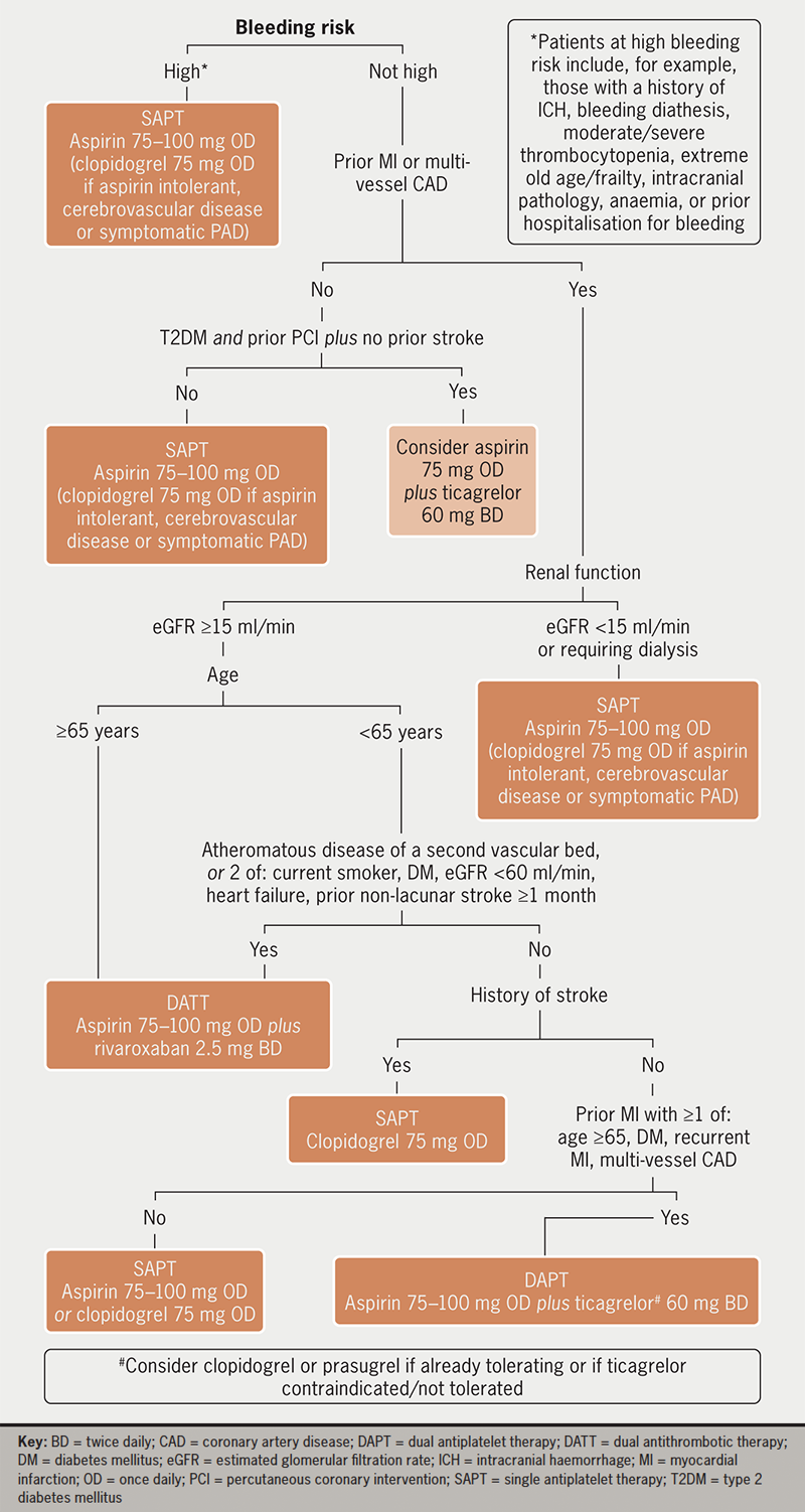
Shaji - Figure 1. Potential algorithm for long-term antithrombotic regimen selection to optimise stroke prevention for patients with chronic coronary syndromes (CCS) without a history of atrial fibrillation or flutter
Figure 1. Potential algorithm for long-term antithrombotic regimen selection to optimise stroke prevention for patients with chronic coronary syndromes (CCS) without a history of atrial fibrillation or flutter

Control of blood pressure

Hypertension is a particularly important risk factor for stroke given it can contribute to both ischaemic and haemorrhagic events. It has been shown to increase risk of stroke in patients with or without ASCVD, particularly those below the age of around 70 years, with a clear relationship between level of hypertension and increase in risk. Current ESC guidelines recommend a target of <140/90 mmHg in all patients, falling to <130/80 mmHg in most patients if initial treatment is well tolerated. A systolic (SBP) target of 120–129 mmHg should be routinely aimed for in those <65 years old, and diastolic blood pressure (DBP) should be kept <80 mmHg in all hypertensive patients.14 Home or ambulatory monitoring of blood pressure can aid accurate assessment of ongoing risk and avoid overtreatment. In general, choice of antihypertensive appears to be less important than the target achieved, though renin–angiotensin–system inhibitors are generally of more benefit if there is prior MI, diabetes mellitus, chronic kidney disease (CKD) or heart failure with reduced ejection fraction, and initial regimens avoiding a beta blocker may have better overall impact on clinical outcomes, possibly related to avoiding an increased risk of diabetes.

Lipid management

Atherogenesis is driven by infiltration of low-density lipoprotein-cholesterol (LDL-C) into the arterial wall, meaning LDL-C is a crucial target for prevention of stroke. Lower levels of LDL-C have been consistently associated with a reduction in both MACE and ischaemic stroke risk, but with an increase in haemorrhagic stroke risk via an, as yet, unclear mechanism.15 In patients with ASCVD, ESC guidelines recommend use of lipid-lowering therapy to achieve a LDL-C <1.4 mmol/L. In those without established CV disease, the use of risk scores, such as SCORE2 or QRISK3, can be useful in determining when to offer therapy.14 Statins remain the mainstay of cholesterol-lowering therapy and have proven dose-dependent efficacy in improving net CV outcomes.16 Where LDL-C targets cannot be met with statins, or in cases of intolerance, additional classes of lipid-lowering drug can be useful. Oral options include the selective cholesterol absorption inhibitor ezetimibe and bempedoic acid, the active metabolite of which inhibits adenosine triphosphate-citrate lyase. Parenteral drugs, such as the proprotein convertase subtilisin kexin type (PCSK9) inhibitors alirocumab and evolocumab, and inclisiran, a long-lasting small-interfering ribonucleic acid against the PCSK9 gene, offer particularly impressive reductions in LDL-C.17 A recent development is the discovery of lipoprotein (a) levels as an independent risk factor for CV events, including stroke, and studies are underway to determine if pharmacological modulation of lipoprotein (a) improves clinical outcomes.

Inflammation

Atherogenesis and thrombosis are processes closely linked to inflammation, which is, therefore, another potential target for prevention of stroke. A meta-analysis of 160,309 patients without ASCVD showed a significant association between C-reactive protein (CRP) levels and risk of ischaemic stroke.18 In the CANTOS (Canakinumab Antiinflammatory Thrombosis Outcome Study) trial, patients with prior MI and high CRP had lower rates of MACE when receiving canakinumab, a monoclonal antibody targeting interleukin-1β, compared with placebo, but with an excess of fatal infections.19 The ability of colchicine to reduce risk of MACE, including stroke, has now been tested in several trials. For example, in the COLCOT (Colchicine Cardiovascular Outcomes Trial) study of patients in the maintenance phase of MI treatment, colchicine impressively and significantly reduced the risk of stroke (a secondary end point) by around 75%, and guidelines acknowledge its consideration to reduce CV risk, though there are still uncertainties given reduction in all-cause mortality has not yet been demonstrated.14 Conversely, methotrexate therapy had no impact on risk of MACE in the CIRT (Cardiovascular Inflammation Reduction Trial) study.2

Optimising glycaemic control in diabetes

Diabetes mellitus is an independent risk factor for cardiovascular diseases including ischaemic and haemorrhagic stroke. In diabetes, each 1% decrease in glycated haemoglobin (HbA1c) towards target is estimated to reduce the relative risk of stroke by 12%.20 Conversely, hypoglycaemia is also associated with increased CV risk, though a specific link to stroke has not been well explored. ESC guidelines recommend a target HbA1c of <7.0% (53 mmol/mol) for reduction in risk of cardiovascular events.14 It is possible that some antidiabetic drug classes play more of a role in stroke prevention than others. For example, in a pooled analysis, glucagon-like peptide-1 (GLP-1) receptor agonists were associated with a significant relative risk reduction of stroke by 13%, whereas sodium-glucose cotransporter-2 (SGLT2) inhibitors were associated with a significant reduction in MACE, but no significant reduction in stroke risk when assessed individually.2

Conclusion

Stroke is a significant cause of mortality and morbidity with a range of risk factors contributing to its pathogenesis. Risk factor assessment and modification form the central basis of stroke prevention. Drugs, alongside lifestyle measures and procedural interventions, can play a key role (figure 2). Stroke risk and prevention should be considered in the context of any concurrent cardiovascular disease or risk factors for adverse effects from therapy, such as bleeding. Ongoing research into stroke prevention strategies will further aid the ability to reduce stroke risk.
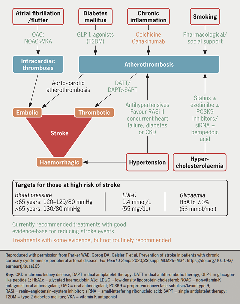
Shaji - Figure 2. Modifiable risk factors for stroke in patients with CCS or peripheral arterial disease (PAD) and evidence-based therapies to address these
Figure 2. Modifiable risk factors for stroke in patients with CCS or peripheral arterial disease (PAD) and evidence-based therapies to address these

Key messages

  • Stroke is responsible for a large burden of mortality and morbidity. Pharmacological strategies to prevent stroke are, therefore, of great importance
  • In patients with a history of atrial fibrillation or flutter, oral anticoagulation is recommended in all but the lowest-risk individuals. Antithrombotic therapy can also reduce stroke in patients with established atherosclerotic cardiovascular disease but without atrial tachyarrhythmia
  • Good control of blood pressure, lipids, glycaemia and chronic inflammation with appropriate therapies also reduces stroke risk
  • When considering whether to initiate drug treatment to prevent stroke, the decision should be made in conjunction with the patient, based on comprehensive assessment of their risk factors

Conflicts of interest

NS: none declared. RFS reports institutional research grants from AstraZeneca, Cytosorbents and GlyCardial Diagnostics; and consultancy fees from AlfaSigma, Alnylam, Amgen, AstraZeneca, Bayer, Bristol Myers Squibb/Pfizer, Chiesi, CSL Behring, Cytosorbents, Daiichi Sankyo, GlyCardial Diagnostics, Hengrui, Idorsia, Novartis, PhaseBio, Sanofi Aventis and Thromboserin. WAEP reports institutional research grants and consultancy fees from AstraZeneca.

Funding

None.

References

1. British Heart Foundation. Heart and circulatory disease statistics 2022. London: BHF, 2022. Available from: https://www.bhf.org.uk/what-we-do/our-research/heart-statistics/heart-statistics-publications/cardiovascular-disease-statistics-2022

2. Parker WAE, Gorog DA, Geisler T et al. Prevention of stroke in patients with chronic coronary syndromes or peripheral arterial disease. Eur Heart J Suppl 2020;22(suppl M):M26–M34. https://doi.org/10.1093/eurheartj/suaa165

3. Hart RG, Pearce LA, Aguilar MI. Meta-analysis: antithrombotic therapy to prevent stroke in patients who have nonvalvular atrial fibrillation. Ann Intern Med 2007;146:857–67. https://doi.org/10.7326/0003-4819-146-12-200706190-00007

4. Hindricks G, Potpara T, Dagres N et al. 2020 ESC guidelines for the diagnosis and management of atrial fibrillation developed in collaboration with the European Association for Cardio-Thoracic Surgery (EACTS): the Task Force for the diagnosis and management of atrial fibrillation of the European Society of Cardiology (ESC) developed with the special contribution of the European Heart Rhythm Association (EHRA) of the ESC. Eur Heart J 2021;42:373–498. https://doi.org/10.1093/eurheartj/ehaa612

5. Antithrombotic Trialists’ (ATT) Collaboration, Baigent C, Blackwell L et al. Aspirin in the primary and secondary prevention of vascular disease: collaborative meta-analysis of individual participant data from randomised trials. Lancet 2009;373:1849–60. https://doi.org/10.1016/S0140-6736(09)60503-1

6. Parker WAE, Storey RF. Antithrombotic therapy for patients with chronic coronary syndromes. Heart 2021;107:925. https://doi.org/10.1136/heartjnl-2020-316914

7. CAPRIE Steering Committee. A randomised, blinded, trial of clopidogrel versus aspirin in patients at risk of ischaemic events (CAPRIE). Lancet 1996;348:1329–39. https://doi.org/10.1016/S0140-6736(96)09457-3

8. Hiatt WR, Fowkes FGR, Heizer G et al.; EUCLID Trial Steering Committee and Investigators. Ticagrelor versus clopidogrel in symptomatic peripheral artery disease. N Engl J Med 2017;376:32–40. https://doi.org/10.1056/NEJMoa1611688

9. Bhatt DL, Fox KAA, Hacke W et al. Clopidogrel and aspirin versus aspirin alone for the prevention of atherothrombotic events. N Engl J Med 2006;354:1706–17. https://doi.org/10.1056/NEJMoa060989

10. Bonaca MP, Bhatt DL, Cohen M et al. Long-term use of ticagrelor in patients with prior myocardial infarction. N Engl J Med 2015;372:1791–800. https://doi.org/10.1056/NEJMoa1500857

11. Yeh RW, Kereiakes DJ, Steg PG et al. Benefits and risks of extended duration dual antiplatelet therapy after PCI in patients with and without acute myocardial infarction. J Am Coll Cardiol 2015;65:2211–21. https://doi.org/10.1016/j.jacc.2015.03.003

12. Armstrong PW. Extending the product label for ticagrelor: looking under the FDA hood. Circulation 2021;144:583–5. https://doi.org/10.1161/CIRCULATIONAHA.121.055907

13. Eikelboom JW, Connolly SJ, Bosch J et al. Rivaroxaban with or without aspirin in stable cardiovascular disease. N Engl J Med 2017;377:1319–30. https://doi.org/10.1056/NEJMoa1709118

14. Visseren FLJ, Mach F, Smulders YM et al. 2021 ESC guidelines on cardiovascular disease prevention in clinical practice: developed by the Task Force for cardiovascular disease prevention in clinical practice with representatives of the European Society of Cardiology and 12 medical societies with the special contribution of the European Association of Preventive Cardiology (EAPC). Eur Heart J 2021;42:3227–337. https://doi.org/10.1093/eurheartj/ehab484

15. Sun L, Clarke R, Bennett D et al. Causal associations of blood lipids with risk of ischemic stroke and intracerebral hemorrhage in Chinese adults. Nat Med 2019;25:569–74. https://doi.org/10.1038/s41591-019-0366-x

16. Baigent C, Blackwell L, Emberson J et al. Efficacy and safety of more intensive lowering of LDL cholesterol: a meta-analysis of data from 170,000 participants in 26 randomised trials. Lancet 2010;376:1670–81. https://doi.org/10.1016/S0140-6736(10)61350-5

17. Pierno S, Musumeci O. Pharmacotherapy of the lipid-lowering drugs: update on efficacy and risk. Int J Mol Sci 2023;24:966. https://doi.org/10.3390/ijms24020996

18. Kaptoge S, Di Angelantonio E, Lowe G et al. C-reactive protein concentration and risk of coronary heart disease, stroke, and mortality: an individual participant meta-analysis. Lancet 2010;375:132–40. https://doi.org/10.1016/S0140-6736(09)61717-7

19. Ridker PM, Everett BM, Thuren T et al. Antiinflammatory therapy with canakinumab for atherosclerotic disease. N Engl J Med 2017;377:1119–31. https://doi.org/10.1056/NEJMoa1707914

20. Stratton IM, Adler AI, Neil HA et al. Association of glycaemia with macrovascular and microvascular complications of type 2 diabetes (UKPDS 35): prospective observational study. BMJ 2000;321:405–12. https://doi.org/10.1136/bmj.321.7258.405
CMR is vital in the management of cardiology inpatients: a tertiary centre experience

Br J Cardiol 2023;30:150doi:10.5837/bjc.2023.041 Leave a comment
Click any image to enlarge
Authors:
First published online 29th November 2023

To review the utility of cardiovascular magnetic resonance (CMR) in the management of hospital inpatients, we performed a retrospective review of all inpatient CMR scans performed over a six-month period at a tertiary referral cardiology centre. Patient demographics, indication for CMR imaging, results of the CMR scans and whether the results changed patient management were recorded. Change in management included medication changes, subsequent invasive procedures, or avoidance of such, and hospital discharge.

Overall, 169 patients were included in the study cohort, 66% were male, mean age was 57.1 years. The most common indication for inpatient CMR was to investigate for cardiomyopathy (53% of patients). The most prevalent diagnosis post-CMR in our cohort was ischaemic heart disease, including ischaemic cardiomyopathy and coronary artery disease. There was a complete change in diagnosis or additional diagnosis found in 29% of patients following CMR. Overall, inpatient CMR led to a change in management in 77% of patients; the most common being changes to medication regimen. CMR was well tolerated in 99% of patients and image quality was diagnostic in 93% of cine scans performed.

In conclusion, CMR is vital for the management of cardiology inpatients, having an impact that is at least as significant as in the management of outpatients.

Introduction

Cardiovascular magnetic resonance (CMR) is an advanced cardiac imaging modality indicated in the assessment of most common cardiology conditions.1 It is a technique that has become increasingly accessible and utilised in clinical practice. CMR is the gold standard in assessing cardiac chamber volume and function with infinite imaging planes, and is not restricted by body habitus, in contrast to transthoracic echocardiography.2 It also allows the non-invasive assessment of the myocardium through tissue characterisation with delayed enhancement following gadolinium-based contrast agents, and more contemporary techniques, such as parametric tissue mapping.3 Myocardial perfusion can also be assessed with CMR and has been shown to be superior to other non-invasive techniques, such as single-photon emission computed tomography (SPECT), with the additional advantage of using non-ionising radiation.4 Given these advantages, CMR is an extremely versatile imaging modality and has an important role in the clinical investigation of cardiomyopathies, ischaemic heart disease and myocarditis.5

The European CMR (EuroCMR) registry reviewed the indications, safety and impact of routine CMR on patient management on a multi-centre and multi-national scale in over 27,000 patients. The study demonstrated that routine CMR is highly impactful on patient management, particularly in the risk stratification of suspected coronary artery disease (CAD) or ischaemia, viability assessment, and in the investigation of cardiomyopathies.6 CMR has been predominantly used in the assessment of stable outpatients, but its increasing availability and clinical utility has resulted in increased inpatient referrals. The EuroCMR registry did not specifically review the utility of CMR for inpatients. Given the unique characteristics of CMR, we hypothesised its clinical use in the acute setting would be highly impactful on inpatient care.

In this single UK tertiary centre study, we sought to evaluate inpatient CMR and whether it similarly impacts patient management as was shown in the EuroCMR registry. Furthermore, as inpatients are often of higher acuity and clinically unstable compared with outpatients, we also aimed to evaluate image quality to see whether high-quality studies can be obtained in this cohort.

Materials and method

This was a clinical audit (AUDI002142) performed at St. George’s Hospital University Hospitals NHS Foundation Trust, London, UK, and, as such, external ethics were not required. We identified all inpatients who had a CMR scan between June and December 2021 at our tertiary referral centre. Scans were performed on a 3 Tesla (T) Phillips Achieva or a 1.5T Siemens Avanto scanner. Data were collected and analysed retrospectively from the electronic notes and imaging reports using a standardised proforma. We included patient demographics, known cardiac comorbidities, including hypertension, ischaemic heart disease, heart failure and atrial fibrillation, and the indication for scan as per the request details. We recorded suspected diagnosis prior to CMR and the subsequent diagnoses made following the scan. We reviewed whether the diagnosis changed following CMR and any other diagnoses made in the event of multiple findings. Change in management was defined as a change in pharmacological therapy (initiation or cessation of treatment), further procedure(s) performed as a consequence of the CMR results, and, conversely, whether the CMR prevented further invasive tests (such as coronary angiography). The data collection was performed using the same terminology as described in the EuroCMR registry to allow comparison. Consultant cardiologists with more than five years’ experience in CMR reported all scans.

Image quality

The quality of the steady-state free-precession (SSFP) cines and delayed enhancement imaging were assessed by a Level 3 EACVI accredited operator with more than five years’ experience in CMR (KDK). Image quality of the cines were rated as good, adequate, or poor. Good was deemed high-quality short-axis and long-axis images with no significant artefact. Adequate, limited artefact but still able to measure cardiac volumes with a high degree of confidence. Poor was deemed insufficient quality to measure cardiac volumes to obtain a reliable ejection fraction. The delayed enhancement images were also rated good, adequate or poor. Similar to the cines, good included studies with long- and short-axis images with no significant artefact and adequate included studies with, limited artefact but still able to make a clinical diagnosis with a high degree of confidence. Poor images were non-diagnostic.

Table 1. Baseline patient demographics

Number %
Gender
Male 112 66%
Female 57 34%
Age, years
<21 9 5%
21–40 28 17%
41–60 51 30%
61–80 68 41%
>80 13 8%
Cardiac history
Hypertension 57 34%
Ischaemic heart disease 30 18%
Heart failure 23 14%
Atrial fibrillation 18 11%
Valvular heart disease 10 6%
Nil known 69 41%

Results

In total, 169 patients were included in this study (baseline patient demographics are included in table 1). The majority of patients were male (66% patients). Mean age was 57.1 ± 19.0 years. The most common pre-existing cardiac conditions were hypertension (34%) and known ischaemic heart disease (18%). The median time from electronic request to scan completion was two days (range 0–11 days) and the median time from scan to hospital discharge was four days (range 0–97 days).

The most common reasons for admission to hospital were chest pain (32%), dyspnoea (14%) and syncope (13%). There were 13% of patients admitted post-cardiac arrest. The primary indication for inpatient CMR was to investigate for cardiomyopathy (53%), including the assessment of left ventricular systolic dysfunction (LVSD) and inherited cardiac conditions, such as hypertrophic cardiomyopathy (HCM), dilated cardiomyopathy (DCM), amyloid and arrhythmogenic cardiomyopathy (AVC) (table 2). Overall, 17% of scans performed were to assess myocardial viability and the indication in 12% of scans was to further evaluate suspected CAD.

Table 2. Primary indication for cardiovascular magnetic resonance (CMR) scan

Indication N %
Cardiomyopathy 89 53%
Viability 29 17%
Suspected CAD 21 12%
Assessment post-cardiac arrest 15 9%
Myocarditis 11 7%
Other 4 2%
Key: CAD = coronary artery disease

The most common diagnosis following CMR imaging was CAD, including myocardial infarction and ischaemic cardiomyopathy (ICM). Non-ischaemic cardiomyopathy (NICM) including, for example, DCM, hypertensive heart disease and tachycardia-mediated cardiomyopathy was diagnosed in 23% patients (table 3). HCM and infiltrative cardiomyopathies were reported in 12% of scans included in this cohort. Myocarditis was detected in 11% of scans. Of note, 12% of scans performed were normal.

Table 3. Diagnosis following inpatient CMR

Diagnosis post-CMR N %
Cardiomyopathy 21 12%
NICM 39 23%
ICM/CAD 58 34%
Myocarditis 18 11%
Thrombus 5 3%
Pericardial disease 5 3%
Note: Cardiomyopathy in this study refers to hypertrophic cardiomyopathy and infiltrative conditions, such as amyloid and cardiac sarcoid
Key: NICM = non-ischaemic cardiomyopathy – including dilated cardiomyopathy, hypertensive cardiomyopathy and tachycardia-induced cardiomyopathies; ICM = ischaemic cardiomyopathy; CAD = coronary artery disease

Impact of CMR on patient management

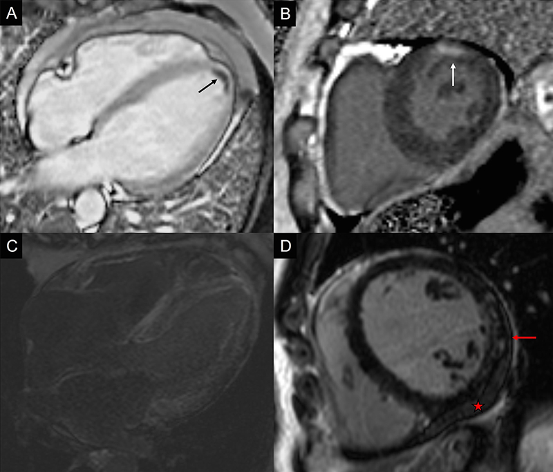
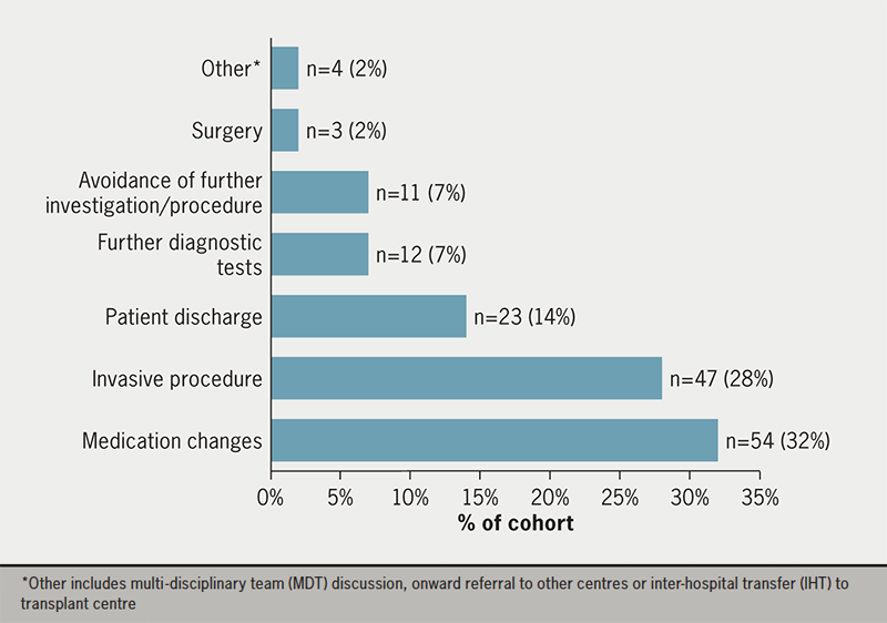
Inpatient CMR led to a new or additional diagnosis in 29% patients (figure 1). In four patients there was a new finding of left ventricular (LV) thrombus. Overall, CMR performed on inpatients impacted clinical management in 77% (130/169) of patients. Most commonly this was a change in pharmacological therapy, which occurred in a total of 54 (32%) patients. A proportion of patients had an invasive procedure, such as cardiac device implantation or coronary angiography following the result of the CMR scan (28% patients). Inpatient CMR led to the avoidance of undertaking further procedures in 11 (7%) patients – predominantly avoiding left heart catheterisation (LHC) in cases with no myocardial viability demonstrated on CMR. Further investigations as a result of the CMR findings were requested in 12 (7%) patients. This included non-invasive tests, such as computed tomography (CT) imaging, genetic testing or electrophysiology tests. The outcome of CMR facilitated patient discharge from hospital in 23 (14%) patients (figure 2).

Image quality and safety profile of CMR

The majority (88%) of scans were performed at 3T with only 12% scans performed at 1.5T. Image quality was diagnostic (good or adequate) in 93% of cine scans and in 87% of scans with late gadolinium enhancement (LGE). Overall, image quality of the cines was good in 67%, adequate in 26% and poor in 5% of patients. For the LGE images, 59% of scans were good, 28% adequate and 6% poor (table 4). In terms of patient acceptability and safety profile of inpatient CMR, 99% scans were well tolerated: two patients did not tolerate the scan, this was due to anxiety during the scan in one case and claustrophobia in the second case, and one patient was unable to proceed due to raised body mass index. There was one case of contrast extravasation.

Hampal - Figure 1. Examples of cases where cardiovascular magnetic resonance (CMR) changed the patient management. A. A 36-year-old man presenting with anterior myocardial infarction. A small left ventricular (LV) thrombus is demonstrated (black arrow). B. A 56-year-old woman presenting with chest pain and unobstructed coronary arteries on coronary angiogram. A small infarct is demonstrated (white arrow), suggestive of an embolic event or small-branch vessel infarction. C. A 72-year-old man with a background of ischaemic heart disease. CMR showed abnormal gadolinium kinetics and extensive enhancement suggestive of cardiac amyloid. D. A 49-year-old man presenting with breathlessness and raised troponin. Coronary arteries were unobstructed. There is patchy late gadolinium enhancement of the lateral wall (red arrow) and a small pericardial effusion (star) suggestive of a myopericarditis
Figure 1. Examples of cases where cardiovascular magnetic resonance (CMR) changed the patient management. A. A 36-year-old man presenting with anterior myocardial infarction. A small left ventricular (LV) thrombus is demonstrated (black arrow). B. A 56-year-old woman presenting with chest pain and unobstructed coronary arteries on coronary angiogram. A small infarct is demonstrated (white arrow), suggestive of an embolic event or small-branch vessel infarction. C. A 72-year-old man with a background of ischaemic heart disease. CMR showed abnormal gadolinium kinetics and extensive enhancement suggestive of cardiac amyloid. D. A 49-year-old man presenting with breathlessness and raised troponin. Coronary arteries were unobstructed. There is patchy late gadolinium enhancement of the lateral wall (red arrow) and a small pericardial effusion (star) suggestive of a myopericarditis
Hampal - Figure 2. Impact of CMR on inpatient management
Figure 2. Impact of CMR on inpatient management

Table 4. CMR scan image quality

Image quality Cine
% (n)
LGE
% (n)
Good 67% (114) 59% (100)
Adequate 26% (44) 28% (48)
Poor 5% (8) 6% (10)
No images 2% (3) 7% (11)
Key: LGE = late gadolinium enhancement

Discussion

In this study we have shown that CMR is crucial for inpatient management in tertiary cardiac centres with 77% of studies resulting in a change in management. To the best of our knowledge, this study is the first to specifically look at inpatient scans and the subsequent impact on patient care.

There were several key findings relating to the indication for CMR, CMR outcomes and consequent impact on patient management worth further discussion. The most common indication in our cohort was to investigate for cardiomyopathy, whereas in the EuroCMR registry, CMR was most commonly used to assess ischaemia. Several reasons for this variance are likely. First, inpatients with troponin-positive events are likely to undergo invasive coronary angiography in our centre, in accordance with the major international society guidelines.7 Urgent diagnostic angiography is readily accessible, and so stress-perfusion imaging is more commonly used in the outpatient setting in patients with stable, chronic coronary disease.8,9 There is increasing evidence that CMR prior to coronary angiography is useful in the inpatient setting as it may help to target intervention, assess viability and prevent repeated angiography.10,11 No stress CMR was performed on an inpatient basis in our cohort. However, at our centre there is routine use of invasive functional assessment with fractional flow reserve (FFR) to assess bystander coronary artery disease, which may then be treated simultaneously.12 However, there is potentially a role for inpatient stress perfusion CMR to assess bystander disease at the same time as assessing myocardial viability.

The most common diagnosis following CMR imaging was ischaemic cardiomyopathy and coronary heart disease in our patient cohort. This is comparable with the EuroCMR registry, which found that CMR was particularly important in suspected CAD and ischaemia. CMR had a high utility in this subgroup of patients with the assessment of viability of myocardium, aetiology of LVSD and in the planning of potential revascularisation and/or device strategies.

Similar to the EuroCMR registry, which evaluated the use of routine CMR, we found that there was a strong impact of CMR on patient management overall, leading to a change in 77% of patients (61.8% in the registry). The proportion of patients who had a change in, or new, diagnosis was also numerically higher in our study; 29% compared with 8.7%. This included four cases of LV thrombus, which had not been previously identified on transthoracic echocardiogram. This highlights the utility of inpatient CMR, even above outpatient CMR, with a greater potential diagnostic yield given the selected cohort of inpatients admitted under cardiology.

In our study, the median time to scan from electronic request was only two days in acute admissions, which may facilitate earlier diagnosis impacting patient management in a more significant way compared with the use of routine CMR. In other centres, or in the non-acute setting, CMR may occur later in the diagnostic pathway, once the diagnosis is provisionally confirmed, reducing the potential impact and utility as a diagnostic method. Early scanning is crucial in the diagnosis of certain conditions, such as myocarditis and Takotsubo syndrome, where the inflammation may resolve or reduce over time, and the appearances on scan are dynamic and time-critical.13 The time from scan to discharge was a key aspect in our evaluation; the median time from scan to discharge was four days, and inpatient CMR had a direct impact on patient discharge in a significant proportion of patients who were scanned (23/169 patients). This highlights the value of CMR as a diagnostic tool among inpatients, and its use may reduce overall length of stay in selected patients and certain presentations.

This study has several implications on clinical practice: CMR findings led to a change in medication regimen in a large number of patients and prompted further diagnostic investigations, such as CT chest scans, positron-emission tomography (PET), exercise tolerance or cardiac genetic testing. In some instances, CMR prevented further investigation, for example, in the cases where patients were scanned early it prevented the need for invasive coronary assessment in 8/169 cases. As such, there is a growing role in risk stratifying patients with suspected ischaemic heart disease or new LVSD to undergo CMR prior to diagnostic angiography. This minimises the procedural risk associated with cardiac catheterisation, may reduce the burden of tests required and, potentially, expedites discharge. One published study found there was a reduced rate of invasive coronary assessment in patients with non-ST-elevation myocardial infarction (NSTEMI) who underwent CMR first, compared with routine clinical care, with overall similar clinical outcomes.11 The specific EuroCMR registry protocol demonstrated that in 1,706 patients who had CMR for suspected CAD,14 the CMR was normal in 866/1,706 patients – which is a significant proportion of patients who could have avoided invasive angiography. Ongoing work is required to assess which patients may benefit the most from CMR performed early in the clinical course.

In terms of the safety and quality of CMR in our study, CMR was very well tolerated in our patient cohort and image quality was diagnostic in 93% cines. This is numerically lower in comparison to the EuroCMR registry (98%) but not unexpected. Inpatients are more likely to be clinically unstable and decompensated compared with outpatients, and so it can be challenging to obtain good quality, diagnostic images due to difficulties associated with breath holding and arrhythmias. It is reassuring that even in this group of patients we were able to make diagnoses in most studies.

This study, to the best of our knowledge, is the first to review the utility of inpatient CMR at a tertiary referral centre. A previous study evaluated routine use of CMR in a district general hospital (DGH) setting.14 Interestingly, the results were similar to our study and the most common indications for CMR were investigation of suspected CAD and in the diagnosis of cardiomyopathy. Of note, more viability studies were performed in our cohort (17%) compared with the patients referred for routine CMR at the DGH in this paper (9.1%), which may reflect accessibility to CMR in the acute instance in those with suspected CAD or ischaemia. There was more stress CMR performed in the outpatient cohort, as would be expected with the assessment of suspected stable CAD. Image quality was deemed diagnostic in 99.9% of cases in this published study, which was higher than we found in our inpatient cohort, but as would be expected with stable patients. Our results are also corroborated by the EuroCMR registry in which CMR was found to change patient management in 62% of cases (similar to 77% in our study).15 They found that importantly, CMR was normal in 11%. This is also reflected in our study where 12% of patient scans were normal studies.

Limitations identified within this study include the relatively short study period of six months and the relatively small sample size (169 patients). The retrospective observational nature of this study increases the risk of information bias. There was a degree of selection bias given only inpatient scans among patients who were admitted under cardiology or requested following discussion with the cardiology team were included. In addition, there was a lack of comparator group in this study, for example inpatients that did not have CMR and had echocardiogram ± angiography alone, a comparator group would have allowed us to further validate our findings. Prospective studies that randomise patients to inpatient and outpatient CMR and compare factors, such as certainty of diagnosis, length of hospital stay, impact on patient management and number of invasive procedures, would be of particular interest to corroborate our findings.

Conclusion

CMR is an essential tool in the early assessment of cardiology inpatients. In this single tertiary centre study, we have shown that inpatient CMR is safe, diagnostic and has a direct impact on patient management, corroborating the EuroCMR registry findings. More widespread use of CMR in the acute setting could prevent unnecessary invasive investigations and treatments, and improve patient care.

Key messages

  • This is the first comprehensive review of the use of cardiovascular magnetic resonance (CMR) in the acute setting at a tertiary cardiology referral centre
  • The main indications for inpatient CMR include investigation of new left ventricular systolic dysfunction, assessment of possible cardiomyopathy and in suspected acute coronary syndrome
  • Inpatient CMR changes patient management and leads to medication changes, further investigation or, interestingly, results in the avoidance of further invasive procedures, such as coronary angiography

Conflicts of interest

None declared.

Funding

None.

Study approval

This was a clinical audit (AUDI002142) performed at St. George’s Hospital University Hospitals NHS Foundation Trust, London, UK, and, as such, external ethics were not required.

References

1. Salerno M, Sharif B, Arheden H et al. Recent advances in cardiovascular magnetic resonance: techniques and applications. Circ Cardiovasc Imaging 2017;10:e003951. https://doi.org/10.1161/CIRCIMAGING.116.003951

2. Assomull RG, Pennell DJ, Prasad SK. Cardiovascular magnetic resonance in the evaluation of heart failure. Heart 2007;93:985–92. https://doi.org/10.1136/hrt.2003.025304

3. Messroghli DR, Moon JC, Ferreira VM et al. Clinical recommendations for cardiovascular magnetic resonance mapping of T1, T2, T2 and extracellular volume: a consensus statement by the Society for Cardiovascular Magnetic Resonance (SCMR) endorsed by the European Association for Cardiovascular Imaging (EACVI). J Cardiovasc Magn Reson 2017;19:75. https://doi.org/10.1186/s12968-017-0389-8

4. Greenwood JP, Maredia N, Radjenovic A et al. Clinical evaluation of magnetic resonance imaging in coronary heart disease: the CE-MARC study. Trials 2009;10:62. https://doi.org/10.1186/1745-6215-10-62

5. Pennell DJ, Sechtem UP, Higgins CB et al. Clinical indications for cardiovascular magnetic resonance (CMR): Consensus Panel report. Eur Heart J 2004;25:1940–65. https://doi.org/10.1081/JCMR-200038581

6. Bruder O, Schneider S, Nothnagel D et al. EuroCMR (European Cardiovascular Magnetic Resonance) registry: results of the German pilot phase. J Am Coll Cardiol 2009;54:1457–66. https://doi.org/10.1186/1532-429X-11-S1-O13

7. Collet JP, Thiele H, Barbato E et al. 2020 ESC guidelines for the management of acute coronary syndromes in patients presenting without persistent ST-segment elevation. Eur Heart J 2021;42:1289–367. https://doi.org/10.1093/eurheartj/ehaa909

8. National Institute for Health and Care Excellence. Recent-onset chest pain of suspected cardiac origin: assessment and diagnosis. CG95. London: NICE, 2016. Available from: https://www.nice.org.uk/guidance/cg95

9. Gulati M, Levy PD, Mukherjee D et al. 2021 AHA/ACC/ASE/CHEST/SAEM/SCCT/SCMR guideline for the evaluation and diagnosis of chest pain: a report of the American College of Cardiology/American Heart Association joint committee on clinical practice guidelines. J Am Coll Cardiol 2021;78:e187–e285. https://doi.org/10.1161/CIR.0000000000001029

10. Kim RJ, Wu E, Rafael A et al. The use of contrast-enhanced magnetic resonance imaging to identify reversible myocardial dysfunction. N Engl J Med 2000;343:1445–53. https://doi.org/10.1056/NEJM200011163432003

11. Smulders MW, Kietselaer BLJH, Wildberger JE et al. Initial imaging-guided strategy versus routine care in patients with non-ST-segment elevation myocardial infarction. J Am Coll Cardiol 2019;74:2466–77. https://doi.org/10.1016/j.jacc.2019.09.027

12. Patricio L, Tonino PAL, De Bruyne B et al. Fractional flow reserve versus angiography for guiding percutaneous coronary intervention. N Engl J Med 2009;360:213–24. https://doi.org/10.1056/NEJMoa0807611

13. Kato K, Daimon M, Sano M et al. Dynamic trend of myocardial edema in Takotsubo syndrome: a serial cardiac magnetic resonance study. J Clin Med 2022;11:987. https://doi.org/10.3390/jcm11040987

14. Abraham G, Shamsi A, Daryani Y. Comprehensive study of routine clinical use of cardiac MRI in a district general hospital setting. Br J Cardiol 2018;25:111–14. https://doi.org/10.5837/bjc.2018.020

15. Bruder O, Wagner A, Lombardi M et al. European cardiovascular magnetic resonance (EuroCMR) registry – multi-national results from 57 centers in 15 countries. J Cardiovasc Magn Reson 2013;15:1–9. https://doi.org/10.1186/1532-429X-15-9

Myocardial revascularisation in complex patients: does it happen as prescribed by the heart team?

Br J Cardiol 2023;30:144–7doi:10.5837/bjc.2023.042 Leave a comment
Click any image to enlarge
Authors:
First published online 29th November 2023

Guidelines recommend decision-making using the heart team (HT) in complex patients considered for myocardial revascularisation, but there are little data on how this approach works in practice. We data-mined our electronic HT database and selected patients in whom the clinical question referred to revascularisation, and documented HT recommendations and their implementation.

We identified 154 patients (117 male), mean age 68.9 ± 11.4 years, discussed between February 2019 and December 2020. The clinical questions were coronary artery bypass graft (CABG) versus percutaneous coronary intervention (PCI) (141 cases, 91%), and medical treatment versus revascularisation by PCI (eight cases, 6%) or by CABG (five cases, 3%).

HT recommended CABG in 55 cases (35%), PCI in 43 (28%), medical treatment in 15 (10%), and equipoise in seven (5%) and further investigations in 34 (22%): non-invasive imaging for ischaemia in 11 (32%), invasive coronary physiology studies in eight (24%), further clinical assessment in seven (20%), structural imaging for five (15%), invasive coronary angiography in two (6%), and an electrophysiology opinion in one case (3%).

Decisions were implemented in 135 cases (89%). The average time between the HT and the implementation of its decision was 80.5 ± 129.3 days. There were 17 deaths: 10 cardiac, six non-cardiac and one of unknown cause. Patients who survived were younger (68.6 ± 11.3 years) than those who died (73.8 ± 10.0 years, p = 0.03).

In conclusion, almost 90% of the decisions of the HT on myocardial revascularisation are implemented, while ischaemia testing is the main investigation required for decision-making. Recent data on the futility of such an approach have not yet permeated clinical practice.

Introduction

Myocardial revascularisation in complex patients: does it happen as prescribed by the heart team?

Contemporary cardiology guidelines often recommend that decision making should take place by discussions within a multi-disciplinary heart team (HT), particularly in complex patients.1,2 However, very little data exist about the effectiveness of such an approach, about the extent to which HT decisions are implemented, or about the outcome of patients being managed according to decisions made by the HT.

We set out to analyse the decisions of our revascularisation HT in a medium-sized regional tertiary centre, in order to optimise our processes, and also to explore the extent to which the HT decisions were actually carried out.

Method

Setting

Morriston Cardiac Centre is a regional tertiary centre that serves a population of approximately one million, in West Wales, UK. Pre-pandemic, it performed approximately 2,500 coronary angioplasties and 750 open-heart procedures per year. There are 10 interventional cardiologists and five cardiac surgeons. A weekly HT meeting has been in operation since the inception of the centre in 1998, but its deliberations are formalised and documented in a dedicated database (Solus, HD Clinical, Stanstead, UK) only since February 2019.

Inclusion criteria

We included all patients discussed at the HT between 1 February 2019 and 31 December 2020 in whom the clinical question was related to myocardial revascularisation. Patients could be referred to the HT both by cardiologists in Morriston and in secondary cardiac centres in the region served by Morriston.

Aim of the study

We documented demographic characteristics of the patients, as well as the decisions of the HT, and we data-mined clinical and electronic records to find out whether, and to what extent, the decisions of the HT were implemented. In cases where the HT could not formulate a treatment recommendation because it required additional information, we documented the type of information required. Where the HT decisions were not carried out, we identified the reasons for that. We documented the time elapsed between the date of the HT decision and its implementation (date of revascularisation procedure); for patients treated without revascularisation we used the date when the HT decision was first mentioned in correspondence about the patient. We identified the survival status of the patients and calculated the length of survival between the date of the HT and our censoring date, 21 December 2021.

Composition of the HT

The quorum required for the HT was two interventional cardiologists and two cardiac surgeons.

Results

Patient characteristics

We identified 152 patients (116 male, 76%), mean age 68.8 ± 8.8 years, discussed between February 2019 and December 2020; 109 referred by Morriston consultants and 43 by external consultants. There were 33 consultants (two cardiac surgeons, 31 cardiologists) submitting an average of 5 ± 3.8, median 4, cases per consultant. Seven cardiology consultants (22%) accounted for 50% of the cases submitted.

Clinical questions

The clinical questions were whether revascularisation should be performed by coronary artery bypass graft (CABG) or percutaneous coronary intervention (PCI) (140 cases, 91%), and whether medical treatment or revascularisation by PCI (eight cases, 6%) or by CABG (four cases, 3%) should be performed.

Additional testing

In 33 patients (22%) the HT required further information in order to formulate a treatment plan. The additional tests included functional testing for inducible myocardial ischaemia (12 cases), followed by intracoronary physiological testing (eight cases), clinical assessment in six cases, structural cardiac imaging in five cases, invasive coronary angiography in two cases, liver function tests and electrophysiology opinion in one case each; one patient had more than one test.

Decisions of the HT

When the HT could formulate a recommendation for treatment without requiring any further information (in 119 cases, 78% of the total number discussed), this was for CABG in 54 cases (45%), for PCI in 42 cases (35%), and for medical treatment in 16 (13%). In seven patients the HT recorded equipoise between CABG and PCI, with the final decision to be taken by the patients involved, after discussion with the cardiac surgeon and cardiologist. In 16 of the patients in whom a treatment plan could be formulated, the HT also requested further tests: functional testing for inducible myocardial ischaemia in six patients, invasive coronary physiological testing in five, structural imaging and invasive coronary angiography in two for each, and biochemical liver function tests in one patient.

Implementation of HT decisions

The decisions of the HT were implemented in 135 cases (89%), while in 17 cases they were not. Of these, eight patients had PCI rather than CABG (patient preference or clinical instability prompting unplanned PCI), three patients switched from medical treatment to PCI because of uncontrolled symptoms, two had CABG instead of PCI, and in each of the following categories there was one patient: CABG instead of medical treatment, medical treatment instead of PCI, and single-vessel PCI instead of two-vessel PCI (failure of chronic total occlusion [CTO] PCI). The average waiting time between the HT and the implementation of the HT decision was 80.5 ± 129.3 days, median 14 days.

Patient survival

There were 17 deaths: 10 with a cardiac cause, six non-cardiac and one of unknown cause. In eight cases the HT decision had been referral for CABG, in three for medical treatment, in another three for further investigations, in two for PCI, and in one there was equipoise and the patient had opted for CABG. Three of the deceased patients had been deemed too frail for any intervention. Patients who survived were younger (68.6 ± 11.3 years) than those who died (73.8 ± 10.0 years, p=0.03). Of the 17 patients who died, eight had interventions, as follows: two had PCI and died before discharge; four had cardiac surgery and died in the intensive therapy unit (ITU); one had only instantaneous wave-free ratio (iFR) and PCI was deferred; one died of complications of limb ischaemia, unrelated to the procedure, 11 months after PCI.

Discussion

In a regional tertiary centre serving a population of one million, the HT could reach a decision on myocardial revascularisation strategy, based on the information provided, in 78% of the cases, with CABG being more often recommended than PCI or conservative treatment. Non-invasive testing for ischaemia and intracoronary physiology testing were the most frequently requested tests before a decision could be reached. In patients in whom the decision of the HT was not implemented, this was due either to conversion from CABG to PCI or from conservative management to revascularisation.

The HT approach is based on the premise that it “… serves the purpose of a balanced multi-disciplinary decision process” (European Society of Cardiology [ESC] guidelines). Both US and European guidelines make a class 1 recommendation for the use of a HT in revascularisation decision-making, although this is supported by evidence of the lowest quality (level C). Thus, guidelines based on low-quality evidence postulate we should use an approach that is very different from the traditional process of referral from an individual cardiologist to an individual cardiac surgeon. The use of a HT discussion has been proposed to prevent inappropriate use or underuse of resources when managing coronary artery disease. Furthermore, with ever advancing subspecialisation within the field of cardiology, such as imaging, complex intervention, electrophysiology, etc., the HT provides a platform for these specialists to provide their valuable input in managing complex cases requiring revascularisation.

This fundamental shift in practice was ushered by the SYNTAX (Synergy between Percutaneous Coronary Intervention with Taxus and Cardiac Surgery) trial,3 which compared CABG versus PCI with drug-eluting stents in patients with complex coronary artery disease. At each participating centre a HT, consisting of an interventional cardiologist and a cardiac surgeon, evaluated patients’ eligibility for the study. However, extrapolating research methods to clinical practice is not necessarily safe or justifiable; priorities are different in these two settings, as is resource allocation and utilisation. Ideally, a rigorous study of outcomes and resource utilisation by the HT model should be undertaken before this approach is enshrined in guidelines.

Evidence is sparse both on the effectiveness of this approach and on its outcomes. A US study has shown that HT decisions correlate well with appropriateness of use criteria for myocardial revascularisation, but these are cumbersome and not really used outside the US.4 More comparable to our experience, a UK study by Pavlidis et al.5 looked at 399 patients discussed in 51 consecutive multi-disciplinary team (MDT) meetings held in a tertiary cardiac centre in London. Medical management was recommended in 30%, CABG in 6%, and PCI in 17%, while further information was needed in 25%; decisions were carried out in 93% of the cases. Of note is the male preponderance, which is noted both in our study as well as by Pavlidis et al.5 and in the SYNTAX trial,3 highlighting what is now a well-known phenomenon of gender disparity in diagnosis, management and treatment of coronary artery disease in women when compared with men. A subgroup of 40 patients was re-discussed using the same clinical data, at least six months after the initial discussion, and the same recommendation as initially provided was formulated in 80% of cases. These results are comparable to ours, with the notable difference that we had a lower rate of recommending medical treatment, perhaps as a reflection of recent progress in stent technology, although these differences may also stem from differences in the patient population; type of presentation (acute vs. stable) or comorbidities of the patient.

It is disturbing that in the study by Pavlidis et al. one in five patients received a different decision when resubmitted to the HT using exactly the same information as the first time, and points to the major weaknesses of any form of collective deliberation, which is susceptible to being highjacked by the participant with ‘the loudest voice’ and is vulnerable to groupthink, a ‘mode of thinking in which individual members of small cohesive groups tend to accept a viewpoint or conclusion that represents a perceived group consensus, whether or not the group members believe it to be valid, correct, or optimal’ (https://www.britannica.com/science/groupthink). It is important to note here that the composition of HT did change to various extents in the study by Pavlidis et al. and, therefore, conclusions regarding inter-observer and intra-observer reliability cannot be inferred. Furthermore, in their study, 25% of the resubmitted cases were recommended to undergo further diagnostic methods, which probably should not be construed as a definitive treatment recommendation.

One in five patients were referred for ischaemia testing (invasive and non-invasive) before a decision could be made. Increasingly strong evidence suggests that, at least in stable patients, revascularisation guided by ischaemia testing (once left main stem disease has been ruled out) does not improve symptoms and outcomes.6 Data are now accruing indicating the same lack of significance of ischaemia in patients with multi-vessel disease at the time of primary PCI.7 It is interesting to see that clinical practice is not yet significantly impacted by these paradigm-shifting recent trials.8

A limitation in our study has been the incomplete documentation of data on review of medical records with baseline characteristics, such as smoking status, diabetes, serum lipid levels, etc., not accurately recorded. This is now changing as the HT discussion is moving towards a redesigned comprehensive online form including all of the patients’ comorbidities to submit in order for the HT to discuss these cases. Another limitation is the limited number of cases discussed with the HT (157) compared with the overall number of patients undergoing revascularisation at our institution annually (over 2,500) at the time of data collection. The situation is now evolving with an increased proportion of cases being referred to the HT currently. It will be interesting to see how the additional data accrued from this affects our recommendations regarding revascularisation in the future.

Conclusion

Systematic research on the processes and outcomes of the HT is needed, as not much data are available to guide its deployment outside the research setting, and its effectiveness and safety have not been formally tested in large numbers of patients. Our own HT processes appear comparable with those in another UK tertiary centre, and can be made more efficient by ensuring the completeness of the clinical datasets used for decision-making. Ischaemia testing remains prevalent in spite of increasing evidence suggesting its lack of usefulness.

Key messages

  • In a regional tertiary centre in West Wales, the use of a heart team multi-disciplinary approach to complex patients requiring revascularisation is comparable with a similar tertiary cardiac centre in the UK
  • There remains a considerable dearth in data validating the heart team approach to revascularisation, and more studies are needed to validate this approach
  • Ischaemia testing was the most prevalent test requested by the multi-disciplinary team for revascularisation, despite increasing evidence against its utility in non-left main lesions. This practice may require further review

Conflicts of interest

None declared.

Funding

None.

Study approval

Requirement for ethical approval and consent was waived by the local hospital audit department as the study is a retrospective analysis of anonymised patient data.

References

1. Lawton JS, Tamis-Holland JE, Bangalore S et al. 2021 ACC/AHA/SCAI guideline for coronary artery revascularization: a report of the American College of Cardiology/American Heart Association Joint Committee on Clinical Practice Guidelines. Circulation 2021;144:e18–e144. https://doi.org/10.1161/CIR.0000000000001038

2. Neumann F-J, Uva M, Ahlsson A et al. 2018 ESC/EACTS guidelines on myocardial revascularization. Eur Heart J 2019;40:87–165. https://doi.org/10.1093/eurheartj/ehy394

3. Serruys PW, Morrice M-C, Kappetein P et al.; for the SYNTAX Investigators. Percutaneous coronary intervention versus coronary-artery bypass grafting for severe coronary artery disease. N Engl J Med 2009;360:961–72. https://doi.org/10.1056/NEJMoa0804626

4. Sanchez CE, Dota A, Badhwar V et al. Revascularization heart team recommendations as an adjunct to appropriate use criteria for coronary revascularization in patients with complex coronary artery disease. Catheter Cardiovasc Interv 2016;88:E103–E112. https://doi.org/10.1002/ccd.26276

5. Pavlidis AN, Perera D, Karamasis GV et al. Implementation and consistency of heart team decision-making in complex coronary revascularisation. Int J Cardiol 2016;206:37–41. https://doi.org/10.1016/j.ijcard.2016.01.041

6. Maron DJ, Hochman JS, Reynolds HR et al. Initial invasive or conservative strategy for stable coronary disease. N Engl J Med 2020;382:1395–407. https://doi.org/10.1056/NEJMoa1915922

7. Puymirat E, Cayla G, Simon T et al. Multivessel PCI guided by FFR or angiography for myocardial infarction. N Engl J Med 2021;385:297–308. https://doi.org/10.1056/NEJMoa2104650

8. Metkus TS, Beckie TM, Cohen MG et al. Heart care team/multidisciplinary team live: the heart team for coronary revascularization decisions: 2 illustrative cases. J Am Coll Cardiol Case Rep 2022;4:115–20. https://doi.org/10.1016/j.jaccas.2021.12.005

Improvement in LV end-diastolic pressure after primary PCI and its impact on patients’ recovery

Br J Cardiol 2023;30:148doi:10.5837/bjc.2023.043 Leave a comment
Click any image to enlarge
Authors:
First published online 29th November 2023

In this study, we evaluated the change in left ventricular end-diastolic pressure (LVEDP) after primary percutaneous coronary intervention (PCI) and its impact on in-hospital outcomes and 30-day and three-month quality of life (SAQ-7), ejection fraction (EF), and major adverse cardiovascular events (MACE). LVEDP ≥19 mmHg was taken as elevated LVEDP. In a sample of 318 patients, 18.9% (n=60) were females and mean age was 55.7 ± 10.52 years. Post-procedure elevated LVEDP was observed in 20.8% (n=66) with a mean reduction of 1.65 ± 4.35 mmHg. LVEDP declined in 39% (n=124) and increased in 10.7% (n=34). In-hospital mortality rate (9.1% vs. 2.4%, p=0.011), 30-day MACE (9.1% vs. 4.0%), and three-month MACE (21.2% vs. 5.6%) were found to be significantly higher among patients with elevated LVEDP, respectively. Elevated LVEDP was found to be associated with a reduced SAQ-7 score (89.84 ± 8.09 vs. 92.29 ± 3.03, p<0.001) and reduced (25–40%) EF (55.6% vs. 22.6%) at three-month follow-up. LVEDP declined acutely in a significant number of patients after primary PCI. Post-procedure elevated LVEDP was found to be associated with poor quality of life and an increased risk of immediate and short-term MACE.

Introduction

Improvement in LV end-diastolic pressure after primary PCI and its impact on patients’ recovery

ST-elevation myocardial infarction (STEMI) is an acute ischaemic event associated with an increased risk of clinical complications, poor recovery, and adverse cardiovascular events.1 Owing to the recent development and advancements in management, outcomes of STEMI patients have improved significantly.2 Primary percutaneous coronary intervention (PCI) remains the recommended treatment option by both European and American clinical practice guidelines.3,4

In addition to improvements in the management strategy, risk stratification of patients with STEMI improved extensively with the introduction of various risk-stratification modalities.5 Over the years, various biomarkers and clinical characteristics have been evaluated for their prognostic role, including gender, age, patient-related comorbid conditions, arrhythmias, location and size of the infarct, haemodynamic complications (cardiogenic shock), and ischaemic mitral regurgitation.1 The prognostic role of left ventricular systolic dysfunction (i.e. left ventricular ejection fraction – LVEF) is well established for patients with STEMI.6 However, the acute event of STEMI causes multiple functional and structural changes at the microcirculation level, which leads to elevated left ventricular end-diastolic pressure (LVEDP). Therefore, left ventricular diastolic dysfunction (i.e. LVEDP) recently gained attention as a prognostic marker for patients with STEMI.7–10

LVEDP is an integrative measure of total left ventricular function, and LVEDP change can be utilised as a significant prognostic indicator to guide medical therapy, and assess risk for post-STEMI adverse events. LVEDP is often measured during primary PCI, and a few studies have been conducted assessing the relationship between LVEDP and myocardial salvage.11 Not many of these studies have been conducted in South Asia, particularly in Pakistan. Therefore, we aimed to assess the improvement in post-procedure LVEDP after PCI and its impact on short-term (three-month) outcomes in terms of quality of life (Seattle Angina Questionnaire [SAQ]-7), ejection fraction (EF), and major adverse cardiovascular events (MACE).

Materials and method

Study setting

This descriptive observational cohort study was conducted at a tertiary care cardiac centre in Karachi, Pakistan. The study was approved by the ethical review board of the hospital (ERC/121/2021) and consent for participation in the study and follow-up was taken from all the patients. Study duration was between January 2022 and June 2022.

Study population

In this study, we included consecutive patients of a first acute event of STEMI, either gender, age ≥18 years, and undergoing primary PCI. Patients with a prior history of coronary artery disease (CAD) or heart failure (HF), patients in cardiogenic shock at the time of presentation to the emergency department, and patients with any structural abnormality that can potentially lead to an increase in LVEDP were excluded from this study.

According to the study conducted by Cap et al.,12 the mean pre-primary PCI LVEDP was 22.1 ± 4.8 mmHg, and the post-primary PCI LVEDP was 19.4 ± 4.8 mmHg; using these statistics to test the hypothesis of significant post-procedure improvement in LVEDP at 5% level significance and 80% power of the test, the minimum required sample size for the study was calculated to be 27 patients. However, considering the expected three-month MACE rate of 15%, at a 95% confidence level (95%CI) and 4% margin of error, the sample size was calculated to be 307 patients. Hence, a total of 318 patients were recruited for this study.

Management and assessment of outcomes

As per the institutional protocol, all the primary PCI procedures were performed free of cost by the on-call team of interventional cardiologists. The pre- and post-LVEDP (mmHg) was measured for all patients as a measurement of pressure within the left ventricle following the completion of diastolic filling, just prior to systole. The primary end point of the study was the assessment of an improvement in post-procedure LVEDP. The secondary end point was the assessment of quality of life, improvement in EF (%), and MACE three months after the procedure. All the patients were followed, by telephone or physically, during their hospital stay, 30 days after discharge, and three months after discharge, and MACE, along with EF on transthoracic echocardiography (TTE) and quality of life using the SAQ-7 were assessed.

Measurements and definitions

STEMI was diagnosed based on positive electrocardiogram (ECG) findings at the time of presentation in the emergency department and a history of typical chest pain for at least 20 minutes. The positive ECG changes included ST-elevation in at least two contiguous leads >2 mm in men or >1 mm in women in leads V2 to V3, and/or >1 mm in other contiguous chest leads or limb leads. In-hospital outcomes included emergency coronary artery bypass grafting (CABG), major bleeding (requiring blood transfusion), stent thrombosis, cerebrovascular accident (CVA)/stroke, and death. The 30-day and three-month cumulative MACE included in-hospital all-cause death, post-discharge all-cause death, re-infarction/myocardial infarction, repeat revascularisation, and hospitalisation due to heart failure.

Data analysis procedure

The total SAQ-7 score was computed as an average of seven elements re-scaled to 0 to 100 from a scale of 1–6 for five elements and 1–5 for two elements. The SAQ score was categorised as fair (<50), good (50–75), and excellent (75–100). The EF was categorised as 25–40%, 41–50%, and more than 50%. Although multiple cut-off values for LVEDP have been used in the literature, a cut-off value of LVEDP >18 mmHg (i.e. ≥19 mmHg) has proven to be a significant predictor of MACE following primary PCI;10 therefore, we categorised patients into two groups with LVEDP ≥19 mmHg as criterion for elevated LVEDP. Clinical characteristics and outcomes were compared between the two groups with the help of appropriate independent sample t-test/Mann-Whitney U-test or Chi-square test/Fisher’s exact test at a 5% level of significance using IBM SPSS version 21.

Results

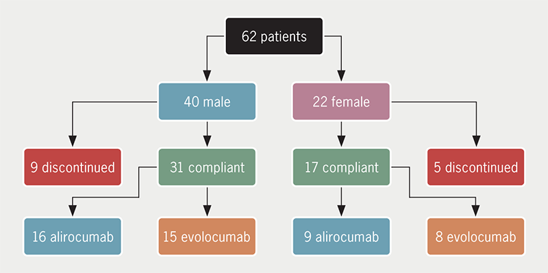
A total of 318 patients were included in this study; the proportion of female patients was 18.9% (n=60), and the mean age of the study sample was 55.7 ± 10.52 years. Elevated post-primary PCI LVEDP was observed in 20.8% (n=66) of the patients. LVEDP declined (by at least 1 mmHg) in 39% (n=124), increased (by at least 1 mmHg) in 10.7% (n=34), and remained the same in the remaining 50.3% (n=160) of the patients. Post-procedure elevated LVEDP was found to be associated with male gender (90.9% vs. 78.6%, p=0.023) and Killip class II (30.3% vs. 8.7%) or III (12.1% vs. 6.3%, p<0.001) (table 1).

Table 1. The comparison of clinical and demographic characteristics for patients with and without elevated left ventricular end-diastolic pressure (LVEDP) after primary percutaneous coronary intervention (PCI)

Total Post-procedure LVEDP p value
<19 mmHg ≥19 mmHg
Total, N (%) 318 252 (79.2%) 66 (20.8%)
Male, n (%) 258 (81.1%) 198 (78.6%) 60 (90.9%) 0.023
Female, n (%) 60 (18.9%) 54 (21.4%) 6 (9.1%)
Height, mean ± SD cm 166.4 ± 8.42 165.49 ± 8.79 169.88 ± 5.61 <0.001
Weight, mean ± SD kg 69.8 ± 10.26 69.4 ± 10.19 71.3 ± 10.45 0.181
Age, mean ± SD years 55.7 ± 10.52 56.43 ± 10.77 52.94 ± 9.03 0.016
Systolic blood pressure,
median (IQR) mmHg
130 (110–145) 130 (110–150) 120 (110–140) 0.018
Diastolic blood pressure,
median (IQR) mmHg
80 (70–90) 80 (70–90) 80 (70–82) 0.245
Heart rate, median (IQR) bpm 86 (76–96) 84 (75–92) 88 (78–100) 0.069
Chest pain to ER time,
median (IQR) minutes
240 (120–360) 233 (120–360) 240 (180–480) 0.094
ER to cath lab time,
median (IQR) minutes
100 (65–130) 100 (65–130) 100 (60–130) 0.724
Killip class, n (%)
I 252 (79.2%) 214 (84.9%) 38 (57.6%) <0.001
II 42 (13.2%) 22 (8.7%) 20 (30.3%)
III 24 (7.5%) 16 (6.3%) 8 (12.1%)
Comorbid conditions, n (%)
Hypertension 174 (54.7%) 132 (52.4%) 42 (63.6%) 0.102
Diabetes mellitus 120 (37.7%) 92 (36.5%) 28 (42.4%) 0.377
Smoking 94 (29.6%) 74 (29.4%) 20 (30.3%) 0.882
Family history of IHD 36 (11.3%) 28 (11.1%) 8 (12.1%) 0.818
Chronic kidney disease 6 (1.9%) 6 (2.4%) 0 (0%) 0.206
Type of myocardial infarction, n (%)
Anterior 166 (52.2%) 122 (48.4%) 44 (66.7%) 0.068
Inferior 108 (34%) 90 (35.7%) 18 (27.3%)
Inferior, posterior 18 (5.7%) 16 (6.3%) 2 (3%)
Lateral 16 (5%) 16 (6.3%) 0 (0%)
Posterior 8 (2.5%) 6 (2.4%) 2 (3%)
Posterior, lateral 2 (0.6%) 2 (0.8%) 0 (0%)
Key: CAD = coronary artery disease; ER = emergency room; IHD = ischaemic heart disease; IQR = interquartile range; LVEDP = left ventricular end-diastolic pressure; SD = standard deviation

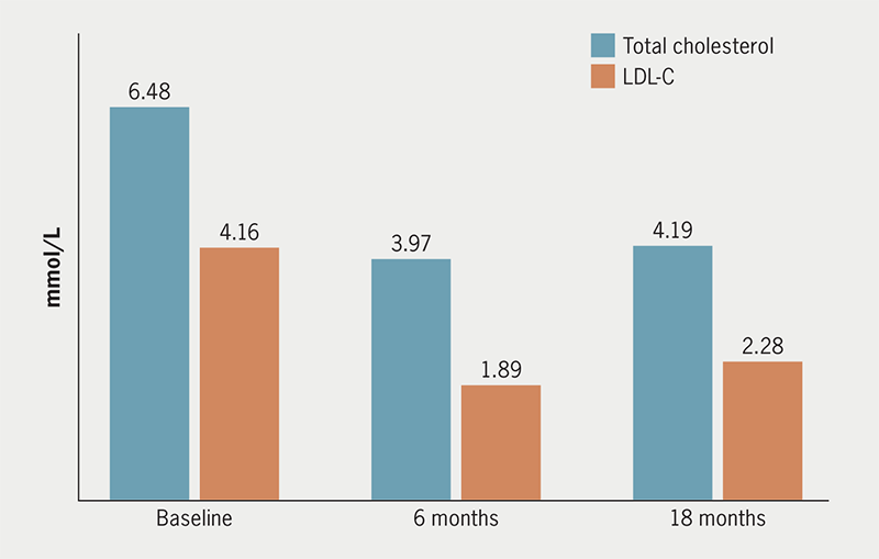
A mean reduction of 1.65 ± 4.35 mmHg in LVEDP was observed after the procedure compared with the pre-procedure LVEDP. Post-procedure elevated LVEDP was found to be associated with pre-procedure TIMI (Thrombolysis in Myocardial Infarction) flow grade 0 (72.7% vs. 46.0%), myocardial blush grade (MBG) 0 (78.8% vs. 46.0%), culprit left anterior descending artery (66.7% vs. 49.2%), elevated pre-procedure LVEDP (22.45 ± 3.67 vs. 15.88 ± 5.71 mmHg), and reduced LVEF (33.64 ± 10.83% vs. 41.98 ± 7.76%) (table 2).

Table 2. The comparison of angiographic findings for patients with and without elevated LVEDP after primary PCI

Total Post-procedure LVEDP p value
<19 mmHg ≥19 mmHg
Total, N (%) 318 252 (79.2%) 66 (20.8%)
Pre-procedure LVEF, mean ± SD % 40.25 ± 9.12 41.98 ± 7.76 33.64 ± 10.83 <0.001
Pre-procedure LVEDP, mean ± SD mmHg 17.25 ± 5.97 15.88 ± 5.71 22.45 ± 3.67 <0.001
Fluoroscopy times, mean ± SD minutes 13.85 ± 6.8 13.07 ± 6.04 16.8 ± 8.59 <0.001
Contrast volume, median (IQR) ml 100 (90–120) 100 (90–120) 100 (100–120) 0.208
Export catheter used, n (%) 16 (5%) 10 (4%) 6 (9.1%) 0.090
Pre-procedure TIMI flow grade, n (%)
0 164 (51.6%) 116 (46%) 48 (72.7%) 0.001
I 26 (8.2%) 22 (8.7%) 4 (6.1%)
II 72 (22.6%) 62 (24.6%) 10 (15.2%)
III 56 (17.6%) 52 (20.6%) 4 (6.1%)
Pre-procedure MBG grade, n (%)
0 168 (52.8%) 116 (46%) 52 (78.8%) <0.001
I 28 (8.8%) 22 (8.7%) 6 (9.1%)
II 82 (25.8%) 76 (30.2%) 6 (9.1%)
III 40 (12.6%) 38 (15.1%) 2 (3%)
Number of involved vessels, n (%)
Single-vessel disease 110 (34.6%) 92 (36.5%) 18 (27.3%) 0.369
Two-vessel disease 106 (33.3%) 82 (32.5%) 24 (36.4%)
Three-vessel disease 102 (32.1%) 78 (31%) 24 (36.4%)
Culprit vessel, n (%)
Left anterior descending artery 168 (52.8%) 124 (49.2%) 44 (66.7%) 0.030
Right coronary artery 98 (30.8%) 86 (34.1%) 12 (18.2%)
Left circumflex artery 46 (14.5%) 36 (14.3%) 10 (15.2%)
Diagonal 6 (1.9%) 6 (2.4%) 0 (0%)
Post-procedure TIMI flow grade, n (%)
0 4 (1.3%) 4 (1.6%) 0 (0%) 0.038
I 4 (1.3%) 4 (1.6%) 0 (0%)
II 12 (3.8%) 6 (2.4%) 6 (9.1%)
III 298 (93.7%) 238 (94.4%) 60 (90.9%)
Post-procedure MBG grade, n (%)
0 0 (0%) 0 (0%) 0 (0%) 0.259
I 4 (1.3%) 4 (1.6%) 0 (0%)
II 26 (8.2%) 18 (7.1%) 8 (12.1%)
III 288 (90.6%) 230 (91.3%) 58 (87.9%)
Post-procedure LVEDP, mean ± SD mmHg 15.59 ± 5.15 13.56 ± 3.16 23.33 ± 3.73 <0.001
Change in LVEDP, mean ± SD mmHg –1.65 ± 4.35 –2.32 ± 4.58 0.88 ± 1.71 <0.001
Key: IQR = interquartile range; LVEDP = left ventricular end-diastolic pressure; LVEF = left ventricular ejection fraction; MBG = myocardial blush grade; SD = standard deviation; TIMI = thrombolysis in myocardial infarction

In-hospital mortality rate (9.1% vs. 2.4%, p=0.011), 30-day MACE (9.1% vs. 4.0%), and three-month MACE (21.2% vs. 5.6%) were found to be significantly higher among patients with elevated LVEDP compared with patients with normal LVEDP level, respectively. Elevated LVEDP was also found to be associated with a reduced LVEF and SAQ-7 score at 30-day and three-month follow-ups (tables 3 and 4).

Table 3. The comparison of post-procedure in-hospital, 30-day, and 3-month outcomes for patients with and without elevated LVEDP after primary PCI

Total Post-procedure LVEDP p value
<19 mmHg ≥19 mmHg
Total, N (%) 318 252 (79.2%) 66 (20.8%)
In-hospital outcomes, n (%)
Successful procedure 312 (98.1%) 250 (99.2%) 62 (93.9%) 0.005
Discharged home 304 (95.6%) 244 (96.8%) 60 (90.9%) 0.037
Emergency CABG 2 (0.6%) 2 (0.8%) 0 (0%) 0.468
Stent thrombosis 0 (0%) 0 (0%) 0 (0%)
Major bleeding 0 (0%) 0 (0%) 0 (0%)
Stroke/CVA 0 (0%) 0 (0%) 0 (0%)
Death 12 (3.8%) 6 (2.4%) 6 (9.1%) 6 (9.1%)
30-day outcome
Available, N (%) 236 (74.2%) 188 (74.6%) 48 (72.7%) 0.756
LVEF %, n (%)
Echo not done 40 (16.9%) 40 (21.3%) 0 (0%) <0.001
25–40% 108 (45.8%) 72 (38.3%) 36 (75%)
41–50% 48 (20.3%) 36 (19.1%) 12 (25%)
>50% 40 (16.9%) 40 (21.3%) 0 (0%)
SAQ-7 score, mean ± SD 91.16 ± 7.3 91.19 ± 7.55 91.07 ± 6.31 0.923
Fair: SAQ-7 score (≤50), n (%) 0 (0%) 0 (0%) 0 (0%) 0.562
Good: SAQ-7 score (51–75), n (%) 14 (5.9%) 12 (6.4%) 2 (4.2%)
Excellent: SAQ-7 score (76–100), n (%) 222 (94.1%) 176 (93.6%) 46 (95.8%)
3-month outcome
Available, N (%) 204 (64.2%) 168 (66.7%) 36 (54.5%) 0.068
LVEF %, n (%)
Echo not done 88 (43.1%) 80 (47.6%) 8 (22.2%) <0.001
25–40% 58 (28.4%) 38 (22.6%) 20 (55.6%)
41–50% 18 (8.8%) 12 (7.1%) 6 (16.7%)
>50% 40 (19.6%) 38 (22.6%) 2 (5.6%)
SAQ-7 score, mean ± SD 91.86 ± 4.44 92.29 ± 3.03 89.84 ± 8.09 <0.001
Fair: SAQ-7 score (≤50), n (%) 0 (0%) 0 (0%) 0 (0%) 0.086
Good: SAQ-7 score (51–75), n (%) 4 (2%) 2 (1.2%) 2 (5.6%)
Excellent: SAQ-7 score (76–100), n (%) 200 (98%) 166 (98.8%) 34 (94.4%)
Key: CABG = coronary artery bypass grafting; CVA = cerebral vascular accident; LVEDP = left ventricular end-diastolic pressure; LVEF = left ventricular ejection fraction; SAQ = Seattle Angina Questionnaire; SD = standard deviation

Table 4. Major adverse cardiovascular events (MACE) at 30 days and 3 months for patients with and without elevated LVEDP after primary PCI

Total Post-procedure LVEDP p value
<19 mmHg ≥19 mmHg
Total, N (%) 318 252 (79.2%) 66 (20.8%)
30-day outcome
Lost to follow-up 70 (22%) 58 (23%) 12 (18.2%) 0.194
No 232 (73%) 184 (73%) 48 (72.7%)
Yes 16 (5%) 10 (4%) 6 (9.1%)
In-hospital mortality 12 (75%) 6 (60%) 6 (100%)
Post-discharge mortality 0 (0%) 0 (0%) 0 (0%)
Hospitalisation due to HF 2 (12.5%) 2 (20%) 0 (0%)
Repeat revascularisation 2 (12.5%) 2 (20%) 0 (0%)
Re-infarction/MI 0 (0%) 0 (0%) 0 (0%)
3-month MACE, n (%)
Lost to follow-up 98 (30.8%) 78 (31%) 20 (30.3%) <0.001
No 192 (60.4%) 160 (63.5%) 32 (48.5%)
Yes 28 (8.8%) 14 (5.6%) 14 (21.2%)
In-hospital mortality 12 (42.9%) 6 (42.9%) 6 (42.9%)
Post-discharge mortality 4 (14.3%) 0 (0%) 4 (28.6%)
Hospitalisation due to HF 6 (21.4%) 4 (28.6%) 2 (14.3%)
Repeat revascularisation 4 (14.3%) 4 (28.6%) 0 (0%)
Re-infarction/MI 2 (7.1%) 0 (0%) 2 (14.3%)
Key: HF = heart failure; LVEDP = left ventricular end-diastolic pressure; MACE = major adverse cardiovascular event; MI = myocardial infarction

Discussion

The LVEDP measures total left ventricular function; it has been observed to be a significant marker of prognosis after acute myocardial infarction. In this study, we evaluated the change in LVEDP after primary PCI in patients with STEMI, and the association of post-procedure elevated LVEDP with quality of life and short-term major adverse outcomes. In summary, an improvement (decline of at least 1 mmHg) in LVEDP was observed in a significant number of patients after primary PCI. However, post-procedure elevated LVEDP manifestation of clinically adverse characteristics was found to be associated with male gender, Killip class II/III at presentation, total occlusion of the culprit artery with pre-procedure TIMI flow grade 0 and MBG grade 0, mainly culprit left anterior descending artery, elevated pre-procedure LVEDP and reduced LVEF. The post-procedure elevated LVEDP was observed to be associated with an increased risk of in-hospital, as well as 30-day and three-month MACE, including all-cause mortality. It has also been associated with a decreased quality of life after three months of primary PCI.

The findings of an increased incidence of MACE during the short-term follow-up after primary PCI of patients with baseline or post-procedure elevated LVEDP are not new to our study. Multiple studies have reported similar observations.7–12 However, poor quality of life among MACE-free patients with post-procedure LVEDP is a point of concern in these patients. Multiple studies have taken both LVEDP (diastolic dysfunction) and LVEF (systolic dysfunction) for the prediction of the short- and long-term fate of patients after primary PCI. A study conducted by Ndrepepa et al.13 reported a ratio of LVEF/LVEDP as an independent and significant predictor of long-term (eight-year) mortality after primary PCI. This ratio has also proved a significant prognostic marker for the prediction of MACE during 43 ± 31 months follow-up after STEMI.8 A LVEDP of >22 mmHg measured during primary PCI is found to be associated with an increased risk of mortality, congestive heart failure, and cardiogenic shock at 90 days after primary PCI.14 Similar to these findings, Planer et al.6 also reported baseline elevated LVEDP as an independent predictor of adverse outcomes on a short- and long-term basis. The association of elevated LVEDP with reduced myocardial salvage and the extent of the ischaemia can be a possible mechanism behind an increased risk of adverse outcomes in patients with STEMI.11 Another index, derived as the ratio of systolic blood pressure to LVEDP, is reported to be an independent predictor of in-hospital mortality at the critical cut-off of ≤4.15 Another combination of criteria of LVEDP >18 mmHg and index of microcirculatory resistance >32 has been found to have added advantage for detecting MACE among patients undergoing primary PCI.10 Two of the recent studies from our population reported the prognostic role of elevated LVEDP. The first by Kumar et al.1 reported LVEDP ≥20 mmHg as an independent predictor of short-term MACE after primary PCI with an adjusted hazard ratio (HR) of 1.81 (95%CI 1.3 to 2.51). The second study by Ammar et al.16 reported LVEDP of ≥20 mmHg as an essential predictor of contrast-induced acute kidney injury after primary PCI, especially in patients with a LVEF ≤40%.

Similar to our finding regarding clinical co-variates of elevated LVEDP, Zhou et al.17 reported that patients with elevated LVEDP had more frequently descending branches as infarct-related arteries, along with the larger left atrial end-systolic and diastolic diameter, higher levels of myocardial necrosis, regional wall motion abnormality, and small ejection fraction, along with the higher incidence of mortality and heart failure. Another study reported a significant relationship between elevated LVEDP and wire-crossing time among patients undergoing primary PCI.18 Very limited data are available regarding the effective treatment options for reducing elevated LVEDP. In a study by Khan et al.,9 the administration of furosemide along with glyceryl trinitrate was a safe and effective strategy for reducing LVEDP in STEMI patients. Similar to our findings of the decline of only 1.65 ± 4.35 mmHg, a study conducted by Khan et al.7 too reported a marginal drop in LVEDP from 18 (interquartile range [IQR] 12 to 22 mmHg) pre-procedure to 15 (IQR 10 to 20 mmHg) post-procedure.

Even though this is the first study of its kind in the Pakistani population, some limitations have to be acknowledged, which included single-centre coverage, the observational nature of the study, the small sample size, and a high rate of loss to follow-up. Large-scale multi-centre studies are warranted to understand the prognostic role of LVEDP, and its association with the quality of life of patients on a long-term basis.

Conclusion

In conclusion, LVEDP declined acutely in a significant number of patients after primary PCI, but the quantum of decline was mostly marginal. Post-procedure elevated LVEDP was found to be associated with poor quality of life and an increased risk of immediate and short-term MACE. Further studies are required to formulate effective strategies for reducing LVEDP levels to minimise its detrimental effects on short- and long-term outcomes after primary PCI.

Key messages

  • Left ventricular end-diastolic pressure (LVEDP) declined acutely in a significant number of patients after primary percutaneous coronary intervention (PCI), but the quantum of decline was mainly marginal
  • Post-procedure elevated LVEDP was found to be associated with poor quality of life and an increased risk of immediate and short-term major adverse cardiovascular events (MACE)
  • Further studies are required to formulate effective strategies for reducing LVEDP levels to minimise its detrimental effects on short- and long-term outcomes after primary PCI

Conflicts of interest

None declared.

Funding

None.

Study approval

This study was approved by the ethical review committee (ERC) of the National Institute of Cardiovascular Diseases (NICVD), Karachi (ERC-121/2021). Verbal informed consent was obtained from all the patients regarding their participation in the study and publication of data, while maintaining confidentiality and anonymity. Due to the observational nature of the study, ERC waived the written consent and verbal consents were approved by the ERC.

Acknowledgement

The authors wish to acknowledge the support of the staff members of the Clinical Research Department of the NICVD, Karachi, Pakistan.

References

1. Kumar R, Shah JA, Solangi BA et al. The burden of short-term major adverse cardiac events and its determinants after emergency percutaneous coronary revascularization: a prospective follow-up study. J Saudi Heart Assoc 2022;34:100–09. https://doi.org/10.37616/2212-5043.1302

2. Osselló X, Huo Y, Pocock S et al. Global geographical variations in ST-segment elevation myocardial infarction management and post-discharge mortality. Int J Cardiol 2017;245:27–34. https://doi.org/10.1016/j.ijcard.2017.07.039

3. Levine GN, Bates ER, Blankenship JC et al. 2015 ACC/AHA/SCAI focused update on primary percutaneous coronary intervention for patients with ST-elevation myocardial infarction: an update of the 2011 ACCF/AHA/SCAI guideline for percutaneous coronary intervention and the 2013 ACCF/AHA guideline for the management of ST-elevation myocardial infarction. J Am Coll Cardiol 2016;67:1235–50. https://doi.org/10.1016/j.jacc.2015.10.005

4. Ibanez B, James S, Agewall S et al. 2017 ESC guidelines for the management of acute myocardial infarction in patients presenting with ST-segment elevation: the Task Force for the management of acute myocardial infarction in patients presenting with ST-segment elevation of the European Society of Cardiology (ESC). Eur Heart J 2018;39:119–77. https://doi.org/10.1093/eurheartj/ehx393

5. Buccheri S, Capranzano P, Condorelli A et al. Risk stratification after ST-segment elevation myocardial infarction. Expert Rev Cardiovasc Ther 2016;14:1349–60. https://doi.org/10.1080/14779072.2017.1256201

6. Planer D, Mehran R, Witzenbichler B et al. Prognostic utility of left ventricular end-diastolic pressure in patients with ST-segment elevation myocardial infarction undergoing primary percutaneous coronary intervention. Am J Cardiol 2011;108:1068–74. https://doi.org/10.1016/j.amjcard.2011.06.007

7. Khan AA, Al-Omary MS, Collins NJ, Attia J, Boyle AJ. Natural history and prognostic implications of left ventricular end-diastolic pressure in reperfused ST-segment elevation myocardial infarction: an analysis of the thrombolysis in myocardial infarction (TIMI) II randomized controlled trial. BMC Cardiovasc Disord 2021;21:243. https://doi.org/10.1186/s12872-021-02046-x

8. Saito D, Nakanishi R, Watanabe I et al. Combined assessment of left ventricular end-diastolic pressure and ejection fraction by left ventriculography predicts long-term outcomes of patients with ST-segment elevation myocardial infarction. Heart Vessels 2018;33:453–61. https://doi.org/10.1007/s00380-017-1080-6

9. Khan AA, Davies AJ, Whitehead NJ et al. Targeting elevated left ventricular end-diastolic pressure following primary percutaneous coronary intervention for ST-segment elevation myocardial infarction – a phase one safety and feasibility study. Eur Heart J Acute Cardiovasc Care 2020;9:758–63. https://doi.org/10.1177/2048872618819657

10. Maznyczka AM, McCartney PJ, Oldroyd KG et al. Risk stratification guided by the index of microcirculatory resistance and left ventricular end-diastolic pressure in acute myocardial infarction. Circ Cardiovasc Interv 2021;14:e009529. https://doi.org/10.1161/CIRCINTERVENTIONS.120.009529

11. Ndrepepa G, Cassese S, Hashorva D et al. Relationship of left ventricular end-diastolic pressure with extent of myocardial ischemia, myocardial salvage and long-term outcome in patients with ST-segment elevation myocardial infarction. Catheter Cardiovasc Interv 2019;93:901–09. https://doi.org/10.1002/ccd.28098

12. Cap M, Erdoğan E, Ali Karagöz et al. Acute change of left ventricular end-diastolic pressure during primary percutaneous coronary intervention and its relationship with early reperfusion parameters. Eastern J Med 2020;25:250–5. https://doi.org/10.5505/ejm.2020.60252

13. Ndrepepa G, Cassese S, Emmer M et al. Relation of ratio of left ventricular ejection fraction to left ventricular end-diastolic pressure to long-term prognosis after ST-segment elevation acute myocardial infarction. Am J Cardiol 2019;123:199–205. https://doi.org/10.1016/j.amjcard.2018.10.007

14. Bagai A, Armstrong PW, Stebbins A et al. Prognostic implications of left ventricular end-diastolic pressure during primary percutaneous coronary intervention for ST-segment elevation myocardial infarction: findings from the Assessment of Pexelizumab in Acute Myocardial Infarction study. Am Heart J 2013;166:913–19. https://doi.org/10.1016/j.ahj.2013.08.006

15. Sola M, Venkatesh K, Caughey M et al. Ratio of systolic blood pressure to left ventricular end‐diastolic pressure at the time of primary percutaneous coronary intervention predicts in‐hospital mortality in patients with ST‐elevation myocardial infarction. Catheter Cardiovasc Interv 2017;90:389–95. https://doi.org/10.1002/ccd.26963

16. Ammar A, Khowaja S, Kumar R et al. Significance of left ventricular end diastolic pressure for risk stratification of contrast-induced acute kidney injury after primary percutaneous coronary intervention. Pak Heart J 2022;55:247–52. https://doi.org/10.47144/phj.v55i3.2135

17. Zhou X, Lei M, Zhou D et al. Clinical factors affecting left ventricular end-diastolic pressure in patients with acute ST-segment elevation myocardial infarction. Ann Palliat Med 2020;9:1834–40. https://doi.org/10.21037/apm.2020.03.22

18. Nugraha IW, Hartopo AB, Taufiq N. Wire crossing time correlate with left ventricular end-diastolic pressure in patients with ST segment elevation myocardial infarction undergoing primary percutaneous coronary intervention. Indo J Cardiol 2020;41:939. https://doi.org/10.30701/ijc.936

Efficacy and tolerability of PCSK9 inhibitors in real-world clinical practice

Br J Cardiol 2023;30:153–6doi:10.5837/bjc.2023.044 Leave a comment
Click any image to enlarge
Authors:
First published online 29th November 2023

Despite widespread use of statins and other lipid-lowering therapies for hypercholesterolaemia, cardiovascular (CV) mortality and morbidity remains high. The proprotein convertase subtilisin/kexin type 9 (PCSK9) inhibitors, alirocumab and evolocumab, have been approved for use in patients with familial hypercholesterolaemia and high CV risk in the UK. We reviewed the records of patients at a large health board in Scotland, who were prescribed these agents, to determine their real-world efficacy and tolerability in routine clinical care.

Introduction

Hypercholesterolaemia, characterised by elevated serum total cholesterol and low-density lipoprotein (LDL), is a crucial factor for atherosclerosis and for the development of cardiovascular diseases (CVD). The hepatic protease proprotein convertase subtilisin/kexin type 9 (PCSK9) targets LDL-receptors for destruction.1,2 Removal of LDL from the blood stream is aided by increased expression of LDL-receptors.3

Statins have been proven to effectively lower LDL-cholesterol (LDL-C) levels and reduce CVD events in many high cardiovascular risk cohorts via 3-hydroxy-3-methylglutaryl coenzyme A (HMG Co-A) reductase inhibition. However, a significant number of patients with hypercholesterolaemia do not achieve their target LDL-C levels despite being on maximally tolerated statin therapy, while others are completely intolerant to statins.4,5

PCSK9 inhibitors (PCSK9i) have emerged as a promising therapeutic agent for treating hypercholesterolaemia and, more importantly, reduction of cardiovascular events. Currently available PCSK9is are either monoclonal antibodies that target and inactivate PCSK9 (alirocumab and evolocumab)6 or small-interfering RNA (siRNA) that inhibits the translation of PCSK9 from mRNA (inclisiran). The common pathway of action is the prevention of PCSK9-mediated LDL-receptor destruction. Statin therapy complements PCSK9i treatment in this regard by increasing LDL-receptor expression.7

Following the results of the landmark FOURIER (Further cardiovascular OUtcomes Research with PCSK9 Inhibition in subjects with Elevated Risk) and ODYSSEY OUTCOMES (Evaluation of Cardiovascular Outcomes After an Acute Coronary Syndrome During Treatment With Alirocumab) trials, where PCSK9i have been shown to be efficacious in reducing serum total cholesterol and LDL-C, these agents have been approved for the treatment of high-risk patients in the National Health Service (NHS).8,9 Outside the confines of a clinical trial, data for the efficacy and tolerability of these agents in real-world routine clinical care is still evolving.

Method

We audited the indications, efficacy, and tolerability of PCSK9i prescribed for 62 patients by the NHS Tayside Cardiovascular Risk (CVR) Clinic between 2017 and 2021. The CVR clinic is a tertiary specialist clinic with a catchment area of approximately 415,000 NHS patients that is responsible for all PCSK9i prescribing in the health board area.

Efficacy was measured by analysing reductions in total cholesterol and calculated LDL-C at six and 18 months following prescription, from baseline (defined as the last cholesterol measurement prior to PCSK9i administration). We did this as LDL-C is a potent surrogate of cardiovascular (CV) events, hence our choice of looking at LDL-C lowering, which is also consistent with the mechanism of action of these drugs.

Statin tolerance status within the cohort was also observed, to determine if statin-intolerant patients reported lower LDL-C levels post-administration of PCSK9i.

Comparative analysis was also made for patients administered with alirocumab and evolocumab to check for a difference in efficacy between the two agents. Similarly, data were analysed to compare LDL-C reduction in patients with familial hypercholesterolaemia (FH) and patients with polyvascular disease, the two currently approved indications for these agents in the UK. At the time of the audit, inclisiran had not been approved for clinical use.

Results

Table 1. Indications for administration of proprotein convertase subtilisin/kexin type 9 inhibitor (PCSK9i)

Indication Number (%)
Heterozygous FH 22 (35)
Polyvascular disease 31 (50)
Deemed as having phenotypic FH 8 (13)
High lipoprotein (a) 1 (2)
Total patients administered PCSK9i 62 (100)
Key: FH = familial hypercholesterolaemia

There were 62 patients, 40 males and 22 females, who were prescribed a PCSK9i (31 alirocumab and 31 evolocumab) over a five-year period, although clinical contact was significantly reduced between March 2020 and December 2020, due to the pandemic. The indication for PCSK9i was heterozygous FH in 22/62 patients and polyvascular disease in 31/62. Eight of the remaining nine patients were treated on the basis of having been deemed as having phenotypic FH (genetics negative) by their treating clinician, while the last was a patient with high lipoprotein (a) who was commenced on PCSK9i therapy by a cardiology multi-disciplinary team, following admission for myocardial infarction (table 1).

A total of 14/62 (9/40 males and 5/22 females) discontinued therapy (figure 1). Four of these discontinued due to non-compliance. In two cases, compliance was not ascertainable due to insufficient data and seven had various reported side effects, including arthralgia, myalgia, rash, rhinosinusitis, sore throat, abdominal pain, diarrhoea, back pain, headaches and palpitations. The majority of the aforementioned side effects were not specified in the clinical trials for these agents, making comparisons inappropriate, although the one patient (1.6%) who discontinued due to rash and the four who discontinued due to myalgia (6.4%), are not dissimilar from the percentage of patients who developed “allergic reaction” (3.1–7.9%) and “muscle-related event” (5%) in the FOURIER and ODYSSEY trials.8,9 In one patient, total cholesterol and LDL-C increased on PCSK9i compared with statin therapy and this was thought to be due to their specific LDL-receptor mutation (heterozygous for LDL-receptor c.313+1G>C). Eleven out of the 14 patients who discontinued PCSK9i therapy were also intolerant of statins, which included all seven who reported side effects on PCSK9i. There were 33/62 classed as statin intolerant, of whom 26/33 tolerated PCSK9i agents, while seven discontinued therapy as stated previously.

Devaiah - Figure 1. Patients prescribed proprotein convertase subtilisin/kexin type 9 inhibitor (PCSK9i) in NHS Tayside Cardiovascular Risk (CVR) clinic
Figure 1. Patients prescribed proprotein convertase subtilisin/kexin type 9 inhibitor (PCSK9i) in NHS Tayside Cardiovascular Risk (CVR) clinic

Absolute values of total cholesterol and LDL-C at baseline and after six and 18 months for the 48 patients taking PCSK9i showed significant reductions in cholesterol levels (figure 2).

Devaiah - Figure 2. Concentration of total cholesterol and low-density lipoprotein-cholesterol (LDL-C), at baseline and after 6 and 18 months of PCSK9i therapy
Figure 2. Concentration of total cholesterol and low-density lipoprotein-cholesterol (LDL-C), at baseline and after 6 and 18 months of PCSK9i therapy

Percentage reductions from baseline cholesterol concentrations at six and 18 months, in this group and specific subgroups of patients, can be seen in table 2. Patient numbers were comparable among subgroups with the exception of female and male distribution (with there being almost twice as many males than females). The lipid-lowering effects seen were similar to each subgroup, with LDL-C concentrations at six and 18 months reduced by 45–55%. After 18 months, both monitoring and adherence deteriorate slightly.

Table 2. Total and low-density lipoprotein-cholesterol (LDL-C) percentage reductions from baseline at 6 and 18 months after PCSK9i therapy in patient subgroup (n denotes number of patients in each subgroup)

Baseline, mmol/L 6-month reduction 18-month reduction
All (n=48)
Total cholesterol 6.48 39% 35%
LDL-C 4.16 55% 45%
Statin intolerant (n=26)
Total cholesterol 6.91 37% 37%
LDL-C 4.69 55% 46%
Alirocumab (n=25)
Total cholesterol 6.31 34% 33%
LDL-C 3.84 46% 41%
Evolocumab (n=23)
Total cholesterol 6.55 43% 37%
LDL-C 4.4 63% 48%
Female (n=17)
Total cholesterol 6.54 30% 31%
LDL-C 4.39 50% 38%
Male (n=31)
Total cholesterol 6.36 43% 38%
LDL-C 3.77 54% 48%
Familial hypercholesterolaemia (n=18)
Total cholesterol 5.83 34% 32%
LDL-C 3.95 54% 44%
Polyvascular disease (n=23)
Total cholesterol 6.9 39% 36%
LDL-C 4.6 55% 45%

A large proportion of patients (>50%) taking PCSK9i who are intolerant of statins and have high CVD risk experience substantial and sustained lipid-lowering effect from these agents. The percentage reductions seen would not be achievable on other conventional therapies.

In comparison, between the two PCSK9is, evolocumab appears to exert a greater lipid-lowering effect than alirocumab. This observation could be due to the different doses of alirocumab available: within this cohort, 14 patients were prescribed 75 mg and 11 patients were prescribed 150 mg once every two weeks. We observed a trend of greater total cholesterol lowering among the subgroup who were prescribed the 150 mg dose after six months of therapy (36% reduction for 150 mg compared with 27% reduction for 75 mg). This was not statistically significant in our data but fits with the observation made by Kastelein et al. when they interrogated data from the ODYSSEY trials and found an additional 14.2% decrease in LDL-C following introduction of the larger dose of alirocumab.10

We observed that males derived greater benefit from PCSK9i therapy than females, and an unpaired t-test showed a significant difference (p=0.0078) between the total cholesterol concentration of men and women after six months of therapy, although this was not observed after 18 months or for LDL-C, due to fewer data points at 18 months. This contrasts with the findings of the major pivotal trials. This could likely be due to the greater variability from the smaller sample size rather than due to any differential pharmacological effect.

Indication for treatment and aetiology of disease had no bearing on the cholesterol-lowering effect of PCSK9i as both FH and PVD patients displayed similar cholesterol reductions after six and 18 months.

Conclusion

Our analysis confirmed the efficacy of PCSK9i in real-world clinical practice with the efficacy outcomes similar to the pivotal trials of these agents. ODYSSEY reported a 54% reduction in LDL-C compared with baseline after six months of alirocumab therapy; for FOURIER and evolocumab, this was 61%.8,9 Our data showed reductions of 46% and 63% in LDL-C compared with baseline, for alirocumab and evolocumab, respectively. A key observation of our study was the strong, sustained, and comparable cholesterol reductions observed between high-risk statin-intolerant patients and other patients with known CVD risks. While males and patients on evolocumab derived greater benefit, dose equivalences and increased sample sizes would be required to confirm this.

A limitation of our audit was the relatively small sample size. However, it does accurately reflect the total uptake of these agents in real-world practice and the impact the pandemic has had as the CVR clinic is the sole prescriber of these agents for the region. Also, non-adherence to PCSK9i for long-term treatment may have contributed to incomplete data in a few excluded patient records.

Despite favourable indications confirming efficacy in real-world practice, future analysis of clinical data will help validate the observations made here and clarify the implications of prolonged use of PCSK9i on reducing cardiovascular morbidity, particularly in less studied populations, such as statin-intolerant patients.

Key messages

  • Familial hypercholesterolaemia and high cardiovascular risk patients administered with proprotein convertase subtilisin/kexin type 9 inhibitors (PCSK9i) showed significant reductions in absolute values of total cholesterol and low-density lipoprotein-cholesterol (LDL-C)
  • Efficacy of the PCSK9i agents is evident in clinically challenging cohorts, such as statin-intolerant patients
  • Future analysis of clinical data from larger datasets will help clarify the implications of prolonged use of PCSK9i on reducing cardiovascular morbidity, particularly in lesser-studied cohorts, such as statin-intolerant patients

Conflicts of interest

None.

Funding

None.

Study approval

Necessary Caldicott approval for this audit was obtained from NHS Tayside.

References

1. Cohen JC, Boerwinkle E, Mosley TH Jr, Hobbs HH. Sequence variations in PCSK9, low LDL, and protection against coronary heart disease. N Engl J Med 2006;354:1264–72. https://doi.org/10.1056/NEJMoa054013

2. Abifadel M, Varret M, Rabès J-P et al. Mutations in PCSK9 cause autosomal dominant hypercholesterolemia. Nat Genet 2003;34:154–6. https://doi.org/10.1038/ng1161

3. Ji E, Lee S. Antibody-based therapeutics for atherosclerosis and cardiovascular diseases. Int J Mol Sci 2021;22:5770. https://doi.org/10.3390/ijms22115770

4. Brugts JJ, Yetgin T, Hoeks SE et al. The benefits of statins in people without established cardiovascular disease but with cardiovascular risk factors: meta-analysis of randomised controlled trials. BMJ 2009;338:b2376. https://doi.org/10.1136/bmj.b2376

5. Iyen B, Akyea RK, Weng S, Kai J, Qureshi N. Statin treatment and LDL-cholesterol treatment goal attainment among individuals with familial hypercholesterolaemia in primary care. Open Heart 2021;8:e001817. https://doi.org/10.1136/openhrt-2021-001817

6. Chaudhary R, Garg J, Shah N, Sumner A. PCSK9 inhibitors: a new era of lipid lowering therapy. World J Cardiol 2017;9:76–91. https://doi.org/10.4330/wjc.v9.i2.76

7. Goldstein JL, Brown MS. The LDL receptor. Arterioscler Thromb Vasc Biol 2009;29:431–8. https://doi.org/10.1161/ATVBAHA.108.179564

8. Sabatine MS, Giugliano RP, Keech AC et al. Evolocumab and clinical outcomes in patients with cardiovascular disease. N Engl J Med 2017;376:1713–22. https://doi.org/10.1056/NEJMoa1615664

9. Schwartz GG, Steg PG, Szarek M et al. Alirocumab and cardiovascular outcomes after acute coronary syndrome. N Engl J Med 2018;379:2097–107. https://doi.org/10.1056/NEJMoa1801174

10. Kastelein JJ, Kereiakes DJ, Cannon CP et al. Effect of alirocumab dose increase on LDL lowering and lipid goal attainment in patients with dyslipidemia. Coron Artery Dis 2017;28:190–7. https://doi.org/10.1097/MCA.0000000000000438

Brachial artery approach for managing retroperitoneal bleed following coronary intervention for STEMI

Br J Cardiol 2023;30:158–60doi:10.5837/bjc.2023.045 Leave a comment
Click any image to enlarge
Authors:
First published online 29th November 2023

Primary percutaneous coronary intervention (PPCI) remains the gold-standard treatment for ST-elevation myocardial infarction (STEMI). Femoral arterial access for the procedure may be an ideal option in patients who are haemodynamically unwell. However, it is associated with rare, but life-threatening, complications such as perforation, leading to retroperitoneal haemorrhage. We present the case of a man in his 50s, admitted with cardiac arrest secondary to inferolateral STEMI. Successful PPCI was performed via right femoral artery, with access gained under ultrasound guidance. However, the patient deteriorated and was diagnosed to have a retroperitoneal haematoma secondary to femoral artery perforation. Additional arterial access via left brachial artery was obtained, and a covered stent was deployed successfully in the right femoral artery with satisfactory haemostasis. The patient recovered successfully and was discharged two weeks later. Early recognition of such complications is imperative to adequate management and percutaneous treatment is a viable option for such situations, in comparison with open surgical repair.

Background

Radial artery is the preferred route of access for percutaneous coronary intervention (PCI) as the femoral artery is associated with a greater incidence of bleeding complications.1–5 Often such complications require open surgical repair.4,6 However, percutaneous treatment with covered stents may represent an alternate option.

Case presentation

A man in his 50s, fit and well and on no routine medications, developed chest pain and posterior ST-elevation myocardial infarction (STEMI) with 2 mm ST-elevation in posterior leads V7, 8 and 9, and 2 mm ST-depression in leads V2 and 3, complicated by cardiogenic shock and out-of-hospital cardiac arrest requiring two defibrillator shocks with a downtime of 20 minutes. Aspirin 300 mg and ticagrelor 180 mg were administered via a nasogastric tube. Since both radial pulses were non-palpable, arterial access via the right femoral artery was gained using ultrasound and fluoroscopy guidance, and a 6-French (Fr) sheath was introduced for emergent angiography, revealing thrombotic occlusions of the left circumflex (LCx) coronary artery and right coronary artery (RCA). Eight thousand units of unfractionated heparin were administered intra-arterially. The LCx lesion was successfully predilated and then stented with a 3.0 × 24 mm biolimus drug-eluting stent (Biomatrix, Biosensors Europe, SA, Switzerland) and RCA was successfully treated with a 3.5 × 19 mm biolimus drug-eluting stent (Biomatrix, Biosensors Europe, SA, Switzerland).

Shah - Figure 1. Right femoral angiogram showing extravasation of contrast confirming diagnosis of active bleeding
Figure 1. Right femoral angiogram showing extravasation of contrast confirming diagnosis of active bleeding
Shah - Figure 2. Fluoroscopy showing positioning of covered stent prosthesis in right femoral artery
Figure 2. Fluoroscopy showing positioning of covered stent prosthesis in right femoral artery
Shah - Figure 3. Digital subtraction angiography showing satisfactory haemostasis with covered stent prosthesis (black arrows represent the proximal and distal ends of the stent prosthesis)
Figure 3. Digital subtraction angiography showing satisfactory haemostasis with covered stent prosthesis (black arrows represent the proximal and distal ends of the stent prosthesis)

The patient remained haemodynamically unstable. A right femoral haematoma was noted at the site of the arterial access. Manual pressure was applied and an angioseal (Terumo, Europe, NV) was attempted to be deployed. However, we were unable to advance it over the guidewire; therefore, the sheath was removed once activated clotting time was less than 150 ms and manual pressure applied. Fluoroscopy showed blunting of the right bladder wall. The patient was immediately resuscitated with intravenous fluids and blood products, but remained too haemodynamically unstable to transfer for urgent computed tomography (CT) angiogram. Therefore, an ultrasound-guided left femoral arterial (LFA) access was gained and femoral angiography performed, revealing extravasation of contrast into the pelvis, proximal to the common femoral artery (CFA) bifurcation (figure 1). The patient remained too unstable to transfer to the vascular department for open surgical repair, located at a different site. Therefore, a covered stent was attempted to be delivered via LFA to control the bleeding. Unfortunately, the acute angle between the common iliac arteries did not allow the passage of equipment. An ultrasound-guided, left brachial access was gained and a 8-Fr sheath was introduced. A multi-purpose (MPA) guide catheter (Medtronic, Minneapolis, MN) was advanced over a 0.035 inch guidewire to the right common femoral artery and a Viabahn® 8 × 100 mm covered stent prosthesis (Gore, Barcelona, Spain) was delivered proximal to RFA bifurcation (figure 2). Subsequent angiography showed that satisfactory haemostasis was achieved (figure 3). A 6-Fr angioseal (Terumo, Europe, NV) was used to close the LFA arteriotomy, and manual pressure was used to achieve haemostasis at the left brachial artery access site. The patient subsequently made good recovery with intact neurological status. Transthoracic echocardiography showed inferior and posterior regional wall motion abnormalities, with an ejection fraction of 54%. He was continued on standard post-acute coronary event treatment and was discharged after two weeks without any further sequalae.

Discussion

Access via radial artery is preferred as the femoral artery is associated with greater bleeding complications.2–4,7 Nevertheless, it may represent the only option in patients with poor upper limb pulses.8 Retroperitoneal bleeding remains a rare but important complication of femoral arterial access and may lead to prolonged hospital stay and significant morbidity and mortality.7,9-12 Risk factors include female gender, low body weight, advanced age, anticoagulation, recurrent intervention and high stick entry location of the sheath.6,12–14 Bleeding may occur at the arteriotomy site as well as higher up.6,11,15

Femoral arterial access via a micropuncture technique, using an initial 21-gauge introducer needle and 4-Fr sheath and then upgrading to 6-Fr sheath, is shown to reduce the rate of vascular complications.16 However, since the smaller lumen of the introducer needle results in much slower blood flow in comparison to standard systems, in situations where the blood pressure is low, there may be uncertainty regarding the needle accessing the true lumen of the artery.17 To avoid this, as well as the need for switching sheath sizes causing further delay, we opted to go with upfront 6-Fr size standard sheath. Micropuncture technique used with ultrasound guidance could potentially be more successful, as the latter is known to improve first pass rates and inadvertent venipuncture,8 thereby, ensuring true lumen access, in addition to early detection of such complications when follow-on femoral angiography is performed prior to sheath upgrade. However, the incidence of significant bleeding or complications was not significantly different when ultrasound guidance was used alone.8

Stable patients with contained bleeding may be treated with conservative therapy, involving fluid resuscitation and manual pressure. Unstable patients, in addition, may require percutaneous or open surgical repair to achieve haemostasis, although the latter is associated with higher morbidity and mortality.5–7

We managed our patient percutaneously. Our attempt via the left femoral artery proved unsuccessful due to the acute angle of the abdominal aortic bifurcation. We decided to attempt via the left brachial artery, which has a larger caliber than the radial artery and may accommodate larger sheath sizes for delivery of the required stents. We delivered and deployed a covered stent prosthesis, which successfully achieved haemostasis. A balloon tamponade was not attempted in the first instance as the clinical impression was that the bleeding site was probably significantly large, evident from the degree of haemodynamic instability and bleeding, and that this alone would not have been successful, and may have caused further delay and harm from uncontrolled bleeding. The patient recovered well and, on follow-up one month later, did not report any significant angina or limb ischaemia, and had good neurological recovery.

Conclusion

Retroperitoneal bleeding remains a major complication associated with femoral artery access in patients undergoing PCI. Percutaneous management represents a feasible option in unstable patients where expertise is available.

Key messages

  • Retroperitoneal bleeding continues to be a rare but major complication of femoral arterial access for percutaneous coronary intervention
  • High index of suspicion is critical to recognise such complications early on for timely management, especially in a patient with altered consciousness where usual symptoms, such as groin or abdominal pain, may not be reported. Micropuncture technique with ultrasound guidance and subsequent angiography may be helpful in early detection and management of such complications
  • Percutaneous treatment of this complication with a covered stent is a feasible option, where facilities and expertise are available, and would avoid the need for open vascular repair, which represents a major surgical procedure
  • Good communication and teamwork with appropriate specialities are key to optimal patient care and management of major procedural complications

Conflicts of interest

None declared.

Funding

None.

Patient consent

Consent for publication was obtained from the patient.

References

1. Ibanez B, James S, Agewall S et al. 2017 ESC guidelines for the management of acute myocardial infarction in patients presenting with ST-segment elevation. Eur Heart J 2018;39:119–77. https://doi.org/10.5603/KP.2018.0041

2. Jolly SS, Yusuf S, Cairns J et al. Radial versus femoral access for coronary angiography and intervention in patients with acute coronary syndromes (RIVAL): a randomised, parallel group, multicentre trial. Lancet 2011;377:1409–20. https://doi.org/10.1016/S0140-6736(11)60404-2

3. Valgimigli M, Gagnor A, Calabró P et al. Radial versus femoral access in patients with acute coronary syndromes undergoing invasive management: a randomised multicentre trial. Lancet 2015;385:2465–76. https://doi.org/10.1016/S0140-6736(15)60292-6

4. Romagnoli E, Biondi-Zoccai G, Sciahbasi A et al. Radial versus femoral randomized investigation in ST-segment elevation acute coronary syndrome. J Am Coll Cardiol 2012;60:2481–9. https://doi.org/10.1016/j.jacc.2012.06.017

5. Kwok CS, Kontopantelis E, Kinnaird T et al. Retroperitoneal hemorrhage after percutaneous coronary intervention. Circ Cardiovasc Interv 2018;11:e005866. https://doi.org/10.1161/CIRCINTERVENTIONS.117.005866

6. Kumar AR, Rajiv A. Percutaneous treatment of severe retroperitoneal hematoma after percutaneous coronary intervention. J Cardiol Cardiovasc Med 2021;6:055–058. https://doi.org/10.29328/journal.jccm.1001119

7. Gündeş E, Aday U, Bulut M et al. Factors affecting treatment, management and mortality in cases of retroperitoneal hematoma after cardiac catheterization: a single-center experience. Postepy Kardiol Interwencyjnej 2017;13:218–24. https://doi.org/10.5114/aic.2017.70189

8. Jolly SS, AlRashidi S, d’Entremont MA et al. Routine ultrasonography guidance for femoral vascular access for cardiac procedures: the UNIVERSAL randomized clinical trial. JAMA Cardiology 2022;7:1110–18. https://doi.org/10.1001/jamacardio.2022.3399

9. Tiroch KA, Arora N, Matheny ME, Liu C, Lee TC, Resnic FS. Risk predictors of retroperitoneal hemorrhage following percutaneous coronary intervention. Am J Cardiol 2008;102:1473–6. https://doi.org/10.1016/j.amjcard.2008.07.039

10. Trimarchi S, Smith DE, Share D et al. Retroperitoneal hematoma after percutaneous coronary intervention: prevalence, risk factors, management, outcomes, and predictors of mortality: a report from the BMC2 (Blue Cross Blue Shield of Michigan Cardiovascular Consortium) registry. JACC Cardiovasc Interv 2010;3:845–50. https://doi.org/10.1016/j.jcin.2010.05.013

11. Farouque HMO, Tremmel JA, Raissi Shabari F et al. Risk factors for the development of retroperitoneal hematoma after percutaneous coronary intervention in the era of glycoprotein IIb/IIIa inhibitors and vascular closure devices. J Am Coll Cardiol 2005;45:363–8. https://doi.org/10.1016/j.jacc.2004.10.042

12. Vilke GM, Kass P. Retroperitoneal hematoma after femoral arterial catheterization. J Emerg Med 2015;49:338–9. https://doi.org/10.1016/j.jemermed.2015.05.002

13. Piper WD, Malenka DJ, Ryan TJ et al. Predicting vascular complications in percutaneous coronary interventions. Am Heart J 2003;145:1022–9. https://doi.org/10.1016/S0002-8703(03)00079-6

14. Wiley JM, White CJ, Uretsky BF. Noncoronary complications of coronary intervention. Catheter Cardiovasc Interv 2002;57:257–65. https://doi.org/10.1002/ccd.10307

15. Liu SY, Zeng B, Deng JB. Massive retroperitoneal hemorrhage secondary to femoral artery puncture: a case report and review of literature. Medicine (Baltimore) 2017;96:e8724. https://doi.org/10.1097/MD.0000000000008724

16. Ben-Dor I, Sharma A, Rogers T et al. Micropuncture technique for femoral access is associated with lower vascular complications compared to standard needle. Catheter Cardiovasc Interv 2021;97:1379–85. https://doi.org/10.1002/ccd.29330

17. Ben-Dor I, Bernardo NL, Satler LF, Pichard AD, Waksman R. Applying micropuncture access. When and how this technique is useful in large-sheath procedures. Cardiac Interventions Today 2012;September/October:53–7. Available from: https://citoday.com/articles/2012-sept-oct/applying-micropuncture-access

Personal recording devices for arrhythmia detection

Br J Cardiol 2023;30:128–31doi:10.5837/bjc.2023.035 Leave a comment
Click any image to enlarge
Authors:
First published online 10th November 2023

Persistent cardiac arrhythmias are readily amenable to detection by performing a standard electrocardiogram (ECG), but detection of transient (paroxysmal) arrhythmias has long been a significant cause of frustration to both doctors and patients. Often a significantly symptomatic arrhythmia is experienced by the patient but terminates before an ECG can be recorded to allow diagnosis. Prognostically important treatment is often delayed, and recurrent symptomatic attacks represent a high morbidity in patients’ lives and result in a burden on emergency services, who often arrive after the arrhythmia has terminated with no resultant progress in making a diagnosis.

Another area of concern has been the presence of asymptomatic, but clinically important, arrhythmias that can go unnoticed by people experiencing them and may result in permanent harm; asymptomatic paroxysmal atrial fibrillation in patients with high CHA2DS2-VASc scores being the most common example.

Both these issues are now being importantly addressed by the widespread availability of portable ECG recording devices, which patients can either manually activate themselves or program to automatically detect abnormal arrhythmias. Information on the range of devices available and their strengths and weaknesses is limited. This article aims to provide a helpful overview for patients and doctors advising them.

Background

Volucke - Personal recording devices for arrhythmia detection

Some paroxysmal arrhythmias are either too short in duration, or result in symptoms too severe, to allow patients to be able to activate and record an electrocardiogram (ECG) on a portable patient-activated monitor. Non-sustained ventricular tachycardia, sinus pauses and transient high-grade atrioventricular block can be examples of this. Many paroxysmal arrhythmias, however, have a duration of at least a few minutes during which, a patient familiar with the use of a personal ECG-recording device, can activate the device and record an ECG that is of sufficient quality for a cardiologist to review the recording and determine the diagnosis, and initiate appropriate investigation and treatment.

Equally, the presence of asymptomatic arrhythmias, perhaps even occurring during sleep, can be detected and recorded by some continuously worn devices, such as smart watches. These are particularly important in the detection of atrial fibrillation (AF). Increasingly, smart watches automatically record such episodes when detected.

Personal technologies for arrhythmia detection

There are two main recording technologies: ECG and photoplethysmography (PPG).1-3 In ECG recording devices, non-12-lead ECGs are recorded by electrodes on the device; participants simply place their fingers on the electrodes, attach the electrodes to their wrists or chest, or hold the electrodes in their hands. These devices provide an actual ECG recording, which can subsequently be shared with a clinician.

PPG is a technology that measures changes in tissue blood volume that enables the recording of each heartbeat; the signal can be detected by any device with a camera monitoring various body parts, including the fingertip, wrist, palm, and face. Such recordings can detect rate and irregularity of the heart rhythm but do not provide an ECG recording.

Both non-12-lead ECG and PPG technologies offer high diagnostic accuracies for AF. Today, artificial intelligence (AI) in cardiac devices detects AF with a high accuracy using ECG or PPG. Demonstrating effectiveness, convenience, and time savings with high diagnostic accuracies, these technologies with built-in automatic interpretation can be used as preliminary screening tools for the detection of AF when the gold-standard method, which generally requires a physician’s interpretation, is not feasible. Some, such as the freely available PPG technology Fibricheck system, can be downloaded as an app on to most smartphones without the need for any supplementary device, and are a useful screening tool for AF, but cannot provide an ECG.4

Wearable personal ECG monitors are effective methods of screening for asymptomatic AF. In the 2020 European Society of Cardiology (ESC) and European Association of Cardio-Thoracic Surgery (EACTS) guidelines for the diagnosis and management of AF,5 it states:

  • Opportunistic screening for AF is recommended in patients ≥65 years old, hypertensive patients, and in patients with obstructive sleep apnoea. Systematic ECG screening should be considered to detect AF in patients aged ≥75 years, or those at high risk of stroke.

Patients with high CHA2DS2-VASc scores may benefit from wearing personal ECG monitoring devices to detect asymptomatic atrial fibrillation and start early preventative treatment with direct oral anticoagulants (DOACs).

The current UK National Screening Committee (UK NSC) policy, based on its last review in August 2019,6 is that population screening for AF should not be offered by the National Health Service (NHS). The UK NSC’s review of AF screening was due to be completed by 31 March 2023 but has not been updated yet. The UK NSC does not recommend a national screening programme for AF because:

  • there are different types of AF and it is not clear if these all have the same risk for stroke
  • it is not known how effective treatment for AF is in people found through screening
  • it is not known if screening is more beneficial for people with AF than the current approach.

A large research project is under way in the UK to find out if screening is more beneficial for people with AF than the current process. This will help the UK NSC to understand more about the benefits and harms of screening.6 The high rate of asymptomatic AF is well documented across many countries, supporting the argument for screening.7–10

A large number of devices are appearing on the market, but, from our own experience, the most common devices in use at present in the UK seem to be those in table 1. Examples of ECGs recorded by patients using personal ECG recording devices are shown in figure 1.

Table 1. Commonly used smart personal devices

Type of device Company Records ECG Requires mobile phone + app Battery life Size Other features Price range
Apple Watch 6, 7 or 8 Apple Yes Yes
Designed for iOS iPhones
24-hour rechargeable 41–45 mm Yes £400–500
Samsung Galaxy Watch 4 or 5 Samsung Yes Yes
Designed for Android and Samsung phones
24-hour rechargeable 40–44 mm Yes £200–250
Withings ScanWatch Withings Yes Yes
iOS and Android compatible
12 months/30 days + 20 days 38–42 mm Yes – limited £250–350
Fitbit Charge 5 Fitbit Yes Yes
Compatible with iOS ≥13 or Android ≥8.0
5 days 1.04 inch display Yes £99–180
AliveCor KardiaMobile and KardiaMobile6L AliveCor Yes Yes, cannot be used without a smartphone 200 hours 8.2 × 3.2 × 0.35 cm
and 9 × 3 × 0.72 cm
No £99–149
EMAY ECG monitor EMAY Yes No, has a basic screen display 500 readings 10 × 4.5 × 1.5 cm No £90–200
Wellue Pulsebit Ex portable ECG monitor; with OLED screen Wellue Yes No, has a screen and can record without phone 100 × 30 second ECG storage
Readings/strap-free – 48 hours
3.5” × 2.2” × 0.5” or
92 × 32 × 8.2 mm
No £100–200
OMRON complete ECG OMRON Yes Yes 90 readings 232 × 123 × 98 mm Yes – measures BP £100–150
Key: BP = blood pressure; ECG = electrocardiogram; iOS = iPhone operating system; OLED = organic light-emitting diode
Volucke - Figure 1. Examples of electrocardiogram (ECG) recordings by patients sent for review by email
Figure 1. Examples of electrocardiogram (ECG) recordings by patients sent for review by email

Discussion

An AliveCor KardiaMobile 6L ECG
AliveCor KardiaMobile 6L ECG
Apple Watch S8
Apple Watch S8
Samsung Galaxy Watch5

Both ECG and PPG technologies offer high diagnostic accuracies for AF, but only the devices recording an ECG can be used reliably for making a diagnosis of other arrhythmias. PPG technologies are of less use when trying to determine the cause of a paroxysmal tachycardia or bradycardia. Some helpful information can, however, be gained from PPG technologies in detecting that a pathological arrhythmia is present; very fast or slow rates of sudden onset and cessation, unassociated with changes in the patients’ activity, are pointers that a clinically significant arrhythmia is occurring.

AF, because of the irregularly, irregular pattern of the heart rate, is particularly suited to detection by PPG devices. Artificial intelligence (AI) in cardiology detects with a high accuracy using ECGs or PPG.3 In a recent meta-analysis1 examining the detection of AF, automatic detection by PPG had a sensitivity and specificity of 94.7% and 97.6%, respectively. While this makes PPG devices good for the detection of AF, the lack of an ECG trace makes identification of other more regular arrhythmias difficult.

Overall, devices providing an ECG recording have wider clinical applicability than PPG devices in determining the cause of different arrhythmias. They are also an excellent way of detecting AF and distinguishing them from similar atrial arrhythmias, such as typical and atypical atrial flutters, and, therefore, appear to be superior overall.

Cost and availability

While some patients will be in a position to purchase their own personal ECG recording devices, the cost will be an issue for many others. Loan of such devices by primary and secondary healthcare facilities for a sufficient time to allow capture of a repeatedly recurring symptomatic paroxysmal arrhythmia may be a valuable investment and cost-effective compared with the insertion of implantable loop recorders or repeated prolonged ambulatory ECG monitor recordings.

The provision of such devices in pharmacies, primary care, supermarkets and other venues frequently visited by high-risk populations has also been shown to be effective in detecting AF when it is either persistent or occurring in prolonged paroxysmal episodes.7 Such locally available options may be helpful for capturing symptomatic arrhythmias as well, if they are not especially transient.

It is important to recognise that the increasing availability of personal ECG-recording devices is not without problems. Abnormal results may cause anxiety, ECG misinterpretation may lead to overdiagnosis and overtreatment, ECG may detect other abnormalities (true or false positives) that may lead to invasive tests and treatments that have the potential for serious harm (e.g. angiography/revascularisation with bleeding, contrast-induced nephropathy and allergic reactions to the contrast). A structured plan for onward management of abnormalities detected on such devices needs to be formulated, and fundamental questions, such as what AF duration results in increased stroke risk and what risks are associated with non-sustained broad complex tachycardia detected by wearable monitoring devices, remain to be answered.

Conclusion

The increasing availability of personal and opportunistic ECG-recording devices and, to a lesser extent, PPG devices represents a very significant step forward in the care of patients presenting with symptomatic arrhythmias. New techniques for digital ECG analysis (e.g. machine learning and AI) and new technologies (e.g. wearables and injectables) have also opened up potentially significant opportunities for the detection and diagnosis of AF. These innovations may help to personalise therapy and risk stratification, but raise questions around whether previous clinical trials based on arrhythmias detected via non-wearable technologies are cross-applicable to arrhythmias detected by these devices. There is an urgent need to harness the potential of such technologies, and to help guide patients in making appropriate choices.

Key messages

  • Portable electrocardiogram (ECG) recording devices, which patients can either manually activate themselves or program to automatically detect abnormal arrhythmias, allow important treatment to be started earlier
  • Both non-12-lead ECG and photoplethysmography (PPG) technologies offer high diagnostic accuracies for atrial fibrillation (AF)
  • An ECG recorded on a smart device is of sufficient quality for a cardiologist to review it and determine the diagnosis

Conflicts of interest

None declared.

Funding

None.

Note

The information about products was as publicly available at the time of article preparation.

References

1. Yang TY, Huang L, Malwade S, Hsu CY, Chen YC. Diagnostic accuracy of ambulatory devices in detecting atrial fibrillation: systematic review and meta-analysis. JMIR Mhealth Uhealth 2021;9:e26167. https://doi.org/10.2196/26167

2. Svennberg E, Sarkar S, Wouters F et al. Will smartphone applications replace the insertable cardiac monitor in the detection of atrial fibrillation? The first comparison in a case report of a cryptogenic stroke patient. Front Cardiovasc Med 2022;9:839853. https://doi.org/10.3389/fcvm.2022.839853

3. Wang YC, Xu X, Hajra A et al. Current advancement in diagnosing atrial fibrillation by utilizing wearable devices and artificial intelligence: a review study. Diagnostics (Basel) 2022;12:689. https://doi.org/10.3390/diagnostics12030689

4. Proesmans T, Mortelmans C, Van Haelst R, Verbrugge F, Vandervoort P, Vaes B. Mobile phone-based use of the photoplethysmography technique to detect atrial fibrillation in primary care: diagnostic accuracy study of the FibriCheck app. JMIR Mhealth Uhealth 2019;7:e12284. https://doi.org/10.2196/12284

5. Hindricks G, Potpara T, Dagres N et al. 2020 ESC guidelines for the diagnosis and management of atrial fibrillation developed in collaboration with the European Association for Cardio-Thoracic Surgery (EACTS). Eur Heart J 2021;42:373–498. https://doi.org/10.1093/eurheartj/ehaa798

6. UK National Screening Committee. Screening in the UK: making effective recommendations 1 April 2019 to 31 March 2020. London: UK NSC, 2021. Available from: https://www.gov.uk/government/publications/uk-national-screening-committee-annual-report-2019-to-2020

7. Kamel Boulos MN, Haywood G. Opportunistic atrial fibrillation screening and detection in “self-service health check-up stations”: a brief overview of current technology potential and possibilities. Mhealth 2021;7:12. https://doi.org/10.21037/mhealth-19-204

8. Freedman B, Camm J, Calkins H et al. Screening for atrial fibrillation. Circulation 2017;135:1851–67.

9. Jonas DE, Kahwati LC, Yun JDY, Cook Middleton J, Coker-Schwimmer M, Asher GN. Screening for atrial fibrillation with electrocardiography: evidence report and systematic review for the US preventive services task force. JAMA 2018;320:485–98. https://doi.org/10.1001/jama.2018.4190

10. Schnabel RB, Wallenhorst C, Engler D et al. Refined atrial fibrillation screening and cost-effectiveness in the German population. Heart 2022;108:451–7. https://doi.org/10.1136/heartjnl-2020-318882

Future-proofing UK echocardiography

Br J Cardiol 2023;30:123–4doi:10.5837/bjc.2023.036 Leave a comment
Click any image to enlarge
Authors:
First published online 10th November 2023

It is no secret that the National Health Service (NHS) is currently screaming along in fifth gear just to stay on a country lane: and we haven’t yet reached the motorway that lies ahead.

The NHS long-term plan couldn’t be more current, but it could perhaps have been more timely.1 Successive governments have watched our population changing shape and ageing over the last 20 years, but a powerful response to that looming ‘motorway’ of healthcare demand has only now materialised. The backlash of COVID-19 and the realisation of the impact of Brexit on NHS staff has become the ‘speed camera’ the NHS needed.

In 2022, the British Society of Echocardiography (BSE) commissioned Professor Alison Leary, Chair of Workforce Modelling at London South Bank University, to design a workforce survey that would allow us to fully understand the challenges facing the echocardiography workforce. This report and the policy report, which models solutions onto these data, can be read in full through our website (bsecho.org).2,3

This is my viewpoint on the messages contained within these gold-dust documents painted within the landscape of three years of fascinating conversations with many members of our profession and the national agencies we interact with.

The problems

First: we are leaking highly skilled workforce

Dr Claire Colebourn, President of the British Society of Echocardiography

The experienced Band 7 echocardiographer is the lynch-pin of every UK NHS echo service, acting as trainers, on-the-ground supervisors and quality assurers. Our data show that, far from recruitment being our major issue, there is a steady drip of senior workforce leaking from the profession. Departments with a high proportion of newly qualified echocardiographers risk a gradual decline in quality without adequate senior presence, the ‘rookie factor’ is simply too high.

Second: echocardiography is an advanced artistic and scientific skill

Performing an echo requires close patient contact and experience – every patient is different and acquiring, particularly transthoracic windows, effectively takes care and skill. If you are doing this more than 10 times a day on several days of the week you develop musculoskeletal injuries, which quickly begin to impact your life and your ability to carry on doing the job you love. Reporting an echocardiogram is an advanced skill requiring scientific and pathophysiological knowledge and experience. The clinical responsibility for each individual echo report lies on the shoulders of the echocardiographer alone.

Third: the profession of echocardiography is multi-disciplinary and has developed organically without a clear plan for advancement of the individual

Our workforce is composed of physiologists, scientists, cardiologists, anaesthetists, intensivists, emergency physicians, general practitioners and more. The only unifying influence on this diverse professional group are the BSE professional accreditations, which gate-keep quality in all clinical arenas and across all echo techniques. Medical members of our profession can access career development, but our scientific colleagues cannot point to a nationally described career development process with a relevant pay structure. Professional people are driven to develop as individuals. When you put individuals into a dead-end career structure they don’t thrive, they leave.

Solutions

There are solutions to these issues, but they are not quick fixes. Historically, quick-fix solutions to workforce issues fail. The quick-fix principles of ‘do more with the same but faster’ and ‘cut corners to work faster’ are fraught with danger, taking risks with patients that have never been considered reasonable before, and because they stress an already exhausted workforce to breaking point. We need to avoid this at all costs.

Instead, we need to intelligently rise above the temptation to focus on waiting-list initiatives and counting the time patients are waiting, and accept the real challenge in front of us: how do we raise the profile of this small but vital profession, and how do we build a career structure that focuses on career longevity and retention?

To answer this call to arms, we need to look at the profession from the inside out and not impose translated one-size-fits-all solutions: professional disengagement flourishes where there is a lack of individual control.

A key aim of our society is to act as the unified voice of echocardiographers across the UK and stop that from happening. Data and feedback from our members have shaped the following four key targeted changes, which we need for the echocardiography workforce to thrive.

Recognition and identity

The defining work of my presidency has been petitioning for a Royal Charter to metamorphose our existing society into a member of the Academy of Royal Colleges. The importance of seeking the Sovereign’s mandate to set up a new college and create a ‘body in perpetuity’ is clear from our numbers and structure. Qualified independently operating echocardiographers across the UK number just 4,800, but the richness of this highly skilled group comes from its diverse professional make-up.

I have no doubt that unifying echocardiographers through chartering of our society and, thereby, also creating the new title of Chartered Echocardiographer, will raise the profile of the profession to where it should be. We have reached this point through ground work and attention to governance, which extends far beyond the work of a society. It is vital to the future of our profession that we continue to seek appropriate recognition for every echocardiographer.

Re-valuing clinical teachers

The majority of Band 8 echocardiographer roles in the UK focus on management or research to elevate the individual out of the Band 7 bracket: and yet we know that the key members of echo communities of practice are the trainers.

The devaluation of trainers within medical clinical practice is a well-recognised phenomenon, which is clearly seen in action in the world of echocardiography. The recognition and re-valuing of clinical teachers is, therefore, key to the future of this workforce.

Two clear mechanisms that could be used to professionally honour this key group are through the formalisation of Band 8 trainer roles and the creation of a central trainer faculty group.

Identifying and up-banding a clear career structure

We need to be clearer about careers. A career structure needs to look like an attractive option for young bright individuals leaving university and looking for a satisfying job opportunity that will take them beyond just the first five years.

We need to attract exactly this group of people into the NHS, and specifically we want them to be interested in echocardiography. Echocardiographers from a scientific and physiologist background face an almost impenetrable bottleneck from Band 7 to Band 8: the current number of Band 8 echocardiographers in the UK being just 150.

This is out of step with the needs of the next generation and out of step with other small NHS professions, which have a clearer career structure and demonstrate career development to Band 9. Creating a national career structure for echocardiographers is a key priority.

Acknowledging the impact of inpatient demand on outpatient services

In 10 years’ time, each NHS Trust should aspire to run an inpatient echo team. Echocardiography is establishing itself as a core part of the care of the sick patient, potently reflected in the recently published Shock to Survival guideline.4

Establishing governance principles for inpatient echo services is a vital next step in preserving outpatient workflow: repeated interruptions to accommodate individual care provision on the wards or at the front door will shortly develop into a very significant issue for all outpatient echo departments. Now is the time to think about this growing demand and plan for it.

Leadership of the inpatient echo team should be expert. Inpatient echo services need national governance standards and should demonstrate equivalent service quality to a footprinted department, providing an echo service ‘without walls’.

Conclusion

Echocardiography sits at a crossroads, but, in fact, we are a microcosm of the crossroads facing the NHS as a whole. We can count, chart and berate; or we can recognise, re-value, develop and plan. I know where we are going.

Conflicts of interest

None declared.

Funding

None.

References

1. NHS England. NHS long term workforce plan. London: NHS England, 2023. Available from: https://www.england.nhs.uk/publication/nhs-long-term-workforce-plan/

2. Punshon G, Leary A. A survey of the echocardiography workforce in the UK. London: British Society of Echocardiography, 2022. Available from: https://www.bsecho.org/Public/Resources/Workforce/Report-1.aspx

3. Leary A, Punshon G. The UK echocardiography workforce. London: British Society of Echocardiography, 2023. Available from: https://www.bsecho.org/Public/Resources/Workforce/Report-2.aspx

4. British Cardiovascular Society and Intensive Care Society. Shock to survival. A framework to improve the care and outcomes of people with cardiogenic shock in the UK. London: Intensive Care Society, 2022. Available from: https://ics.ac.uk/resource/shock-to-survival-report.html

Stent, balloon and hybrid in de novo PCI: could the whole be greater than the sum of its parts?

Br J Cardiol 2023;30:149doi:10.5837/bjc.2023.037 Leave a comment
Click any image to enlarge
Authors:
First published online 10th November 2023

Andreas Grüntzig, an ardent angiologist crafted an indeflatable sausage-shaped dual-lumen balloon-catheter, designed its delivery to the heart, launched minimally invasive coronary intervention and taught by beaming live demonstration. Subsequent advances are just incremental tweaks and tinkers around this fully formed framework from 1978. The near-immediate or instant feedback learning process by which the heart responds to any new invasive procedural variation facilitates each new change; be it drug-eluting stent, drug-coated balloon, or both in different combinations and permutations. Now with Grüntzig’s balloon armed with an antiproliferative drug, it could dominate the field once more, as he originally envisaged.

Introduction

Pitt O Lim, Consultant Cardiologist, Department of Cardiology, St. George’s Hospital, London
Dr Pitt O Lim, Consultant Cardiologist

The use of drug-coated balloon (DCB) in de novo coronary artery disease has seeped through into routine practice in recent years.1 Largely unnoticed by the mainstream community, ignored by multi-national device companies and rarely discussed at international meetings. Its development actually parallels that of first-generation drug-eluting stent (DES) from the early 2000s; pioneered, propagated and instructed by expert German operators.2,3 Its efficacy is proven for in-stent re-stenosis (ISR),4 small vessel disease,5,6 high-risk bleeder,7 and where stenting might be avoided, such as in Takayasu arteritis.8 It has been safely trialled in ST-elevation myocardial infarction (STEMI),9,10 side-branch involving left main,11–13 multi-vessel,14 large-vessel15 and calcified16,17 disease. Simply put, the whole spectrum of ischaemic heart disease is amenable to DCB, notwithstanding ectatic or aneurysmal coronary lesion that is too large to stent.18,19

DCB practice

Searching with the terms “DCB”, “coronary” and “de novo” in Google Scholar brings up publications reaching 100 per month in 2022, compared with 140 for DES. Although the current drive for DCB is mainly in Far Eastern and Nordic countries. However, the evidence-base and strut/polymer/drug technology for DES have matured and its price has been driven down by competition. DCB costs two to three times more than DES. With the realisation that the bulk of coronary interventions could be completed with DCB, it is conceivable that big players will soon enter the foray. Cordis Corporation, which implanted the first DES in 1999 and launched it in 2003, has just reportedly acquired a DCB being studied for de novo use for nearly £1 billion. Its first-generation DES was hamstrung by thick stent strut, polymer allergy, stent thrombosis and excess mortality after two years.20 It might lead again, using the same antiproliferative drug, this time delivered not with stent, but by balloon. Hence, leaving nothing behind, that is, no stent, no stent thrombosis.

In 2022 alone, over one million DCB-only or 20–25% of coronary interventions were undertaken in China. In the UK, DCB use was 8.1%, mainly for ISR but with a small de novo component, according to the British Cardiovascular Intervention Society (BCIS) national audit data from 2021/2022. If 70% of coronary cases are done with DCB in the UK, it would be an estimated £30 million market per annum in the next five to 10 years (see below for DCB vs. DES split). The DCB journey is akin to that of radial approach adoption, which is presently the dominant vascular access by diktat following a 25-year journey from fringe to the forefront. Each technique has its own performance bias. With radial access, the mortality benefit in acute coronary syndrome is only apparent in the above 70% high-volume operators.21,22 DCB is similar, and its quarter-century voyage reaches its zenith by 2028, as coronary intervention did in 2003, when the author started his training.23

The beginning

Percutaneous coronary transluminal angioplasty or plain-old balloon angioplasty (POBA) was first reported in The Lancet by Andreas Grüntzig (1939–85), a visionary German angiologist.24 He improved on Charles Dotter’s technique performing peripheral angioplasty with his patented double-lumen balloon-catheter. He forged his sausage-shaped balloon with polyvinyl chloride (PVC) plastic after consulting with Heinrich Hopff, a retired German organic chemist in Zurich.25 He designed a system with pressured-and-contrasted balloon indeflation, blood pressure transducing and radiographic-dye injection for visualisation to perform coronary angioplasty. This was with low-resolution cine-fluoroscopy without the replay button. He first tested his balloon on dogs, followed by patients undergoing coronary artery bypass grafting (CABG). It was a theatrical performance then, as it is today, when he imparted his technique broadcasted to a live audience starting on 7–10 August 1978; four such meetings took place in Zurich while he was there.25–27

POBA has an instant feedback for success or failure, with 10% abrupt vessel closure (AVC), which Grüntzig had a tough time explaining to seniors at his institution, who were less than sympathetic, culminating with him leaving Zurich for Atlanta in 1980; entering the US as a national treasure.23 He was offered a professorship and unlimited funding at Emory University, courtesy of a 100 million dollar grant from Coca Cola, which is headquartered at Atlanta.27 Grüntzig’s Lancet series describes five patients. One was a 44-year-old man with an undilatable calcified circumflex artery. Another man, Dölf Bachmann who was 38, coincidentally the same age as Grüntzig at the time, was symptom free from his proximal left anterior descending (LAD) POBA for 23 years. He was also Grüntzig’s first coronary patient treated on 16 September 1977.23 At 61 years of age, he had angina again with borderline pressure-wire fractional flow reserve of 0.84 and he had a bare-metal stent placed, which failed within two months. This was balloon dilated. He was well for another 14 years until 75 years old when he had a DES for recurrent ISR. One could speculate that he would have had fewer repeat procedures if Bernhard Meier, Grüntzig’s protégé had access to DCB. He detailed this man’s clinical course in his Andreas Grüntzig Lecture at the 35th European Society of Cardiology (ESC) Congress in London on 30 August 2015. Early adopters of POBA had a 6–10% failure rate necessitating emergency CABG.25,28,29 There was a learning curve in case selection and skilful lesion preparation. For instance, Geoffrey Hartzler’s institutional failure rate was under 1% after a decade’s practice, from 10%.28

Stent issues

DES is recommended by guidelines for STEMI.4 The CADILLAC (Controlled Abciximab and Device Investigation to Lower Late Angioplasty Complications) study, which was conducted over 20 years ago, seemingly confirmed the superiority of stenting.30 But this was on the basis of planned target-vessel revascularisation at six months, which was not clinically driven. Nevertheless, experience gained over the last two decades has uncovered some downsides to stenting in STEMI. These include no reflow from distal embolisation, causing microvascular obstruction that extends infarct size, as with side-branch occlusion.31,32 In the mid-LAD, which is replete with side branches, the septal and diagonal branches are vulnerable to occlude with stent optimisation from plaque or thrombotic shift. Conversely, stent mal-apposition could result from stent under-sizing from vasoconstriction during STEMI.33

With stenting in general, beyond nine months there is a continuous risk of ISR, from 5–15% within the year to a year-on-year rise to 25–30% in three to five years; this curve continues to climb, even with third-generation DES.20,34–37 The ideal stenting rate is probably around 30%, according to Bernhard Meier, as per law of diminishing return for POBA and bare-metal stent.36 Conjecturally, the same ratio holds true for DCB and DES. Target-vessel failure might partly be driven by vascular endothelial dysfunction that is exacerbated by the stent.38 There is also renewed interest in the coronary microvasculature.39 It plays a central role in coronary blood flow autoregulation, which explains the disconnect between epicardial coronary artery disease, symptoms and mortality in patients with stable disease,40,41 chronic kidney disease (CKD),42 and heart failure;43 where DES imparts salutary and no lasting benefit, nor does it impact survival in the unselected population.44

In another Andreas Grüntzig Lecture at the 42nd ESC Congress on 27 August 2022 in Barcelona, Javier Escaned cautioned that a third of daily coronary interventional practice now deals with stent failures. These are mainly caused by the metallic stent strut and neo-atheroma. Neither thinner strut nor abluminal biodegradable polymer coating offers durable outcomes, and the search for a bioresorbable stent remains elusive. Abbott’s Scaffold, a short-lived experiment with a bioresorbable poly-L-lactide strut, instead gave rise to the imaging-guided stenting approach. This involves extensive stenting for more complete plaque coverage, possibly contributing to further stent failures, as stenting length of over 40 mm is a potent predictor.34,45

Plaque stabilisation?

The shortcomings of DES might be avoided by DCB. The large SCAAR (Swedish Coronary Angiography and Angioplasty Registry)46 and other47,48 research registries in the mode of post-marketing surveillance have demonstrated long-term efficacy and safety of DCB in all-comers, including STEMI. What is remarkable is that the target-vessel failure curve flattens after three years, reminiscent of a ‘cure’. This is also observed in some randomised-controlled trials (RCTs) comparing DES and DCB.5,49 Not unlike that of POBA, in the author’s 20-year practice, only an 89-year-old woman re-presented for surveillance of her aortic stenosis. She had POBA 30 years prior in 1993 and has been angina free since. Perhaps this is because, without a stent-caged artery, POBA or DCB preserves vascular endothelial function,50,51 regresses atherosclerotic plaque,52 and, in an animal experiment, stabilises vulnerable plaque.53 The latter is being examined in the DEBuT-LRP (Intravascular Identification and Drug-Eluting Balloon Treatment of Vulnerable Lipid-Rich Plaques) study (NCT04765956).

Abrupt vessel closure

There is undoubtedly a learning curve with DCB harking back to the POBA era.24,28 It is clear that AVC is mostly thrombotic,54 and rarely is due to occlusive dissection.55 The former is uncommon with modern antiplatelet drugs pretreatment, while the latter requires stenting, and could be anticipated with practice, such that it becomes less frequent than acute stent thrombosis.1,55 Otherwise, ambulatory DCB practice would not be feasible.56 Pressure-wire guided DCB,57 and controlled plaque-modification,8 provide added confidence. Although the fear of AVC without stenting is real, be that as it may, advanced chronic-total occlusion (CTO) operators are quite content to leave a dissected vessel unstented as an investment procedure (Invest CTO PCI Trial: NCT04774913). On the other hand, stent thrombosis occurs in 0.8% (one in 125) and 1% of cases within 30 days and one year, respectively, according to the BCIS national audit data. There are patient and procedural factors. Technically, stent under expansion and mal-apposition are probably less common with current practice, with the increased awareness for these, and the plethora of imaging and calcium modification devices we have in our armamentarium. However, the reverse might be happening. Stent oversizing, aggressive post-dilatation or stenting into a diseased segment causes stent-edge dissection (SED) in up to 20% of cases, detectable with optical coherence tomography (OCT), which carries a 5% mortality within three months.58 This is because the resulting flap of tissue beyond the distal stent edge goes against the blood flow, extending the dissection, impeding blood circulation and precipitating subacute stent thrombosis (figure 1).

Lim - Figure 1. A stent was deployed from the distal right coronary artery into the posterolateral branch, sized angiographically to the proximal vessel. The patient re-presented a few days later with a collapse. The emergency coronary angiogram showed stent thrombosis and involvement of the atrioventricular branch, which explains the presentation. An optical coherence tomography (OCT) was carried out following thrombectomy. This showed residual clots within the distal stent segment (upper panel and lower panel, right cross-section) with a counterflow coronary flap with an arc of >60° and a cavity depth of >1 mm into the vascular media layer with intramural haematoma, within 5 mm from the stent edge (lower panel, left 3 cross-sections). The stent edge dissection was caused by stent and vessel size mismatch by a factor of 1.3
Figure 1. A stent was deployed from the distal right coronary artery into the posterolateral branch, sized angiographically to the proximal vessel. The patient re-presented a few days later with a collapse. The emergency coronary angiogram showed stent thrombosis and involvement of the atrioventricular branch, which explains the presentation. An optical coherence tomography (OCT) was carried out following thrombectomy. This showed residual clots within the distal stent segment (upper panel and lower panel, right cross-section) with a counterflow coronary flap with an arc of >60° and a cavity depth of >1 mm into the vascular media layer with intramural haematoma, within 5 mm from the stent edge (lower panel, left 3 cross-sections). The stent edge dissection was caused by stent and vessel size mismatch by a factor of 1.3

POBA without stenting could be achieved in over 95% of STEMI cases in RCTs.10,30–32 In the DANAMI-DEFER (Deferred versus Conventional Stent Implantation in Patients with ST-segment Elevation Myocardial Infarction) study,31 deferring stenting for three days (612 stenting and 603 defer), AVC in the second half of study occurred in one (<1%) case versus 11 (1.5%) cases indicating a learnable process. Echocardiographic left ventricular ejection fraction was significantly better in the defer group (60% vs. 57%, p=0.042). In the Super-MIMI (Minimalist Immediate Mechanical Intervention) study,32 there was 1.2% AVC when stenting was delayed to one week, these cases did not receive a glycoprotein IIb/IIIa inhibitor. In the more up-to-date EROSION III (Effective Anti-Thrombotic Therapy Without Stenting: Intravascular Optical Coherence Tomography Based Management in Plaque Erosion) study,59 patients with plaque erosion on OCT had medical treatment in nearly 60% of cases, suggesting that stenting is optional in STEMI. No AVC occurred, despite a 20% rate of glycoprotein IIb/IIIa inhibitor use, compared with a third in the DANAMI-DEFER study, but this study included a direct-acting third-generation P2Y12 inhibitor. STEMI AVC is, therefore, as predictable as in other settings.1,56,57 Importantly, intravascular imaging with OCT has highlighted the diverse aetiologies of STEMI; plaque rupture, plaque erosion, calcium protrusion and a whole host of MINOCA (Myocardial Infarction with Non-Obstructive Coronary Artery) presentations, where stenting is not advisable, such as in spontaneous coronary artery dissection (SCAD) with spreading intramural haematoma, bridging segment, thrombotic or embolic occlusion, epicardial and microvascular vasospasm, etc. STEMI intervention, hence, could be tailored and individualised. The STEMI guidelines written in 2018 will need to be updated.4

Hybrid provisional stenting strategy

Stent use was mostly off-label. The instruction for use (IFU) of DES dictated one stent for a straightforward lesion. In which case, ISR, metal allergy, CTO, STEMI, long lesion requiring two-stent overlap, vein graft, left main, bifurcation, ostial and three-vessel disease were all treated at the operator’s discretion. In spite of this, it drove technical innovation and refinement. The best attended sessions at meetings are case reviews on unforeseen challenges and get-out-of-trouble solutions, apparently worked out on the hoof. The heart is a highly-sensitive life-sustaining organ reacting with immediacy to any invasive procedure. This allows the instant feedback learning process. Any complication arises self-evidently and if not prompt, would be within a short time span. For example, the double-barrel or simultaneous-kissing stenting for bifurcation lesion went out of favour swiftly because of early stent failure.60 Instead, crush stenting remains in use.61 Lim and Dzavik’s crucial description of balloon crush of side-branch stent in 200462 allows for a stepwise approach, giving rise to double-kiss (DK) crush stenting, which is the standard for two-stent strategy.63 In Europe, single-stent technique is favoured and, with DCB, it could be hybrid provisional stenting for complex bifurcation disease as illustrated in figure 2.

Lim - Figure 2. A 76-year-old man presented with a late anterior ST-elevation myocardial infarction (STEMI) with pulmonary oedema, atrial fibrillation, hypotension and severe left ventricular dysfunction. He was taken directly to the cardiac catheter laboratory. Image a: significant left main disease with a subtotally occluded proximal left anterior descending (LAD) lesion and diffuse mid-circumflex artery disease. Image b: a dilated left ventricle with apical thrombus, the filling pressure was 21 mmHg but this was at the end of his procedure after intravenous frusemide and with inotropic support. He had T-stenting to his proximal LAD. The left main into the circumflex artery was next stented, the long segment of disease in the mid-circumflex artery was treated with drug-coated balloon (DCB). Image c: the LAD was recrossed and treated with DCB into the left main, ending with its proximal optimisation. Image d: he had a planned restudy at 10 months with coronary physiology and intravascular imaging which showed no flow limitation in both LAD and circumflex artery. He remains well at 26 months in New York Heart Association class 2 and his echocardiogram showed moderate left ventricular dysfunction with resolution of left ventricular thrombus
Figure 2. A 76-year-old man presented with a late anterior ST-elevation myocardial infarction (STEMI) with pulmonary oedema, atrial fibrillation, hypotension and severe left ventricular dysfunction. He was taken directly to the cardiac catheter laboratory. Image a: significant left main disease with a subtotally occluded proximal left anterior descending (LAD) lesion and diffuse mid-circumflex artery disease. Image b: a dilated left ventricle with apical thrombus, the filling pressure was 21 mmHg but this was at the end of his procedure after intravenous frusemide and with inotropic support. He had T-stenting to his proximal LAD. The left main into the circumflex artery was next stented, the long segment of disease in the mid-circumflex artery was treated with drug-coated balloon (DCB). Image c: the LAD was recrossed and treated with DCB into the left main, ending with its proximal optimisation. Image d: he had a planned restudy at 10 months with coronary physiology and intravascular imaging which showed no flow limitation in both LAD and circumflex artery. He remains well at 26 months in New York Heart Association class 2 and his echocardiogram showed moderate left ventricular dysfunction with resolution of left ventricular thrombus

The hybrid provisional stenting strategy limits stenting and allows underfilled and diseased vessel to remodel, as demonstrated by Carlo Di Mario in a CTO case report.64 It is being appraised in bifurcation RCTs.65,66 Any two-stent technique creates stiff-and-thick multi-layered struts, mal-apposition and flow turbulence in the neo-carina, as nidus for stent failure. In contrast, one-stent layer allows conformation to the artery lumen minimising ISR. Further, T-stenting the side branch first guarantees its access after main-branch stent trapping, the sliver of uncovered ostium is then treated with DCB. This technique is quick, it enables easier future re-intervention and it is 5-French compatible, as well as suitable for the unstable patient who tolerates myocardial ischaemia poorly.

Conclusion

It is a good idea to heed many stent manufacturers’ advice to restrict to one non-overlapping DES if stenting is planned, in a straight segment, well-prepared and compliant if calcified, avoiding any bifurcation unless it involves the left main coronary artery. The rest is complemented with DCB, refraining from the spurious or purist argument of using either DES or DCB. Use of DES with DCB at once simplifies and hastens coronary intervention. DCB might be the primary focus for STEMI in the future, as its varied underlying mechanisms are considered. AVC is uncommon with newer antiplatelet drugs, and is both preventable and predictable with practice. DES delays, rather than prevents, target-vessel failure; from POBA that occurs within three to five months to three to five years and continues to fail in due course. With DCB the plaque appears to stabilise with plateauing event rate. Finally, DCB-alone strategy carries no systematic penalty. For the stent enthusiasts, it could be regarded as a defer-stent technique, even if it fails it results in limited stenting in a larger remodelled vessel, but stent deferment tends to be a long-term investment.

Key messages

  • Drug-coated balloon reduces coronary restenosis from plain-old-balloon-angioplasty, matching that of drug-eluting stent in de novo small vessel disease
  • It has been evaluated in the whole range of coronary artery disease and its use is increasing, especially in the Far East
  • It causes luminal enlargement due to positive vascular remodelling, preserves endothelial function, potentially stabilises the plaque and speculatively improves cardiac function. Although its procedural outcome, as with stenting, depends critically on lesion modification, i.e. ‘failure to prepare is preparing to fail’
  • Abrupt vessel closure after drug-coated balloon is preventable, predictable and is no more frequent than with stent thrombosis
  • When drug-coated balloon is used with drug-eluting stent it limits stenting and simplifies complex coronary artery anatomy

Conflicts of interest

None declared.

Funding

None.

Patient consent

The patients have kindly given their informed consent for their cases to be shared for learned communication.

Acknowledgements

The author trained at the Toronto General Hospital (2003–2005) under Vladimir Dzavik. This started with the severe acute respiratory syndrome (SARS CoV-1) outbreak, albeit at the tail end. Procedures were performed with personal protection equipment as was the case with SARS CoV-2 or COVID-19 in the UK. Over the course of his fellowship, the prevailing philosophy of procedural success there, as was elsewhere, was defined by stenting. Brachytherapy was employed for repeated bare-metal stent ISR and the first-generation DES use started to pick up. Reports of late-acquired stent mal-apposition and thrombosis emerged with post-marketing surveillance after two years, resulting in National Institute for Health and Care Excellence (NICE) guidelines in 2008, which also took into account the costs; restricting second-generation DES to small vessel <3.0 mm in diameter and lesion >15 mm in length. Under this circumstance, the author’s use of DES was <30% and the rest were bare-metal stent and POBA plus thrombectomy in STEMI. The author’s first use of DCB was a DIOR in a 65-year-old man with CKD and right coronary artery ISR on 10 December 2009. It failed within six weeks. The second case was for a de novo ostial obtuse marginal branch lesion in a 59-year-old man with stable angina on 11 January 2010 and he remains well beyond 12 years. The author attended Jim Nolan’s Transradial Masterclass in Manchester on 20–21 November 2014. Simon Eccleshall presented an interesting de novo DCB case in the evening angiographic review session. The author asked him which product he used. Alex Grimster (former head of physiology) put it on the shelves within six weeks. The author participated in Simon Eccleshall’s DCB meeting in Birmingham on 4 September 2015 and the next one on 29 September 2016 sharing his early DCB experience. In the following meeting on 28 September 2017, the author presented his LAD STEMI series and postulated on the stented LAD’s impact on cardiac function. The author thanks the clinical seniors; Stephen Brecker, Rajan Sharma and Manav Sohal for their insightful and progressive leadership; also expresses gratitude to Mary Keal (Matron), Dinesh Sajnani (Head of Radiology), the patients treated by the author including the left main bifurcation case described in this manuscript, Klio Konstantinou and EnHui Yong (trainees) who assisted in the procedure, as illustrated in this report, and all the cardiology staff who selflessly looked after these patients. Finally, this article is the author’s personal viewpoint written from his own experience, interpretation of literature and through interactions with and listening to talks given by experts in the field; Bruno Scheller, Franz Kleber, Klaus Bonaventura, Simon Eccleshall, Sandeep Basavarajaiah, Bernardo Cortese and Tuomas Rissanen.

References

1. Jeger RV, Eccleshall S, Wan Ahmad WA et al. Drug-coated balloons for coronary artery disease: third report of the International DCB Consensus Group. JACC Cardiovasc Interv 2020;13:1391–402. https://doi.org/10.1016/j.jcin.2020.02.043

2. Kleber FX, Schulz A, Waliszewski M et al. Local paclitaxel induces late lumen enlargement in coronary arteries after balloon angioplasty. Clin Res Cardiol 2015;104:217–25. https://doi.org/10.1007/s00392-014-0775-2

3. Scheller B, Speck U, Abramjuk C, Bernhardt U, Bohm M, Nickenig G. Paclitaxel balloon coating, a novel method for prevention and therapy of restenosis. Circulation 2004;110:810–14. https://doi.org/10.1161/01.CIR.0000138929.71660.E0

4. Neumann FJ, Sousa-Uva M, Ahlsson A et al. 2018 ESC/EACTS guidelines on myocardial revascularization. Eur Heart J 2019;40:87–165. https://doi.org/10.1093/eurheartj/ehy855

5. Latib A, Ruparelia N, Menozzi A et al. 3-year follow-up of the Balloon Elution and Late Loss Optimization Study (BELLO). JACC Cardiovasc Interv 2015;8:1132–4. https://doi.org/10.1016/j.jcin.2015.04.008

6. Jeger RV, Farah A, Ohlow MA et al. Drug-coated balloons for small coronary artery disease (BASKET-SMALL 2): an open-label randomised non-inferiority trial. Lancet 2018;392:849–56. https://doi.org/10.1016/S0140-6736(18)31719-7

7. Rissanen TT, Uskela S, Eranen J et al. Drug-coated balloon for treatment of de-novo coronary artery lesions in patients with high bleeding risk (DEBUT): a single-blind, randomised, non-inferiority trial. Lancet 2019;394:230–9. https://doi.org/10.1016/S0140-6736(19)31126-2

8. Chiew KLX, Lim PO. Three-year outcome with drug-coated balloon percutaneous coronary intervention in coronary Takayasu arteritis: a case review. Catheter Cardiovasc Interv 2021;97:841–6. https://doi.org/10.1002/ccd.29099

9. Li QY, Chang MY, Wang XY et al. Efficacy and safety of drug-coated balloon in the treatment of acute myocardial infarction: a meta-analysis of randomized controlled trials. Sci Rep 2022;12:6552. https://doi.org/10.1038/s41598-022-10124-z

10. Vos NS, Fagel ND, Amoroso G et al. Paclitaxel-coated balloon angioplasty versus drug-eluting stent in acute myocardial infarction: the REVELATION randomized trial. JACC Cardiovasc Interv 2019;12:1691–9. https://doi.org/10.1016/j.jcin.2019.04.016

11. Erdogan E, Li Z, Zhu YX et al. DCB combined with provisional DES implantation in the treatment of De Novo Medina 0,1,0 or 0,0,1 left main coronary bifurcation lesions: a proof-of-concept study. Anatol J Cardiol 2022;26:218–25. https://doi.org/10.5152/AnatolJCardiol.2021.1157

12. Gunawardena TD, Corballis N, Merinopoulos I et al. Drug-coated balloon vs. drug-eluting stents for de novo unprotected left main stem disease: the SPARTAN-LMS study. J Cardiovasc Dev Dis 2023;10:84. https://doi.org/10.3390/jcdd10020084

13. Jiang ZM, Liu L. Drug-coated versus uncoated balloon for side branch protection in coronary bifurcation lesions treated with provisional stenting using drug-eluting stents: a meta-analysis. Int J Clin Pract 2022;2022:5892589. https://doi.org/10.1155/2022/5892589

14. Shin ES, Jun EJ, Kim S et al. Clinical impact of drug-coated balloon-based percutaneous coronary intervention in patients with multivessel coronary artery disease. JACC Cardiovasc Interv 2023;16:292–9. https://doi.org/10.1016/j.jcin.2022.10.049

15. Yu X, Wang X, Ji F et al. A non-inferiority, randomized clinical trial comparing paclitaxel-coated balloon versus new-generation drug-eluting stents on angiographic outcomes for coronary de novo lesions. Cardiovasc Drugs Ther 2022;36:655–64. https://doi.org/10.1007/s10557-021-07172-4

16. Aziz A, Bhatia G, Pitt M et al. Intravascular lithotripsy in calcified-coronary lesions: a real-world observational, European multicenter study. Catheter Cardiovasc Interv 2021;98:225–35. https://doi.org/10.1002/ccd.29263

17. Rissanen TT, Uskela S, Siljander A et al. Percutaneous coronary intervention of complex calcified lesions with drug-coated balloon after rotational atherectomy. J Interv Cardiol 2017;30:139–46. https://doi.org/10.1111/joic.12366

18. Shiraishi J, Koshi N, Matsubara Y et al. Stent-less percutaneous coronary intervention using rotational atherectomy and drug-coated balloon: a case series and a mini review. Cardiovasc Revasc Med 2018;19:705–11. https://doi.org/10.1016/j.carrev.2018.02.007

19. Zheng B, Yi T, Wu Q, Bai F, Li J. Drug-coated balloon treatment for possible sequelae of Kawasaki disease evaluated by multi-modalities. Int Heart J 2022;63:773–6. https://doi.org/10.1536/ihj.21-593

20. Volz S, Angeras O, Odenstedt J et al. Sustained risk of stent thrombosis and restenosis in first generation drug-eluting stents after one decade of follow-up: a report from the Swedish Coronary Angiography and Angioplasty Registry (SCAAR). Catheter Cardiovasc Interv 2018;92:E403–E409. https://doi.org/10.1002/ccd.27655

21. Jolly SS, Yusuf S, Cairns J et al. Radial versus femoral access for coronary angiography and intervention in patients with acute coronary syndromes (RIVAL): a randomised, parallel group, multicentre trial. Lancet 2011;377:1409–20. https://doi.org/10.1016/S0140-6736(11)60404-2

22. Valgimigli M, Frigoli E, Leonardi S et al. Radial versus femoral access and bivalirudin versus unfractionated heparin in invasively managed patients with acute coronary syndrome (MATRIX): final 1-year results of a multicentre, randomised controlled trial. Lancet 2018;392:835–48. https://doi.org/10.1016/S0140-6736(18)31714-8

23. Meier B, Bachmann D, Luscher T. 25 years of coronary angioplasty: almost a fairy tale. Lancet 2003;361:527. https://doi.org/10.1016/S0140-6736(03)12470-1

24. Grüntzig A. Transluminal dilatation of coronary-artery stenosis. Lancet 1978;311:263. https://doi.org/10.1016/S0140-6736(78)90500-7

25. Barton M, Grüntzig J, Husmann M, Rosch J. Balloon angioplasty – the legacy of Andreas Grüntzig, M.D. (1939–1985). Front Cardiovasc Med 2014;1:15. https://doi.org/10.3389/fcvm.2014.00015

26. Anderson HVS. Andreas R. Gruentzig, MD (1939–1985). Cardiology 2022;147:107–12. https://doi.org/10.1159/000519303

27. Meier B. His master’s art, Andreas Grüntzig’s approach to performing and teaching coronary angioplasty. EuroIntervention 2017;13:15–27. https://doi.org/10.4244/EIJV13I1A2

28. Hartzler GO. PTCA in evolution: why is it so popular? Cleve Clin J Med 1990;57:121–4. https://doi.org/10.3949/ccjm.57.2.121

29. Sowtgon E, de Bono D, Gribbin B, Man FS, Silverton P. Coronary angioplasty in the United Kingdom. Report of a Working Party of the British Cardiac Society. Br Heart J 1991;66:325–31. https://doi.org/10.1136/hrt.66.4.325

30. Stone GW, Grines CL, Cox DA et al. Comparison of angioplasty with stenting, with or without abciximab, in acute myocardial infarction. N Engl J Med 2002;346:957–66. https://doi.org/10.1056/NEJMoa013404

31. Kelbaek H, Hofsten DE, Kober L et al. Deferred versus conventional stent implantation in patients with ST-segment elevation myocardial infarction (DANAMI 3-DEFER): an open-label, randomised controlled trial. Lancet 2016;387:2199–206. https://doi.org/10.1016/S0140-6736(16)30072-1

32. Mester P, Bouvaist H, Delarche N et al. At least seven days delayed stenting using minimalist immediate mechanical intervention (MIMI) in ST-segment elevation myocardial infarction: the SUPER-MIMI study. EuroIntervention 2017;13:390–6. https://doi.org/10.4244/EIJ-D-16-00667

33. Adriaenssens T, Joner M, Godschalk TC et al. Optical coherence tomography findings in patients with coronary stent thrombosis: a report of the PRESTIGE Consortium (Prevention of Late Stent Thrombosis by an Interdisciplinary Global European Effort). Circulation 2017;136:1007–21. https://doi.org/10.1161/CIRCULATIONAHA.117.026788

34. Kong MG, Han JK, Kang JH et al. Clinical outcomes of long stenting in the drug-eluting stent era: patient-level pooled analysis from the GRAND-DES registry. EuroIntervention 2021;16:1318–25. https://doi.org/10.4244/EIJ-D-19-00296

35. Kereiakes DJ, Windecker S, Jobe RL et al. Clinical outcomes following implantation of thin-strut, bioabsorbable polymer-coated, everolimus-eluting SYNERGY stents. Circ Cardiovasc Interv 2019;12:e008152. https://doi.org/10.1161/CIRCINTERVENTIONS.119.008152

36. Brophy JM, Belisle P, Joseph L. Evidence for use of coronary stents. A hierarchical bayesian meta-analysis. Ann Intern Med 2003;138:777–86. https://doi.org/10.7326/0003-4819-138-10-200305200-00005

37. Brodie BR, Pokharel Y, Garg A et al. Very late hazard with stenting versus balloon angioplasty for ST-elevation myocardial infarction: a 16-year single-center experience. J Interv Cardiol 2014;27:21–8. https://doi.org/10.1111/joic.12082

38. Lim PO. Angina with coronary microvascular dysfunction and its physiological assessment: a review with cases. Br J Cardiol 2022;29:13. https://doi.org/10.5837/bjc.2022.013

39. Boden WE, Marzilli M, Crea F et al. Evolving management paradigm for stable ischemic heart disease patients: JACC review topic of the week. J Am Coll Cardiol 2023;81:505–14. https://doi.org/10.1016/j.jacc.2022.08.814

40. Al-Lamee R, Thompson D, Dehbi HM et al. Percutaneous coronary intervention in stable angina (ORBITA): a double-blind, randomised controlled trial. Lancet 2018;391:31–40. https://doi.org/10.1016/S0140-6736(17)32714-9

41. Maron DJ, Hochman JS, Reynolds HR et al. Initial invasive or conservative strategy for stable coronary disease. N Engl J Med 2020;382:1395–407. https://doi.org/10.1056/NEJMoa1915922

42. Herzog CA, Simegn MA, Xu Y et al. Kidney transplant list status and outcomes in the ISCHEMIA-CKD trial. J Am Coll Cardiol 2021;78:348–61. https://doi.org/10.1016/j.jacc.2021.05.001

43. Perera D, Clayton T, O’Kane PD et al. Percutaneous revascularization for ischemic left ventricular dysfunction. N Engl J Med 2022;387:1351–60. https://doi.org/10.1056/NEJMoa2206606

44. Coughlan JJ, Raber L, Brugaletta S et al. Sex differences in 10-year outcomes after percutaneous coronary intervention with drug-eluting stents: insights from the DECADE cooperation. Circulation 2023;147:575–85. https://doi.org/10.1161/CIRCULATIONAHA.122.062049

45. Onuma Y, Chevalier B, Ono M et al. Bioresorbable scaffolds versus everolimus-eluting metallic stents: five-year clinical outcomes of the randomised ABSORB II trial. EuroIntervention 2020;16:e938–e941. https://doi.org/10.4244/EIJ-D-20-00024

46. Venetsanos D, Omerovic E, Sarno G et al. Long term outcome after treatment of de novo coronary artery lesions using three different drug coated balloons. Int J Cardiol 2021;325:30–6. https://doi.org/10.1016/j.ijcard.2020.09.054

47. Lee SY, Cho YK, Kim SW et al. Clinical results of drug-coated balloon treatment in a large-scale multicenter Korean registry study. Korean Circ J 2022;52:444–54. https://doi.org/10.4070/kcj.2021.0261

48. Merinopoulos I, Gunawardena T, Wickramarachchi U et al. Long-term safety of paclitaxel drug-coated balloon-only angioplasty for de novo coronary artery disease: the SPARTAN DCB study. Clin Res Cardiol 2021;110:220–7. https://doi.org/10.1007/s00392-020-01734-6

49. Scheller B, Vukadinovic D, Jeger R et al. Survival after coronary revascularization with paclitaxel-coated balloons. J Am Coll Cardiol 2020;75:1017–28. https://doi.org/10.1016/j.jacc.2019.11.065

50. Kawai T, Watanabe T, Yamada T et al. Coronary vasomotion after treatment with drug-coated balloons or drug-eluting stents: a prospective, open-label, single-centre randomised trial. EuroIntervention 2022;18:e140–e148. https://doi.org/10.4244/EIJ-D-21-00636

51. Kim S, Lee JS, Kim YH et al. Favorable vasomotor function after drug-coated balloon-only angioplasty of de novo native coronary artery lesions. J Clin Med 2022;11:299. https://doi.org/10.3390/jcm11020299

52. Yamamoto T, Sawada T, Uzu K, Takaya T, Kawai H, Yasaka Y. Possible mechanism of late lumen enlargement after treatment for de novo coronary lesions with drug-coated balloon. Int J Cardiol 2020;321:30–7. https://doi.org/10.1016/j.ijcard.2020.07.028

53. Chowdhury MM, Singh K, Albaghdadi MS et al. Paclitaxel drug-coated balloon angioplasty suppresses progression and inflammation of experimental atherosclerosis in rabbits. JACC Basic Transl Sci 2020;5:685–95. https://doi.org/10.1016/j.jacbts.2020.04.007

54. Rothberg MB, Celestin C, Fiore LD, Lawler E, Cook JR. Warfarin plus aspirin after myocardial infarction or the acute coronary syndrome: meta-analysis with estimates of risk and benefit. Ann Intern Med 2005;143:241–50. https://doi.org/10.7326/0003-4819-143-4-200508160-00005

55. Huber MS, Mooney JF, Madison J, Mooney MR. Use of a morphologic classification to predict clinical outcome after dissection from coronary angioplasty. Am J Cardiol 1991;68:467–71. https://doi.org/10.1016/0002-9149(91)90780-O

56. Merinopoulos I, Wickramarachchi U, Wardley J et al. Day case discharge of patients treated with drug coated balloon only angioplasty for de novo coronary artery disease: a single center experience. Catheter Cardiovasc Interv 2020;95:105–08. https://doi.org/10.1002/ccd.28217

57. Shin ES, Ann SH, Balbir Singh G, Lim KH, Kleber FX, Koo BK. Fractional flow reserve-guided paclitaxel-coated balloon treatment for de novo coronary lesions. Catheter Cardiovasc Interv 2016;88:193–200. https://doi.org/10.1002/ccd.26257

58. Jinnouchi H, Sakakura K, Yanase T et al. Impact of stent edge dissection detected by optical coherence tomography after current-generation drug-eluting stent implantation. PLoS One 2021;16:e0259693. https://doi.org/10.1371/journal.pone.0259693

59. Jia H, Dai J, He L et al. EROSION III: a multicenter RCT of OCT-guided reperfusion in STEMI with early infarct artery patency. JACC Cardiovasc Interv 2022;15:846–56. https://doi.org/10.1016/j.jcin.2022.01.298

60. Morris PD, Iqbal J, Chiastra C, Wu W, Migliavacca F, Gunn JP. Simultaneous kissing stents to treat unprotected left main stem coronary artery bifurcation disease; stent expansion, vessel injury, hemodynamics, tissue healing, restenosis, and repeat revascularization. Catheter Cardiovasc Interv 2018;92:E381–E392. https://doi.org/10.1002/ccd.27640

61. Raphael CE, O’Kane PD, Johnson TW et al. Evolution of the crush technique for bifurcation stenting. JACC Cardiovasc Interv 2021;14:2315–26. https://doi.org/10.1016/j.jcin.2021.08.048

62. Lim PO, Dzavik V. Balloon crush: treatment of bifurcation lesions using the crush stenting technique as adapted for transradial approach of percutaneous coronary intervention. Catheter Cardiovasc Interv 2004;63:412–16. https://doi.org/10.1002/ccd.20179

63. Chen X, Li X, Zhang JJ et al. 3-year outcomes of the DKCRUSH-V trial comparing DK crush with provisional stenting for left main bifurcation lesions. JACC Cardiovasc Interv 2019;12:1927–37. https://doi.org/10.1016/j.jcin.2019.04.056

64. Ciardetti N, Nardi G, Di Mario C. Direct high-frequency intravascular ultrasound visualization of drug delivery after paclitaxel urea eluting balloon angioplasty. Eur Heart J 2023;44:1096. https://doi.org/10.1093/eurheartj/ehad020

65. Gao XF, Ge Z, Kan J et al. Rationale and design for comparison of non-compliant balloon with drug-coating balloon angioplasty for side branch after provisional stenting for patients with true coronary bifurcation lesions: a prospective, multicentre and randomised DCB-BIF trial. BMJ Open 2022;12:e052788. https://doi.org/10.1136/bmjopen-2021-052788

66. Guo Q, Peng L, Rao L et al. The “L-Sandwich” strategy for true coronary bifurcation lesions: a randomized clinical trial. J Interv Cardiol 2023;2023:6889836. https://doi.org/10.1155/2023/6889836

Correspondence: ECG changes in right- and left-sided pneumothoraces

Br J Cardiol 2023;30:125doi:10.5837/bjc.2023.038 Leave a comment
Click any image to enlarge
Authors:
First published online 10th November 2023

Dear Sirs,

We read with interest the article by Yamamoto et al.,1 regarding the distinct electrocardiographic (ECG) manifestations in a large primary spontaneous right-sided pneumothorax. We concur that physicians’ awareness of possible right-sided pneumothorax associated ECG manifestations remains insufficient and not well reported.1

Yamamoto et al. highlighted multiple distinct ECG manifestations including phasic voltage variation, P-pulmonale and vertical P-wave axis.1 Our case, recently published in the Journal of Electrocardiology,2 also reported similar unique ECG changes, but in a smaller right-sided pneumothorax. We recognised a new vertical P-wave axis, increased P-wave amplitude in the inferior leads (which did not fluctuate on deep inspiration) and ST-elevation in V1–V2. All ECG changes resolved following decompression with an intercostal drain.

The mechanism behind these ECG manifestations is not entirely clear. It has been suggested the right-sided pneumothorax depressed the right hemi-diaphragm causing right atrial stretching, which results in a vertical P-wave axis and increased P-wave amplitude;1 this suggested pathophysiological mechanism appears to be supported by the correlation between diaphragm levels and P-wave axis and amplitude.3 Also, Kataoka et al. speculated the ST-segment elevation is due to the build-up of intrapleural air, shifting the cardiac silhouette and exerting pressure on the coronary vessels resulting in ischaemia.4 Similar to Yamamoto et al., we have not found a vertical P-wave axis being reported in left-sided pneumothoraces. We concur with Yamamoto et al. that this ECG manifestation can, therefore, be considered specific to the right-sided pneumothorax.

ECG manifestations in a large left-sided pneumothorax have been well described, with phasic voltage variation being a specific sign.5,6 Other manifestations include: right QRS axis deviation, low QRS voltage, reduced precordial R-wave voltage, ST-segment elevation and T-wave inversion.7,8 The mechanism causing the ECG changes has been attributed to displacement of the heart around its longitudinal axis; sudden increase in pulmonary vascular resistance resulting in right ventricular dilation; and interference with conduction of electrical signals by retrosternal air mass.9,10

Patients with a primary spontaneous pneumothorax are at risk of severe cardio-pulmonary collapse; thus, early recognition is important. The unique ECG changes described can be useful for physicians in emergency and ward-based settings where ECGs are routinely done on patients with acute chest symptoms and dyspnoea. We, therefore, strongly encourage our medical colleagues to be aware of the reported left and right pneumothorax-associated ECG manifestations.

Conflicts of interest

None declared.

Funding

None.

References

1. Yamamoto H, Satomi K, Aizawa Y. Electrocardiographic manifestations in a large right-sided pneumothorax. BMC Pulm Med 2021;21:101. https://doi.org/10.1186/s12890-021-01470-1

2. Sooltan I, Khan S, Dzhakhangirli F, Bulugahapitiya S, Khalid T. Electrocardiographic changes in a right-sided pneumothorax. J Electrocardiol 2023;80:7–10. https://doi.org/10.1016/j.jelectrocard.2023.04.005

3. Shah NS, Koller SM, Janower ML, Spodick DH. Diaphragm levels as determinants of P axis in restrictive vs obstructive pulmonary disease. Chest 1995;107:697–700. https://doi.org/10.1378/chest.107.3.697

4. Kataoka E, Kimura M, Iwai T, Sawada T. ST-segment elevation mimicking inferior wall myocardial infarction caused by right tension pneumothorax. Circ J 2022;86:1590. https://doi.org/10.1253/circj.CJ-22-0135

5. Hallengren B. Phasic voltage alternation in spontaneous left-sided pneumothorax. Acta Med Scand 1979;205:143–4. https://doi.org/10.1111/j.0954-6820.1979.tb06020.x

6. Kurisu S, Inoue I, Kawagoe T. Phasic voltage variation on electrocardiogram in pneumothorax. Intern Med 2008;47:471–2. https://doi.org/10.2169/internalmedicine.47.0785

7. Kozelj M, Rakovec P, Sok M. Unusual ECG variations in left-sided pneumothorax. J Electrocardiol 1997;30:109–11. https://doi.org/10.1016/S0022-0736(97)80018-0

8. Karaüzüm K, Gökçek MD, Kalaş B, Karaüzüm İ, Ural E. A transient inferolateral ST-segment elevation on the electrocardiogram due to an iatrogenic left-sided pneumothorax after an urgent tracheostomy in a patient with metastatic hypopharynx cancer. Turk Kardiyol Dern Ars 2023;51:72–5. https://doi.org/10.5543/tkda.2022.28589

9. Klin B, Gueta I, Bibi H, Baram S, Abu-Kishk I. Electrocardiographic changes in young patients with spontaneous pneumothorax: a retrospective study. Medicine (Baltimore) 2021;100:e26793. https://doi.org/10.1097/MD.0000000000026793

10. Walston A, Brewer DL, Kitchens CS, Krook JE. The electrocardiographic manifestations of spontaneous left pneumothorax. Ann Intern Med 1974;80:375–9. https://doi.org/10.7326/0003-4819-80-3-375