Where are we getting it wrong? Prevalence and causes of unplanned SAVR in a UK regional cardiac centre

Br J Cardiol 2023;30:105doi:10.5837/bjc.2023.026 Leave a comment
Click any image to enlarge
Authors:
First published online 5th September 2023

Surgical aortic valve replacement (SAVR) prolongs life and improves its quality in patients with severe aortic stenosis (AS). Unplanned SAVR is a failure of AS screening and follow-up programmes. We identified all elective, first, isolated SAVRs performed between 1 January and 31 December 2019 in a Welsh tertiary cardiac centre, and documented the clinical and echocardiographic variables, and reasons for unplanned SAVR.

Of 140 isolated SAVR, 37 (26%) were unplanned (16 female, mean age 72.3 ± 8.4 years). Twenty had been on the SAVR waiting list and had expedited operations because of concerns about the severity of the AS (12 patients), or because of acute (four patients) or chronic (four patients) left ventricular failure (LVF). Of the 17 not on the waiting list, AS was known in seven: three had acute pulmonary oedema while under follow-up with ‘moderate AS’, one had been referred but developed pulmonary oedema while waiting for a surgical outpatient appointment, one refused SAVR but was subsequently admitted with acute pulmonary oedema and accepted SAVR, one was admitted directly from home because concerns about worsening AS, and one had infective endocarditis with severe aortic regurgitation. Of 10 patients with a new diagnosis of AS, five presented with LVF, four with angina and in three there was a history of syncope (p=0.003 vs. known AS; multiple symptoms). Survival, age, Canadian Cardiovascular Society (CCS) and New York Heart Association (NYHA) class, number of risk factors, peak and mean aortic valve (AV) gradients, AV area, and stroke volume index were not different between patients who had planned versus unplanned SAVR, or with known or new AS. Patients with a new diagnosis of AS had longer pre-operative wait (22.3 ± 9.3 vs. 6.0 ± 10.3 days, p<0.001).

In conclusion, a quarter of SAVRs are unplanned and half are in patients without a prior diagnosis of AS. Unplanned SAVR is associated with prolonged length of hospital stay and with a history of syncope, but other conventional clinical and echocardiographic parameters do not differ between patients undergoing planned versus unplanned SAVR.

Introduction
Where are we getting it wrong? Prevalence and causes of unplanned SAVR in a UK regional cardiac centre

Surgical aortic valve replacement (SAVR) is the best established treatment for severe, symptomatic aortic valve stenosis (AS), where it restores life-expectancy to levels seen in the general population.1 Ideally, patients with AS should be followed-up in a valve clinic, so that the optimal timing for performing SAVR can be determined, based on a combination of periodically assessed symptoms, signs, imaging and laboratory tests.2,3 Operating too late carries an increased risk of death and of peri-operative complications, related to (potentially irreversible) deterioration of left ventricular (LV) function from afterload mismatch.4

From this perspective, any instance of SAVR performed as an unplanned procedure is a missed opportunity to provide timely care with the minimal possible risk.

We set out to analyse urgent, unplanned SAVR procedures in our centre, in an effort to understand factors that explain why some patients ‘slip through the net’ and are operated on in an unplanned manner, to help us determine what can be done to avoid this in the future.

Method

Setting

Morriston cardiac centre provides tertiary cardiology and cardiac surgical care to a population of about one million in West Wales, UK; there are five cardiac surgeons operating in two cardiac theatres, performing approximately 750 open-heart procedures annually.

Inclusion and exclusion criteria

We selected all unplanned (urgent or emergency) isolated, first SAVR procedures performed for AS in 2019, the last full year before the COVID-19 pandemic. We excluded SAVRs that had associated procedures, such as coronary artery bypass graft (CABG), root replacement, or concomitant intervention of any kind for another heart valve pathology.

Data analysis

The urgency of the operation was ascertained from our electronic surgical database (Dendrite Medical, Reading, UK). We also used the Welsh Clinical Portal to document patient- and procedure-related variables, as well as the pathway each patient followed between diagnosis and urgent/emergency SAVR, in order to identify factors that may have led to unplanned SAVR. Specifically, we documented presence and severity of AS symptoms on admission, whether patients were on a waiting list for SAVR at the time of their unplanned operation, whether the diagnosis of AS pre-dated the admission or not, which of the major cardiovascular risk factors were present, echocardiographic descriptors of the severity of the AS (peak aortic velocity, mean aortic gradient, aortic valve area by continuity, stroke volume index) and LV ejection fraction (LVEF) on the transthoracic echo closest to the date of SAVR, stage of AS-related myocardial damage,5 time lag between admission and operation, and the length of total and of postoperative hospital stay, as well as survival status (with a censoring date of 21 December 2021). We compared clinical and echo parameters between patients on, and those not on, the SAVR waiting list, and between patients with known AS versus those with a new diagnosis of AS during the index admission, using the Chi-square statistic for nominal variables and Student’s t statistic for ordinal, continuous variables; p<0.05 was deemed significant. Where the assumption of equal variance was violated we used the Welch and Mann-Whitney tests. We used JASP 0.16.0.0, an open-source statistical package, (https://jasp-stats.org/) for the statistical analysis.

Results

Patients

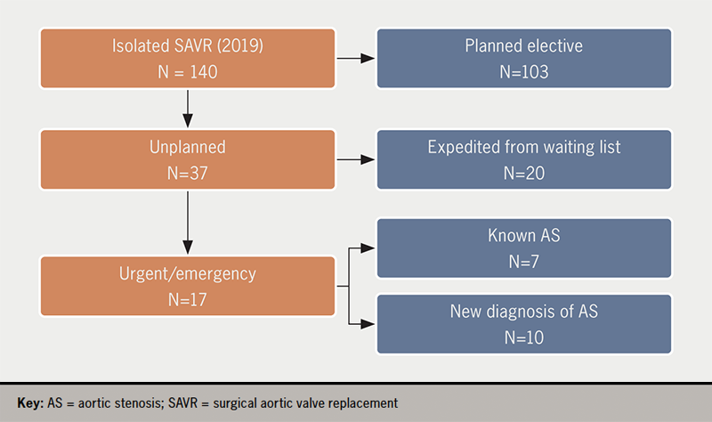
In 2019 there were 103 elective, first-time, isolated SAVRs and 37 (16 female, mean age 72.3 ± 8.4 years) non-elective, isolated, first SAVR (26% of total 140 SAVRs). Of these, 20 patients were already on the waiting list for SAVR and 17 were not (figure 1).
Ali - Figure 1. Flow chart of the study
Figure 1. Flow chart of the study

Clinical characteristics

Clinical characteristics of the patients are summarised in table 1, which is classified by patients on the waiting list versus patients not on the waiting list, and patients with a pre-existing diagnosis of AS versus patients without such a diagnosis. Frequency and severity of angina and of heart failure, prevalence of risk factors (table 2), aortic valve area, mean gradient and peak velocity, as well as stroke volume index and LVEF, were not different between those who were on the waiting list for SAVR, compared with those who were not, although LVEF was numerically lower and mean gradient numerically higher in patients who were not on the waiting list. The same findings apply to patient groups defined by whether a diagnosis of AS was known before the index admission. Almost one in three patients without a pre-existing diagnosis of AS presented with syncope, a much higher proportion than in those with known AS, but the significance of this finding is uncertain given the small size of our sample.

Table 1. Clinical and echocardiographic characteristics of patients stratified according to whether they were on the surgical waiting list for surgical aortic valve replacement (SAVR) or not, and whether they had been diagnosed with aortic stenosis (AS) or not prior to the index admission for SAVR. Variables in bold achieved statistical significance for the difference between the groups

Variable, mean ± SD Not on W/L for SAVR On W/L for SAVR p AS not known Known AS p
Age at admission, years 72.4 ± 8.7 72.2 ± 8.3 0.98 72.8 ± 9.1 72.1 ± 8.3 0.84
CCS angina class 1.6 ± 0.9 1.6 ± 0.8 0.87 1.4 ± 0.7 2.7 ± 0.8 0.04
NYHA heart failure class 2.8 ± 0.9 2.6 ± 0.7 0.40 2.7 ± 0.9 2.7 ± 0.8 0.80
Number of cardiac risk factors 1.7 ± 0.8 1.8 ± 1.1 0.80 1.9 ± 0.7 1.7 ± 1.1 0.75
Peak velocity across AV, m/s 4.5 ± 0.8 4.1 ± 0.9 0.18 4.5 ± 0.8 4.1 ± 0.9 0.31
Mean aortic valve gradient, mmHg 50.1 ± 20.0 40.8 ± 19.3 0.15 50.5 ± 19.7 43.1 ± 20.0 0.31
Aortic valve area, cm2 0.6 ± 0.2 0.6 ± 0.1 0.55* 0.6 ± 0.2 0.6 ± 0.1 0.43
LV ejection fraction, % 49.4 ± 13.3 54.2 ± 10.3 0.22 52.2 ± 11.3 51.9 ± 12.3 0.95
Stroke volume index,
ml/m2
34.5 ± 7.6 33.2 ± 8.2 0.61 33.4 ± 7.7 33.9 ± 8.0 0.87
Pre-operative hospital stay, days 20.0 ± 11.8 2.2 ± 4.3 0.001* 22.3 ± 9.3 6.0 ± 10.3 0.001
Postoperative hospital stay, days 16.4 ± 16.2 9.4 ± 6.7 0.008 12.5 ± 10.0 12.7 ± 3.3 0.9
Total hospital stay, days 36.5 ± 21.9 11.7 ± 7.7 0.01* 34.8 ± 13.9 18.77 ± 20.4 0.029
*Levene’s test was significant (p<0.05), suggesting a violation of the equal variance assumption, but p values remained the same with Welch’s and Mann-Whitney’s tests.
Key: AS = aortic stenosis; AV = aortic valve; CCS = Canadian Cardiac Society; LV = left ventricle; NYHA = New York Heart Association; SAVR = surgical aortic valve replacement; SD = standard deviation; W/L = waiting list

Table 2. Distribution by gender, main symptom at presentation and cardiovascular risk factors according to whether patients were on the waiting list for SAVR or not and whether patients were diagnosed with AS before presentation or not

Patient characteristics and symptoms Not on W/L for SAVR On W/L for SAVR p AS not known Known AS p
Gender F 9 7 0.27 5 11 0.60
M 8 13 5 16
Angina No 12 16 0.50 6 22 0.17
Yes 5 4 4 5
Heart failure No 8 6 0.28 6 8 0.09
Yes 9 14 4 19
Syncope No 14 20 0.05 7 27 0.03
Yes 3 0 3 0
T2DM No 12 12 0.50 8 16 0.24
Yes 5 8 2 11
High BP No 7 7 0.62 2 12 0.62
Yes 10 13 8 14
Current smoking No 15 20 0.11 9 26 0.45
Yes 2 0 1 1
Family history of CAD No 16 17 0.3 10 23 0.3
Yes 1 3 0 4
Chronic renal disease No 14 18 0.4 9 23 0.70
Yes 3 2 1 4
Key: AS = aortic stenosis; BP = blood pressure; CAD = coronary artery disease; SAVR = surgical aortic valve replacement; T2DM = type 2 diabetes mellitus; W/L = waiting list

Length of hospital stay

Length of hospital stay was significantly longer in patients who were not on the waiting list: total length of stay (mean of 36.5 ± 21.9 days vs. 11.7 ± 7.7 days, p<0.01); pre- and post-operative length of stay was also longer in patients not on the waiting list (table 1). Findings were similar for a comparison between patients with, versus those without, a pre-existing diagnosis of AS (table 1).

Patient pathways to SAVR

Patients with a previous diagnosis of AS

There were 27 patients with known AS. Of these, 20 were on the waiting list and seven were not. Reasons leading to expedited SAVR in the 20 patients on the waiting list included concern about how severe the AS was (12 patients – operation brought forward by the surgeon, after the initial surgical clinic appointment), and the development of a clinical emergency in eight: four cases of acute pulmonary oedema and four cases of congestive heart failure. Of seven patients not on the waiting list, three had acute pulmonary oedema while under routine cardiology follow-up for ‘moderate AS’, one had been referred but developed pulmonary oedema while waiting for a surgical outpatient appointment, one refused SAVR but was subsequently admitted with acute pulmonary oedema, one was admitted directly from home because of concerns from the GP about the severity of AS, and one had infective endocarditis on a background of mild AS, and was admitted with severe acute aortic regurgitation.

Patients without a previous diagnosis of AS

In 10 patients the diagnosis of AS was first established during the admission that led to SAVR: five presented with LV failure, four with angina and in three there was a history of syncope (p=0.003 vs. known AS; total >10 as some patients had multiple symptoms).

Table 3. Survival status according to whether patients were on the waiting list or not, and to whether they had been diagnosed with AS before the index admission

Not on W/L for SAVR On W/L for SAVR p AS not known Known AS p
Alive 15 16 0.49 9 22 0.53
Dead 2 4 1 5
Key: AS = aortic stenosis; SAVR = surgical aortic valve replacement; W/L = waiting list

Table 4. Time between initial diagnosis of AS and SAVR in years and the number of patients with and without a prior diagnosis of AS, according to whether patients were on the waiting list or not

Not on W/L for SAVR On W/L for SAVR
Time between initial diagnosis of AS and SAVR in years (p=0.49)
N 17 20
Mean ± SD 1.8 ± 2.3 2.4 ± 2.3
Minimum 0.0 0.2
Maximum 5.8 8.9
Prior diagnosis of AS (p<0.001)
No 10 0
Yes 7 20
Total 17 20
Key: AS = aortic stenosis; SAVR = surgical aortic valve replacement; SD = standard deviation; W/L = waiting list

Both patients not on the waiting list and those with a new diagnosis of AS had numerically worse indices of AS, but the differences were not significant. There was no difference in mortality, or in the stage of AS-related myocardial damage at presentation between patients on, or not on, the waiting list, or those with a prior versus a new diagnosis of AS (tables 3, 4 and 5). The extent of AS-related myocardial damage was not different between survivors and deceased (table 5).

Discussion

In a tertiary UK cardiac centre serving a population of one million, 12% of SAVR (17 of 140) were urgent, unplanned, and had increased pre-operative length of hospital stay. Almost half of these (7/17, 44%) unplanned SAVRs were in patients without a previous diagnosis of AS.

Another 20 operations (14% of SAVR) were expedited from the waiting list. These were patients who had been accepted for SAVR, but whose operations were brought forward by surgeons concerned about the severity of the disease, or who decompensated with heart failure while waiting for their procedure. For service improvement, studying patients who had unplanned operations is likely to be most informative.

Patients with operations expedited while on the waiting list for SAVR

Patients on the waiting list for SAVR have had pre-operative investigations completed and, in principle, could receive their SAVR at any time after they have been placed on the waiting list. If the NHS had the capacity to perform timely operations, this category of patients would not exist, or their number would be negligible. Limited capacity means that – unavoidably – some patients will decompensate while waiting for elective SAVR.

Table 5. Stage of myocardial damage by whether patients were on the waiting list for SAVR, had known AS, and mortality

Stage of myocardial damage Not on W/L for SAVR On W/L for SAVR AS not known Known AS Alive Dead
0 3 2 2 3 5 0
1 4 2 7 9 11 0
2 6 9 4 11 11 4
3 4 1 2 3 3 2
4 0 1 0 1 1 0
Total (N=37) 17 20 10 27 31 6
p value 0.37 0.79 0.16
Key: AS = aortic stenosis; SAVR = surgical aortic valve replacement; W/L = waiting list

In our sample of 37 patients, 20 were identified by their physicians as needing an expedited operation, and thus they ‘jumped the queue’ to an early SAVR, which is a compromise solution for the delivery of effective care within a constrained resources setting. Broadly speaking, these patients can be considered a ‘surrogate’ for the larger group of patients having genuinely elective, planned procedures, which do not need to be brought forward. However, of these 20, only 12 were clinically stable and had expedited surgery because of concerns about the severity of the AS, while the other eight had developed heart failure that prompted early surgery. Although recent guidelines6 still emphasise presence of symptoms as an important trigger for SAVR, heart failure (or any symptoms) in AS is an ‘end-stage’ manifestation with a dire outlook in a condition with a long asymptomatic phase. However, the tide may be about to turn, with recent evidence from randomised-controlled trials that early surgery is associated with markedly improved survival in asymptomatic patients with very severe7 or severe asymptomatic AS,8 when compared with conventional, guideline-driven practice. In light of these latest data, having patients develop heart failure after they have already been accepted for SAVR will become an untenable proposition.

Patients with AS not on the waiting list for SAVR

All seven such patients were under yearly cardiology follow-up with a diagnosis of ‘moderate aortic stenosis’, and three had been seen in a cardiology or valve clinic within three months of the index admission. The only difference we could identify was a longer total, pre- and postoperative hospital stay compared with patients who were on the waiting list (table 1). Patients in this group had numerically more severe indices of AS, and lower LVEF, suggesting more advanced disease, but the differences were not significant, presumably due to the low numbers involved. Clearly, the follow-up delivered by our valve clinics is imperfect, having failed to predict decompensation, albeit in a small number of patients. Potential reasons remain speculative due to the small size of the group, but may include underestimation of AS severity (although the higher gradients observed would identify these as cases of genuinely severe AS), reluctance of the valve clinic to refer for SAVR for clinical reasons (although the fact that patients ended up having SAVR mitigates against such a possibility) or under-reporting of symptoms by stoical patients. Whether a wider use of N-terminal proB-type naturietic peptide (NT-proBNP) levels and of aortic valve calcium scoring by computed tomography (CT)6 for monitoring AS progression can prevent such ‘near-misses’ remains to be seen.

Patients with AS not diagnosed until the index admission

The 10 patients in this category had a life-threatening complication of their valve disease as its first manifestation. No improvement in the efficiency of our current model of care for patients with AS would reduce this proportion; to achieve that, active screening for AS would be needed. In a previous study from the same geographic area, we found a prevalence of 8% of at least moderate mitral regurgitation (MR), and 5% of at least moderate AS (total prevalence 13%) in asymptomatic people over the age of 75,9 slightly higher than in the similar Ox-Valve study, which found a total population prevalence of moderate or severe valvular heart disease (VHD) of 11.3% in slightly younger subjects.10 The cost/benefit implications of such an approach need further study, but a primary care-based population-screening programme may turn out to be cost-effective and clinically feasible by potentially avoiding the long hospital stays associated with SAVR in patients with previously undetected AS.

Length of hospital stay (LOHS)

Consistently, patients with the least degree of planning of their SAVR (those with known AS but not on the waiting list, and those without a previous diagnosis of AS) spent longer in hospital than either those on the waiting list or those with a previous diagnosis of AS. The differences in the length of the pre-operative stay were most striking between patients on, versus those not on, the waiting list, with a mean of 2.2 ± 4.3 versus 20.0 ±11.8 days (p<0.001) (table 1), and were less marked for the postoperative stay. Patients without, versus those with, a diagnosis of AS had a mean pre-operative hospital stay of 22.3 ± 9.3 versus 6.0 ± 10.3 days (p<0.001), while the postoperative stay was not different. Our data are not granular enough to allow us to understand the exact causes of these differences, but they are likely to be a composite of delays relating to the availability of a bed to allow transfer from the admitting hospital to the surgical centre, and of the time it takes to stabilise and investigate acutely unwell patients.

Paradoxically, the finding of vastly longer LOHS in patients undergoing expedited or unplanned SAVR may be the most important motivator for improving the process of detection and monitoring of AS in the NHS. Further research is needed in order to model the net effect on healthcare costs of a screening programme for heart valve disease in the community.

Referral to treatment time (RTT)

The RTT for isolated SAVR in our hospital during the study period was 25 weeks. The significance of our findings is evidently dependent on the broader context of the healthcare system: patients who were already on the waiting list would not have had to present in extremis if the valve intervention could have been delivered sooner after diagnosis. Moreover, the goalposts are moving all the time, with the availability of transcatheter aortic valve replacement (TAVR) now reducing waiting list time to weeks after the diagnosis is made and the indication for valve intervention established.

Limitations

Size of the study

This is a small study, as it was designed and performed by busy clinicians rather than by dedicated research staff, and, as such, potentially significant differences between the various subgroups may not have reached the significance threshold. However, in spite of the low numbers, it offers an accurate ‘snapshot’ of the patient journey in a system with limited resources. We did not have the resources to collect detailed data on patients who had genuinely elective procedures, but our category of ‘expedited SAVR’ is likely to be broadly equivalent to those.

Assessment of myocardial damage

A limitation of our study is that imaging markers of myocardial damage, such as T1 mapping or late gadolinium enhancement (LGE) by cardiac magnetic resonance (CMR), were not available; such markers may allow further risk stratification of patients being followed-up for AS.

Relevance of SAVR in the TAVR era

The advent of percutaneous aortic valve replacement has changed the landscape of valve intervention beyond recognition,11 and the indications are expanding rapidly to encompass low-risk patients.12 It is likely that we shall see a further diminution of the proportion of patients undergoing SAVR, simultaneous to a major increase in the total number of aortic valve interventions, with open surgery reserved in the future for patients with concomitant pathologies, such as coronary artery disease not suitable for percutaneous coronary intervention (PCI) or aortic disease in need of surgical treatment. The proportion of patients for whom our findings are relevant will probably diminish, but SAVR is unlikely to become extinct, and the durability of TAVR valves is still not fully established, so our findings can still generate useful impetus for service improvement.

Conclusion

With the current model of care in the NHS, a significant proportion of patients have SAVR, the life-saving treatment for AS, performed in an unplanned manner, precipitated by clinical decompensation; such patients have a higher incidence of syncope. Unplanned surgery leads to prolonged hospital stay, an undesirable outcome. A small proportion of the unplanned operations are performed in patients without a prior diagnosis of AS; such patients could only be detected by rolling out a screening programme for heart valve disease in the community.

Key messages

  • Intervening in cases of aortic stenosis requires a programme of close surveillance of the patients’ symptomatology, echocardiographic parameters and suitability for contemporary aortic valve replacement procedures
  • While surgical aortic valve replacement in a suitable candidate offers unmatched results in terms of durability, performing this procedure in an expedited or unplanned fashion may lead to unnecessary morbidity in terms of length of hospital stay
  • In a resource-limited public health programme, valvular screening programmes need to be enhanced to pick out cases of silent valvular degeneration and facilitate timely intervention of progressive aortic valve disease

Conflicts of interest

None declared.

Funding

None.

Study approval

Requirement for ethical approval and consent was waived by the local hospital audit department as the study is a retrospective analysis of anonymised patient data.

References

1. Vahanian A, Beyersdorf F, Praz F et al. 2021 ESC/EACTS guidelines for the management of valvular heart disease. EuroIntervention 2022;17:e1126–e1196. https://doi.org/10.4244/EIJ-E-21-00009

2. Chambers JB. How to run a specialist valve clinic: the history, examination and exercise test. Echo Res Pract 2019;6:T23–T28. https://doi.org/10.1530/ERP-19-0003

3. Chambers JB, Parkin D, Rimington H et al. Specialist valve clinic in a cardiac centre: 10-year experience. Open Heart 2020;7:e001262. https://doi.org/10.1136/openhrt-2020-001262

4. Ross J. Afterload mismatch in aortic and mitral valve disease: implications for surgical therapy. J Am Coll Cardiol 1985;4:811–26. https://doi.org/10.1016/S0735-1097(85)80418-6

5. Vollema EM, Amanullah MR, Ng ACT et al. Staging cardiac damage in patients with symptomatic aortic valve stenosis. J Am Coll Cardiol 2019;74:538–49. https://doi.org/10.1016/j.jacc.2019.05.048

6. Otto CM, Nishimura RA, Bonow RO et al. 2020 ACC/AHA guideline for the management of patients with valvular heart disease: executive summary. A report of the American College of Cardiology/American Heart Association Joint Committee on Clinical Practice Guidelines. Circulation 2021;143:e35–e71. https://doi.org/10.1161/CIR.0000000000000932

7. Kang DH, Park SJ, Lee SA et al. Early surgery or conservative care for asymptomatic aortic stenosis. N Engl J Med 2020;382:111–19. https://doi.org/10.1056/NEJMoa1912846

8. Banovic M, Putnik S, Penicka M et al. Aortic valve replacement versus conservative treatment in asymptomatic severe aortic stenosis: the AVATAR trial. Circulation 2022;145:648–58. https://doi.org/10.1161/CIRCULATIONAHA.121.057639

9. Williams C, Mateescu A, Rees E et al. Point-of-care echocardiographic screening for left-sided valve heart disease: high yield and affordable cost in an elderly cohort recruited in primary practice. Echo Res Pract 2019;6:71–9. https://doi.org/10.1530/ERP-19-0011

10. d’Arcy JL, Coffey S, Loudon MA et al. Large-scale community echocardiographic screening reveals a major burden of undiagnosed valvular heart disease in older people: the Ox-VALVE population cohort study. Eur Heart J 2016;37:3515–22. https://doi.org/10.1093/eurheartj/ehw229

11. Arora S, Misenheimer J, Ramaraj R. Transcatheter aortic valve replacement: comprehensive review and present status. Tex Heart Inst J 2017;44:29–38. https://doi.org/10.14503/THIJ-16-5852

12. Spears J, Al-Saiegh Y, Goldberg D, Manthey S, Goldberg S. TAVR: a review of current practices and considerations in low-risk patients. J Interv Cardiol 2020;2020:2582938. https://doi.org/10.1155/2020/2582938

Navigating the research landscape in cardiology. Part 1: research – career necessity or bonus?

Br J Cardiol 2023;30:91–4doi:10.5837/bjc.2023.027 Leave a comment
Click any image to enlarge
Authors:
First published online 5th September 2023

Undertaking a period of research in cardiology is considered a vital part of training. This has many advantages including enhancing skills that better equip the clinician for patient care. However, in modern cardiology training, the feasibility and necessity of undertaking a period of formal research during training should be considered on an individual basis. The first of this four-part editorial series will explore the benefits of and obstacles to pursuing research in cardiology, with the aim of equipping the reader with an understanding of the options around research during cardiology training in the UK.

Introduction

For those in cardiology training, finding research is often a daunting and multi-faceted process. The objective of this four-part series is to explore research in cardiology and will aim to serve as a reference point from finding the research, to applying for funding, straight through to the finish line (table 1). Although these editorials are targeted mainly at cardiology registrars and have a UK focus, they may be of interest to any medical or allied-health professionals looking to undertake research in the field of cardiology. The first part of this series aims to explore the role of research as part of cardiology training in the context of current training challenges.

Table 1. An overview of this navigating the research landscape in cardiology series

Editorial number Editorial content
Part 1: research – career necessity or bonus? Exploring the role of research in contemporary cardiology
Part 2: finding the right research Approaching a research group, different options for research degrees (MD vs. PhD, clinical vs. basic science)
Part 3: the application process Funding bodies, ethics processes/approvals
Part 4: beyond the finish line What to do on the ‘other side’ of finishing research

New changes to the curriculum and impact on research

Navigating the research landscape in cardiology. Part 1: research – career necessity or bonus?

At the heart of patient-centered medicine is research, which drives progress and ultimately enhances care.1 However, with the recent changes to the cardiology curriculum and training structure, the question arises as to whether pursuing a formal research degree is both necessary and feasible. The new curriculum, which has been approved by the General Medical Council (GMC) and implemented from August 2022, places a greater emphasis on general medicine, yet it remains unclear how this will be balanced with the practical, skills-based curriculum required for cardiology training. Although participating in the general medicine rota can be beneficial, the time constraints it presents pose a challenge. However, Joint Royal College of Physicians Training Board’s (JRCPTB) curriculum outline document2 still highlights the importance of research, with many trainees considering a research component as a means of staying competitive in the cardiology job market.

Despite such suggestions that research is central to modern cardiology training, it is important to note that academic cardiology is no longer a distinct sub-specialty area as it was in the previous system. To pursue a career in academia, one must complete training in general internal medicine, full sub-specialty cardiology, and research, which is a more demanding route than before.

Research: career necessity or bonus

Despite the challenges posed by the new training structure, many still consider a period of research during medical training to be an essential step in career development. Such a period is viewed as a valuable CV-enhancing exercise, offering opportunities for career networking and skill development that may not be available in a standard cardiology training programme (table 2). Additionally, a research period offers the advantage of being able to remain in one location for several years, building rapport and relationships that are otherwise difficult to cultivate during frequent rotations. Furthermore, time undertaking research may be necessary for those seeking certain types of jobs, such as in tertiary or quaternary care centres. The experience gained during a research period can be invaluable, equipping trainees with the expertise and credentials needed to stand out in a competitive job market and to excel in academic or clinical settings.

Table 2. Pros and cons of research

Pros Cons
CV enhancing: publications, presentations, teaching and leadership opportunities Extends training time
Opportunity to network in your chosen field No on-call supplement to salary in research
Learn transferable skills, e.g. literature searching, team work, project management skills, time management De-skill in practical procedures unless part of your research time
Opportunity to learn research-specific skills, e.g. lab skills, coding, statistical analysis, patient recruitment, ethics writing Potential toxic environment of some research groups/academia

Is a research degree necessary for tertiary/quaternary jobs?

Whether a research degree is necessary to work as a cardiologist in a tertiary or quaternary hospital centre in the UK depends on individual hospital policies, requirements, and career aspirations. While research and academic medicine are significant aspects of medical practice, some hospitals may prioritise clinical experience and practical skills over research training. Ultimately, the decision to pursue a research degree should be based on individual goals, interests, and aptitudes.

Arguments for a research degree include enhancing clinical expertise, managing complex cases, and meeting hospital requirements for academic and research responsibilities. However, many experienced cardiologists work in tertiary or quaternary hospitals without research degrees and provide excellent patient care. A research degree may not necessarily be directly relevant to clinical practice or guarantee better patient care, and excessive emphasis on research training may detract from the importance of clinical training and practical skills.

Is research right for me?

Cardiology is a dynamic specialty that prides itself on a large evidence base. But, how does one decide whether this is the correct path? While the answer is unlikely to be the same for everyone, exploring a number of avenues and considering the following areas, provides a structured approach.

When is the right time?

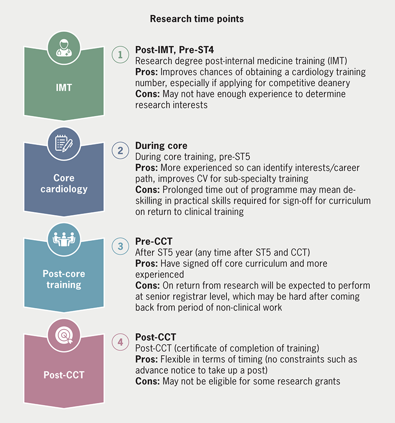
Training programmes are critical in shaping the careers of medical professionals and promoting consistent clinical services across institutions. In the UK, the JRCPTB2,3 sets guidelines and standards for cardiology training programmes in collaboration with other organisations, such as the GMC. Each deanery may have their own requirements for out-of-programme requests, which will be covered in a subsequent part of this editorial series. Figure 1 outlines common research time points, and the new curriculum does not strictly define core training years.

Kurdi - Figure 1. Is there a right time for research? Summary of the various time points that trainees can opt to take time out for research
Figure 1. Is there a right time for research? Summary of the various time points that trainees can opt to take time out for research

Your motivation behind undertaking research

Research fellowships offer a unique lifestyle that differs from that of a typical medical professional, as they usually involve working in an office or laboratory setting, rather than on a clinical ward. One of the benefits of this type of position is that it often does not require participation in an on-call rota. This can be particularly appealing for medical professionals who are looking to prioritise family time or raise young children. By not being on-call, individuals may have more flexibility to arrange their work schedules and to plan their personal lives, without the unpredictability and potential interruptions of being called into work at odd hours. This can also help to reduce the overall stress and demands of the job, allowing individuals to focus more on their research, and to achieve a better work–life balance. While there are certainly trade-offs involved in pursuing a research fellowship, the opportunity to prioritise family and personal time is often a significant benefit that should be carefully considered when making career decisions.

Despite the perception that research offers a slower pace of work, this can be challenging at times. Unlike the immediate feedback and sense of accomplishment that comes from working directly with patients in a clinical setting, research often requires a long-term investment of time and effort before any tangible results can be seen. This can lead to a sense of frustration and a lack of purpose, particularly for those who enjoy the day-to-day interactions with patients. It is important, therefore, to be clear about one’s motivations for pursuing research and to have a clear understanding of the long-term goals and benefits of the work. This can help to maintain motivation and a sense of purpose, even in the face of the day-to-day challenges and minutiae of research work. Ultimately, the rewards of research may be less immediate, but they can be significant and long-lasting, both in terms of personal and professional fulfilment, and in the contributions that research can make to improving patient care and advancing medical knowledge. Some of the advantages and disadvantages of research are listed in table 2.

What can’t you compromise on?

The quest to finding research in cardiology is helped if you know what you are willing to negotiate on. Narrowing things, such as location, is helpful to provide attainable research goals. It is helpful to also understand your long-term career aspirations and how much time out of programme you want to take (two year MD vs. three year PhD). Although the topic of your research is not always vital; having a focus may help to improve career trajectory. For example, if you are an aspiring imaging consultant then you may want to tailor your research towards a particular imaging modality.

If academia is the long-term goal, then obtaining a grant becomes more important. Grants from professional research bodies, such as British Heart Foundation (BHF) and National Institute for Health Research (NIHR), are impressive when applying for academia roles. Applying for funding will be described in more detail in Part 2 of the editorial series. However, for most consultant cardiology jobs in the UK this is not a strict pre-requisite.

What are the alternatives to a formal research degree?

Alternatives to a formal research degree during cardiology training include fellowships, which offer specialised training in a particular area of cardiology, and can be clinical or research-based, and can be taken during or after cardiology training. Taught degrees, such as master’s degrees in leadership and management, can also provide valuable opportunities to gain a deeper understanding of healthcare systems and how to affect change in the NHS, although cost can be a barrier. Diplomas and postgraduate certificates can offer flexibility and remote-learning options. Overall, finding opportunities that align with interests and career aspirations, and seeking guidance and support from mentors and colleagues, can help to build a well-rounded set of skills and experiences during cardiology training without the need for a formal research degree.

Conclusion

While research may not be necessary for every cardiology trainee, it can provide valuable opportunities for personal and professional growth. There is no one-size-fits-all approach to research, and individuals should be prepared to seize opportunities and adjust plans as needed. Securing research is the first step, what follows then is a minefield of research terms and jargon. Cath lab and crash calls are swapped for costing research equipment, service level agreements and substantial amendments. Twitter can be helpful, so can other websites including the Good Clinical Practice, NIHR and Health Research Authority (HRA) websites. There are also excellent resources such as the “The ABCs of developing a clinical study” talk by Dr Rasha Al Lamee on the British Junior Cardiologists’ Association website.5 Research is not the right choice for everyone. And, in this new era of cardiology, is not necessarily a pre-requisite for securing a consultant job. However, with the right support, determination, and resources, those wishing to pursue research should not be discouraged by the challenges that may arise, as they are not insurmountable.

Key messages

  • Cardiology trainees have various options to enhance their training experience beyond academia and research, such as fellowships, postgraduate certificates/diplomas, and taught degrees
  • The new cardiology curriculum’s emphasis on general internal medicine presents a challenge to trainees pursuing a formal research degree, as it may require an extended period of training to attain the same level of clinical expertise as their predecessors. Additionally, academic routes are now more demanding as academic cardiology is no longer a distinct sub-specialty area
  • While PhDs and MDs are unlikely to be strict requirements for tertiary/quaternary centre jobs, those with a passion for research and academia should not be deterred from pursuing formal research degrees

Conflicts of interest

None declared.

Funding

None.

References

1. Kasivisvanathan V, Tantrige PM, Webster J, Emberton M. Contributing to medical research as a trainee: the problems and opportunities. BMJ 2015;350:h515. https://doi.org/10.1136/bmj.h515

2. Joint Royal College of Physicians Training Board. Curriculum for cardiology training. London: JRCPTB, 2022. Available from: https://www.jrcptb.org.uk/specialties/cardiology

3. Joint Royal College of Physicians Training Board. Annual review of competence progression (ARCP) decision aids. Available from: https://www.jrcptb.org.uk/training-certification/arcp-decision-aids

4. Weissberg P. Training in academic cardiology: prospects for a better future. Heart 2002;87:198–200. https://doi.org/10.1136/heart.87.3.198

5. Al Lamee R. The ABCs of developing a clinical study. Available from: https://bjca.tv/video/research-career-development/page/2

NICE guidelines in the Sunderland RACPC cohort study: one size does not fit all

Br J Cardiol 2023;30:106–7doi:10.5837/bjc.2023.028 Leave a comment
Click any image to enlarge
Authors:
First published online 5th September 2023

At least 5% of GP and accident and emergency (A&E) attendances are undifferentiated chest pain. Rapid access chest pain clinics (RACPC) offer urgent guideline-directed management of suspected cardiac chest pain. The National Institute for Health and Care Excellence (NICE) recommends computed tomography coronary angiography (CTCA) as a first-line investigation. We evaluated the effectiveness and efficiency of a local RACPC.

Retrospective analysis of unselected referrals to a RACPC in the Northeast of England was conducted for 2021. Baseline demographics and major adverse cardiovascular events (MACE) were compared between typical, atypical and non-angina. Anatomical and functional imaging results were recorded. Backward stepwise binary logistic regression modelled obstructive coronary artery disease (CAD) incidence.

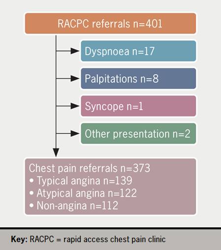
There were 373/401 (93.0%) patients with chest pain; 139 (37.3%) typical angina, 122 (32.8%) atypical angina and 112 (30.0%) non-angina. Typical angina patients were older (p<0.001) with more cardiovascular risk factors (p<0.001) and increased risk of obstructive CAD (adjusted odds ratio [OR] 6.27, 95% confidence interval [CI] 2.93 to 13.38) and MACE (9.4%, p=0.029). In total, 164 (44.0%) had invasive coronary angiography (ICA) within 7.4 ± 4.8 weeks; 19.5% had normal coronary arteries, 26.2% had obstructive CAD and 22.6% proceeded to invasive haemodynamic assessment ± PCI without major procedural complications. There were 39 (10.5%) who had CTCA within 34.6 ± 18.1 weeks; 25.6% needed ICA to clarify diagnosis.

In conclusion, typical angina patients were at heightened risk of cardiovascular events. In the absence of adequate CTCA capacity, greater reliance on ICA still facilitated accurate diagnosis with options for immediate revascularisation, timely and safely, in the right patients. Better risk stratification and expansion of non-invasive imaging can improve local RACPC service delivery in the wider Northeast cardiology network.

Introduction

Undifferentiated chest pain places a significant burden on the UK National Health Service (NHS). Up to 50% of the general population experiences chest pain in their lifetime contributing to at least 1% of GP consultations and 5% of accident and emergency (A&E) attendances.1 Chest pain patients have a twofold higher mortality versus age-matched asymptomatic controls.1 One reason is undiagnosed obstructive coronary artery disease (CAD), which has effective treatments to prolong life and improve symptoms.1 The challenge is identifying the patients at greatest risk, providing a timely diagnosis and starting effective treatment.

The national service framework for coronary heart disease set out a 10-year strategy to improve the care and outcomes of coronary heart disease patients in 2000.2 Part of the mandate was the establishment of rapid access chest pain clinics (RACPC) to provide expert cardiology consultation for patients with chest pain of suspected cardiac origin within two weeks of referral.2 An important advantage is the access to specialist cardiac tests to investigate CAD. In 2016, the revised guidelines from the National Institute of Health and Care Excellence (NICE) recommended first-line computed tomography (CT) coronary angiography (CTCA) in patients without known CAD, presenting with typical angina, atypical angina or non-angina chest pain, but where clinical suspicion of cardiac ischaemia remains.1,3

Reasons in favour of CTCA included relatively low cost and less radiation, a strong negative-predictive value and the prospect of improved medical management of non-obstructive atheromatous plaque.4 However, the provision of a national CTCA service would require access to more high-quality CT scanners with greater slice frequency and prospective gating, higher numbers of appropriately trained clinical staff and a geographically more equitable service delivery.5

The aim of our service evaluation was to assess the use of anatomical and functional imaging modalities to investigate angina at a RACPC against NICE recommendations. We wished to better understand the epidemiology and risk level of the RACPC population to make informed recommendations on the local triage system and provision of investigations for obstructive CAD.

Method

Study population and design

Patients referred to the RACPC at Sunderland Royal Hospital (SRH), a district general hospital in the Northeast of England in 2021 were retrospectively identified. Data were collected from three distinct periods: January to February, April to May and August to October 2021.

Eligible patients presented with typical, atypical or non-angina chest pain. Typical angina was defined by a constricting/heavy discomfort in the front of the chest, neck, shoulders, jaws or arms; worsened by physical activity; and relieved by rest or glyceryl trinitrate spray within five minutes.3 Any two of the three features defined atypical angina and one or no features characterised non-angina chest pain.3 Exclusion criteria were symptoms other than chest pain.

Patients were stratified by presenting complaint into typical angina, atypical angina or non-angina. Another important comparison was drawn between patients with ‘normal coronaries’, non-obstructive CAD and obstructive CAD. Obstructive CAD was defined by visually assessed ≥70% stenosis of the three epicardial coronary arteries (the left anterior descending [LAD], the left circumflex [LCx] and the right coronary artery [RCA]), ≥50% stenosis of the left main coronary artery (LMCA) and/or fractional flow reserve (FFR) or ctFFR positive (≤0.80) moderate lesions. Non-obstructive CAD ranged from minor and eccentric plaque disease to moderate stenoses of 50–69%.6

Data were collected from the local hospital’s electronic patient records, MediTech 6.0. Ethical approval was waived as anonymised retrospective data were used.

Variables of interest were patient age, sex, cardiovascular risk factors, clinical observations and signs, investigations and prescription of cardiac medication. Relevant investigations were electrocardiogram (ECG), echocardiogram and levels of haemoglobin (g/L), creatinine (μmol/L), estimated glomerular filtration rate (ml/min/1.73 m2), total and non-high-density lipoprotein (non-HDL) cholesterol (mmol/L). Important medications were lipid-lowering therapy, antithrombotic and anti-anginal agents. The imaging modality to investigate CAD was also recorded.

End points

The primary composite end point of major adverse cardiovascular events (MACE) included all-cause death, myocardial infarction (MI), or ischaemia-driven repeat revascularisation at maximum follow-up. In line with the fourth universal definition, MI was diagnosed by a rise in cardiac troponin (cTn) above the 99th percentile with evidence of ischaemia from symptoms, ECG changes, imaging and/or angiography.7 Ischaemia-driven revascularisation included any percutaneous coronary intervention (PCI) or coronary artery bypass graft surgery (CABG) for refractory or unstable angina.

Additional secondary end points were the components of the primary end point, re-referral to cardiology and hospital re-attendance.

Statistical analysis

Numerical variables were reported as mean ± standard deviation (SD) for parametric data, median (interquartile range [IQR]) for non-parametric data and compared using either analysis of variance (ANOVA) or the Kruskal-Wallis test for multiple group comparisons. Categorical variables were reported as number (percentage) and compared using the Pearson’s chi-square test.

We modelled the incidence of obstructive CAD using binary logistic regression. Significant variables on univariate analysis were retained for multi-variate analysis and regressed against obstructive CAD using backward stepwise methodology.

Statistical analysis was performed on IBM SPSS version 2020. Statistical significance was assessed at the conventional two-sided 5% level (p<0.05).

Results

Hesse - Figure 1. Patient selection
Figure 1. Patient selection

From 401 sampled RACPC referrals in 2021, 373 were due to chest pain, including 139 (37.3%) typical angina, 122 (32.8%) atypical angina and 112 (30.0%) non-angina (figure 1).

Patient characteristics

Typical angina patients were older (65.1 ± 11.4 vs. 61.1 ± 13.1 and 57.3 ± 15.2 years) and more likely to have major cardiovascular risk factors, including hypertension, diabetes, hypercholesterolaemia and a positive family history, compared with patients presenting with atypical angina and non-angina (table 1). Typical angina presentations were associated with higher rates of known CAD (28.1% vs. 16.4% and 10.7%) and abnormal echocardiograms (33.3% vs. 10.0% and 15.4%) than atypical angina and non-angina presentations. There was a trend to lower total cholesterol levels in patients with typical angina (4.6 ± 1.2 vs. 4.8 ± 1.2 and 3.5 ± 1.0 mmol/L).

Primary and secondary end points

The mean length of follow-up was 61.3 ± 16.7 weeks. The primary composite end point of MACE occurred in 13/139 (9.4%) patients with typical angina in comparison to 3/122 (2.5%) and 4/112 (3.6%) patients with atypical and non-angina (p=0.029). There was no difference in the secondary outcomes between patients presenting with typical, atypical and non-angina chest pain (p>0.05) (table 2).

Table 1. Baseline clinical characteristics

Typical angina (n=139) Atypical angina (n=122) Non-angina (n=112) p value
Age, years 65.1 ± 11.4 61.1 ± 13.1 57.3 ± 15.2 <0.001
Male 82 (59.0) 58 (47.5) 57 (50.9) 0.16
Current smoker 23 (16.5) 19 (15.6) 13 (11.6) 0.52
Hypertension 85 (61.2) 62 (50.8) 42 (37.5) <0.001
Diabetes mellitus 36 (25.9) 20 (16.4) 13 (11.6) 0.011
Hypercholesterolaemia 102 (73.4) 69 (56.6) 53 (47.3) <0.001
Obesity 50 (36.0) 45 (36.9) 29 (25.9) 0.14
Known CAD 39 (28.1) 20 (16.4) 12 (10.7) 0.002
FH of CAD 69 (49.6) 64 (52.5) 40 (35.7) 0.023
Known to cardiology 17 (12.2) 13 (10.7) 13 (11.6) 0.92
Previous echocardiogram 39 (28.1) 30 (24.6) 26 (23.2) 0.66
LVSD and/or RWMA 13 (33.3) 3 (10.0) 4 (15.4) 0.044
Referral ECG available 69 (49.6) 58 (47.5) 58 (51.8) 0.81
Clinic ECG done 126 (90.6) 103 (84.4) 107 (95.5) 0.017
BBB pattern 5 (4.0) 4 (3.9) 6 (5.6)
ST changes 5 (4.0) 2 (1.9) 1 (0.9)
T-wave changes/q-waves 19 (15.1) 12 (11.7) 9 (8.4)
Sinus tachycardia/AF/ectopics 14 (11.1) 13 (12.6) 11 (10.3)
Heart rate, bpm* 77 ± 14 75 ± 13 76 ± 12 0.50
Systolic BP, mmHg* 138 ± 20 135 ± 19 134 ± 17 0.46
Murmur* 6 (5.7) 7 (8.3) 8 (9.1) 0.63
Haemoglobin, g/L* 139 ± 17 142 ± 15 141 ± 15 0.27
Creatinine, μmol/L* 86 ± 22 81 ± 23 84 ± 24 0.28
eGFR, ml/min/1.73 m2* 73 ± 15 77 ± 15 77 ± 14 0.11
Total cholesterol, mmol/L* 4.6 ± 1.2 4.8 ± 1.2 4.9 ± 1.0 0.05
Non-HDL-C, mmol/L* 3.2 ± 1.2 3.4 ± 1.2 3.5 ± 1.0 0.14
Data are in mean ± standard deviation (SD) for numerical variables and n (%) for categorical variables unless otherwise stated.
*>5% missing data.
Key: AF = atrial fibrillation; BBB = bundle branch block; BP = blood pressure; CAD = coronary artery disease; ECG = electrocardiogram; eGFR = estimated glomerular filtration rate; FH = family history; LVSD = left ventricular systolic dysfunction; non-HDL-C = non-high-density lipoprotein cholesterol; RWMA = regional wall motion abnormality

Table 2. Patient outcomes

Typical angina (n=139) Atypical angina (n=122) Non-angina (n=112) p value
Primary composite end point MACE 13 (9.4) 3 (2.5) 4 (3.6) 0.029
Secondary end points
All-cause death 6 (4.3) 2 (1.6) 3 (2.7) 0.43
Myocardial infarction 3 (2.2) 0 (0.0) 1 (0.9) 0.23
Ischaemia-driven revascularisation 5 (3.6) 1 (0.8) 0 (0.0) 0.055
Re-referral to cardiology 6 (4.3) 6 (4.9) 5 (4.5) 0.97
Hospital re-attendance 11 (7.9) 10 (8.2) 8 (7.1) 0.95
Data are n (%) unless otherwise shown.
Key: MACE = major adverse cardiovascular event

Investigations, diagnosis and treatment

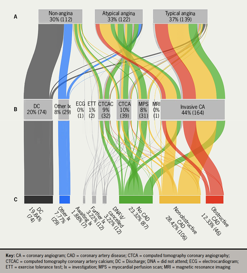
The cumulative diagnoses stratified by presenting complaint and choice of investigation are presented in figure 2.

Overall, 164 (44.0%) underwent invasive coronary angiography (ICA), 39 (10.5%) CTCA, 32 (8.6%) CT coronary artery calcium score (CTCAC) and 31 (8.3%) myocardial perfusion scan (MPS). The mean time to investigation was 7.4 ± 4.8, 34.6 ± 18.1, 10.5 ± 8.8 and 11.4 ± 8.7 weeks, respectively. Another 29 (7.8%) were investigated for non-ischaemic heart disease and 74 (19.8%) were discharged without follow-up.

Hesse - Figure 2. The presenting complaint (A), choice of investigation (B) and final diagnosis (C) of rapid access chest pain clinic (RACPC) attendances
Figure 2. The presenting complaint (A), choice of investigation (B) and final diagnosis (C) of rapid access chest pain clinic (RACPC) attendances

In total, 46 (12.3%) had obstructive CAD, 106 (28.4%) non-obstructive CAD and 87 (23.3%) normal coronary arteries. From 164 ICA, 32 (19.5%) had normal coronary arteries, 82 non-obstructive CAD (50.0%) and 43 (26.2%) obstructive CAD; 37 (22.6%) proceeded to FFR assessment and/or PCI. No major complications were reported. From 39 CTCA, 10 (25.6%) needed ICA to clarify diagnosis. Of the 31 MPS, two (6.4%) demonstrated reversible ischaemia and required ICA, which did not identify any stent targets.

At least 9 out of 10 patients with obstructive and non-obstructive CAD were prescribed primary and secondary prevention therapy before and after their diagnosis. Fewer than 50% of patients with normal coronary arteries were prescribed statin therapy after their diagnosis. We are unable to comment on whether this treatment was continued in the community.

Obstructive CAD risk

On univariate and multi-variate analysis, obstructive CAD was predicted by typical angina (odds ratio [OR] 6.27), age >61 years (OR 3.33), male sex (OR 2.09), known CAD (OR 2.64) and Hb >141 g/L (OR 2.83) (table 3).

Table 3. Univariate and multi-variate logistic regression to predict obstructive coronary artery disease (CAD)

Unadjusted odds ratio 95%CI Adjusted odds ratio 95%CI
Typical angina 6.82 3.33 to 13.97 6.27 2.93 to 13.38
Age >61 years 3.12 1.53 to 6.36 3.33 1.50 to 7.38
Male 2.85 1.43 to 5.70 2.09 0.94 to 4.61
Current smoker 1.18 0.47 to 2.92
Hypertension 2.21 1.15 to 4.25
Diabetes mellitus 1.67 0.82 to 3.43
Hypercholesterolaemia 1.81 0.92 to 3.56
Obesity 0.77 0.39 to 1.52
FH of CAD 1.07 0.58 to 1.98
Known CAD 3.72 1.93 to 7.18 2.64 1.27 to 5.53
Abnormal ECG and/or echo 1.78 0.91 to 3.47
Hb >141 g/L 1.96 1.03 to 3.71 2.83 1.31 to 6.09
Creatinine >84 μmol/L 2.61 1.39 to 4.92
Total cholesterol >5 mmol/L 0.67 0.34 to 1.32
Data are n (%) unless otherwise shown.
Key: CAD = coronary artery disease; CI = confidence interval; ECG = electrocardiogram; FH = family history; Hb = haemoglobin

Discussion

Non-specific chest pain is a common clinical presentation with an associated increased mortality rate that may be attributed to undiagnosed obstructive CAD.1–4 The RACPC aims to triage high-risk patients who require urgent intervention and intermediate-risk patients who need outpatient ischaemia testing from the bulk of low-risk ‘non-cardiac chest pain’ patients who can be safely discharged.1–4 In a service evaluation of a RACPC at a busy district general hospital in the Northeast of England, approximately 10% of patients had obstructive CAD. Typical angina patients were significantly more likely to have obstructive CAD with a higher one-year risk of MACE than other chest pain patients. Contrary to NICE recommendations, less than 10% had CTCA and instead, close to 50% of patients had ICA within two months of presentation, with a diagnostic rate of 20% normal coronary arteries and 25% obstructive CAD.

Similar to the populations of the CTCA in patients with suspected angina due to coronary heart disease (SCOT-HEART) trial and the Outcomes of Anatomical versus Functional Testing for Coronary Artery Disease (PROMISE) trial, our RACPC population was on average 60 years or older and 50% male with at least two modifiable cardiovascular risk factors.4,8 Our 37% typical angina burden was comparable with the 35% reported in SCOT-HEART.4

Our results are consistent with the published literature on the prognosis of patients presenting with and without typical angina.9–11 In an English multi-centre and a Scottish single-centre cohort study, angina patients had a 10–20% risk of a major cardiac event in comparison with less than 4% of patients with non-cardiac chest pain.10,11 We reported at least one in 10 patients with typical angina experiencing death of any cause, MI or ischaemia-driven revascularisation versus less than one in 20 patients with atypical or non-angina. The very low event rate among patients not presenting with typical angina should encourage stricter triaging of referrals. At least one fourth of chest pain patients could be safely discharged at triage.

In the SCOT-HEART trial, the addition of CTCA to the standard management of patients presenting with suspected angina clarified the diagnosis of CAD and angina, reduced further stress imaging, increased preventative therapy and was associated with reduced rates of cardiac death and MI at five years.4,12 Our high rate of ICA similarly improved our management of patients with suspected CAD.

There are likely to have been several reasons for our high ICA and low CTCA rate. First, a timely diagnosis is paramount. It can alleviate patient anxiety and pre-empt future cardiovascular events through appropriate treatment and intervention. In a cohort study of 172,180 first presentation chest pain patients, 4.6% of the 8,260 with a clinical diagnosis of angina experienced an MI in the first six months.9 Over 50% of our MACE events occurred within six months of RACPC attendance. On average, we provided ICA within two months of presentation versus CTCA within nine months. Second, ICA offers the opportunity to precisely quantify the haemodynamic significance of intermediate coronary lesions and to proceed directly to revascularisation in a ‘one-stop’ strategy in patients who remain symptomatic despite optimal medical anti-anginal therapy. One in five patients proceeded to FFR assessment and/or PCI after ICA.

The disadvantage of ICA in comparison with CTCA is the increased risk of serious complications including stroke, MI, contrast nephropathy and vascular access bleeding. Although we did not report any major complications, a significant number of ICA demonstrated normal coronary arteries. CTCA also facilitates better characterisation of plaque composition and risk stratification, which may be of greater relevance in a population at low risk of obstructive CAD.13 However, accurate interpretation is heavily dependent on patient factors (e.g. weight, heart rate control and breathing) and technical parameters (e.g. slice width and number, Z-axis coverage, reconstruction algorithms and ctFFR assessment).14 NICE recommends a minimum 64-slice CT scanner.3 Despite our use of a 128-slice CT scanner, a high proportion of patients who initially had CTCA required ICA to clarify diagnosis. The advantages of CTCA, including lower cost and less radiation were then obviated. A recently commissioned 320-slice CT scanner will be used in patients with sub-optimal heart rate control.

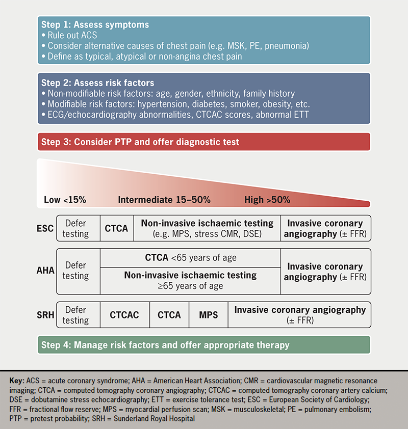
An alternative to anatomical imaging is non-invasive functional testing. The 2019 European Society of Cardiology (ESC) and the 2021 American Heart Association (AHA) guidelines on stable angina recommended non-invasive functional imaging to investigate stable CAD in intermediate-to-high risk patients, including those ≥65 years of age.15,16 At least 30% of our cohort met eligibility criteria, but less than 10% underwent stress testing, including MPS and cardiovascular magnetic resonance imaging. We recognise that our reliance on ICA is because of an underdeveloped CTCA service, rather than greater perceived benefits of an invasive strategy. Despite this seeming compromise, the ICA service has reassuringly still delivered timely, safe diagnostic information for patients and clinicians. In figure 3 we have summarised the management of patients with chest pain of suspected cardiac origin according to national, international guidelines and local practice.

Hesse - Figure 3. Diagnostic algorithm for patients presenting with chest pain of suspected cardiac origin
Figure 3. Diagnostic algorithm for patients presenting with chest pain of suspected cardiac origin

Our observations lend weight to the recommendations made in the Getting It Right First Time (GIRFT) review of cardiology in 2021, and the goals set out in NHS Long Term Plan, published in 2019.17–19 Both reports identified significant regional variation in access to and quality of cardiac care, particularly CTCA in the ‘stable chest pain pathway’.17–19 Important next steps in the delivery of a more equitable national CTCA service are a more than 100% increase in CT-scanning capacity and a further 2,000 radiologists and/or cardiologists accredited in CTCA reporting.19 Our data show that a full capacity CTCA service may avoid 75% of elective invasive coronary angiograms. Local PCI and complex device services could then be expanded, reducing the workload on tertiary centres as their volume of transcutaneous aortic valve implantations (TAVI) and other structural heart interventions, complex device extractions and ventricular tachycardia (VT) ablations increases. Ultimately, this would facilitate the development of a regional cardiology network, dictated by function and not geography, in Sunderland and the Northeast.18

Finally, this study has the inherent limitations of a retrospective analysis of clinical practice during a virus pandemic. We contend that the impact was small and that the data reflected current practice.

Conclusion

Our service evaluation of a RACPC showed that typical angina patients had a significantly higher likelihood of underlying obstructive CAD with increased risk of MACE than other chest pain patients. Despite NICE recommendations, a limited CTCA service led to a greater volume of ICA, which reassuringly still facilitated accurate diagnosis with the option of immediate revascularisation, timely and safely, in the right patients. However, a stricter referral triage system, as well as investment in and expansion of non-invasive anatomical and functional imaging capacity, may improve RACPC service delivery, support the larger cardiology network in the Northeast and, most importantly, offer safer and more equitable patient care.

Key messages

  • In a major paradigm shift away from the modified Diamond-Forrester pre-test probability model, the National Institute for Health and Care Excellence (NICE), in 2016, recommended first-line computed tomography coronary angiography (CTCA) for patients presenting with chest pain of suspected cardiac origin
  • Our service evaluation of a rapid access chest pain clinic (RACPC) provides valuable contemporary insights into local challenges, alternative management strategies and their impact on patient care and outcomes
  • Our study emphasises the importance of considering other equally safe and effective anatomical and functional imaging techniques as CTCA service capacity continues to expand nationally

Conflicts of interest

None declared.

Funding

None.

Study approval

Ethical approval was waived as anonymised retrospective data were used.

Acknowledgement

We thank the staff at SRH for their high standard of care of the patients included in the study.

References

1. Skinner JS, Smeeth L, Kendall JM, Adams PC, Timmis A, Chest Pain Guideline Development Group. NICE guidance. Chest pain of recent onset: assessment and diagnosis of recent onset chest pain or discomfort of suspected cardiac origin. Heart 2010;96:974–8. https://doi.org/10.1136/hrt.2009.190066

2. National Health Service. Coronary heart disease. National service framework for coronary heart disease. London: Department of Health, 2000. Available from: https://assets.publishing.service.gov.uk/government/uploads/system/uploads/attachment_data/file/198931/National_Service_Framework_for_Coronary_Heart_Disease.pdf

3. National Institute for Health and Care Excellence. Recent-onset chest pain of suspected cardiac origin: assessment and diagnosis. London: NICE, 2016. Available from: https://www.nice.org.uk/guidance/cg95/evidence/full-guideline-pdf-245282221

4. SCOT-HEART Investigators. CT coronary angiography in patients with suspected angina due to coronary heart disease (SCOT-HEART): an open-label, parallel-group, multicentre trial. Lancet 2015;385:2383–91. https://doi.org/10.1016/S0140-6736(15)60291-4

5. Dreisbach JG, Nicol ED, Roobottom CA, Padley S, Roditi G. Challenges in delivering computed tomography coronary angiography as the first-line test for stable chest pain. Heart 2018;104:921–7. https://doi.org/10.1136/heartjnl-2017-311846

6. Neumann FJ, Sousa-Uva M, Ahlsson A et al. 2018 ESC/EACTS guidelines on myocardial revascularization. Eur Heart J 2019;40:87–165. https://doi.org/10.1093/eurheartj/ehy394

7. Thygesen K, Alpert JS, Jaffe AS et al. Fourth universal definition of myocardial infarction (2018). J Am Coll Cardiol 2018;72:2231–64. https://doi.org/10.1016/j.jacc.2018.08.1038

8. Douglas PS, Hoffmann U, Patel MR et al. Outcomes of anatomical versus functional testing for coronary artery disease. N Engl J Med 2015;372:1291–300. https://doi.org/10.1056/NEJMoa1415516

9. Jordan KP, Timmis A, Croft P et al. Prognosis of undiagnosed chest pain: linked electronic health record cohort study. BMJ 2017;357:j1194. https://doi.org/10.1136/bmj.j1194

10. Sekhri N, Feder GS, Junghans C, Hemingway H, Timmis AD. How effective are rapid access chest pain clinics? Prognosis of incident angina and non-cardiac chest pain in 8762 consecutive patients. Heart 2007;93:458–63. https://doi.org/10.1136/hrt.2006.090894

11. Taylor GL, Murphy NF, Berry C et al. Long-term outcome of low-risk patients attending a rapid-assessment chest pain clinic. Heart 2008;94:628–32. https://doi.org/10.1136/hrt.2007.125344

12. Newby DE, Adamson PD, Berry C et al. Coronary CT angiography and 5-year risk of myocardial infarction. N Engl J Med 2018;379:924–33. https://doi.org/10.1056/NEJMoa1805971

13. Otsuka K, Fukuda S, Tanaka A et al. Prognosis of vulnerable plaque on computed tomographic coronary angiography with normal myocardial perfusion image. Eur Heart J Cardiovasc Imaging 2014;15:332–40. https://doi.org/10.1093/ehjci/jet232

14. Royal College of Radiologists. Standards of practice of computed tomography coronary angiography (CTCA) in adult patients. London: Royal College of Radiologists, 2014. Available from: https://www.rcr.ac.uk/system/files/publication/field_publication_files/BFCR14%2816%29_CTCA.pdf

15. Gulati M, Levy PD, Mukherjee D et al. 2021 AHA/ACC/ASE/CHEST/SAEM/SCCT/SCMR guideline for the evaluation and diagnosis of chest pain: a report of the American College of Cardiology/American Heart Association Joint Committee on Clinical Practice Guidelines. Circulation 2021;144:e368–e454. https://doi.org/10.1161/CIR.0000000000001029

16. Knuuti J, Wijns W, Saraste A et al. 2019 ESC guidelines for the diagnosis and management of chronic coronary syndromes. Eur Heart J 2020;41:407–77. https://doi.org/10.1093/eurheartj/ehz425

17. National Health Service. The NHS long term plan. London: Department of Health, 2019. Available from: https://www.longtermplan.nhs.uk/wp-content/uploads/2019/08/nhs-long-term-plan-version-1.2.pdf

18. Clarke S, Ray S. Cardiology: GIRFT programme national specialty report. London: GIRFT, 2021. Available from: https://www.gettingitrightfirsttime.co.uk/wp-content/uploads/2021/09/Cardiology-Jul21k-NEW.pdf

19. Richards M. Diagnostics: recovery and renewal. Report of the independent review of diagnostic services for NHS England. London: NHS England, 2020. Available from: https://www.england.nhs.uk/wp-content/uploads/2020/11/diagnostics-recovery-and-renewal-independent-review-of-diagnostic-services-for-nhs-england-2.pdf

Large fusiform aneurysm of the superior vena cava: CT findings

Br J Cardiol 2023;30:119–20doi:10.5837/bjc.2023.029 Leave a comment
Click any image to enlarge
Authors:
First published online 5th September 2023

A 62-year-old man presented complaining of atrial fibrillation.  Plain chest radiography and contrast-enhanced computed tomography (CT) revealed a large fusiform aneurysmal dilatation of the upper segment of the superior vena cava (SVC) without evidence of rupture, thrombosis, or pulmonary embolism. It was decided to treat the patient conservatively with follow-up imaging recommended.

Case presentation

Batouty - Figure 1. Chest radiograph showing widened mediastinum by a right para-tracheal soft tissue shadow (arrow)
Figure 1. Chest radiograph showing widened mediastinum by a right para-tracheal soft tissue shadow (arrow)

A 62-year-old male patient complaining of atrial fibrillation was referred for pre-ablation workup. Plain chest radiography demonstrated a right paratracheal soft tissue shadow mimicking a mediastinal mass (figure 1). Contrast-enhanced computed tomography (CT) revealed fusiform aneurysmal dilatation of the upper segment of the superior vena cava (SVC) starting from its origin at the confluence of the right and left brachiocephalic veins and ending 3 cm above the cavo-atrial junction (figure 2), with maximal axial dimensions 6.5 x 5 x 6 cm. The azygous vein was seen to drain into the lower part of the aneurysmal SVC. There was no evidence of complications such as rupture, thrombus formation or pulmonary embolism. The patient was reassured and managed conservatively with follow-up imaging recommended.

Discussion

Congenital aneurysms of the SVC are very rare findings caused by congenital weakness of the venous wall or absence of the longitudinal muscle layer.1 Congenital aneurysms are more commonly fusiform, while saccular aneurysms are thought to be post-inflammatory or resulting from trauma or intervention. In most described cases, the cause is unknown.2,3 Most SVC aneurysms are asymptomatic, discovered incidentally during chest X-ray, simulating mediastinal mass. They may be associated with a ruptured aneurysm, or thrombosis and subsequent pulmonary embolisation.2,3 There are no established guidelines for the management of SVC aneurysms but it is generally influenced by aneurysm type and size. Most fusiform aneurysms are managed conservatively with follow-up imaging since complications are uncommon. Prophylactic anticoagulation may be given to prevent thrombosis. Saccular aneurysms, on the other hand, are usually candidates for surgical resection even if asymptomatic, due to the higher risk of rupture or thrombosis. Development of symptoms or complications or significant aneurysm growth on follow-up may warrant interventions.2,3

Batouty - Figure 2. Contrast-enhanced computed tomography (CT) showing aneurysmal dilatation of the superior vena cava. A: axial image, B: coronal-reformatted image, and C: volume-rendered image. Arrow shows the superior vena cava aneurysm, the asterisk shows the left brachiocephalic vein
Figure 2. Contrast-enhanced computed tomography (CT) showing aneurysmal dilatation of the superior vena cava. A: axial image, B: coronal-reformatted image, and C: volume-rendered image. Arrow shows the superior vena cava aneurysm, the asterisk shows the left brachiocephalic vein

Conflicts of interest

None declared.

Funding

None.

Patient consent

Patient consent was obtained for anonymous publishing of images.

References

1. Sonavane SK, Milner DM, Singh SP, et al. Comprehensive imaging review of the superior vena cava. Radiographics 2015;35:1873–92. https://doi.org/10.1148/rg.2015150056

2. Soares Souza LV, Souza Jr AS, Morales MM, Marchiori E. Superior vena cava aneurysm: an unusual mediastinal mass. Eur J CardioThorac Surg 2021;5:276–7. https://doi.org/10.1093/ejcts/ezaa271

3. Kapoor H, Gulati V, Pawley B, Lee JT. Massive fusiform superior vena cava aneurysm in a 47-year-old complicated by pulmonary embolism: a case report and review of literature. Clin Imaging 2022;81:43–5. https://doi.org/10.1016/j.clinimag.2021.08.008

The delusion of measuring blood pressure

Br J Cardiol 2023;30:83–5doi:10.5837/bjc.2023.023 Leave a comment
Click any image to enlarge
Authors:
First published online 9th August 2023

Hypertension affects over a billion people worldwide and is a leading cause of premature death and disability. However, it continues to remain a silent epidemic, with the majority of patients undiagnosed or untreated. The World Health Organisation reports that only 42% of individuals with hypertension receive a diagnosis and appropriate treatment. Furthermore, only one in five adults have their blood pressure under control.1 These statistics reflect a grave failure in identifying and managing a condition that has far-reaching health consequences. The misdiagnosis and undertreatment of blood pressure pose substantial risks to individuals and impose a tremendous burden on healthcare systems worldwide.

Measurement

The delusion of measuring blood

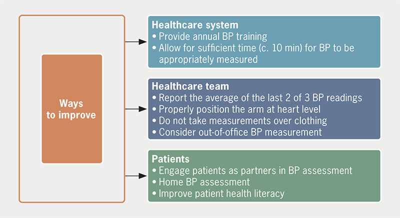
Blood pressure measurement is a quintessential part of healthcare, and accurate measurement is paramount for proper diagnosis and treatment of hypertension. However, studies indicate that inaccuracies in blood pressure monitoring are prevalent, even among healthcare professionals.2–5 Flawed measurement techniques, such as relying on a single reading, insufficient time for measurement, and incorrect arm positioning contribute to misdiagnoses.

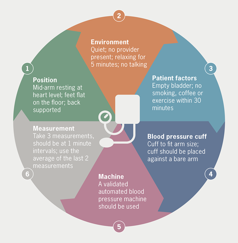
Our prior work has highlighted the inadequate adherence to accurate blood pressure measurement protocols among healthcare professionals, including even cardiologists.2 Key factors contributing to inaccuracies included failure to measure blood pressure in both arms, insufficient time before measurement, and reliance on a single reading. Other contributors to inaccuracies were a preference for measurements ending in zero, which accounted for 60–80% of readings, improper positioning of the arm in relation to the heart, and patient-related factors, such as crossing the legs, making measurements over long-sleeve clothing, or addressing the need to urinate prior to the blood pressure measurement. What is even more concerning was that, despite these inaccuracies in measurement, eight of every 10 healthcare professionals trusted their blood pressure measurements, indicating a significant disconnect between accurate measurement and perceived accuracy. Opinions varied among medical assistants, nurses, and doctors regarding the need and frequency of practising blood pressure measurement skills, and they had differing knowledge about the reliability of their blood pressure measurement equipment to ensure accuracy.

Where does this leave the effort to combat hypertension?

Do the inaccuracies of in-office blood pressure measurement emphasise the need for better education and training among healthcare professionals, or is it time to shift gears towards out-of-office blood pressure assessment? The answer is likely yes to both. Out-of-office blood pressure assessment involves home blood pressure measurement and ambulatory blood pressure measurement, both of which are recommended by the National Institute for Health and Care Excellence (NICE), European Society of Cardiology (ESC)/European Society of Hypertension (ESH), and the American College of Cardiology (ACC)/American Heart Association (AHA) guidelines as complementary methods for measuring blood pressure and diagnosing hypertension.6–8 Although each method comes with its own disadvantages, including the potential for measurement error, both home and ambulatory blood pressure measurements can diagnose white-coat and masked hypertension. Furthermore, these modalities better prognosticate future cardiovascular events compared with office measurement of blood pressure.9,10

While out-of-office methods will likely alleviate some of the misdiagnoses of hypertension, healthcare professionals must adhere to the most recent blood pressure measurement guidelines.6–8 These guidelines recommend the use of validated automated devices, regular calibration, measurements taken in both arms, and the recording of multiple readings with proper intervals. Graphical recording of blood pressure values within ranges can help reduce the impact of inaccuracies.3 Physicians should consider using automated office blood pressure (AOBP) readings as the preferred method for routine blood pressure monitoring to ensure accurate measurements and reduce the potential impact of the white-coat effect. AOBP readings, taken with the patient sitting alone in a quiet place, are similar in accuracy to ambulatory readings and more accurate than routine office readings.11

What can be done?

Combatting blood pressure misdiagnosis, clinical inertia and undertreatment require a collaborative effort between healthcare professionals, patients, and emerging technology (figure 1). Patient education plays a crucial role, ensuring individuals understand the importance of accurate measurements, the risks associated with hypertension, and the significance of treatment adherence (figure 2). Similarly, additional physician and healthcare professional education is clearly required. Standardised blood pressure management protocols should be implemented, and clinic personnel should be trained, and preferably certified, in standardised blood pressure measurement, and receive regular refresher training.12 Healthcare systems must allocate resources to establish preventive care programmes, integrate routine blood pressure screenings into regular check-ups, and launch community-based initiatives for early detection.13–16 Leveraging technological advancements, such as telemedicine and mobile health applications, can further enhance accuracy and patient engagement.17,18

Hong - Figure 1. How do we implement change to improve blood pressure (BP) measurement?
Figure 1. How do we implement change to improve blood pressure (BP) measurement?
Hong - Figure 2. Factors for correct blood pressure measurement
Figure 2. Factors for correct blood pressure measurement

The delusion and inaccuracies surrounding blood pressure measurement undermines the diagnosis and treatment of hypertension, leading to significant health risks and economic burdens. By promoting accurate measurement techniques, enhancing patient education, and fostering collaboration among healthcare professionals and policymakers, we can bridge the gap in blood pressure management. Improving diagnostic accuracy and treatment adherence will not only save lives but also reduce healthcare costs and alleviate the burden on healthcare systems, fostering a healthier future for all.

Conflicts of interest

None declared.

Funding

None.

References

1. World Health Organization. Hypertension. Available at: https://www.who.int/news-room/fact-sheets/detail/hypertension#:~:text=An%20estimated%2046%25%20of%20adults%20with%20hypertension%20are,adults%20%2821%25%29%20with%20hypertension%20have%20it%20under%20control [accessed 19 May 2023].

2. Gulati M, Peterson LA, Mihailidou A. Assessment of blood pressure skills and belief in clinical readings. Am J Prev Cardiol 2021;8:100280. https://doi.org/10.1016/j.ajpc.2021.100280

3. Kallioinen N, Hill A, Horswill MS, Ward HE, Watson MO. Sources of inaccuracy in the measurement of adult patients’ resting blood pressure in clinical settings: a systematic review. J Hypertens 2017;35:421–41. https://doi.org/10.1097/HJH.0000000000001197

4. Sebo P, Pechère-Bertschi A, Herrmann FR, Haller DM, Bovier P. Blood pressure measurements are unreliable to diagnose hypertension in primary care. J Hypertens 2014;32:509–17. https://doi.org/10.1097/HJH.0000000000000058

5. Sandoya-Olivera E, Ferreira-Umpiérrez A, Machado-González F. Quality of blood pressure measurement in community health centres. Calidad de la medida de la presión arterial en centros de salud comunitarios. Enferm Clin 2017;27:294–302. https://doi.org/10.1016/j.enfcli.2017.02.001

6. National Institute for Health and Care Excellence. Hypertension in adults: diagnosis and management. NG136. London: NICE, 2019. Available from: https://www.nice.org.uk/guidance/ng136/chapter/recommendations

7. Williams B, Mancia G, Spiering W et al. 2018 ESC/ESH guidelines for the management of arterial hypertension: the Task Force for the management of arterial hypertension of the European Society of Cardiology and the European Society of Hypertension. J Hypertens 2018;36:1953–2041. [Published correction appears in J Hypertens 2019;37:226.] https://doi.org/10.1097/HJH.0000000000001940

8. Flack JM, Adekola B. Blood pressure and the new ACC/AHA hypertension guidelines. Trends Cardiovasc Med 2020;30:160–64. https://doi.org/10.1016/j.tcm.2019.05.003

9. Ward AM, Takahashi O, Stevens R, Heneghan C. Home measurement of blood pressure and cardiovascular disease: systematic review and meta-analysis of prospective studies. J Hypertens 2012;30:449–56. https://doi.org/10.1097/HJH.0b013e32834e4aed

10. Fagard RH, Celis H, Thijs L et al. Daytime and nighttime blood pressure as predictors of death and cause-specific cardiovascular events in hypertension. Hypertension 2008;51:55–61. https://doi.org/10.1161/HYPERTENSIONAHA.107.100727

11. Roerecke M, Kaczorowski J, Myers MG. Comparing automated office blood pressure readings with other methods of blood pressure measurement for identifying patients with possible hypertension: a systematic review and meta-analysis. JAMA Intern Med 2019;179:351–62. https://doi.org/10.1001/jamainternmed.2018.6551

12. Woods JL, Jacobs MD, Sheeder JL. Improving blood pressure accuracy in the outpatient adolescent setting. Pediatr Qual Saf 2021;6:e416. https://doi.org/10.1097/pq9.0000000000000416

13. Rossi GP, Bisogni V, Rossitto G et al. Practice recommendations for diagnosis and treatment of the most common forms of secondary hypertension. High Blood Press Cardiovasc Prev 2020;27:547–60. https://doi.org/10.1007/s40292-020-00415-9

14. Severin R, Sabbahi A, Albarrati A, Phillips SA, Arena S. Blood pressure screening by outpatient physical therapists: a call to action and clinical recommendations. Phys Ther 2020;100:1008–19. https://doi.org/10.1093/ptj/pzaa034

15. Zhou Q, Yu M, Jin M, Zhang P, Qin G, Yao Y. Impact of free hypertension pharmacy program and social distancing policy on stroke: a longitudinal study. Front Public Health 2023;11:1142299. https://doi.org/10.3389/fpubh.2023.1142299

16. Valdés González Y, Campbell NRC, Pons Barrera E et al. Implementation of a community-based hypertension control program in Matanzas, Cuba. J Clin Hypertens (Greenwich) 2020;22:142–9. https://doi.org/10.1111/jch.13814

17. Omboni S, McManus RJ, Bosworth HB et al. Evidence and recommendations on the use of telemedicine for the management of arterial hypertension: an international expert position paper. Hypertension 2020;76:1368–83. https://doi.org/10.1161/HYPERTENSIONAHA.120.15873

18. Mabeza RMS, Maynard K, Tarn DM. Influence of synchronous primary care telemedicine versus in-person visits on diabetes, hypertension, and hyperlipidemia outcomes: a systematic review. BMC Prim Care 2022;23:52. https://doi.org/10.1186/s12875-022-01662-6

Mechanical life support algorithm for the emergency management of patients with left-sided Impella

Br J Cardiol 2023;30:117–8doi:10.5837/bjc.2023.024 Leave a comment
Click any image to enlarge
Authors:
First published online 9th August 2023

We sought to remedy the limited guidance that is available to support the resuscitation of patients with the Impella Cardiac Power (CP) and 5.0 devices during episodes of cardiac arrest or life-threatening events that can result in haemodynamic decompensation.

In a specialist tertiary referral centre we developed, by iteration, a novel resuscitation algorithm for Impella emergencies, which we validated through simulation and assessment by our multi-disciplinary team. A mechanical life support course was established to provide theoretical and practical education, combined with simulation to consolidate knowledge and confidence in algorithm use. We assessed these measures using confidence scoring, a key performance indicator (the time taken to resolve a suction event) and a multiple-choice question (MCQ) examination.

Following this intervention, median confidence score increased from 2 (interquartile range [IQR] 2 to 3) to 4 (IQR 4 to 4) out of a maximum of 5 (n=53, p<0.0001). Theoretical knowledge of the Impella, as assessed by median MCQ score, increased from 12 (IQR 10 to 13) to 13 (12 to 14) out of a maximum of 17 (p<0.0001).

The use of a bespoke Impella resuscitation algorithm reduced the mean time taken to identify and resolve a suction event by 53 seconds (95% confidence interval 36 to 99, p=0.0003).

In conclusion, we present an evidence-based resuscitation algorithm that provides both technical and medical guidance to clinicians responding to life-threatening events in Impella recipients.

Introduction

The Johnson and Johnson Inc. (formerly Abiomed Inc.) Impella left ventricular assist devices (LVADs) are catheter-based, intravascular, transaortic micro-axial flow pumps that supplement native ventricular function while providing ventricular offloading. There are a range of left ventricular Impella devices of increasing size from Impella 2.5, Impella Cardiac Power (CP), Impella 5.0 and Impella 5.5. The Impella 2.5 and CP are inserted via a percutaneous femoral access route, and provide up to 3.5 L/min blood flow, whereas the Impella 5.0 is deployed surgically via a branch graft to the subclavian artery and provides up to 5 L/min. Impella support is increasingly applied both during primary coronary intervention,1 and for medium-term use during cardiogenic shock.2-4 The Impella 5.5 provides up to 5.5 L/min support, and a right-sided device is also available. Within the Impella CP catheter, a pressure transducer allows the proximal aortic pressure to be continuously monitored. This is referred to as the placement signal. In contrast, for the Impella 5.0 the placement signal represents the pressure gradient profile across the aortic valve. This reaches a maximal value in ventricular diastole and a minimum in ventricular systole. For all Impella LVADs, systemic perfusion is normally supplemented by residual left ventricular output. The P-level corresponds to the level of blood flow through the device, and ranges from no flow at P0 to maximum flow at P9. The purge system supplies constant pressurised fluid (typically 5% dextrose containing heparin) through the device, maintaining high pH, anticoagulation and fluid-blood barrier to protect the pump.

Catheter laboratory staff in centres that deploy the Impella have become experienced in its preparation and insertion using the rapid imaging methods of fluoroscopy and echocardiography. However, for the therapy to be effective in both the catheter laboratory and in the intensive-care setting (if prolonged support is required), complications must be recognised early and remedial actions implemented without delay.

Impella therapy-associated complications, in our experience, fall into five broad categories: misplacement of the catheter during insertion resulting in impaired blood flow; unintentional displacement of the catheter, particularly as a result of skin-care and patient transfer manoeuvres; impaired flow through the Impella as a result of inflow occlusion (suction) or pump thrombosis; excessive blood trauma; and compromise to the integrity of the catheter and/or the purge system.

The manufacturer provides comprehensive online troubleshooting guides for the Impella systems,5 including remedial instructions for common issues. With the advent of the Impella connect platform,6 console alarms can be accessed remotely and alert physicians in a timely manner. However, a quick reference tool that embodies the principles of both resuscitation using the airway, breathing, circulation (ABC) approach for deteriorating patients or those in cardiac arrest, and device troubleshooting, is not yet available.

We have previously described the development of emergency resuscitation algorithms for patients with implantable LVADs for use in the pre-hospital and inpatient settings.7,8 We applied a similar strategy in the development of an Impella resuscitation algorithm and training structure, as described.

Method

This initiative was conducted between November 2021 and August 2022 at Harefield Hospital, part of Guy’s and St. Thomas’ NHS Foundation Trust, a regional transplant and mechanical circulatory support (MCS) centre with an active primary angioplasty service. We deploy the Impella CP for peri-procedural use in the catheter laboratory, and the Impella CP and 5.0 for cardiogenic shock, as a bridge to transplantation.9 The Impella CP may also be deployed for left ventricular decompression during veno-arterial extracorporeal membrane oxygenation (VA-ECMO).4 The guidance described only pertains to the use of isolated Impella LVADs, i.e. it is not applicable when the Impella is deployed in combination with other MCS devices.

Algorithm design

We convened a working group consisting of key medical, nursing and allied healthcare professional stakeholders in cardiology, intensive care, perfusion, cardiac surgery and resuscitation. Through a process of simulation and testing, the algorithm was iterated until it was deemed to be safe and clinically efficacious (figure 1). The algorithm was refined following presentation at hospital governance meetings, was approved for internal use in February 2022, and has since been attached to all cardiac arrest trollies in clinical areas where the Impella is used.

Akhtar - Figure 1. Impella emergency resuscitation algorithm
Figure 1. Impella emergency resuscitation algorithm

Algorithm features

The deployment of the Impella resuscitation algorithm is intended to be coordinated by a team leader whose first task should be to divide available staff into ‘Impella’ and ‘patient’ teams to allow concurrent management of MCS and medical issues, respectively.

Initial response

The algorithm starts with the identification of an Impella CP or 5.0 recipient who is unresponsive and/or not breathing normally. This leads to the activation of a ‘Ventricular Assist Device (VAD) Cardiac Arrest’ call, which alerts, not only the cardiac arrest team, but also our specialist ventricular assist device nursing team members who have a specialised knowledge of the Impella systems. We also encourage the use of the emergency call if there is any unexplained clinical deterioration.

The next step is to determine whether cardiopulmonary resuscitation (CPR) should be initiated according to a mean arterial pressure (MAP) <30 mmHg and/or an endotracheal tube end-tidal carbon dioxide (ETCO2) of <2 kPa (if recorded). The MAP threshold was determined through expert consensus and is included because all Impella recipients routinely undergo invasive arterial line monitoring. Not all patients are intubated and, thus, the ETCO2 may not always be monitored. However, when an endotracheal ETCO2 is present, a threshold of <2 kPa is recommended for the initiation of CPR in MCS.10 Prior to the initiation of CPR, it is important to reduce the Impella speed/power setting to P2 to avoid damage to the aortic valve, which could occur as a result of CPR-induced catheter displacement. Aside from aortic valve entrainment, CPR is not without risk in these patients, with chest compression potentially leading to device dislodgement,11 or pericardial perforation.12 However, pragmatically, the priority remains providing cerebral blood flow in this situation and, thus, CPR is advised until inadequate circulation is addressed.

Impella and patient teams

Irrespective of whether CPR has been initiated, the patient team should follow a standard ABC approach to secure the airway, manage any respiratory issues and attach electrocardiography monitoring and defibrillator electrodes. The Impella team should proceed to the device troubleshooting section and respond appropriately to the alarm displayed on the Impella console as detailed below.

Suction alarm: This is typically caused by inadequate blood volume within the left ventricle relative to the Impella speed (P-level) setting or by mechanical obstruction of the Impella catheter inflow. This should be managed by a reduction in the P-level by 1–2 levels and targeted fluid bolus delivery at 2.5 ml/kg. An alternative strategy would be to reduce the power setting to P-level 2 and then increase it in increments. However, selection of P-level values of less than 2 are not recommended as these can cause retrograde blood flow within the Impella (from aorta to left ventricle). Echocardiography is essential to confirm appropriate catheter position, as suction events can be caused by obstruction of the Impella catheter inflow by the ventricular septal wall/mitral valve apparatus. If malpositioned, the catheter should be repositioned by a suitably trained team member. This entails unlocking the extracorporeal portion of the catheter to allow repositioning. It is essential that the catheter is re-secured once it has been correctly repositioned. Alternative causes of suction include bleeding from the catheter access site resulting in hypovolaemia, or pump thrombosis, which is frequently associated with an occlusion alarm. Having successfully repositioned the Impella catheter, it is vital that the P-level is returned to its baseline value in order to restore full circulatory support.

Having verified correct Impella catheter placement and restored the Impella speed (P-level) to the baseline level, it is also important to maintain the MAP within the 60–90 mmHg range for two reasons: it reduces the pressure gradient across the Impella, thereby, maximising its blood flow; and it reduces left ventricular afterload and, thereby, maximises stroke volume. Inotropes should be considered if the predominant problem is right ventricular failure resulting in inadequate left ventricular filling.

Impella stopped – controller failure: When this alarm is displayed, the Impella team should follow the sequential instructions for console replacement.

Impella stopped – restart Impella: The team should restart the Impella at the previous P-level if the device has stopped. If this fails, two re-attempts should be made. If these fail, the team should wait for one minute before restarting at P-level 2, as per the manufacturer’s recommendation.5 The resumption of device operation can be checked by chest auscultation.

Position unknown/warning alarm: The Impella CP catheter is particularly vulnerable to displacement. The management of this issue requires the presence of appropriately trained staff who should be familiar with both echocardiography and the catheter repositioning process. For the Impella CP, if a left ventricular placement signal is present and the motor current trace is non-pulsatile, the speed should be adjusted to P-level 2 to avoid damaging the aortic valve, and the catheter should be withdrawn under echocardiography until an aortic pressure waveform is first observed, followed by further withdrawal, typically of approximately 4 cm. If an Impella CP displays an aortic placement signal and a non-pulsatile or almost non-pulsatile motor current waveform, the catheter should be advanced under echocardiography, but only if the pigtail remains on the ventricular aspect of the aortic valve. Otherwise, the catheter cannot be repositioned safely and a replacement Impella or alternative MCS device should be deployed.

For both the Impella CP and 5.0, a non-pulsatile placement signal in combination with a non-pulsatile motor current can be indicative of cardiac akinesia. For the Impella 5.0, non-pulsatile placement and motor current waveforms are suggestive of catheter misplacement, necessitating urgent repositioning. The Impella 5.0 placement signal can be counter-intuitive to clinicians, and so interpretation requires expert review and echocardiography to determine whether the catheter is malpositioned. The surgical placement of Impella 5.0 via a subclavian vessel side graft normally offers sustained positional stability making catheter displacement extremely unlikely, even during patient transfer and rehabilitation manoeuvres.

ECG shows VT/VF: A rapid fluctuation in placement and motor signals waveforms may be indicative of life-threatening ventricular arrhythmias, i.e. ventricular tachycardia/fibrillation (VT/VF), which can be confirmed by electrocardiographic (ECG) monitoring. If the patient is unresponsive, they should be defibrillated using three escalating stacked shocks. If the patient remains responsive, which can occur in LVAD recipients with VF/VT,6 treatment options include medical therapy (a trial of amiodarone or lignocaine) or direct current cardioversion after appropriate assessment and sedation.

Is there adequate circulation?

After initial troubleshooting there should be a re-assessment of circulatory adequacy. We have defined the following criteria to be representative of a normal circulation in an Impella recipient: the patient should be responsive; without cyanosis; with normal capillary refill (<3 seconds); MAP between 60 and 90 mmHg; a humming sound should be present on chest auscultation; there should be a normal console display with an Impella flow rate over 2 L/min; and an ETCO2 >2 kPa. If all, or the majority, of these criteria are met, circulation is deemed to be adequate and staff should proceed to a standard ‘Airway to Exposure’ assessment and exclude common causes of haemodynamic compromise, such as stroke, bleeding and sepsis.

If a number of these parameters are not met, inadequate circulation can be confirmed by clinical judgement. If, by this stage, CPR has not already been implemented, it should be commenced, after adjustment of the Impella P-level to 2. The Impella should be repositioned, if necessary. If the Impella was placed in a surgical context and post-operative day <10, then chest re-opening should be performed, as per the ‘Cardiac Advanced Life Support (CALS)’ approach. The emergency deployment of VA-ECMO should also be considered.

Echocardiography

Echocardiography is indispensable in the management of an Impella recipient. The catheter position is deemed to be optimised when the distance from the inlet of the Impella to the aortic valve leaflets is 3.5 cm, with the distal end of the catheter in the central region of the left ventricle, free from obstruction by the septal wall or mitral valve apparatus (figure 2). Under conditions of unexplained clinical deterioration, echocardiography should be used to assess right ventricular function, left ventricular suction, tamponade and pump thrombosis. Echocardiography should always be used to confirm an appropriate Impella position following all resuscitative interventions. Impella therapy can also be titrated according to echocardiography parameters, such as degree of left ventricular distention, severity of mitral regurgitation and pulmonary pressures measured at differing P-levels. Complications can also be detected, such as aortic valve damage and insufficiency, aortic dissection, pericardial perforation and tamponade.

Akhtar - Figure 2. Correct Impella positioning on transthoracic echocardiography, parasternal long-axis view
Figure 2. Correct Impella positioning on transthoracic echocardiography, parasternal long-axis view

Mechanical Life Support© (MLS) course

The Impella algorithm, which details commonly encountered clinical complications, was implemented to facilitate troubleshooting by initial responders and implement appropriate remedial actions. The proposed algorithm assumes a basic level of understanding of the device, including familiarity with the connections of the catheter to the console, controls and normal operating parameters. By means of a half-day course we sought to consolidate a foundational understanding of Impella therapy.

Candidates, structure, simulator, environment

The course was advertised to all hospital staff and 14 participants from nursing, medical and allied healthcare professional backgrounds attended each course. The breadth of experience ranged from no previous Impella experience to daily exposure. The venue for the course was the hospital’s simulation suite. We used the Castle Andersen SimMon app, in combination with an Apple iPad, to display simulated vital sign parameters. A purpose-made pulsatile lumped parameter mock circulation model incorporating an Impella CP catheter was constructed to allow the simulation of representative clinical conditions during normal and abnormal device operation.

Lectures provided an introduction to advanced heart failure, temporary and long-term mechanical support, and transplantation. Subsequently, the modus operandi of Impella was demonstrated to groups of five attendees using the mock circulation model. These groups subsequently participated in a wide range of clinical simulations, including left ventricular suction, ventricular arrhythmia, and ventricular and aortic catheter displacement. The full day also included long-term LVAD training, as previously described,8 and extracorporeal membrane oxygenation (ECMO) training.

Feedback and assessment

Pre- and post-course multiple-choice questions (MCQs) and ongoing observational assessments were conducted to determine if attendees had gained adequate competency. Additional teaching was provided to those who needed further support after the course on a one-to-one basis. Course effectiveness was assessed according to the change in a self-assessed confidence score graded by Likert scale (1 = not confident at all to
5 = very confident) for both the Impella and ECMO course components and by means of a MCQs examination for Impella (maximum score 17), which focused on basic patient assessment, Impella components and algorithm use in clinical emergencies.

Key performance indicator

We identified a key performance indicator (KPI): the time taken for a member of staff to recognise a suction alarm causing hypotension, and respond by reducing the Impella CP P-level and administering an appropriate fluid bolus. The time taken was defined as the duration from initiation of the simulation until resolution of the problem, as described above, up to a maximum of 180 seconds, at which time the test was stopped. A simulation of an intensive-care patient room using the equipment described above was provided. Initially, each staff member was randomised to one of two groups using an online random number generator: 1 = standard practice; 2 = use of the algorithm. The instructor who selected the candidate and supervised the simulation was blinded to the randomisation until the simulation was commenced.

Statistical analysis

All numerical variables are presented as median (interquartile range, IQR). The unit of the KPI (time taken to resolve a suction event) was seconds.

Comparisons between standard practice and algorithm use, pre- and post-training confidence rating, and MCQ scores were made using the Wilcoxon rank-sum test.

The difference in KPI times between the two methods was estimated with the Hodges-Lehmann estimate and reported with 95% confidence intervals (CI) due to non-parametric distribution of the population.

The Hodges-Lehmann estimator for matched pairs is defined as the median of the set of n(n+1)/2 Walsh averages.13 More specifically, the process entails estimating the average difference in outcomes (x – y) for every possible n(n+1)/2 pair and then deriving the overall median of all averages (the Hodges-Lehmann estimator). A distribution-free confidence interval is estimated using large-sample approximation.14

The analysis was performed using the statistical software Stata version 17 (StataCorp LLC, Texas).

Results

A total of 53 candidates attended the MLS course and completed the pre- and post-course MCQ and confidence assessments. Following training, the median confidence scores assessing the half-day course, including ECMO and Impella, increased from 2 (IQR 2 to 3) to 4 (IQR 4 to 4) (p<0.0001). Theoretical knowledge of the Impella, assessed by median MCQ score, increased from 12 (IQR 10 to 13) to 13 (IQR 12 to 14) (p<0.0001) (table 1).

Table 1. Mechanical Life Support course confidence and multiple-choice examination results. Key performance indicator results

Number Percentile
50th 25th 75th p value
Mechanical Life Support course Confidence (Likert 1–5 score)
Pre-course 53 2 2 3
Post-course 53 4 4 4 <0.0001
Multiple-choice questions (max 17 score)
Pre-course 53 12 10 12
Post-course 53 13 13 14 <0.0001
Key performance indicator Timing to resolved problem (seconds)
Baseline 18 93 82 196
Algorithm used 18 45.5 35 49

The KPI was assessed in 36 participants working in our intensive-care unit, with 18 in each arm. Use of the Impella algorithm reduced the time taken to identify and resolve a suction event from a median of 93 (IQR 82 to 169) to 45.5 (IQR 35 to 49) seconds. The mean reduction in the time taken to resolve the suction event in the Impella algorithm group was 53 seconds (95%CI 36 to 99, p=0.0003). Four of the candidates (22%) failed to perform the correct intervention in the standard practice group and none in the algorithm group, in the time allotted.

Discussion

We identified an urgent unmet clinical need for comprehensive guidelines to assist clinicians in managing precipitous haemodynamic decompensation in patients supported with Impella LVADs. However, we encountered a number of challenges in the development of the algorithm, which aims to fulfil this unmet need. Foremost, there is very limited published guidance for clinicians faced with an Impella/cardiac arrest scenario, not least, the most basic consideration, i.e. the recognition of cardiac arrest per se. In spite of the ETCO2 value being a reasonable indicator of the adequacy of circulation, not all Impella recipients are intubated at time of arrest.15 Thus, we included a MAP value of <30 mmHg as an unequivocal marker of cardiac arrest, through expert consensus at our centre, until a more appropriate marker is identified.

Second, though we have attempted to simplify the resolution of commonly encountered problems in Impella recipients, the clinical management of such patients may be complex, necessitating a high level of staff skill and experience. Notably, malpositioning of the Impella can only safely be managed by experts. Nevertheless, we prioritised the early recognition of malposition of the Impella by all staff, including more junior members, in order to facilitate timely resolution of the issue.

Aside from the issues addressed by the algorithm, there are other potential causes of sudden haemodynamic compromise. The maintenance of purge fluid pressure and flow is essential to prevent blood ingress between the rotating impeller and its catheter in order to prevent thrombus formation, motor seizure and loss of circulatory support. Such a situation is a clinical emergency. However, purge system failure is a relatively infrequent occurrence subject to adherence to the protocol of: infusion bag replacement, inspection of the purge system and regular inspection of the extracorporeal Impella components. Irrespectively, purge system failure is a life-threatening event and demands timely resolution.

In spite of the demonstrated effectiveness of the MLS training course, it is noteworthy that self-assessed staff confidence scores and multiple-choice examination scores were comparatively low prior to training, even in experienced staff. This highlights the importance of considering the meaning of competency in the clinical setting, particularly in relation to complex medical devices, and organising adequate training.

Limitations of this initiative include a single-centre study design and inadequate evidence to provide prescriptive guidance in advanced life support involving Impella use. We sought to overcome these limitations by extensive multi-disciplinary team engagement and repeated evaluation of the algorithm. While the evidence-base remains limited, we believe that the algorithm fulfils an important, previously unmet, clinical need by facilitating the prompt recognition of both technical and medical issues and the timely implementation of remedial actions

Key messages

  • Limited guidance is available to support the resuscitation of patients with the Impella CP and 5.0 devices during episodes of cardiac arrest or life-threatening events that can result in haemodynamic decompensation
  • We present an evidence-based resuscitation algorithm that provides both technical and medical guidance to clinicians responding to life-threatening events in Impella recipients
  • While the evidence-base remains limited, we believe that the algorithm fulfils an important, previously unmet, clinical need by facilitating the prompt recognition of both technical and medical issues, and the timely implementation of remedial actions

Conflicts of interest

WA: Abbott/Medtronic/Abiomed educational grant. SP: Abiomed honoraria. AR: Abiomed honoraria. VP: education and fellowship grant and Abiomed honoraria. KK, AW-Z, AKHC, WB, CTB, JD: none declared.

Funding

None.

Study approval

Ethical approval was not required for the algorithm development and the implementation of an educational course.

Acknowledgement

We would like to thank the medical, nursing and allied healthcare professionals who contributed to the development of this algorithm.

References

1. Amin AP, Spertus JA, Curtis JP et al. The evolving landscape of Impella use in the United States among patients undergoing percutaneous coronary intervention with mechanical circulatory support. Circulation 2020;141:273–84. https://doi.org/10.1161/CIRCULATIONAHA.119.044007

2. Panuccio G, Neri G, Macrì LM, Salerno N, de Rosa S, Torella D. Use of Impella device in cardiogenic shock and its clinical outcomes: a systematic review and meta-analysis. Int J Cardiol Heart Vasc 2022;40:101007. https://doi.org/10.1016/j.ijcha.2022.101007

3. Affas ZR, Touza GG, Affas S. A meta-analysis comparing venoarterial (VA) extracorporeal membrane oxygenation (ECMO) to Impella for acute right ventricle failure. Cureus 2021;13:e19622. https://doi.org/10.7759/cureus.19622

4. Schrage B, Becher PM, Bernhardt A et al. Left ventricular unloading is associated with lower mortality in patients with cardiogenic shock treated with venoarterial extracorporeal membrane oxygenation: results from an international, multicenter cohort study. Circulation 2020;142:2095–106. https://doi.org/10.1161/CIRCULATIONAHA.120.048792

5. Abiomed. Impella ventricular support systems for use during cardiogenic shock and high-risk PCI: Impella 2.5TM, Impella 5.0TM, Impella LDTM, and Impella CP® (Shock) Impella 2.5TM and Impella CP® (HRPCI). Instructions for use and clinical reference manual (United States only). Danvers, MA: Abiomed, 2017. Available from: https://www.accessdata.fda.gov/cdrh_docs/pdf14/p140003s018d.pdf [accessed 19 September 2022].

6. Abiomed. Impella Connect. Available at: https://impellaconnect.com/login [accessed 16 October 2022].

7. Bowles CT, Hards R, Wrightson N et al. Algorithms to guide ambulance clinicians in the management of emergencies in patients with implanted rotary left ventricular assist devices. Emerg Med J 2017;34:842–9. https://doi.org/10.1136/emermed-2016-206172

8. Akhtar W, Gamble B, Kiff K et al. Mechanical life support algorithm developed by simulation for inpatient emergency management of recipients of implantable left ventricular assist devices. Resusc Plus 2022;10:100254. https://doi.org/10.1016/j.resplu.2022.100254

9. Monteagudo-Vela M, Panoulas V, García-Saez D, de Robertis F, Stock U, Simon AR. Outcomes of heart transplantation in patients bridged with Impella 5.0: comparison with native chest transplanted patients without preoperative mechanical circulatory support. Artif Organs 2021;45:254–62. https://doi.org/10.1111/aor.13816

10. Peberdy MA, Gluck JA, Ornato JP et al. Cardiopulmonary resuscitation in adults and children with mechanical circulatory support: a scientific statement from the American Heart Association. Circulation 2017;135:e1115–e1134. https://doi.org/10.1161/CIR.0000000000000504

11. Aggarwal S, Bannon S. Displacement of impella post chest compressions. Heart Views 2014;15:127–8. https://doi.org/10.4103/1995-705X.151090

12. Peritz DC, Linstroth L, Selzman CH, Gilbert EM. Left ventricular perforation after Impella® placement in a patient with cardiogenic shock. Catheter Cardiovasc Interv 2018;91:894–6. https://doi.org/10.1002/ccd.27329

13. Walsh JE. Applications of some significance tests for the median which are valid under very general conditions. J Am Stat Assoc 1949;44:342–55. https://doi.org/10.1080/01621459.1949.10483311

14. Hollander M, Wolfe DA, Chicken E. Nonparametric Statistical Methods (3rd ed). Hoboken, NJ: John Wiley and Sons, 2014. https://doi.org/10.1002/9781119196037

15. Paiva EF, Paxton JH, O’Neil BJ. The use of end-tidal carbon dioxide (ETCO2) measurement to guide management of cardiac arrest: a systematic review. Resuscitation 2018;123:1–7. https://doi.org/10.1016/j.resuscitation.2017.12.003

BCS 2023: future-proofing cardiology for the next 10 years

Br J Cardiol 2023;30:86–9 Leave a comment
Click any image to enlarge
Authors:
First published online 9th August 2023

The British Cardiovascular Society (BCS) annual conference returned to the Manchester Central Convention Complex on the 5th–7th June 2023. The focus this year was on workforce, resilience, sustainability and multi-disciplinary working. Dr J. Aaron Henry reports selected highlights from BCS 2023.

What is the future of cardiovascular health?

NHS Medical Director Professor Sir Stephen Powis opened the conference by outlining the growing need to provide high quality cardiovascular care. With a quarter of deaths in England attributable to cardiovascular disease and a wider cost to the economy of £15.8 billion per year,1 there is an urgent need for innovative care pathways and new technologies.

He showcased virtual wards as one example of innovation, with over 100,000 patients having been managed remotely in 2022.2 In Liverpool, a Telehealth team has successfully utilised a medical monitoring app to manage patients at home, leading to a 10% decrease in all-cause readmissions and a 10% decrease in 30-day all-cause mortality. Similar initiatives are being implemented across the country.

Professor Powis emphasised the importance of preventive medicine. Data from 2019 reveal that 42% of poor health and premature deaths in England were linked to modifiable risk factors, such as diet, alcohol, and tobacco.3 He urged local health leaders to prioritise dedicated secondary prevention programmes to address hypertension, dyslipidaemia, obesity and atrial fibrillation. Such programmes have already shown promise, with a 7% decrease in the incidence of type 2 diabetes observed in practices implementing the NHS diabetes prevention program.

Finally, Professor Powis emphasised the importance of research and innovation in cardiovascular health. Artificial intelligence (AI) is already impacting the field, with 86% of stroke units using AI-assisted image analysis and new technologies such as CaRiHeart being developed to harness AI-image analysis to predict coronary event risk.

Professor Dame Anna Dominiczac delivers the BCS lecture

Can precision medicine become medicine?

This year’s BCS lecture was delivered by Professor Dame Anna Dominiczak (University of Glasgow). She discussed how genetics has revolutionised our understanding of hypertension, with over 30 genes associated with monogenic hypertension and over 1,477 single nucleotide polymorphisms (SNPs) identified as polygenic risk factors.4 Such knowledge may be utilised in generating polygenic risk scores to predict risk of disease or to identify patients who may respond to certain therapies. The ongoing BHF UMOD trial seeks to achieve the latter by investigating a genotype-based treatment strategy for loop diuretic use in hypertension.5 Professor Dominiczak outlined that with the increasing amounts of multi-omic data and network modelling techniques, precision medicine may simply become routine medical practice.

Can genetics give promise to CureHeart?

In the Thomas Lewis BCS Lecture, Professor Hugh Watkins (University of Oxford) presented the CureHeart project, winner of the British Heart Foundation (BHF) Big Beat Challenge and £30 million in funding to find a cure for inherited cardiomyopathies. The project is a multinational collaboration of principally eight research groups with expertise in cardiomyopathy genetics and emerging gene editing techniques.

Professor Watkins outlined the two broad categories of challenges the project would seek to overcome.

Firstly, identifying who to treat, when to treat them and how to monitor treatment effects, given that patients with similar genetic mutations have a heterogeneous clinical presentation and disease progression. A combination of rare variant mutations, polygenic risk scores and family history are being utilised to inform patient identification. Advances in cardiac magnetic resonance imaging (CMR) techniques such as myocardial energetics and blood oxygenation dependent (BOLD) imaging are currently showing promise in detecting biofilament defects before changes in volumes.

Secondly, there is a need for gene editing techniques to allow gene supplementation or silencing, with the team investigating both reversible (e.g. antisense) and one-time (e.g. editing) approaches for each. One promising approach is that of base editing, with recent work showing that a single intrathoracic injection of an adenosine base editor prevented hypertrophy and fibrosis in a mouse model of hypertrophic cardiomyopathy.6

BCS 2023

New frontiers in interventional cardiology

Is RDN the cure for hypertension?

Hypertension is the leading modifiable risk factor for mortality but only half of those affected are aware of their condition, and only half of those receive adequate treatment.7 Professor Andrew Sharp (University Hospital of Wales, Cardiff) highlighted the need for alternative strategies to address hypertension, such as renal denervation (RDN).

First attempted over 70 years ago, sympathectomy procedures result in significant reductions in blood pressure but also a high side-effect burden. This led to more selective strategies to target renal innervation. A randomised, sham-controlled trial, SYMPLICITY HTN-3, investigated RDN for resistant hypertension, and whilst finding a significant decrease in blood pressure in those undergoing RDN, this was similar to the reduction in the sham procedure group.8 More recently, however, a meta-analysis of three sham-controlled ultrasound RDN trials (RADIANCE II, SOLO, TRIO) showed the reduction in ambulatory blood pressure at two months was greater in those undergoing RDN.9 New National Institute of Health and Care Excellence (NICE) guidance is now in place permitting RDN in cases of resistant hypertension under special arrangement.10

Tricuspid intervention – the forgotten valve?

Tricuspid regurgitation (TR) is increasing in prevalence and has a significant morbidity and mortality, as does tricuspid valve surgery. Dr Rob Smith (Royal Brompton and Harefield Hospitals, Harefield) outlined the current transcatheter intervention options, including leaflet devices, coaptation devices, annuloplasty systems, caval valve implantation and tricuspid valve replacement. Particular focus was paid to the transcatheter edge-to-edge repair (TEER) procedure. In the randomised, controlled TRILUMINATE study, TEER reduced the severity of TR and improved quality of life compared to medical therapy.11 Further work must be conducted to identify which patients should be offered treatment, and whether we should intervene at an earlier stage in lower-risk patients.

Should a cardiologist offer MT in acute ischaemic stroke?

Currently only 3% of patients in the UK receive emergency mechanical thrombectomy (MT) due to limited staffing and training. Dr Helen Routledge (Worcestershire Acute Hospitals NHS Trust, Worcester) discussed the possible role for interventional cardiologists in providing this service, with a General Medical Council credentialling service for acute stroke interventions being finalised. This is particularly important given the limited effectiveness of thrombolysis for those with large vessel occlusions, and that MT has a number needed-to-treat of 2.6 for a 1-unit improvement in the modified Rankin scale.12

How to manage epidemics: innovative primary care service models

Innovative primary care service models were discussed in a session looking at how to manage the epidemics of atrial fibrillation (AF), heart failure and dyslipiadaemia in primary care.

Atrial fibrillation

Professor Ahmet Fuat (GPSI, Darlington; and Durham University) outlined the ‘Detect, Protect, Perfect’ pathway for AF.

  • Detect – raising public awareness and targeted detection using a pulse check
  • Protect – ensuring those who would benefit from anticoagulation receive it
  • Perfect – ensuring those requiring anticoagulation are on the correct dose

He highlighted the use of a stroke prevention in AF tool – Oberoi SPAF – in Darlington which can identify patients with AF in the medical record and optimise their anticoagulation treatment.

Heart failure

Mr Jaya Authunuri (Clinical Pharmacist, Bridlington Primary Care Network) promoted pharmacist-led in-practice heart failure clinics for uptitration of prognostic medicines and patient education. These have brought:

  • Regular liaison with the heart failure multidisciplinary team (MDT) for challenging cases
  • Planning for this to be replicated across surgeries in the primary care network.

Dyslipidaemia

Dr Peter Carey (South Tyneside and Sunderland NHS Foundation Trust) showcased the use of electronic patient records to identify those at high risk of familial hypercholesterolaemia. This has led to the development of a Northern England Evaluation and Lipid Intensification (NEELI) guideline to ensure those undergoing lipid-lowering therapy are treated to target.

Michael Davies Early Career Award

Professor Vanessa Ferreira, the Michael Davies Early Career Award winner

Professor Vanessa Ferreira (University of Oxford) received the Michael Davies Early Career Award for her work on quantitative tissue characterisation using CMR imaging. CMR T1 mapping techniques can be used to non-invasively detect particular changes in myocardial water content, which can occur in ischaemia and fibrosis. Professor Ferreira’s early work focused on the development of a Shortened Modified Look-Locker Inversion (ShMOLLI) recovery method of generating T1 maps, shortening the breath hold to 10s and therefore allowing wider clinical applications.13 Subsequent work has shown the utility of the technique in detecting infarction and myocarditis, and also subclinical myocardial inflammation in systemic conditions such as rheumatoid arthritis, systemic sclerosis and systemic lupus erythematosus.

Professor Ferreira’s more recent work has focused on the development of contrast-free CMR techniques. Using an AI-based, deep-learning model trained on contrast-free T1 maps and cine images, Professor Ferreira and her team developed a virtual native enhancement imaging technology. This allowed generation of ‘virtual’ late gadolinium images with lower costs, quicker scan times and without the need for gadolinium.14 This technique has now been validated in ischaemic and non-ischaemic pathologies, with future work planned in different patient populations.

Young Investigator Award

Five candidates gave excellent presentations in the competition for the Young Investigator Award.

Ms Konstantina Amoiradaki (King’s College London) presented work using fibrosis-specific MR contrast agents to quantify cardiac fibrosis in a mouse model of myocardial infarction. She also showed that intramyocardial delivery of a novel cardioprotective and antifibrotic factor, Chrdl1, was able to reduce infarct size, fibrosis area and preserve cardiac function.

Dr Maddalena Ardissino (University of Cambridge; Imperial College London) presented work on modelling the predicted effect of lowering low-density lipoprotein cholesterol (LDL-C) in pregnancy on congenital malformations. Using Mendelian randomisation, Dr Ardissino found an association between LDL-C lowering via PCSK9 inhibition with congenital malformations, providing evidence to support the avoidance of PCSK9 inhibitors in pregnancy.

Dr Amrit Chowdhary (University of Leeds) presented work using advanced CMR imaging to investigate the effect of liraglutide on myocardial function in patients with type 2 diabetes. Dr Chowdhary showed that liraglutide improved myocardial stress perfusion, myocardial energetics and six-minute walk distance.

Dr Krishnaraj Rathod (Queen Mary University of London) presented work from the NITRATE-OCT study investigating the effects of dietary nitrate on stent restenosis in patients with stable angina. In this randomised control trial of 300 patients, Dr Rathod found that a once-daily shot of beetroot juice for six months resulted in a significant reduction in late lumen loss and a trend towards a reduction in major adverse cardiovascular events at two years.

Dr Arunashis Sau (Imperial College London) presented work utilising neural networks to identify ECG morphological features associated with adverse outcomes. From 1.6 million ECGs, three phenogroups emerged, with one phenogroup being associated with a poorer prognosis, even when only ECGs designated as normal were included.

Congratulations to the final winner Dr Arunashis Sau, and to all candidates for their excellent work and presentations.

Top 10 trials

The top 10 recent cardiovascular trials were discussed at the meeting and summarised in table 1.

Table 1. Top 10 trials discussed at the BCS

Trial Take home message
REVIVED15 In patients with severe ischaemic left ventricular dysfunction, revascularisation by PCI did not reduce all-cause death or heart failure hospitalisation
BIOVASC16 In patients with acute coronary syndrome and multivessel disease, immediate complete revascularisation was non-inferior to delayed complete revascularisation, and associated with a lower risk of MI
ADVOR17 Acetazolamide in addition to loop diuretics in patients with acute decompensated heart failure resulted in a greater incidence of successful decongestion
STRONG-HF18 A rapid uptitration of heart failure medication following an acute admission improved quality of life and reduced the 180-day all cause death or heart failure readmission
DANCAVAS19 After five years, undergoing comprehensive cardiovascular screening did not significantly reduce all-cause mortality, but did reduce a composite end point of death, stroke or MI by 7%
ERASE-AF20 In patients with persistent AF, atrial low-voltage myocardium ablation in addition to pulmonary vein isolation (PVI) was superior to PVI alone in reducing atrial arrhythmia recurrence
LBBP-RESYNC21 In heart failure patients with non-ischaemic cardiomyopathy and left bundle branch block, left bundle pacing demonstrated a greater improvement in LVEF than biventricular pacing
DISCHARGE22 In patients with stable chest pain and an intermediate pre-test probability of CAD, the risk of death, stroke and MI was similar between those undergoing a CTCA vs. invasive coronary angiogram
National Trends in Coronary Artery Disease Imaging23 CTCA is increasing in use and its greater regional use is associated with fewer hospitalisations for MI and a more rapid decline in CAD mortality
CLEAR Outcomes24 In statin-intolerant patients with or at high risk of atherosclerotic CVD, bempedoic acid was associated with reduced major adverse cardiovascular events
Key: AF = atrial fibrillation; CAD = coronary artery disease; CTCA = computed tomography coronary angiography; LVEF = left ventricular ejection fraction; MI = myocardial infarction; PCI = percutaneous coronary intervention

Training the next generation at BCS

This year the conference featured:

  • An imaging village for transthoracic echocardiography (TTE), transoesophageal echocardiolography (TOE), computed tomography (CT), CMR and nuclear
  • a simulator village for transcatheter aortic valve implantation, percutaneous coronary intervention, pacing and pericardiocentesis
  • sessions on cardiac laboratory emergency medical simulations (CLEMS); introductory nuclear and CT courses and lifelong learning interactive sessions.

Much discussion was had at the conference on the new curriculum for cardiology. These discussions focused on the most notable change, namely the requirement to dual accredit in general internal medicine (GIM) and the difficulty in finding GIM supervisors. Concerns were raised as to how this requirement would impact upon trainees’ ability to obtain sufficient competencies in cardiology. The British Junior Cardiologists’ Association (BJCA) encouraged trainees to make use of their newly developed GIM calculator to prospectively monitor time spent on GIM rotas (https://bjca.tv/gim-training-time-calculator/).

The BJCA also announced the results of the 2023 trainee survey. This was the 19th year of the survey, generating over 400 responses from trainees across the UK. Headline figures were:

  • 30% of trainees stated that the new curriculum has made them less likely to consider academia
  • 20% of trainees felt burnt out, with GIM requirement being the most common reason
  • 11% of trainees reported that they had been bullied, the majority by consultant cardiologists.

Take home messages from BCS 2023

  • Cardiovascular disease is an essential priority and must be tackled by:
    • developing innovative care models such as virtual wards
    • targeting primary and secondary prevention
    • embracing new technologies and research
  • The CureHeart project to find a cure for inherited cardiomyopathies will face two principal challenges:
    • who to treat, when to treat them and how to measure treatment response
    • how to effectively manipulate the genetics of inherited cardiomyopathies
  • Emerging technologies and international collaboration are helping to overcome these challenges
  • Precision medicine, which seeks to deliver treatment tailored to individuals’ molecular characteristics, may become common place in the coming years
  • Renal denervation is a safe and effective alternative treatment for resistant hypertension
  • Transcatheter tricuspid interventions are evolving, and randomised control data are now available to support transcatheter edge-to-edge repair (TEER) for severe tricuspid regurgitation
  • Interventional cardiologists may play a key role in filling the unmet need in delivering mechanical thrombectomy, with a GMC credentialling service and possible training academies being set up to facilitate this.

Diary date

Next year’s BCS meeting will be held on the 3rd–5th June 2024 in Manchester Central Convention Complex.

Conflicts of interest

None declared.

J. Aaron Henry
Academic Foundation Doctor
Oxford Centre for Clinical Magnetic Resonance Research
University of Oxford
([email protected])

References

1. British Heart Foundation. Heart & Circulatory Disease Statistics 2023. https://www.bhf.org.uk/what-we-do/our-research/heart-statistics/heart-statistics-publications/cardiovascular-disease-statistics-2023 (last accessed 5th July 2023)

2. O’Dowd A. Virtual wards: Staffing gaps threaten national plan, warn leaders. BMJ 2023;381:1205. https://doi.org/10.1136/bmj.p1205

3. Institute for Health Metrics and Evaluation (IHME). United Kingdom Profile. Data from Global Burden of Disease Study 2019. https://ghdx.healthdata.org/gbd-2019

4. Padmanabhan S, Dominiczak AF. Genomics of hypertension: the road to precision medicine. Nat Rev Cardiol 2021;18:235–50. https://doi.org/10.1038/s41569-020-00466-4

5. McCallum L, Brooksbank K, McConnachie A, et al. Rationale and design of the Genotype-Blinded Trial of Torasemide for the Treatment of Hypertension (BHF UMOD). Am J Hypertens 2021;34:92–9. https://doi.org/10.1093/ajh/hpaa166

6. Reichart D, Newby GA, Wakimoto H, et al. Efficient in vivo genome editing prevents hypertrophic cardiomyopathy in mice. Nat Med 2023;29:412–21. https://doi.org/10.1038/s41591-022-02190-7

7. Mills KT, Stefanescu A, He J. The global epidemiology of hypertension. Nat Rev Nephrol 2020;16:223–37. https://doi.org/10.1038/s41581-019-0244-2

8. Bhatt DL, Kandzari DE, O’Neill WW, et al. A controlled trial of renal denervation for resistant hypertension. N Engl J Med 2014;370:1393–1401. https://doi.org/10.1056/nejmoa1402670

9. Kirtane AJ, Sharp ASP, Mahfoud F, et al. Patient-level pooled analysis of ultrasound renal denervation in the sham-controlled RADIANCE II, RADIANCE-HTN SOLO, and RADIANCE-HTN TRIO trials. JAMA Cardiology 2023;8:464. https://doi.org/10.1001/jamacardio.2023.0338

10. National Institute for Health and Care Excellence (NICE). Percutaneous transluminal renal sympathetic denervation for resistant hypertension. Interventioanl procedures guidance (IPG 754). Published 1 March 2023. https://www.nice.org.uk/guidance/ipg754 (last accessed 5th July 2023)

11. Sorajja P, Whisenant B, Hamid N, et al. Transcatheter repair for patients with tricuspid regurgitation. N Engl J Med 2023;388:1833–42. https://doi.org/10.1056/nejmoa2300525

12. Goyal M, Menon BK, van Zwam WH, et al. Endovascular thrombectomy after large-vessel ischaemic stroke: a meta-analysis of individual patient data from five randomised trials. Lancet 2016;387:1723–31. https://doi.org/10.1016/s0140-6736(16)00163-x

13. Piechnik SK, Ferreira VM, Dall’Armellina E, et al. Shortened Modified Look-Locker Inversion recovery (ShMOLLI) for clinical myocardial T1-mapping at 1.5 and 3 T within a 9 heartbeat breathhold. J Cardiovasc Magn Reson 2010;12:69. https://doi.org/10.1186/1532-429x-12-69

14. Zhang Q, Burrage MK, Lukaschuk E, et al. Toward replacing late gadolinium enhancement with artificial intelligence virtual native enhancement for gadolinium-free cardiovascular magnetic resonance tissue characterization in hypertrophic cardiomyopathy. Circulation 2021;144:589–99. https://doi.org/10.1161/circulationaha.121.054432

15. Perera D, Clayton T, O’Kane PD, et al. Percutaneous revascularization for ischemic left ventricular dysfunction. N Engl J Med 2022;387:1351–60. https://doi.org/10.1056/nejmoa2206606

16. Diletti R, Den Dekker WK, Bennett J, et al. Immediate versus staged complete revascularisation in patients presenting with acute coronary syndrome and multivessel coronary disease (BIOVASC): a prospective, open-label, non-inferiority, randomised trial. Lancet 2023;401:1172–82. https://doi.org/10.1016/s0140-6736(23)00351-3

17. Mullens W, Dauw J, Martens P, et al. Acetazolamide in acute decompensated heart failure with volume overload. N Engl J Med 2022;387:1185–95. https://doi.org/10.1056/nejmoa2203094

18. Mebazaa A, Davison B, Chioncel O, et al. Safety, tolerability and efficacy of up-titration of guideline-directed medical therapies for acute heart failure (STRONG-HF): a multinational, open-label, randomised, trial. Lancet 2022;400:1938–52. https://doi.org/10.1016/s0140-6736(22)02076-1

19. Lindholt JS, Søgaard R, Rasmussen LM, et al. Five-year outcomes of the Danish cardiovascular screening (DANCAVAS) trial. N Engl J Med 2022;387:1385–94. https://doi.org/10.1056/nejmoa2208681

20. Huo Y, Gaspar T, Schönbauer R, et al. Low-voltage myocardium-guided ablation trial of persistent atrial fibrillation. NEJM Evid 2022;1. https://doi.org/10.1056/evidoa2200141

21. Wang Y, Zhu H, Hou X, et al. Randomized trial of left bundle branch vs biventricular pacing for cardiac resynchronization therapy. J Am Coll Cardiol 2022;80:1205–16. https://doi.org/10.1016/j.jacc.2022.07.019

22. Maurovich-Horvat P, Bosserdt M, Kofoed KF, et al. CT or invasive coronary angiography in stable chest pain. N Engl J Med 2022;386:1591–1602. https://doi.org/10.1056/nejmoa2200963

23. Weir-McCall JR, Williams MC, Shah ASV, et al. National trends in coronary artery disease imaging: associations with health care outcomes and costs. JACC Cardiovasc Imaging 2023;16:659–71. https://doi.org/10.1016/j.jcmg.2022.10.022

24. Nissen SE, Lincoff AM, Brennan D, et al. Bempedoic acid and cardiovascular outcomes in statin-intolerant patients. N Engl J Med 2023;388:1353–64. https://doi.org/10.1056/nejmoa2215024

The usefulness of initial serum ferritin level as a predictor of in-hospital mortality in STEMI

Br J Cardiol 2023;30:113–6doi:10.5837/bjc.2023.020 1 Comment
Click any image to enlarge
Authors:

Several studies have shown that elevated serum ferritin level is associated with a higher risk of coronary artery disease. Recently, it has been shown that high serum ferritin levels in men are independently correlated with an increased risk of cardiovascular mortality. This study aimed to investigate the possible correlation between the initial serum ferritin level and in-hospital mortality in patients presenting with ST-elevation myocardial infarction (STEMI).

This retrospective cohort study included 890 patients who presented with acute STEMI and underwent successful primary percutaneous coronary intervention (PPCI) according to the standard techniques during the period from 1 May 2020 to 1 May 2021. At the time of admission, an initial serum ferritin level was measured in all patients. Comparison between initial ferritin levels was made between two groups: died and survived. Propensity matching was performed to exclude confounding factors effect.

Forty-one patients had in-hospital mortality. There was no significant difference between both groups regarding baseline clinical characteristics. Initial serum ferritin levels were higher in deceased patients, even after propensity matching.

In conclusion, even after propensity matching, initial ferritin levels were significantly higher in patients who died after being admitted for STEMI.

Introduction

Ferritin is an essential protein in iron metabolism that reflects body iron homeostasis and is considered a biomarker for inflammation. The clinical significance of serum ferritin levels resides in its strong correlation with adverse outcomes in the general population and patients with various pathological conditions. In this regard, both low and high ferritin values were associated with all-cause mortality in some general population studies.1 Regarding cardiovascular morbidity and mortality, different studies showed that a high serum ferritin level is associated with a higher rate of all-cause and cardiovascular mortality.2,3 Besides all-cause and cardiovascular mortality, ferritin was also linked to a higher burden of cardiovascular risk factors, including metabolic syndrome. A recent meta-analysis that analysed 14 studies observed a higher risk of metabolic syndrome in patients with increased serum ferritin.4 The correlation between ferritin levels and the risk of ischaemic heart disease is still debatable. One study did not observe any correlation between ferritin and coronary heart disease or stroke during long-term follow-up (17 years), while in another study, a more than five-fold higher risk of acute myocardial infarction (AMI) in patients with ferritin values above 200 µg/L was found.5,6 This study aimed to investigate the correlation between initial ferritin levels and in-hospital mortality in patients presenting with STEMI.

Method

This study was a retrospective observational cohort analysis of all patients admitted to a tertiary-level hospital with acute STEMI between 1 May 2020 to 1 May 2021 who underwent primary percutaneous coronary intervention (PPCI) according to the standard techniques. Baseline clinical and laboratory measures were documented. Exclusion criteria included patients with a previous history of anaemia or on any supplemental iron therapy, patients with a previous history of PCI or coronary artery bypass grafting (CABG), patients with a concomitant diagnosis of sepsis, and/or COVID-19 infection. Initial serum ferritin level was measured in all patients at the time of admission.

Statistical analyses were performed using IBM SPSS software version 22 (SPSS Inc., Chicago, IL, USA) and its related materials. Categorical data are presented as frequency and percentages while continuous data are presented as a mean and standard deviation for normally distributed variables or as a median and interquartile range for non-normally distributed variables. A p value of less than 0.05 was considered significant. Propensity matching was done to exclude the effect of any confounding factors. Matching included age, gender, and cardiovascular risk factors, as well as the left ventricular ejection fraction.

The study protocol was approved by the medical ethics committee of the hospital and, as it was a retrospective observational cohort study, there was no need for informed consent. The study is compatible with the Declaration of Helsinki.

Results

The study included 890 patients with acute STEMI, 41 patients died (4.61%). Patients were divided into two groups, those who survived (n=849) and who died (n=41). Baseline patient clinical characteristics of both groups are illustrated in table 1.

Table 1. Comparison between both groups regarding baseline clinical and laboratory characteristics

Survived
(N=849)
Died
(N=41)
p value
Mean age ± SD, years 59.3 ± 13 60.5 ± 12.2 0.56
Male sex, n (%) 636 (74.9) 29 (70.7) 0.55
Anterior STEMI, n (%) 670 (78.9) 30 (73.2) 0.38
Inferior STEMI, n (%) 153 (18) 11 (26.8) 0.16
Lateral STEMI, n (%) 17 (2) 0 (0) 0.36
Posterior STEMI, n (%) 9 (1.1) 0 (0) 0.5
Diabetes mellitus, n (%) 466 (54.9) 22 (53.7) 0.88
Hypertension, n (%) 634 (74.7) 30 (73.2) 0.83
Smoking, n (%) 556 (65.5) 25 (61) 0.55
Mean ejection fraction ± SD, % 45 ± 12 44 ± 15 0.61
Mean pain to balloon ± SD, minutes 120.1 ± 65 115.3 ± 62 0.64
Killip class I–II, n (%) 645 (76) 31 (75.6) 0.95
Killip class III–IV, n (%) 204 (24) 10 (24.4) 0.95
Mechanical complications, n (%) 16 (1.9) 2 (4.9) 0.18
Post-PCI TIMI flow I–II, n (%) 130 (15.3) 8 (19.5) 0.47
Post-PCI TIMI flow III–IV, n (%) 719 (84.7) 33 (80.5) 0.47
Mean haemoglobin ± SD, g/dL 12.8 ± 1.1 12.8 ± 1.5 1
Mean serum creatinine ± SD, mg/dL 0.7 ± 1.1 0.85 ± 0.8 0.39
Mean platelet count ± SD, 103/ml3 250 ± 114 231 ± 93 0.29
Mean serum ferritin ± SD, ng/ml 255 ± 286 457 ± 361 0.0004*
*p<0.05, significant
Key: PCI = percutaneous coronary intervention; SD = standard deviation; STEMI = ST-elevation myocardial infarction; TIMI = thrombolysis in myocardial infarction

Comparing baseline clinical characteristics between both groups, no statistically significant difference was noted. Initial serum ferritin level was 457 ± 361 ng/ml in those who survived and 255 ± 286 ng/ml in those who died, which was statistically significant (p=0.004). After propensity matching of both groups, this difference was still significant (p=0.039) (table 2).

Table 2. Comparison between both groups after propensity matching

Survived
(N=849)
Died
(N=41)
p value
Mean age ± SD, years 60.3 ± 12.5 60.5 ± 12.2 0.64
Male sex, n (%) 29 (70.7) 29 (70.7) 1
Anterior STEMI, n (%) 30 (73.2) 30 (73.2) 1
Inferior STEMI, n (%) 11 (26.8) 11 (26.8) 1
Lateral STEMI, n (%) 0 (0) 0 (0) 1
Posterior STEMI, n (%) 0 (0) 0 (0) 1
Diabetes mellitus, n (%) 22 (53.7) 22 (53.7) 0.88
Hypertension, n (%) 30 (73.2) 30 (73.2) 0.83
Smoking, n (%) 25 (61) 25 (61) 0.55
Mean ejection fraction ± SD, % 44 ± 14 44 ± 15 1
Mean pain to balloon ± SD, minutes 116 ± 63 115.3 ± 62 0.96
Mean haemoglobin ± SD, g/dL 12.8 ± 1.3 12.8 ± 1.5 1
Mean serum creatinine ± SD, mg/dL 0.86 ± 0.9 0.85 ± 0.8 0.96
Mean platelet count ± SD, 103/ml3 233 ± 99 231 ± 93 0.93
Mean serum ferritin ± SD, ng/ml 311 ± 261 457 ± 361 0.039*
*p<0.05, significant
Key: SD = standard deviation; STEMI = ST-elevation myocardial infarction

Discussion

Data from our retrospective observational study showed that higher serum ferritin levels were associated with increased in-hospital mortality in patients presented with acute STEMI undergoing PPCI. Results from population-based studies, including 8,988 individuals, revealed a stepwise correlation between increasing concentrations of ferritin and the risk of premature mortality.3 Specifically, serum ferritin levels ≥600 μg/L, compared with <200 μg/L, were associated with higher cardiovascular mortality (adjusted hazard ratio 1.5, 95% confidence interval [CI] 1.1 to 2.0).3 A large meta-analysis of 35,799 individuals from three Danish population-based studies revealed a stepwise increase in the risk of atrial fibrillation with increasing levels of ferritin, particularly with ferritin ≥600 μg/L, without significant effect modification by gender.4 However, subgroup analyses based on gender only showed statistically significant stepwise association in men. Another population-based study comprising 5,471 individuals showed that men in the highest ferritin quartile, without major comorbidities, had a higher risk of cardiovascular mortality; however, the significance was not seen in women.2 These results suggest a sex-specific association between circulating ferritin levels and cardiovascular morbidity/mortality.

Being an acute-phase reactant protein, ferritin plays a role in both acute and chronic inflammatory conditions.7 Hepatic synthesis of ferritin is upregulated, mainly by interleukin-6, to sequester free iron in serum, thereby reducing iron availability to bacteria and cancer cells.7 Free intracellular iron is associated with hydroxyl radical production and oxidative stress via Fenton/Haber-Weiss reactions.8 These detrimental reactions are prevented by rapid sequestration of free cytosolic iron by ferritin, rendering it redox-inactive.9

Serum ferritin levels are increased in the setting of preclinical ischaemia/reperfusion (I/R) injury models, including intestines and kidneys.9,10 Data from an in-vivo study conducted in mice subjected to transient cerebral I/R injury showed that higher serum ferritin levels, which reflected higher body iron stores, were associated with worse ischaemic damage and haemorrhagic transformation.11 However, the levels of serum ferritin did not change following acute ischaemic induction in a study of mice with brain I/R injury.11

Recent evidence suggests that impaired iron homeostasis and lipid peroxidation are associated with a programmed cell death known as ferroptosis.8 Ferroptosis has been postulated to associate with many pathological conditions, including I/R injury, stroke, neurodegenerative diseases, and cancers.8 Excessive iron levels not only increase oxidative stress, but also cellular susceptibility to ferroptosis inducers.12 Administration of iron was associated with impaired ejection fraction and cardiac output, together with increased lipid peroxide levels, following cardiac I/R injury in rabbits. This detrimental effect was effectively mitigated by giving an iron chelator, deferoxamine.13

A limitation of the study is its retrospective nature. The study included a relatively large number of patients. Although the number of patients who died was small, after propensity matching, the ferritin level difference was still significant.

Conclusion

Our study showed that serum ferritin levels were significantly higher in patients presenting with acute STEMI undergoing PPCI who had in-hospital mortality compared with survivors. It is unknown whether elevated ferritin levels were a result of acute inflammation following I/R injury, hence more severe myocardial damage, or a reflection of excessive baseline iron levels in those patients. Further studies are required to elucidate whether the causal relationship exists and a clear cut-off value.

Key messages

  • Ferritin is an essential protein in iron metabolism that reflects body iron homeostasis and is considered a biomarker for inflammation
  • Serum ferritin levels are associated with adverse outcomes in the general population and various pathological conditions. Both low and high ferritin values were associated with all-cause mortality in some general population studies
  • Different studies showed that a high serum ferritin level was associated with a higher rate of all-cause and cardiovascular mortality. Besides all-cause and cardiovascular mortality, ferritin was also linked to a higher burden of cardiovascular risk factors, including metabolic syndrome
  • This current report shows that ferritin levels were significantly higher in patients presenting with acute ST-elevation myocardial infarction (STEMI) undergoing primary percutaneous coronary intervention (PPCI) who had in-hospital mortality compared with survivors

Conflicts of interest

None declared.

Funding

None.

Study approval

The study protocol was approved by the medical ethics committee with approval number 2021/112/9392 and, as it was a retrospective observational cohort study, the committee waived the need for informed consent. The study is compatible with the Declaration of Helsinki.

References

1. Cornelissen A, Guo L, Sakamoto A et al. New insights into the role of iron in inflammation and atherosclerosis. EBioMedicine 2019;47:598–606. https://doi.org/10.1016/j.ebiom.2019.08.014

2. Kadoglou NPE, Biddulph JP, Rafnsson SB et al. The association of ferritin with cardiovascular and all-cause mortality in community-dwellers: the English longitudinal study of ageing. PLoS One 2017;12:e0178994-e. https://doi.org/10.1371/journal.pone.0178994

3. Ellervik C, Marott JL, Tybjærg-Hansen A et al. Total and cause-specific mortality by moderately and markedly increased ferritin concentrations: general population study and meta-analysis. Clin Chem 2014;60:1419–28. https://doi.org/10.1373/clinchem.2014.229013

4. Mikkelsen LF, Nordestgaard BG, Schnohr P et al. Increased ferritin concentration and risk of atrial fibrillation and heart failure in men and women: three studies of the Danish general population including 35799 individuals. Clin Chem 2019;65:180–8. https://doi.org/10.1373/clinchem.2018.292763

5. Holay MP, Choudhary AA, Suryawanshi SD. Serum ferritin – a novel risk factor in acute myocardial infarction. Indian Heart J 2012;64:173–7. https://doi.org/10.1016/S0019-4832(12)60056-X

6. Knuiman MW, Divitini ML, Olynyk JK et al. Serum ferritin and cardiovascular disease: a 17-year follow-up study in Busselton, Western Australia. Am J Epidemiol 2003;158:144–9. https://doi.org/10.1093/aje/kwg121

7. Wang W, Knovich MA, Coffman LG et al. Serum ferritin: past, present and future. Biochim Biophys Acta 2010;1800:760–9. https://doi.org/10.1016/j.bbagen.2010.03.011

8. Li J, Cao F, Yin H-L et al. Ferroptosis: past, present and future. Cell Death Dis 2020;11:1–13. https://doi.org/10.1038/s41419-020-2298-2

9. Park Y-Y. Ischemia/reperfusion lung injury increases serum ferritin and heme oxygenase-1 in rats. Korean J Physiol Pharmacol 2009;13:181–7. https://doi.org/10.4196/kjpp.2009.13.3.181

10. Xie G-L, Zhu L, Zhang Y-M et al. Change in iron metabolism in rats after renal ischemia/reperfusion injury. PLoS One 2017;12:e0175945. https://doi.org/10.1371/journal.pone.0175945

11. García-Yébenes I, Sobrado M, Moraga A et al. Iron overload, measured as serum ferritin, increases brain damage induced by focal ischemia and early reperfusion. Neurochem Int 2012;61:1364–9. https://doi.org/10.1016/j.neuint.2012.09.014

12. Chen X, Yu C, Kang R et al. Iron metabolism in ferroptosis. Front Cell Dev Biol 2020;8:590226. https://doi.org/10.3389/fcell.2020.590226

13. Ravingerová T, Kindernay L, Barteková M et al. The molecular mechanisms of iron metabolism and its role in cardiac dysfunction and cardioprotection. Int J Mol Sci 2020;21:7889. https://doi.org/10.3390/ijms21217889

Cardiac catheterisation: avoiding common pitfalls with transradial vascular access

Br J Cardiol 2023;30:99–103doi:10.5837/bjc.2023.021 Leave a comment
Click any image to enlarge
Authors:

Cardiac catheterisation is a common invasive procedure. Transradial vascular access is the default approach due to a reduced risk of vascular and bleeding complications. Although transradial vascular access complications are infrequent it is important to identify, mitigate and manage them appropriately when they arise. Several techniques have been identified to try to reduce their occurrence pre- and post-procedurally, as well as manage any complication sequalae. This review article summarises the incidence, type, prevention and management of complications encountered in transradial vascular access.

Introduction

Introduced by Dr Lucien Campeau in 1989, transradial vascular access (TRA) is now the standard approach for diagnostic coronary angiography due to a reduced incidence of complications compared with femoral access, increased patient satisfaction, a quicker recovery time and a reduction in mortality in those with ST-elevation myocardial infarction (STEMI).1 Radial access is associated with a 77% reduction in major vascular complications compared with transfemoral access, and is, therefore, recommended as the default access for patients presenting with acute coronary syndromes in current European Society of Cardiology (ESC) guidelines.2

There are, however, important potential complications associated with TRA. These include both intra-procedural complications, such as radial artery spasm or perforation, and post-procedural complications, such as compartment syndrome or radial arterio-venous fistula. This article aims to discuss the incidence, risk factors and management of these complications, which are especially important to know when discussing consent for angiographic procedures.

Radial access complications

Vascular complications following transradial artery access are uncommon. Major complications occur in about 0.2% of cases and significant bleeding complications in about 1% of cases.

Complications of radial access can be divided into intra-procedural and post-procedural:1 these are detailed in table 1.

Table 1. Complications of transradial access and their incidence

Intra-procedural Incidence
Radial artery spasm (RAS) and equipment entrapment ~5% (severe 2.7%, entrapment <1%)
Radial artery perforation/dissection <1%
Catheter kinks <1%
Vasovagal events 6%
Post-procedural
Access-site haematoma 2.6%
Compartment syndrome 0.004%
Radial artery occlusion without adverse sequelae 1–33%
Pseudoaneurysm 0.06%
Arteriovenous fistulae <0.01%

Intra-procedural complications

Radial artery spasm

Radial artery spasm (RAS) is the abrupt, spasmodic narrowing of the radial artery. Quoted incidence ranges widely from 4 to 20%,1 with larger trials suggesting about 5%.3 RAS occurs due to traumatic irritation of the artery, which is highly vasoactive, containing a high number of alpha-adrenoreceptors in the adventitia. This leads to localised spasm at the site of trauma.4

Clinically, RAS presents as pain or discomfort for the patient, with increased resistance while manipulating the sheath, wire or catheter. The majority of events are mild, but severe spasm (2.7%)5 can occasionally lead to more significant complications, including catheter entrapment (<1%) or, very rarely, radial eversion endarterectomy.1 Several techniques may be employed to minimise the risk of RAS.

  1. Selection of suitable patients is important. Risk factors for RAS include female gender, small arterial diameter (<2 mm),6 arterial anomaly seen on ultrasound, previous access failure and vaso-occlusive disease (e.g. vasculitis or Raynaud’s phenomenon). If there are multiple risk factors present, femoral access may be preferred.
    Routine ultrasound can be used to assess the artery for size and aberrant anatomy. The most common anomaly seen is a high-bifurcation radial origin (7%) associated with a small diameter vessel (<3 mm). Radial artery loops (2.3%) and severe tortuosity (2%) are occasionally found: these are rarer but associated with a significantly higher failure rate (37% and 23%, respectively) than high origin bifurcation (5%). If identified, the use of hydrophilic wires (e.g. Terumo Glidewire) can be used to navigate cautiously during the procedure.
  2. Consideration of pre-procedural sedation (e.g. midazolam) and analgesia (e.g. fentanyl) in higher risk patients. Studies have shown that patients given sedation and analgesia have a significantly lower (2.6% vs. 8.3%) occurrence of RAS.3
  3. Intra-procedural techniques. Adequate local anaesthetic use can help to reduce the risk of RAS. Administration of glyceryl trinitrate (GTN) sublingually or topically/subcutaneously at the site of intended access dilates the artery allowing for easier palpation and visualisation on ultrasound. Subcutaneous GTN has been shown to reduce the risk of RAS (1% vs. 8% without),7 but there is less evidence for sublingual and topical use. There are contraindications to GTN use, such as severe aortic stenosis, hypertrophic cardiomyopathy and hypotension.
  4. While gaining access, ultrasound use during arterial puncture is especially helpful if there are small vessels, or to minimise repeated attempts if there is concern about challenging access. There is no evidence of benefit from routine ultrasound use over palpation alone.8 Puncture of the anterior arterial wall alone (Seldinger technique) minimises trauma.
  5. Sheath choice also affects RAS: long radial sheaths with the smallest possible diameter and hydrophilic coating give the best outcome. Evidence suggests that compared with non-coated sheaths, hydrophilic-coated sheaths reduce RAS incidence by about 50% (19% vs. 40%, odds ratio [OR] 2.87).9
  6. A vasodilatory intra-arterial cocktail can also help to minimise RAS. Nitrates (e.g. isosorbide dinitrate, GTN) and calcium-channel blockers (CCBs, e.g. verapamil, diltiazem, nicardipine) are used alone or in combination. The American Heart Association (AHA) endorses the use of a CCB (verapamil/diltiazem 2.5–5 mg) with GTN (100–200 µg) at sheath insertion ± at removal.10
  7. If RAS occurs prior to sheath insertion, it can be reduced by heating of the radial site (either palm-mediated or a warm compress with towels), blood pressure (BP) cuff inflation for three minutes at 30 mmHg above systolic pressure to cause reactive vasodilation, or subcutaneous GTN.

Management of RAS

Management of RAS depends on the degree of spasm. Initially, the anatomy should be assessed with gentle contrast injection. This can allow the site of spasm to be identified, and exclude vascular abnormalities, arterial dissection, or perforation. Mild spasm can be treated by sedation and analgesia, and further intra-arterial spasmolytic therapy.

More severe RAS can be associated with equipment entrapment. Techniques for mild spasm should first be attempted. If these are unsuccessful then forearm heating, BP cuff vasodilation and subcutaneous nitrate injection at the site of spasm should be tried. In severe cases with catheter entrapment, ViperSlide lubricant can be used to remove entrapped equipment.11

Sadler - Figure 1. Balloon-assisted tracking. Following coronary guidewire advancement, a coronary balloon is positioned half inside and half outside the guide catheter and then the equipment is advanced together, reducing the shearing effect on the arterial wall
Figure 1. Balloon-assisted tracking. Following coronary guidewire advancement, a coronary balloon is positioned half inside and half outside the guide catheter and then the equipment is advanced together, reducing the shearing effect on the arterial wall

If initial interventions are unsuccessful, balloon-assisted tracking (BAT) can be used (figure 1). This involves expansion of a balloon half out of the catheter, which can help traverse the area of spasm and reduce the need for crossover. In cases where alternative options have failed, deep conscious sedation (propofol) or general anaesthesia can be used: this causes widespread sympathetic inhibition and vasodilation, facilitating equipment removal. Alternatively, axillary, brachial plexus or radial nerve block can provide a temporary functional sympathectomy and allow sufficient vasodilation to remove entrapped equipment. However, this is not well studied in anticoagulated patients and should be done as a last resort. If all non-invasive techniques are exhausted, surgical endarterectomy should be considered. After managing severe RAS, crossover to femoral access should be considered, depending on the indication and urgency of the case.

If the very rare, but serious, complication of radial artery endarterectomy or avulsion occurs on sheath removal, a vascular surgical opinion should be sought as an emergency.

Radial artery perforation

Radial artery perforation is an uncommon complication of radial artery access seen in <1% of procedures.12 It is often associated with wire advancement into a small arterial side branch causing trauma, perforation or avulsion. Artery dissection without perforation can also occur, and has the same risk factors and management.

Techniques to reduce the risk of radial artery perforation include careful advancement of the J wire: if any resistance or patient discomfort occurs, angiography should be used to assess for abnormal anatomy or complications. Occasionally, the advancement of the catheter, rather than the wire, causes arterial injury and perforation due to the razor effect – the stiff edge of the catheter slicing into the arterial wall. To reduce this risk, balloon-assisted tracking can be used to overcome coronary spasm, tortuosity and loops, as well as reducing the risk of dissection/perforation.

Management of radial artery perforation

Management of radial artery perforation is dependent on whether the wire is distal or proximal to where the perforation has occurred. If the wire is proximal, it is usually possible to seal the perforation with either a longer sheath or guide catheter. Often the procedure can be completed and the perforation site reassessed with a final angiogram. If the perforation persists then management should include protamine administration to reverse peri-procedural heparin, BP cuff inflation proximal to the perforation, and discussion with the vascular surgical team. Interventional options can include intra-arterial prolonged balloon inflation (up to 20 minutes), use of a covered stenting or vascular surgery.

If the wire is distal to the site of perforation, cautious attempts can be made to cross the site and allow for balloon inflation or use of a covered stenting. Protamine and BP cuff compression should then be undertaken as before, and further management based on discussion with surgical colleagues.

Catheter kinks

Catheter kinks are generated by excessive manipulation and torque of the catheter during the procedure. This can be due to tortuous anatomy or entrapment due to spasm. It can be readily identified by sudden loss of the aortic pressure tracing. Screening of the entire catheter length should then be undertaken to look for kinks.

If a kink is identified, the catheter must be untwisted. Gentle advancement of the 035 guide wire can be attempted. If this fails, then distal fixation of the catheter, which can be achieved by BP cuff inflation if the kink is in the forearm, can allow for untwisting of the knot. Alternative techniques are to use a long sheath to engulf or straighten the kink, or use of a snare introduced via femoral access to grasp the kinked catheter allowing rotation of both ends to untwist the kink.13

Vasovagal events

Vagal stimulation is reported to occur in 6% of cardiac catheterisation cases.14 Indicated by bradycardia and hypotension, with the potential for atrioventricular block. Incidence can be reduced by adequate pre-procedural hydration. Mostly triggered by access site acquisition or sheath removal, in coronary angiography vasovagal reactions can specifically be triggered by catheter manipulation activating aortic arch/carotid baroreceptors or coronary intubation and contrast injection suppressing sinus node function (predominantly right coronary artery [RCA]). Management of intra-procedural events involves withdrawal of the noxious stimulation driving the vagal reaction (i.e. catheter manipulation/contrast injection), intravenous fluid bolus and chronotropic agents, such as atropine, to treat any significant bradycardia. Rarely, more invasive management, such as temporary pacing, is required for persistent bradyarrhythmia. It is important to exclude more serious causes for a sudden drop in blood pressure (i.e. haemorrhage, coronary artery dissection/occlusion or pericardial effusion) dependent on the clinical context.

Post-procedural complications

Access site haematoma

Clinically significant haematomas occur in approximately 1.2–2.6% of cases, but can be seen on ultrasound in 23%.15 Categorisation follows EASY trial grading:16 Grade I (<5 cm, local haematoma), Grade II (<10 cm, local with moderate muscular involvement), Grade III (<10 cm below elbow with moderate muscular involvement), Grade IV (above the elbow with moderate muscular involvement) and Grade V (anywhere with threat of compartment syndrome/ischaemia).

Compartment syndrome occurs very rarely (0.004%).17 Bleeding into soft tissues can increase intra-compartmental pressures, reducing arterial perfusion and venous return, which leads to ischaemia, tissue necrosis and further swelling. If untreated, muscle contractures, paralysis, deformity, rhabdomyolysis and, potentially, mortality can occur. Treatment often requires surgical fasciotomy.

Early detection of haematomas prevents progression and further complications. Small haematomas usually only require elevation of the limb and alteration of the radial compression device. If a larger haematoma is identified, then prompt BP cuff inflation to provide haemostasis with gradual release of pressure can be employed, or, alternatively, the use of a compression dressing covering the forearm.

Anticoagulation should be held and reviewed after 24 hours. Good control of hypertension and pain is important, and plethysmography should be used to monitor for development of compartment syndrome. If concerned, direct intra-compartmental pressure measurement should be undertaken with involvement of the surgical team.

Radial artery occlusion

Radial artery occlusion (RAO) has been reported with a wide range of incidence (1–33%),1 but with optimal preventative measures rates of <1% can be achieved.18 Most patients are asymptomatic, and symptomatic RAO requiring medical management is very uncommon (0.2%).19 It is useful to preserve radial artery patency for future angiography, graft conduit or fistulae formation, but occlusion has been shown to have no impact on functional outcomes or digital blood supply.20

RAO can be prevented by avoiding spasm, use of small sheaths, adequate intra-arterial anticoagulation, patient haemostasis of reduced (two-hour) duration and ipsilateral ulnar compression.17 Treatment with anticoagulation or percutaneous intervention can be offered if symptomatic, however, spontaneous recanalisation often occurs over time.

Pseudoaneurysm

Pseudoaneurysm is a rare complication (0.03–0.09%) that usually presents with a pulsatile swelling at the access site, sometimes with pain. Diagnosis is made using ultrasound. The need for closure is low (0.2%).21 Management can include compression, thrombin injection, covered stent or surgical repair.

Arteriovenous fistulae

Iatrogenic arteriovenous fistulae (AVF) are formed when simultaneous puncture of an adjacent artery and vein leads to a persistent communication. Femoral AVF are seen more commonly, especially following therapeutic catheterisation. Although much rarer, AVF following cardiac catheterisation using a radial approach are also reported (0–0.08%). Diagnosis is made by duplex ultrasound (figure 2).

Sadler - Figure 2. Colour Doppler ultrasound of the right wrist showing a 1.7 mm communication between a dilated (4.5 mm) radial artery and the distal cephalic vein branch (indicated by star)
Figure 2. Colour Doppler ultrasound of the right wrist showing a 1.7 mm communication between a dilated (4.5 mm) radial artery and the distal cephalic vein branch (indicated by star)

The risk of AVF can be reduced by experienced operators, using sheath sizes less than the arterial diameter, minimising repeated access and using ultrasound guidance. Typically, 5 or 6 FG sheaths are used for radial artery access. Using sheaths where the ratio of the inner radial artery diameter to the outer sheath diameter is >1.0 increases the risk of severe flow reduction in the artery from 4% to 13%, with an ideal ratio being 0.9.22

Distal transradial access

An emerging technique for vascular access, distal transradial access (dTRA) involves puncture of the radial artery on the dorsum of the hand at the anatomical snuff box. Compared with proximal radial access, dTRA has several proposed benefits including an ergonomic benefit to the patient and operator, association with lower incidence of RAO (0.91% vs. 0.31%) and shorter haemostasis time (c. 180 vs. 153 minutes). Limitations include increased incidence of RAS (2.7% vs. 5.4%), slightly longer access time and higher crossover rates (3.5% vs. 7.4%).23 It offers a viable option in cases where haemostasis or RAO is of concern, at the potential trade-off with higher crossover and RAS rates.

Contrast-induced nephropathy

Contrast-induced nephropathy (CIN) is an increase in serum creatinine post-contrast administration in the absence of an alternative explanation. It is seen in about 6% of patients undergoing angiography, and defined as a 25% relative increase or 0.5 mg/dL absolute increase of serum creatinine.24 Due to vasoconstrictive renal medullary hypoxia and direct contrast toxicity to tubular cells, it can rarely lead to the need for renal replacement therapy (0.5%).24 Risk factors include chronic kidney disease (especially estimated glomerular filtration rate [GFR] <40 ml/min), diabetes, dehydration, haemodynamic instability (requiring inotropic or mechanical support), prior CIN and large-volume contrast exposure (>4 ml/kg).24 Methods to attempt to prevent CIN in patients at risk include:25

  • Discontinuation of nephrotoxic medications for 48 hours pre-procedure.
  • Intravenous pre-hydration (0.9% saline 1–1.5 ml/kg for 12 hours or 3 ml/kg for one hour in elective cases).
  • Intravenous post-hydration (0.9% saline for 1–1.5 ml/kg for 12–24 hours).
  • Use of contrast agents that are isosmotic (i.e. Visipaque) with lowest possible volume use, including non-contrast assessment of coronary anatomy (i.e. intravascular ultrasound).

Repeat serum creatinine testing should be undertaken at 48–72 hours post-contrast exposure. If CIN is diagnosed, then it should be managed using recommended acute kidney injury (AKI) guidelines, such as those provided by the National Institute for Health and Care Excellence (NICE).26 Although contrast use is similar between femoral and radial access routes, incidence of AKI is about 34% lower with radial access, in part due to reduced incidence of bleeding events and athero-embolisation from femoral catheter transversal of thoraco-abdominal aorta.27

Conclusion

Transradial access is now the standard method for obtaining arterial access for coronary angiography. There are several preventive methods that can be employed to reduce the peri-procedural risk of complications. Although complications are rare, with early identification and prompt management, their incidence and severity can be decreased.

Key messages

  • Transradial vascular access for cardiac catheterisation is the favoured approach due to shorter admissions and fewer complications
  • It is important to detect transradial access complications early to allow for prompt, appropriate management. Delayed diagnosis can compound the severity of complications and lead to a requirement for surgical management
  • Intra-procedural complications include radial artery spasm (RAS), perforation and catheter kinks
  • Post-procedural complications include haematoma, radial artery occlusion, pseudoaneurysm and, rarely, arteriovenous fistulae

Conflicts of interest

None declared.

Funding

None.

Acknowledgement

We acknowledge radiologist Dr Ignotus for his contributions in US duplex image acquisition and interpretation.

References

1. Sandoval Y, Bell MR, Gulati R. Transradial artery access complications. Circ Cardiovasc Interv 2019;12:e007386. https://doi.org/10.1161/CIRCINTERVENTIONS.119.007386

2. Ibanez B, James S, Agewall S et al.; ESC Scientific Document Group. 2017 ESC guidelines for the management of acute myocardial infarction in patients presenting with ST-segment elevation: the Task Force for the management of acute myocardial infarction in patients presenting with ST-segment elevation of the European Society of Cardiology (ESC). Eur Heart J 2018;39:119–77. https://doi.org/10.1093/eurheartj/ehx393

3. Deftereos S, Giannopoulos G, Raisakis K et al. Moderate procedural sedation and opioid analgesia during transradial coronary interventions to prevent spasm: a prospective randomized study. JACC Cardiovasc Interv 2013;6:267–73. https://doi.org/10.1016/j.jcin.2012.11.005

4. Caputo RP, Tremmel JA, Rao S et al. Transradial arterial access for coronary and peripheral procedures: executive summary by the Transradial Committee of the SCAI. Catheter Cardiovasc Interv 2011;78:823–39. https://doi.org/10.1002/ccd.23052

5. Goldsmit A, Kiemeneij F, Gilchrist IC et al. Radial artery spasm associated with transradial cardiovascular procedures: results from the RAS registry. Catheter Cardiovasc Interv 2014;83:E32–E36. https://doi.org/10.1002/ccd.25082

6. Dieter RS, Akef A, Wolff M. Eversion endarterectomy complicating radial artery access for left heart catheterization. Catheter Cardiovasc Interv 2003;58:478–80. https://doi.org/10.1002/ccd.10441

7. Ezhumalai B, Satheesh S, Jayaraman B. Effects of subcutaneously infiltrated nitroglycerin on diameter, palpability, ease-of-puncture and pre-cannulation spasm of radial artery during transradial coronary angiography. Indian Heart J 2014;66:593–7. https://doi.org/10.1016/j.ihj.2014.05.023

8. Seto AH, Roberts JS, Abu-Fadel MS et al. Real-time ultrasound guidance facilitates transradial access: RAUST (Radial Artery access with Ultrasound Trial). JACC Cardiovasc Interv 2015;8:283–91. https://doi.org/10.1016/j.jcin.2014.05.036

9. Rathore S, Stables RH, Pauriah M et al. Impact of length and hydrophilic coating of the introducer sheath on radial artery spasm during transradial coronary intervention: a randomized study. JACC Cardiovasc Interv 2010;3:475–83. https://doi.org/10.1016/j.jcin.2010.03.009

10. Mason PJ, Shah B, Tamis-Holland JE et al. An update on radial artery access and best practices for transradial coronary angiography and intervention in acute coronary syndrome: a scientific statement from the American Heart Association. Circ Cardiovasc Interv 2018;11:e000035. https://doi.org/10.1161/HCV.0000000000000035

11. Fidone E, Price J, Gupta R. Use of ViperSlide lubricant to extract entrapped sheath after severe radial artery spasm during coronary angiography. Tex Heart Inst J 2018;45:186–7. https://doi.org/10.14503/THIJ-17-6394

12. Sanmartín M, Cuevas D, Goicolea J et al. Vascular complications associated with radial artery access for cardiac catheterization. Rev Esp Cardiol 2004;57:581–4. https://doi.org/10.1016/S0300-8932(04)77150-X

13. Ben-Dor I, Rogers T, Satler LF, Waksman R. Reduction of catheter kinks and knots via radial approach. Catheter Cardiovasc Interv 2018;92:1141–6. https://doi.org/10.1002/ccd.27623

14. Gedela M, Kumar V, Shaikh KA, Stys A, Tomasz S. Bradycardia during transradial cardiac catheterization due to catheter manipulation: resolved by catheter removal. Case Rep Vasc Med 2017;2017:8538149. https://doi.org/10.1155/2017/8538149

15. Riangwiwat T, Blakenshio JC. Vascular complications of transradial access for cardiac catheterization. US Cardiol 2021;15:e04. https://doi.org/10.15420/usc.2020.23

16. Bertrand OF, Larose E, Rodés-Cabau J et al. Incidence, predictors, and clinical impact of bleeding after transradial coronary stenting and maximal antiplatelet therapy. Am Heart J 2009;157:164–9. https://doi.org/10.1016/j.ahj.2008.09.010

17. Tizón-Marcos H, Barbeau GR. Incidence of compartment syndrome of the arm in a large series of transradial approach for coronary procedures. J Interv Cardiol 2008;21:380–4. https://doi.org/10.1111/j.1540-8183.2008.00361.x

18. Pancholy SB, Bernat I, Bertrand OF, Patel TM. Prevention of radial artery occlusion after transradial catheterization: the PROPHET-II randomized trial. JACC Cardiovasc Interv 2016;9:1992–9. https://doi.org/10.1016/j.jcin.2016.07.020

19. Jolly SS, Yusuf S, Cairns J et al.; RIVAL Trial Group. Radial versus femoral access for coronary angiography and intervention in patients with acute coronary syndromes (RIVAL): a randomised, parallel group, multicentre trial. Lancet 2011;377:1409–20. https://doi.org/10.1016/S0140-6736(11)60404-2

20. Rymer JA, Rao SV. Preventing acute radial artery occlusion: a battle on multiple fronts. JACC Cardiovasc Interv 2018;11:2251–3. https://doi.org/10.1016/j.jcin.2018.09.018

21. Kim D, Orron DE, Skillman JJ et al. Role of superficial femoral artery puncture in the development of pseudoaneurysm and arteriovenous fistula complicating percutaneous transfemoral cardiac catheterization. Cathet Cardiovasc Diagn 1992;25:91–7. https://doi.org/10.1002/ccd.1810250203

22. Saito S, Ikei H, Hosokawa G et al. Influence of the ratio between radial artery inner diameter and sheath outer diameter on radial artery flow after transradial coronary intervention. Catheter Cardiovasc Interv 1999;46:173–8. https://onlinelibrary.wiley.com/doi/10.1002/%28SICI%291522-726X%28199902%2946%3A2%3C173%3A%3AAID-CCD12%3E3.0.CO%3B2-4

23. Aminian A, Sgueglia G, Wiemer M et al. Distal versus conventional radial access for coronary angiography and intervention. JACC Interv 2022;15:1191–201. https://doi.org/10.1016/j.jcin.2022.04.032

24. Rear R, Bell RM, Hausenloy DJ. Contrast-induced nephropathy following angiography and cardiac interventions. Heart 2016;102:638–48. https://doi.org/10.1136/heartjnl-2014-306962

25. Windecker S, Kolh P, Alfonso F et al. 2014 ESC/EACTS guidelines on myocardial revascularization: the task force on myocardial revascularization of the European Society of Cardiology (ESC) and the European Association for Cardio-Thoracic Surgery (EACTS) developed with the special contribution of the European Association of Percutaneous Cardiovascular Interventions (EAPCI). Eur Heart J 2014;35:2541–619. https://doi.org/10.1093/eurheartj/ehu278

26. National Institute for Health and Care Excellence. Acute kidney injury: prevention, detection and management. NG148. London: NICE, 2019. Available from: https://www.nice.org.uk/guidance/ng148

27. Wang C, Chen W, Yu M, Yang P. Comparison of acute kidney injury with radial vs. femoral access for patients undergoing coronary catheterization: an updated meta-analysis of 46,816 patients. Exp Ther Med 2020;20:42. https://doi.org/10.3892/etm.2020.9170

The effect of icosapent ethyl on left atrial and left ventricular morphology

Br J Cardiol 2023;30:108–12doi:10.5837/bjc.2023.022 Leave a comment
Click any image to enlarge
Authors:

Atrial fibrillation (AF) is a common arrhythmia associated with poor outcomes. N-3 fatty acids have been shown to provide significant cardiovascular risk reduction, but they may exacerbate the risk of AF. The pathway by which N-3 fatty acids may be arrhythmogenic is unknown. One possible mechanism involves cardiac chamber morphology alteration. The purpose of this study was to investigate the effect of icosapent ethyl (IPE) on left atrial (LA) size and left ventricular (LV) mass.

This study used coronary computed tomographic angiography images gathered from the Effect of Icosapent Ethyl on Progression of Coronary Atherosclerosis (EVAPORATE) trial. EVAPORATE was a randomised, double-blind, placebo-controlled study finding a significant reduction in coronary atherosclerosis progression in patients with residually elevated triglycerides despite statin therapy on 4 g IPE daily versus 4 g placebo daily. Computed tomography images were used to measure LA size and LV mass at 0 and 18 months.

Of 80 enrolled patients, 68 were included in the final analysis. Baseline demographics and risk factors were similar between IPE and placebo cohorts. LA anterior-posterior diameter measured on axial (p=0.51) and sagittal (p=0.52) orientations were not different over time. Also, there was no difference between groups in the change in LA volume (p=0.84). Change in LV mass was similar between groups (p=0.13).

In conclusion, this study did not detect differences in LA size or LV mass over 18 months between patients on 4 g daily IPE versus placebo.

Introduction

Atrial fibrillation (AF) is a common arrhythmia with significant associated morbidity, mortality, and healthcare costs.1 N-3 fatty acids may influence the risk of AF, but previous studies show conflicting evidence on whether N-3 fatty acids are pro- or anti-arrhythmogenic. Given the significant cardiovascular disease risk reduction associated with N-3 fatty acids, there has been interest in delineating their risk profile.

In 2004, Mozaffarian et al. noted that increased dietary fish intake was associated with lower incidence of AF.2 Two separate studies suggested that higher levels of circulating long-chain N-3 fatty acid and docosahexaenoic acid (DHA) levels were associated with lower risk of incident AF.3,4 In contrast, the Cardiovascular Risk Reduction with Icosapent Ethyl for Hypertriglyceridemia (REDUCE-IT) trial, Long-Term Outcomes Study to Assess Statin Residual Risk With Epanova in High Cardiovascular Risk Patients With Hypertriglyceridemia (STRENGTH) trial, and the Effects of N-3 Fatty Acid Supplements in Elderly Patients After Myocardial Infarction (OMEMI) trial showed an increase in rates of AF in the N-3 group.5–7 Of note, REDUCE-IT used icosapent ethyl (IPE), which consists almost entirely of eicosapentaenoic acid (EPA), while STRENGTH and OMEMI used a combination of EPA and DHA. A systematic review of randomised-controlled trials concluded that increased N-3 fatty acid intake reduced the risk of AF, but noted there was only marginal statistical significance, substantial heterogeneity, and very low quality evidence.8 Additionally, this same review noted that there seemed to be a protective effect of N-3 fatty acids in shorter trials, but harm in longer trials.

Given these nebulous findings, it is important to continue examinations. One outstanding question is the mechanism by which N-3 fatty acids may alter the incidence of AF. There are many risk factors for AF, including both left atrial enlargement (LAE) and left ventricular hypertrophy (LVH).9–11 Previously, a population study showed no difference in cardiac chamber size associated with serum levels of EPA and DHA.12 Then, Heydari et al. (2016) showed that N-3 fatty acid supplementation started in patients presenting with acute myocardial infarction led to a reduction in left ventricular remodelling.13 A randomised-controlled trial on the effects of N-3 fatty acid therapy on cardiac chamber morphology in an outpatient setting has not been previously published.

The purpose of this study was to investigate the impact of IPE on LA size and LV mass using coronary computed tomographic angiography (coronary CTA) images from the Effect of Icosapent Ethyl on Progression of Coronary Atherosclerosis in Patients with Elevated Triglycerides on Statin Therapy trial (EVAPORATE).14

Materials and method

The study design, rationale, patient selection, and coronary CTA protocol for EVAPORATE have been published previously.15 Briefly, the objective of EVAPORATE was to study the effects of 4 g of IPE per day as an adjunct to diet and statin therapy, in patients with elevated fasting triglyceride levels, on coronary CTA plaque volumes.

Study end points

The purpose of this study was to investigate the impact of IPE (4 g/day) on LA size (as assessed by LA diameter and LA volume) and LV mass using coronary CTA images from the EVAPORATE trial. The primary end point was the change in LA diameter, LA volume, and LV mass.

Study population

A total of 80 patients were enrolled in this randomised, double-blind, placebo-controlled trial. Inclusion criteria comprised age between 30 and 85 years, known coronary artery disease, elevated fasting triglyceride levels (135–499 mg/dL), and low-density lipoprotein (LDL) levels between ≥40 and ≤115 mg/dL. All patients were to be on stable statin therapy, with or without ezetimibe, diet, and exercise for at least four weeks before entering the study. All patients were instructed to maintain a low cholesterol diet and to continue current statin therapy.

Study design

EVAPORATE was a multi-centre, randomised, double-blind, placebo-controlled trial that evaluated the effect of IPE 4 g/day on coronary plaque progression determined by coronary CTA compared with pharmaceutical grade mineral oil placebo. Patients were randomised 1:1 to IPE or placebo to evaluate progression rates of plaque volume on coronary CTA. Participants underwent a coronary CTA scan at 0 and 18 months. This study was approved by the Institutional Review Board at each site and was conducted in accordance with the principles of Good Clinical Practice and the trial conformed to the principles outlined in the Declaration of Helsinki. All patients provided written informed consent prior to randomisation.

Chamber measurement protocol

Quantitative data analyses were performed using automated methods on a workstation and software (Philips Intellispace Portal, Philips Healthcare, Cleveland, OH) that used a Hounsfield unit-based endocardial border detection technique and method that was previously validated.16 Images were reconstructed with a 1.25 mm slice thickness. The mid-diastolic phases were chosen for measurements of LA volume, LA diameter, and LV mass (figure 1). The LA appendage and pulmonary veins were not included in the LA volume measurement. LA volume and LV mass were simultaneously calculated automatically. LA diameter was manually measured at the maximum anterior-posterior diameter of the midline in its middle 50% on axial and sagittal orientations, similar to a validated method (figure 1).17

Kitchen - Figure 1. Data extraction from coronary computed tomography angiography imaging. Top left: Fully automated method of obtaining left atrial volumes and left ventricular mass. Top right: Measurement of the anterior-posterior diameter of the midline of the left atrium in the middle 50% from sagittal view. Bottom left: Measurement of the anterior-posterior diameter of the midline of the left atrium in the middle 50% from axial view. Bottom right: Virtual three-dimensional rendering of left atrium derived from a fully automated method of obtaining cardiac chamber volumes
Figure 1. Data extraction from coronary computed tomography angiography imaging. Top left: Fully automated method of obtaining left atrial volumes and left ventricular mass. Top right: Measurement of the anterior-posterior diameter of the midline of the left atrium in the middle 50% from sagittal view. Bottom left: Measurement of the anterior-posterior diameter of the midline of the left atrium in the middle 50% from axial view. Bottom right: Virtual three-dimensional rendering of left atrium derived from a fully automated method of obtaining cardiac chamber volumes

Placebo composition and icosapent ethyl

The total daily dose of placebo was 4 g soft gelatin capsules, to be taken as two capsules twice daily with meals. Active treatment consisted of 4 g IPE, which is a highly purified, stable, ethyl ester of EPA, per day.

Statistical analysis

Chamber size measures are presented as mean ± standard deviation. ANOVA was used to examine the changes in measures between the IPE versus placebo group. A two-sided p value of 0.05 was considered statistically significant. Multi-variate linear regression models adjusted for cardiovascular risk factors were used to evaluate the IPE effect on change in cardiac chamber measures. Outcome variables were log transformed if appropriate. All statistical calculations were performed using SAS (Version 9.4, SAS Inc., Cary, NC).

Results

A total of 80 eligible subjects were enrolled with 68 subjects completing the 18-month visit and having an interpretable coronary CTA at baseline and the 18-month visit. The baseline demographics, risk factors for AF, and laboratory results stratified by arm (IPE group n=31 and placebo group n=37) of the study participants were initially presented in EVAPORATE, and are displayed here in a slightly modified format (table 1).14 Baseline demographics, risk factors, and laboratory results were not significantly different between the IPE and placebo groups.14

Table 1. Baseline characteristics of the EVAPORATE cohort (adapted from Budoff et al. 202014). Comparison of baseline characteristics of the icosapent ethyl (IPE) and placebo groups. There was no difference in baseline demographics or risk factors for atrial fibrillation (AF) between groups

Characteristic Total
N=68
IPE
N=31
Placebo
N=37
p value
Mean age ± SD, years 57.4 ± 8.7 56.5 ± 8.9 58.3 ± 8.6 0.394a
Male gender, n (%) 37 (54.4) 17 (54.8) 20 (54.1) 0.948b
Mean BMI ± SD, kg/m2 33.7 ± 6.7 34.1 ± 6.5 33.3 ± 6.9 0.632a
Mean time between visit 1 and 5, months 17.8 ± 3.8 17.2 ± 4.0 18.2 ± 3.6 0.275a
Diabetes mellitus, n (%) 47 (69.1) 22 (71.0) 25 (67.6) 0.763b
Statin use, n (%) 68 (100) 31 (100) 37 (100)
Hypertension, n (%) 52 (76.5) 24 (77.4) 28 (75.7) 0.866b
Past smoking, n (%) 29 (42.6) 13 (41.9) 16 (43.2) 0.986b
a Independent t-test; b Chi-square test
Key: BMI = body mass index; SD = standard deviation

Baseline inflammatory markers for treatment and placebo groups, including fibrinogen (467.8 ± 105.4 mg/dL vs. 474.8 ± 118.8 mg/dL), lipoprotein-associated phospholipase A2 (122.7 ± 33.0 ng/mL vs. 123.7 ± 45.5 ng/mL), myeloperoxidase (379.1 ± 158.2 µg/L vs. 389.6 ± 152.4 µg/L), and high-sensitivity C-reactive protein (4.2 ± 4.2 mg/L vs. 4.5 ± 4.2 mg/L) were similar between groups. All inflammatory markers, except high-sensitivity C-reactive protein trended down over time in both groups. High-sensitivity C-reactive protein was stable in the treatment group (+0.0 ± 3.6 mg/L) and in the placebo group (+1.1 ± 5.9 mg/L). There was no difference in change in any of the inflammatory markers between groups over time.

Kitchin - Figure 2. Change in left atrial diameter over time. Left atrial anterior-posterior diameter taken by axial (a) and sagittal (b) measurements over the initial and final visits for icosapent ethyl (IPE) and placebo groups. There was no difference in the change in left atrial diameter between groups
Figure 2. Change in left atrial diameter over time. Left atrial anterior-posterior diameter taken by axial (a) and sagittal (b) measurements over the initial and final visits for icosapent ethyl (IPE) and placebo groups. There was no difference in the change in left atrial diameter between groups
Kitchin - Figure 3. Left atrial volume change over time. Left atrial volume over the initial and final visit for IPE and placebo groups. There was no difference in change in left atrial volume between groups
Figure 3. Left atrial volume change over time. Left atrial volume over the initial and final visit for IPE and placebo groups. There was no difference in change in left atrial volume between groups

The IPE group had a mean difference of +0.4 ± 2.3 mm and +0.3 ± 3.4 mm in axial and sagittal measurements of LA anterior-posterior diameter over 18 months, respectively. The placebo group had a mean difference of +0.3 ± 3.4 mm and +0.9 ± 3.4 mm in axial and sagittal measurements of LA anterior-posterior diameter, respectively, over the same time period. There was no difference in LA diameter by axial (p=0.51) or sagittal (p=0.52) measurement between groups (figure 2). Mean LA volume change over 18 months was +4.7 ± 11.3 ml in the IPE group and +4.1 ± 11.8 ml in the placebo group, with no between-group difference for LA volume (p=0.84) (figure 3). Mean LV mass mean difference over time was +8.9 ± 26.3 g in the IPE group, and +0.2 ± 8.9 g in the placebo group (figure 4). There was no difference in the change in LV mass over time between groups (p=0.13). In adjusted models, changes in cardiac chamber measures did not demonstrate an IPE effect (p>0.05).

Discussion

N-3 supplementation, whether EPA alone or EPA with DHA, has shown both cardiovascular disease risk benefit and increased risk of AF. Historical data pertaining to the influence of IPE on the risk of AF is unclear. Given the significant cardiovascular disease benefit, it is important to characterise the potential risks of this therapy. IPE did not significantly change LA volume, LA diameter, or LV mass over 18 months as compared with placebo in this trial.

Kitchin - Figure 4. Left ventricular mass change over time. Left ventricular mass over the initial and final visits for IPE and placebo groups. There was no difference in the change in LV mass between groups
Figure 4. Left ventricular mass change over time. Left ventricular mass over the initial and final visits for IPE and placebo groups. There was no difference in the change in LV mass between groups

One commonly cited risk factor associated with AF is LA size, assessed by both LA volume and LA diameter.9–11,18–21 LAE, which negatively impacts LA function, is a maladaptive response to a variety of stressors. Such maladaptive remodelling promotes electrical changes in the atria, which may make the LA more prone to re-entry tachycardias.22 Our study did not detect any between-group differences for LA diameter and LA volume. Similarly, a prior population study did not show any echocardiographic evidence of changes to LA size associated with increased serum levels of EPA, though we have extended these findings with a randomised design and using CT imaging.12

LVH is another risk factor that is associated with AF.11,18,23 Several aetiologies for the increased arrhythmogenicity in LVH have been proposed. Electrophysiologic studies have considered the impact of prolongation, or non-uniform propagation, of the action potential throughout the myocardium, as well as slowing and fractionation of ventricular conduction.24,25 Ischaemia, fibrosis, neuroendocrine pathways, and ventricular wall stress have also been identified.24,25 The data presented here show no change in LV mass with IPE therapy as compared with placebo. Other literature on N-3 fatty acids and LVH are sparse and conflicting.

One study showed no significant associations between serum EPA levels and LV mass, but did show a positive correlation between DHA and LV mass in women.26 However, other trials have suggested that higher DHA and EPA serum levels or dietary intake were significantly associated with reduced LV mass.27,28 N-3 fatty acids have shown cardiovascular disease benefit, and LV mass is a marker of cardiovascular disease (CVD) outcomes. Therefore, one may have expected an inverse relationship between N-3 fatty acids and LV mass.29,30 There are several reasons why our study may have different results from previous studies, including trial design, population size, and omega-3 formulation.

Other investigations on LV mass and N-3 fatty acids include population and dietary intervention trials, compared with the present randomised pharmacologic trial. Next, the various inclusion criteria of these studies were different from EVAPORATE; they did not include having known coronary artery disease or taking statin medication. Also, the serum levels of EPA may have been different between these other studies and EVAPORATE. Although EVAPORATE did not trend serum N-3 fatty acid levels, these were recorded at baseline and at one year in REDUCE-IT, in a similar population with the same dosage and drug, as 26.1 µg/ml at baseline and 144 µg/ml at one year.5 Only one of the aforementioned studies on LVH published absolute baseline serum EPA levels as 70.9 µg/ml and 63.2 µg/ml in the two cohorts, respectively.27 It is unlikely that these other studies reached similar levels of serum EPA as EVAPORATE or REDUCE-IT. Lastly, EVAPORATE used IPE, which is almost exclusively derived from EPA, while the other studies on N-3 fatty acids and LV mass assessed both DHA and EPA.

It is important to delineate the exact omega-3 formulation assessed in each study, because different formulations have shown different clinical outcomes. For example, STRENGTH, which was similarly designed to REDUCE-IT but used a different omega-3 formulation containing both EPA and DHA, was a negative trial. Interestingly, there does not seem to be much difference in the risk of AF so far between EPA alone or EPA in combination with DHA, since REDUCE-IT, STRENGTH, and OMEMI all used different N-3 fatty acid therapies, and all showed increased risk of AF. Interestingly, REDUCE-IT showed a significant reduction in stroke in the treatment group while STRENGTH and OMEMI did not.

Two variables in study design that do seem to influence AF outcomes are duration of follow-up and sample size. This is consistent with the formerly mentioned meta-analysis, which had found evidence that N-3 fatty acids were more likely to be pro-arrhythmic in longer trials.8 EVAPORATE (median follow-up of 1.5 years) did not show an increase in incidence of AF, REDUCE-IT, STRENGTH, and OMEMI had longer median follow-up (5 years, 3 years, 2 years, respectively). The two longer and larger trials, REDUCE-IT (n=8,179) and STRENGTH (n=12,633), both showed an increased risk of AF in the treatment group, although the absolute risk increase was small for both REDUCE-IT (1.4%) and STRENGTH (0.9%).5,7 Meanwhile, there was a trend towards increased risk of AF in OMEMI (n=1,014), but this did not achieve statistical significance.6

Given cardiac chamber morphologies remained stable between groups, alternative risk factors and pathways by which N-3 fatty acids influence AF risk must be scrutinised. Inflammation, cardiac fibrosis, and oxidative stress are commonly cited, closely linked risk factors for AF, which together represent the effects of a variety of clinical conditions, such as obesity, diabetes, age, hypertension, and alcohol use.31-34 These pathways seem less likely to be exacerbated by N-3 fatty acids. Indeed, N-3 fatty acids have been shown to be anti-inflammatory across multiple pathways.35-37 As an example, REDUCE-IT and the Effect of Omega-3 Acid Ethyl Esters on Left Ventricular Remodeling After Acute Myocardial Infarction (OMEGA-REMODEL) showed a reduction in serum biomarkers of inflammation.5,13 Interestingly, our study showed no difference in inflammatory markers between groups, which is likely a function of a smaller sample size and different clinical population as compared with these other studies. OMEGA-REMODEL assessed patients suffering an acute myocardial infarction, while EVAPORATE was an outpatient study. The significant pro-inflammatory state in the setting of acute myocardial infarction may have contributed to a larger effect size of the anti-inflammatory N-3 fatty acids. N-3 fatty acids are also thought to have both anti-fibrotic and anti-oxidant properties based on cell, animal, and human studies.13,38-43 However, data on the end effect of N-3 fatty acids on myocardial tissue characteristics and AF substrate formation is very limited.

One of the limitations of this study is the relatively small sample size. Further, the duration of EVAPORATE was originally designed to detect changes in plaque progression, for which 18 months is reasonable. In this instance, 18 months may have been insufficient to detect changes in cardiac chamber sizes. Moreover, LA functional analysis and myocardial tissue assessments were not done in this investigation. Alterations in both measures may predict and/or precede AF, even in the absence of morphologic changes.

Conclusion

This is the first study to evaluate the longitudinal effect of IPE on LA size and LV mass in an outpatient population. There was no effect of IPE on LA size or LV mass compared with placebo. Thus, changes in LA and LV gross anatomy are unlikely to be the culprit for the observed increase in AF incidence in studying omega-3 supplements.

Key messages

  • N-3 fatty acids influence the risk of atrial fibrillation
  • Icosapent ethyl is a therapeutic N-3 fatty acid that consists of nearly entirely eicosapentaenoic acid and has been found to both reduce cardiovascular risk and increase the rate of atrial fibrillation, via an unknown mechanism, in patients with residually elevated triglycerides despite maximally tolerated statin therapy
  • The present study evaluated left atrial and left ventricular morphologic changes over time in patients on icosapent ethyl compared with patients on placebo over an 18-month period, utilising computed tomography images, and found no difference in left atrial diameter, left atrial volume, or left ventricular mass
  • Alternative mechanisms by which icosapent ethyl causes an increased risk of atrial fibrillation should be further investigated to more completely understand the risk profile of this drug

Conflicts of interest

MGR: Consultant for HeartFlow. DLB: Advisory boards for Boehringer Ingelheim, Cardax, CellProthera, Cereno Scientific, Elsevier Practice Update Cardiology, Janssen, Level Ex, Medscape Cardiology, MyoKardia, NirvaMed, Novo Nordisk, PhaseBio, PLx Pharma, Regado Biosciences, and Stasys; Board of Directors for Boston VA Research Institute, DRS.LINQ (stock options), Society of Cardiovascular Patient Care, and TobeSoft; Inaugural Chair, American Heart Association Quality Oversight Committee; Data Monitoring Committees: Acesion Pharma, Assistance Publique-Hôpitaux de Paris, Baim Institute for Clinical Research (formerly Harvard Clinical Research Institute, for the PORTICO trial, funded by St. Jude Medical, now Abbott), Boston Scientific (Chair, PEITHO trial), Cleveland Clinic (including for the ExCEED trial, funded by Edwards), Contego Medical (Chair, PERFORMANCE 2), Duke Clinical Research Institute, Mayo Clinic, Mount Sinai School of Medicine (for the ENVISAGE trial, funded by Daiichi Sankyo), Novartis, Population Health Research Institute; Honoraria: American College of Cardiology (Senior Associate Editor, Clinical Trials and News, ACC.org; Chair, ACC Accreditation Oversight Committee), Arnold and Porter law firm (work related to Sanofi/Bristol-Myers Squibb clopidogrel litigation), Baim Institute for Clinical Research (formerly Harvard Clinical Research Institute; RE-DUAL PCI clinical trial steering committee funded by Boehringer Ingelheim; AEGIS-II executive committee funded by CSL Behring), Belvoir Publications (Editor in Chief, Harvard Heart Letter), Canadian Medical and Surgical Knowledge Translation Research Group (clinical trial steering committees), Cowen and Company, Duke Clinical Research Institute (clinical trial steering committees, including for the PRONOUNCE trial, funded by Ferring Pharmaceuticals), HMP Global (Editor in Chief, Journal of Invasive Cardiology), Journal of the American College of Cardiology (Guest Editor; Associate Editor), K2P (Co-Chair, interdisciplinary curriculum), Level Ex, Medtelligence/ReachMD (CME steering committees), MJH Life Sciences, Piper Sandler, Population Health Research Institute (for the COMPASS operations committee, publications committee, steering committee, and USA national co-leader, funded by Bayer), Slack Publications (Chief Medical Editor, Cardiology Today’s Intervention), Society of Cardiovascular Patient Care (Secretary/Treasurer), WebMD (CME steering committees); Other: Clinical Cardiology (Deputy Editor), NCDR-ACTION Registry Steering Committee (Chair), VA CART Research and Publications Committee (Chair); Research Funding: Abbott, Afimmune, Aker Biomarine, Amarin, Amgen, AstraZeneca, Bayer, Beren, Boehringer Ingelheim, Bristol-Myers Squibb, Cardax, CellProthera, Cereno Scientific, Chiesi, CSL Behring, Eisai, Ethicon, Faraday Pharmaceuticals, Ferring Pharmaceuticals, Forest Laboratories, Fractyl, Garmin, HLS Therapeutics, Idorsia, Ironwood, Ischemix, Janssen, Javelin, Lexicon, Lilly, Medtronic, Moderna, MyoKardia, NirvaMed, Novartis, Novo Nordisk, Owkin, Pfizer, PhaseBio, PLx Pharma, Recardio, Regeneron, Reid Hoffman Foundation, Roche, Sanofi, Stasys, Synaptic, The Medicines Company, 89Bio; Royalties: Elsevier (Editor, Cardiovascular Intervention: A Companion to Braunwald’s Heart Disease); Site Co-Investigator: Abbott, Biotronik, Boston Scientific, CSI, St. Jude Medical (now Abbott), Philips, Svelte; Trustee: American College of Cardiology; Unfunded research: FlowCo, Merck, Takeda. MJB: Amarin grant support and speakers bureau, General electric grant support. SSK, SL, AK, SSM: None declared.

Funding

EVAPORATE, from which this study utilised data and images, was funded by Amarin Pharma, Inc. (Bridgewater NJ, USA). As an investigator-initiated study (MJB), the company had no input in analysis, end point adjudication, or study performance or measures. Otherwise, this research did not receive any specific grant from funding agencies in the public, commercial, or not-for-profit sectors.

Study approval

This study was approved by the Institutional Review Board at each site and was conducted in accordance with the principles of Good Clinical Practice and the trial conformed to the principles outlined in the Declaration of Helsinki. All patients provided written informed consent prior to randomisation.

References

1. Wolf PA, Mitchell JB, Baker CS, Kannel WB, D’Agostino RB. Impact of atrial fibrillation on mortality, stroke, and medical costs. Arch Intern Med 1998;158:229–34. https://doi.org/10.1001/archinte.158.3.229

2. Mozaffarian D, Psaty BM, Rimm EB et al. Fish intake and risk of incident atrial fibrillation. Circulation 2004;110:368–73. https://doi.org/10.1161/01.CIR.0000138154.00779.A5

3. Wu JHY, Lemaitre RN, King IB et al. Association of plasma phospholipid long-chain ω-3 fatty acids with incident atrial fibrillation in older adults: the cardiovascular health study. Circulation 2012;125:1084–93. https://doi.org/10.1161/CIRCULATIONAHA.111.062653

4. Virtanen JK, Mursu J, Voutilainen S, Tuomainen TP. Serum long-chain n-3 polyunsaturated fatty acids and risk of hospital diagnosis of atrial fibrillation in men. Circulation 2009;120:2315–21. https://doi.org/10.1161/CIRCULATIONAHA.109.852657

5. Bhatt DL, Steg PG, Miller M et al. Cardiovascular risk reduction with icosapent ethyl for hypertriglyceridemia. N Engl J Med 2019;380:11–22. https://doi.org/10.1056/NEJMoa1812792

6. Kalstad AA, Myhre PL, Laake K et al. Effects of n-3 fatty acid supplements in elderly patients after myocardial infarction. Circulation 2021;143:528–39. https://doi.org/10.1161/CIRCULATIONAHA.120.052209

7. Nicholls SJ, Lincoff AM, Garcia M et al. Effect of high-dose omega-3 fatty acids vs corn oil on major adverse cardiovascular events in patients at high cardiovascular risk: the STRENGTH randomized clinical trial. JAMA 2020;324:2268–80. https://doi.org/10.1001/jama.2020.22258

8. Abdelhamid AS, Martin N, Bridges C et al. Polyunsaturated fatty acids for the primary and secondary prevention of cardiovascular disease. Cochrane Database Syst Rev 2018;11:CD012345. https://doi.org/10.1002/14651858.CD012345.pub3

9. Henry WL, Morganroth J, Pearlman AS et al. Relation between echocardiographically determined left atrial size and atrial fibrillation. Circulation 1976;53:273–9. https://doi.org/10.1161/01.CIR.53.2.273

10. Keren G, Etzion T, Sherez J et al. Atrial fibrillation and atrial enlargement in patients with mitral stenosis. Am Heart J 1987;114:1146–55. https://doi.org/10.1016/0002-8703(87)90190-6

11. Vaziri SM, Larson MG, Benjamin EJ, Levy D. Echocardiographic predictors of nonrheumatic atrial fibrillation. The Framingham heart study. Circulation 1994;89:724–30. https://doi.org/10.1161/01.CIR.89.2.724

12. Reinders I, van Ballegooijen AJ, Visser M et al. Associations of serum n-3 and n-6 polyunsaturated fatty acids with echocardiographic measures among older adults: the Hoorn study. Eur J Clin Nutr 2013;67:1277–83. https://doi.org/10.1038/ejcn.2013.167

13. Heydari B, Abdullah S, Pottala JV et al. Effect of omega-3 acid ethyl esters on left ventricular remodeling after acute myocardial infarction. Circulation 2016;134:378–91. https://doi.org/10.1161/CIRCULATIONAHA.115.019949

14. Budoff MJ, Bhatt DL, Kinninger A et al. Effect of icosapent ethyl on progression of coronary atherosclerosis in patients with elevated triglycerides on statin therapy: final results of the EVAPORATE trial. Eur Heart J 2020;41:3925–32. https://doi.org/10.1093/eurheartj/ehaa652

15. Budoff M, Brent Muhlestein J, Le VT, May HT, Roy S, Nelson JR. Effect of Vascepa (icosapent ethyl) on progression of coronary atherosclerosis in patients with elevated triglycerides (200–499 mg/dL) on statin therapy: rationale and design of the EVAPORATE study. Clin Cardiol 2018;41:13–19. https://doi.org/10.1002/clc.22856

16. Mao SS, Li D, Vembar M et al. Model-based automatic segmentation algorithm accurately assesses the whole cardiac volumetric parameters in patients with cardiac CT angiography: a validation study for evaluating the accuracy of the workstation software and establishing the reference values. Acad Radiol 2014;21:639–47. https://doi.org/10.1016/j.acra.2014.01.010

17. Lick AN, Danrad R, Smith DL, Lammi MR. Left atrium measurements via computed tomography pulmonary angiogram as a predictor of diastolic dysfunction. J Comput Assist Tomogr 2017;41:792–7. https://doi.org/10.1097/RCT.0000000000000597

18. Shenasa M, Shenasa H, El-Sherif N. Left ventricular hypertrophy and arrhythmogenesis. Card Electrophysiol Clin 2015;7:207–20. https://doi.org/10.1016/j.ccep.2015.03.017

19. McManus DD, Yin X, Gladstone R et al. Alcohol consumption, left atrial diameter, and atrial fibrillation. J Am Heart Assoc 2016;5:e004060. https://doi.org/10.1161/JAHA.116.004060

20. Abhayaratna WP, Seward JB, Appleton CP et al. Left atrial size: physiologic determinants and clinical applications. J Am Coll Cardiol 2006;47:2357–63. https://doi.org/10.1016/j.jacc.2006.02.048

21. Wang TJ, Parise H, Levy D et al. Obesity and the risk of new-onset atrial fibrillation. JAMA 2004;292:2471–7. https://doi.org/10.1001/jama.292.20.2471

22. Thomas L, Abhayaratna WP. Left atrial reverse remodeling: mechanisms, evaluation, and clinical significance. JACC Cardiovasc Imaging 2017;10:65–77. https://doi.org/10.1016/j.jcmg.2016.11.003

23. Seko Y, Kato T, Haruna T et al. Association between atrial fibrillation, atrial enlargement, and left ventricular geometric remodeling. Sci Rep 2018;8:6366. https://doi.org/10.1038/s41598-018-24875-1

24. Chatterjee S, Bavishi C, Sardar P et al. Meta-analysis of left ventricular hypertrophy and sustained arrhythmias. Am J Cardiol 2014;114:1049–52. https://doi.org/10.1016/j.amjcard.2014.07.015

25. Wolk R. Arrhythmogenic mechanisms in left ventricular hypertrophy. EP Eur 2000;2:216–23. https://doi.org/10.1053/eupc.2000.0110

26. Anderson JS, Nettleton JA, Hundley WG et al. Associations of plasma phospholipid omega-6 and omega-3 polyunsaturated fatty acid levels and MRI measures of cardiovascular structure and function: the Multiethnic Study of Atherosclerosis. J Nutr Metab 2011;2011:e315134. https://doi.org/10.1155/2011/315134

27. Yano Y, Hoshide S, Tamaki N et al. Regional differences in hypertensive cardiovascular remodeling between fishing and farming communities in Japan. Am J Hypertens 2011;24:437–43. https://doi.org/10.1038/ajh.2010.263

28. Haufe S, Utz W, Engeli S et al. Left ventricular mass and function with reduced-fat or reduced-carbohydrate hypocaloric diets in overweight and obese subjects. Hypertens Dallas Tex 1979;59:70–5. https://doi.org/10.1161/HYPERTENSIONAHA.111.178616

29. Quiñones MA, Greenberg BH, Kopelen HA et al. Echocardiographic predictors of clinical outcome in patients with left ventricular dysfunction enrolled in the SOLVD registry and trials: significance of left ventricular hypertrophy. J Am Coll Cardiol 2000;35:1237–44. https://doi.org/10.1016/S0735-1097(00)00511-8

30. de Simone G, Izzo R, Aurigemma GP et al. Cardiovascular risk in relation to a new classification of hypertensive left ventricular geometric abnormalities. J Hypertens 2015;33:745–54. https://doi.org/10.1097/HJH.0000000000000477

31. Guo Y, Lip GYH, Apostolakis S. Inflammation in atrial fibrillation. J Am Coll Cardiol 2012;60:2263–70. https://doi.org/10.1016/j.jacc.2012.04.063

32. Schnabel RB, Larson MG, Yamamoto JF et al. Relation of multiple inflammatory biomarkers to incident atrial fibrillation. Am J Cardiol 2009;104:92–6. https://doi.org/10.1016/j.amjcard.2009.02.053

33. Rudolph V, Andrié RP, Rudolph TK et al. Myeloperoxidase acts as a profibrotic mediator of atrial fibrillation. Nat Med 2010;16:470–4. https://doi.org/10.1038/nm.2124

34. Li J, Solus J, Chen Q et al. Role of inflammation and oxidative stress in atrial fibrillation. Heart Rhythm 2010;7:438–44. https://doi.org/10.1016/j.hrthm.2009.12.009

35. Calder PC. Omega-3 polyunsaturated fatty acids and inflammatory processes: nutrition or pharmacology? Br J Clin Pharmacol 2013;75:645–62. https://doi.org/10.1111/j.1365-2125.2012.04374.x

36. Bloomer RJ, Larson DE, Fisher-Wellman KH, Galpin AJ, Schilling BK. Effect of eicosapentaenoic and docosahexaenoic acid on resting and exercise-induced inflammatory and oxidative stress biomarkers: a randomized, placebo controlled, cross-over study. Lipids Health Dis 2009;8:36. https://doi.org/10.1186/1476-511X-8-36

37. Ebrahimi M, Ghayour-Mobarhan M, Rezaiean S et al. Omega-3 fatty acid supplements improve the cardiovascular risk profile of subjects with metabolic syndrome, including markers of inflammation and auto-immunity. Acta Cardiol 2009;64:321–7. https://doi.org/10.2143/AC.64.3.2038016

38. Harada M, Van Wagoner DR, Nattel S. Role of inflammation in atrial fibrillation pathophysiology and management. Circ J Off J Jpn Circ Soc 2015;79:495–502. https://doi.org/10.1253/circj.CJ-15-0138

39. Oppedisano F, Mollace R, Tavernese A et al. PUFA supplementation and heart failure: effects on fibrosis and cardiac remodeling. Nutrients 2021;13:2965. https://doi.org/10.3390/nu13092965

40. Van Wagoner DR. Oxidative stress and inflammation in atrial fibrillation: role in pathogenesis and potential as a therapeutic target. J Cardiovasc Pharmacol 2008;52:306–13. https://doi.org/10.1097/FJC.0b013e31817f9398

41. Oppedisano F, Macrì R, Gliozzi M et al. The anti-inflammatory and antioxidant properties of n-3 PUFAs: their role in cardiovascular protection. Biomedicines 2020;8:306. https://doi.org/10.3390/biomedicines8090306

42. Liao C-H, Akazawa H, Tamagawa M et al. Cardiac mast cells cause atrial fibrillation through PDGF-A-mediated fibrosis in pressure-overloaded mouse hearts. J Clin Invest 2010;120:242–53. https://doi.org/10.1172/JCI39942

43. Samman Tahhan A, Sandesara PB, Hayek SS et al. Association between oxidative stress and atrial fibrillation. Heart Rhythm 2017;14:1849–55. https://doi.org/10.1016/j.hrthm.2017.07.028