Triglyceride-rich lipoproteins and their role in cardiovascular disease

Br J Cardiol 2023;30(suppl 2):S4–S9doi:10.5837/bjc.2023.s06 Leave a comment
Click any image to enlarge
Authors:
Sponsorship Statement: Amarin UK Limited has funded this supplement through an independent grant and has had no control or input into the education content of this activity. Editorial and content decisions were made solely by the BJC.

Epidemiological and genetic studies have revealed that plasma triglyceride (TG) levels are strongly and causally related to atherosclerotic cardiovascular disease (ASCVD). The concentration of this lipid in the circulation is a biomarker of the abundance of triglyceride-rich lipoproteins (TRLs) and their cholesterol-enriched remnants which are generated during lipolysis. Furthermore, both clinical trials and registry data have shown that elevated levels of TRLs contribute significantly to the ‘residual’ risk in individuals treated with statins. Addressing this residual risk is an unmet need in strategies to prevent cardiovascular disease in populations and individuals. Plasma TG levels can be lowered using diet and lifestyle interventions through known actions on the production and clearance of chylomicrons and very low-density lipoprotein (VLDL). Novel pharmacotherapy to regulate TG levels is under development; however, clinical trials have already identified agents that can reduce the risk of ASCVD in individuals with elevated plasma TG levels.

Introduction

Triglycerides (TG) and cholesterol are water-insoluble molecules that are transported through the aqueous medium of plasma in large quantities from sites of absorption, synthesis or storage to tissues that require them for cell functions such as energy production. To enable transport to occur, these lipids are solubilised into plasma lipoproteins which have the general structure of a lipid-filled core and a surface of phospholipid and specific proteins. It is the proteins that direct the fate of the contained lipid by interacting with key lipolytic enzymes and cell membrane receptors. Figure 1 depicts the general structure of lipoproteins and their size and density. They can be categorised by density interval into chylomicrons (density <0.95 g/mL) – intestinally derived, large TG-rich particles that appear during lipid absorption; very low-density lipoproteins (VLDL, density 0.95–1.006 g/mL) that are made by the liver on a continuous basis; low-density lipoproteins (LDL, density 1.006–1.063 g/mL) – LDL is the major cholesterol-carrying lipoprotein; lipoprotein(a), a lipoprotein of unknown function that is a complex of an LDL particle and a large protein, apo(a); and high-density lipoproteins (HDL, density 1.063–1.21 g/mL), which have a role in transporting cholesterol from peripheral tissues to the liver.
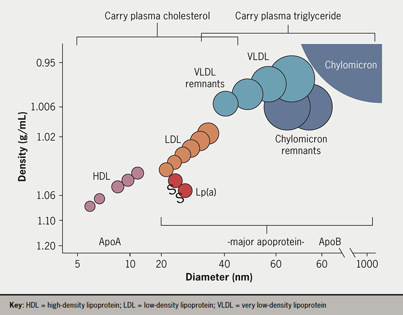
BJC supplement 2 2023 - Packard - Figure 1. Plasma lipoprotein spectrum. Apolipoprotein B-containing plasma lipoproteins comprise the triglyceride transporting chylomicrons and VLDL and their respective remnants; the cholesterol transporting LDL and lipoprotein(a) – a lipoprotein of unknown function that is formed as a complex of an LDL particle with apolipoprotein(a) – a large protein synthesised in the liver. The apoA-containing HDLs are involved in the transport of cholesterol from tissues to liver
Figure 1. Plasma lipoprotein spectrum. Apolipoprotein B-containing plasma lipoproteins comprise the triglyceride transporting chylomicrons and VLDL and their respective remnants; the cholesterol transporting LDL and lipoprotein(a) – a lipoprotein of unknown function that is formed as a complex of an LDL particle with apolipoprotein(a) – a large protein synthesised in the liver. The apoA-containing HDLs are involved in the transport of cholesterol from tissues to liver

In most populations, the levels of plasma TG and cholesterol far exceed the concentrations needed to support physiological processes, which lead to pathological consequences, such as atherosclerotic cardiovascular disease (ASCVD) and pancreatitis.1,2 Additionally, LDL is recognised as a causal risk factor for ASCVD;3,4 there is a strong, positive relationship between its plasma level and ASCVD risk that extends across the range seen in the population. Intervention trials have established that LDL-lowering reduces risk and the benefits of intensive reductions in this lipoprotein are now clear.4,5 Plasma TG, carried within chylomicrons and VLDL (collectively termed triglyceride-rich lipoproteins, TRL), has long been regarded as an important risk factor for the development of atherosclerosis; however, it is only recently that evidence has emerged supporting the causal role of TRL in ASCVD.1,2,6 Specifically, genetic variants that influence the level of plasma TG (and hence TRL) are linked with an altered risk of ASCVD events.1,6,7 Triglyceride itself, unlike cholesterol, is not an abundant constituent of atherosclerotic plaques and the current working hypothesis is that it is the TG-depleted, cholesterol-enriched ‘remnants’ of chylomicron and VLDL metabolism that contribute to atherogenesis.1,2

Definition, causes and consequences of hypertriglyceridaemia

Large epidemiological studies have reported consistently that plasma TG levels are strongly associated with ASCVD risk in populations (figure 2).1,2,6,8,9 As plasma TG concentration increases, metabolic and structural perturbations arise throughout the lipoprotein spectrum; TRL remnants are generated in increasing numbers and there are changes in LDL and HDL composition, notably the formation of small, dense LDL.1,10 In a recent consensus report from the European Atherosclerosis Society,1 definitions of the degrees of hypertriglyceridaemia were developed to reflect the nature of the relationship with clinical disorders such as ASCVD and pancreatitis (table 1). Since disadvantageous changes start to become evident at TG levels below the population average (which is ~1.5 mmol/L [~133 mg/dL]), an ‘optimal’ level was defined as <1.2 mmol/L (<106 mg/dL). In this range, production and clearance rates of TRL are entirely sufficient to maintain physiological health.
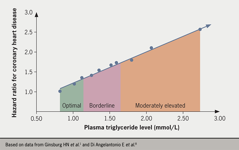
Figure 2. Relationship of plasma triglyceride (TG) levels to coronary heart disease risk. Using data from the Emerging Risk Factors Collaboration report, the hazard ratio for coronary heart disease (corrected for age and sex) was plotted against the usual mean plasma TG level (in mmol/L) for each decile in the pooled cohort examined. Decile 1, with the lowest TG level, was the reference group. The coloured segments relate to the degree of hypertriglyceridaemia, as defined in the European Atherosclerosis Society Consensus statement (the number of individuals in a general population who have severe or extreme hypertriglyceridaemia is small)
Figure 2. Relationship of plasma triglyceride (TG) levels to coronary heart disease risk. Using data from the Emerging Risk Factors Collaboration report,8 the hazard ratio for coronary heart disease (corrected for age and sex) was plotted against the usual mean plasma TG level (in mmol/L) for each decile in the pooled cohort examined. Decile 1, with the lowest TG level, was the reference group. The coloured segments relate to the degree of hypertriglyceridaemia, as defined in the European Atherosclerosis Society Consensus statement (the number of individuals in a general population who have severe or extreme hypertriglyceridaemia is small)1

Table 1. Hypertriglyceridaemia: definition and clinical sequelae

Category Plasma triglyceride level (mmol/L)/[mg/dL] Primary metabolic abnormality Clinical correlates
Optimal <1.2 [<106]
Borderline 1.2–1.7 [106–150] VLDL overproduction Overweight
ASCVD
Moderate 1.7–5.7 [150–504] VLDL overproduction plus defective clearance Obesity
Fatty liver disease
Type 2 diabetes
ASCVD
Severe 5.7–10.0 [504–885] Substantially defective clearance of VLDL and chylomicrons Obesity
Fatty liver disease
Type 2 diabetes
ASCVD
Extreme >10.0 [>885] Combined defective lipolysis of VLDL and chylomicrons Enlarged liver and spleen
Pancreatitis
Key: ASCVD = atherosclerotic cardiovascular disease; VLDL = very low-density lipoprotein

‘Borderline’ hypertriglyceridaemia is classified as 1.2 to 1.7 mmol/L (106 to 150 mg/dL), while ‘moderate’ and ‘severe’ forms of the condition are defined by plasma TG concentrations in the range 1.7 to 5.7 mmol/L (150 to 504 mg/dL) and 5.7 to 10.0 mmol/L (504 to 885 mg/dL), respectively. The rationale for setting the optimal level so low is that the association with increased risk of ASCVD is continuous and graded across the range of values seen in most countries (figure 2)1,2,6,8 in analogy with the observations for LDL cholesterol (LDL-C). ‘Extreme’ hypertriglyceridaemia was defined as a TG level greater than 10.0 mmol/L (885 mg/dL), which is a risk for developing the major clinical sequela of acute pancreatitis.

Metabolic changes that give rise to hypertriglyceridaemia are a combination of TRL overproduction, slower lipolysis rates and delayed clearance of particles from the circulation.1,10 Chylomicrons are produced in a wave during absorption of dietary fat in the intestine. These particles rapidly undergo lipolysis, delivering the TGs in their core mainly to adipose tissue, while the remnants formed are cleared by the liver (figure 3). VLDL is assembled using TG released from hepatic stores or made from fatty acids taken up from the circulation. It is secreted by the liver in response to the need to have TG present in the bloodstream in the periods between meals to provide an energy source for cardiac and skeletal muscle. Being overweight or obese is associated with increased production of VLDL (table 1).11,12 Higher rates of VLDL production are also a feature of both type 2 diabetes (since insulin is a major regulator of VLDL assembly),13 and fatty liver disease (as the organ attempts to export excess fat stored in cell droplets).1,12
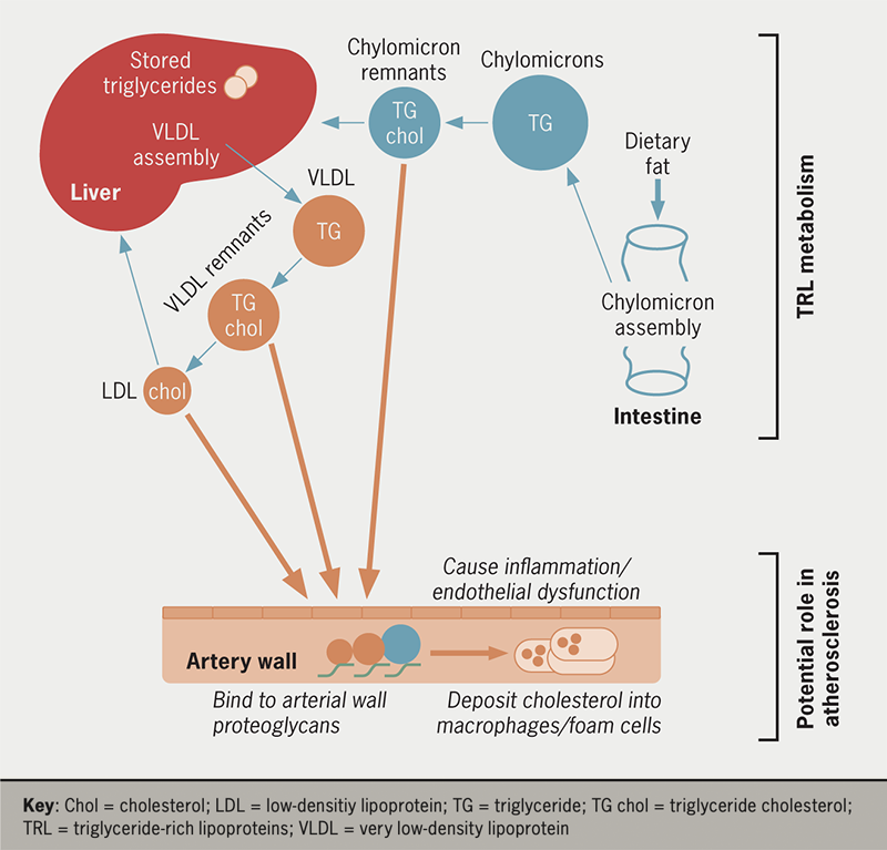
BJC Supplement 2 2023 - Packard - Figure 3. Metabolism of TRLs and their role in atherosclerosis. Chylomicrons are released in a ‘wave’ during fat absorption in the intestine. They are large, triglyceride-rich particles that rapidly undergo lipolysis by the action of lipoprotein lipase in capillary beds. During lipolysis, remnants are formed which normally are cleared by the liver. The liver assembles (using stored TG and fatty acids taken up from the circulation) and secretes VLDL to supply tissues with a source of TG in the periods between meals. VLDL undergoes lipolysis to become remnants, again via the action of lipoprotein lipase. VLDL remnants undergo two fates; a proportion of VLDL is converted into LDL while the remainder is cleared directly by the liver. As TRLs undergo lipolysis, the particles lose TG and acquire cholesterol so that the remnants become relatively cholesterol-enriched. These cholesterol-rich remnants, like LDL, can be taken up and retained by the artery wall. They have the ability to bind to arterial wall constituents, such as proteoglycans, and can contribute their cholesterol to resident macrophages to promote the formation of lipid-filled ‘foam’ cells. They may also act to promote local inflammation
Figure 3. Metabolism of TRLs and their role in atherosclerosis. Chylomicrons are released in a ‘wave’ during fat absorption in the intestine. They are large, triglyceride-rich particles that rapidly undergo lipolysis by the action of lipoprotein lipase in capillary beds. During lipolysis, remnants are formed which normally are cleared by the liver. The liver assembles (using stored TG and fatty acids taken up from the circulation) and secretes VLDL to supply tissues with a source of TG in the periods between meals. VLDL undergoes lipolysis to become remnants, again via the action of lipoprotein lipase. VLDL remnants undergo two fates; a proportion of VLDL is converted into LDL while the remainder is cleared directly by the liver.1,10 As TRLs undergo lipolysis, the particles lose TG and acquire cholesterol so that the remnants become relatively cholesterol-enriched.1,10 These cholesterol-rich remnants, like LDL, can be taken up and retained by the artery wall. They have the ability to bind to arterial wall constituents, such as proteoglycans, and can contribute their cholesterol to resident macrophages to promote the formation of lipid-filled ‘foam’ cells.1,10 They may also act to promote local inflammation1

More marked degrees of hypertriglyceridaemia are caused by defects in TRL lipolysis and clearance.1,10,14,15 Impaired lipase action slows the delipidation of both chylomicrons and VLDL and increases the generation of remnant lipoproteins (figure 3). Remnants are depleted in TG in the lipid core and become enriched in cholesterol. In individuals with optimal plasma TG levels, the concentration of remnants in the circulation is low but in hypertriglyceridaemic subjects, these particles accumulate and, like LDL-C, they have the ability to be taken up and retained by the artery wall (figure 3).1,2 Remnants appear to have a number of potentially deleterious effects leading to inflammation, endothelial dysfunction, and due to their high cholesterol content per particle, can contribute to macrophage ‘foam’ cell formation (so called because of the high lipid content seen on histology) in growing atherosclerotic plaques.1

Knowledge of the metabolic aberrations that lead to elevated plasma TG levels enables an informed approach to interventions designed to lower TRL concentrations and potentially reduce the risk of ASCVD and pancreatitis. Reduction in body weight results in lower liver fat and decreased rates of VLDL production.1,11 Likewise, improved diabetic glycaemic control is associated with lower VLDL secretion rates.1,13 In an alternate approach focusing on stimulating lipolysis, a number of novel agents are in development that promote the action of lipoprotein lipase, the major enzyme responsible for TRL delipidation.7 Both intervention routes – decreased production and increased lipolysis – are likely to lower the level of atherogenic remnant particles. The major defects in lipolysis seen in extreme hypertriglyceridaemia have been addressed by agents that stimulate lipase activity.16

Approaches to lipid lowering to prevent ASCVD

Raised plasma lipid levels are strongly associated with an increased risk of ASCVD, alongside smoking and high blood pressure. Intervention trials, especially those addressing the benefits of LDL-C lowering, have underpinned the concept that the relationship is causal; lipoproteins have a direct role in the development of atherosclerotic plaques and in the precipitation of a major clinical event, such as myocardial infarction and ischaemic stroke. International guidelines have been developed that provide evidence-based recommendations regarding efficacious treatment options and therapeutic goals.4,17 These guidelines recognise the long-term nature of the disease process – atherosclerosis is a silent disease of the arterial wall that develops over decades and then manifests in the form of an acute coronary or cerebral event – and the need to promote population-based healthy lifestyle advice on diet and behaviours. Another feature of the recommendations is the adoption of a graded, risk-based approach with the aggressiveness of the intervention tailored according to the predicted ongoing risk.

Nutrition and lifestyle advice

Nutrition and lifestyle advice

The cornerstone intervention in both primary and secondary prevention of ASCVD is the adoption of a prudent diet and the avoidance of behaviours known to increase risk, such as smoking and sedentary lifestyle. The metabolism of cholesterol and TG is intimately linked, as shown in figure 3, and the same general advice applies to lowering both plasma lipids through dietary changes. Guideline-recommended steps for lowering plasma TG levels1,4,17 are first to reduce overall calorie intake to achieve weight loss and reduce adiposity. Second, to alter the types of fat consumed by increasing the intake of fatty fish and by substituting saturated fat (animal fat and dairy) with mono- and polyunsaturated fat sources (plant oils and nuts) and to keep the intake of trans-fatty acids to a minimum. Third, avoid low-fat, highly refined, carbohydrate-rich foods and excess consumption of alcohol. Fourth, eat larger amounts of fibre-rich foodstuffs, such as vegetables and whole grain products.

Most people with hypertriglyceridaemia fall into the overweight or obese categories and weight reduction can often produce a much greater benefit in terms of lowering plasma TG than it does in reducing LDL-C. This is in part due to the close connection between visceral and hepatic fat stores and VLDL production as outlined above.12 In people with type 2 diabetes, the initial goal is to achieve optimum glycaemic control, which in itself, can result in substantial reductions in plasma TG levels.1,4,17

Managing raised LDL-C effectively

Three decades of clinical trials have demonstrated not only that lowering LDL-C reduces the risk of major coronary and cerebral atherothrombotic events but also that the association with risk extends to very low levels of LDL.5 That is, further clinical benefit is seen when LDL-C is reduced to <1.4 mmol/L (<54 mg/dL) or even <1.0 mmol/L (<39 mg/dL) compared to previous goals of approximately 1.8 mmol/L (69.5 mg/dL). No safety issues have arisen when LDL has been profoundly lowered; potential concerns relating to haemorrhagic stroke, cognitive function and cancer were examined and no evidence was seen of treatment-emergent adverse effects of the drugs used or the low LDL level achieved. Accordingly, the latest revision of the European guidelines sets aggressive goals for LDL-C lowering of <1.4 mmol/L (<54 mg/dL) for those at highest risk, typically individuals with a history of clinical ASCVD.4

In the risk-based strategy set out in these guidelines, individuals without evidence of clinical ASCVD are first offered lifestyle and diet advice, followed by moderate-dose statin therapy for those whose LDL-C remains above the threshold for pharmacologic intervention. In secondary prevention, statin monotherapy is first-line, and international surveys show the universality of this approach.18 However, this strategy has been found to be insufficient to allow the majority of patients to meet the previous goal for LDL-C of <1.8 mmol/L (<69.5 mg/dL) and only a minority are able to reach the revised lower target level. Recognition of this fact has led to the promotion of combination LDL-lowering therapy with agents such as ezetimibe and proprotein convertase subtilisin/kexin 9 (PCSK9) inhibitors. The latter class of drugs are particularly effective in reducing LDL and with their use, most people can achieve an LDL-C of <1.4 mmol/L (<54 mg/dL), if required.4,17,19 A strategy of early use/first-line combination therapy19 recognises that treatment with statins alone, even at maximally tolerated doses, does not achieve the full benefits of lipid lowering. Where the initial LDL-C level is high, it will predictably remain above the goal on monotherapy and the early use of combined drug regimens (as in hypertension treatment) delivers the optimum benefit quickly and efficiently.

Beyond LDL – addressing residual risk on statin therapy

Clinical trial evidence shows that in secondary prevention, patients on statin monotherapy with a high residual risk can usefully be divided into (i) patients in whom LDL-C is persistently high (who require combination LDL-lowering therapy as set out above), (ii) patients with elevated plasma TG levels that contribute to ongoing risk, (iii) patients in whom lipoprotein(a) is high and (iv) those with chronic inflammation).6 Analyses from clinical trials and ‘real-world’ surveys highlight the part that TRL and their remnants play in the high residual risk in individuals on statin monotherapy.1,20 In a Canadian registry study, approximately 25% of subjects on statins had high ASCVD risk associated with elevated plasma TG levels.20 Similarly, a Danish study reported an analysis of lipid-associated predictors of ASCVD risk in participants on statins.21 Those with above-the-median non-HDL cholesterol but below-the-median LDL-C – i.e., elevated TRL/remnant cholesterol – had a high continuing risk of ASCVD.

An active question in ASCVD prevention is how best to reduce the residual risk attributable to elevated plasma TG levels (and TRL) in patients on optimised statin therapy. Clinical trials have given mixed signals. The Reduction of Cardiovascular Events With Icosapent Ethyl–Intervention Trial (REDUCE-IT) revealed that administration of high-dose purified eicosapentaenoic acid was associated with a strong positive effect, reducing the risk of ASCVD by about 25% in statin-treated participants with established, or at high risk for, ASCVD who had plasma TG levels in the range of 1.5 to 5.0 mmol/L (i.e., moderately elevated levels [133 to 442 mg/dL]).22 Other recent trials in cohorts with similar risk and lipid profiles using an eicosapentaenoic/docosahaexanoic acid mix or a fibrate type-drug gave negative results for reasons that are presently unclear.23

Summary

Raised plasma TG levels are a biomarker of the presence of an increased concentration of TRL – chylomicrons, VLDL and their remnants – in the circulation and are associated with an elevated risk of ASCVD. The relationship is continuous and graded across the range of values seen in populations and accordingly, only low levels of plasma TG (<1.2 mmol/L [<106 mg/dL]) are considered optimal. Genetic studies indicate that the relationship of TRL with disease is causal and that lowering levels of plasma TG is likely to be a useful strategy for ASCVD risk reduction, especially for individuals on statins who have a high residual risk attributable to elevated TG levels.

Key messages

  • Individuals with moderately to severely elevated plasma triglyceride (TG) levels (1.7 to 10.0 mmol/L [150 to 885 mg/dL]) are at a high risk of developing atherosclerotic cardiovascular disease
  • An elevated plasma TG level is a biomarker of high levels of triglyceride-rich lipoproteins (TRLs) in the circulation; regulating these is a target for intervention, especially in patients with high residual risk on statin therapy
  • Diet and lifestyle interventions are the first steps required to reduce plasma TG levels; however, when this is insufficiently effective, pharmacotherapy is recommended in international guidelines to address the risk associated with raised plasma TG levels

Conflicts of interest

CP has received grants/honoraria from Amarin, Amgen, Daiichi-Sankyo, Dalcor, MSD, Novartis, Pfizer, and Response Therapeutics.

Chris J Packard
Honorary Senior Research Fellow and Professor of Vascular Biochemistry
Institute of Cardiovascular and Medical Sciences, Glasgow University, 126 University Place, Glasgow, G12 8TA
([email protected])

Articles in this supplement

Introduction
The evidence for fish oils and eicosapentaenoic acid in managing hypertriglyceridaemia
REDUCE-IT: findings and implications for practice
Icosapent ethyl use in clinical practice: current and future directions

References

1. Ginsberg HN, Packard CJ, Chapman MJ, et al. Triglyceride-rich lipoproteins and their remnants: metabolic insights, role in atherosclerotic cardiovascular disease, and emerging therapeutic strategies – a consensus statement from the European Atherosclerosis Society. Eur Heart J 2021;42:4791–806. https://doi.org/10.1093/eurheartj/ehab551

2. Nordestgaard BG, Varbo A. Triglycerides and cardiovascular disease. Lancet 2014;384:626–35. https://doi.org/10.1016/S0140-6736(14)61177-6

3. Borén J, Chapman MJ, Krauss RM, et al. Low-density lipoproteins cause atherosclerotic cardiovascular disease: pathophysiological, genetic and therapeutic insights: a consensus statement from the European Atherosclerosis Society Consensus Panel. Eur Heart J 2020;41:2313–30. https://doi.org/10.1093/eurheartj/ehz962

4. Mach F, Baigent C, Catapano AL, et al. ESC Scientific Document Group. 2019 ESC/EAS Guidelines for the management of dyslipidaemias: lipid modification to reduce cardiovascular risk. Eur Heart J 2020;41:111–88. https://doi.org/10.1093/eurheartj/ehz455

5. Packard C, Chapman MJ, Sibartie M, Laufs U, Masana L. Intensive low-density lipoprotein cholesterol lowering in cardiovascular disease prevention: opportunities and challenges. Heart 2021;107:1369–75. https://doi.org/10.1136/heartjnl-2020-318760

6. Libby P. The changing landscape of atherosclerosis. Nature 2021;592:524–33. https://doi.org/10.1038/s41586-021-03392-8

7. Tokgözoğlu L, Libby P. The dawn of a new era of targeted lipid-lowering therapies. Eur Heart J 2022;43:3198–208. https://doi.org/10.1093/eurheartj/ehab841

8. Di Angelantonio E, Sarwar N, Perry P, et al.; The Emerging Risk Factors Collaboration. Major lipids, apolipoproteins, and risk of vascular disease. JAMA 2009;302:1993–2000. https://doi.org/10.1001/jama.2009.1619

9. Sarwar N, Danesh J, Eiriksdottir G, et al. Triglycerides and the risk of coronary heart disease: 10,158 incident cases among 262,525 participants in 29 Western prospective studies. Circulation 2007;115:450–8. https://doi.org/10.1161/CIRCULATIONAHA.106.637793

10. Borén J, Taskinen MR, Björnson E, Packard CJ. Metabolism of triglyceride-rich lipoproteins in health and dyslipidaemia. Nat Rev Cardiol 2022;19:577–92. https://doi.org/10.1038/s41569-022-00676-y

11. Watts GF, Chan DC, Barrett PH, Susekov AV, Hua J, Song S. Fat compartments and apolipoprotein B-100 kinetics in overweight–obese men. Obes Res 2003;11:152–9. https://doi.org/10.1038/oby.2003.24

12. Borén J Watts GF, Adiels M, et al. Kinetic and related determinants of plasma triglyceride concentration in abdominal obesity: multicenter tracer kinetic study. Arterioscler Thromb Vasc Biol 2015;35:2218–24. https://doi.org/10.1161/ATVBAHA.115.305614

13. Taskinen M-R, Borén J. New insights into the pathophysiology of dyslipidemia in type 2 diabetes. Atherosclerosis 2015;239:483–95. https://doi.org/10.1016/j.atherosclerosis.2015.01.039

14. Taskinen M-R, Adiels M, Westerbacka J, et al. Dual metabolic defects are required to produce hypertriglyceridemia in obese subjects. Arterioscler Thromb Vasc Biol 2011;31:2144–50. https://doi.org/10.1161/ATVBAHA.111.224808

15. Baass A, Paquette M, Bernard S, Hegele RA. Familial chylomicronemia syndrome: an under-recognized cause of severe hypertriglyceridaemia. J Intern Med 2020;287:340–8. https://doi.org/10.1111/joim.13016

16. Shamsudeen I, Hegele RA. Safety and efficacy of therapies for chylomicronemia. Expert Rev Clin Pharmacol 2022;15:395–405. https://doi.org/10.1080/17512433.2022.2094768

17. Grundy SM, Stone NJ, Bailey AL, et al. 2018 AHA/ACC/AACVPR/AAPA/ABC/ACPM/ADA/AGS/APhA/ASPC/NLA/PCNA Guideline on the management of blood cholesterol: a report of the American College of Cardiology/American Heart Association Task Force on Clinical Practice Guidelines. Circulation 2019;139:e1082–143. https://doi.org/10.1161/CIR.0000000000000625

18. Ray KK, Molemans B, Schoonen WM, et al. EU-wide cross-sectional observational study of lipid-modifying therapy use in secondary and primary care: the DA VINCI study. Eur J Prev Cardiol 2021;28:1279–89. https://doi.org/10.1093/eurjpc/zwaa047

19. Ray KK, Reeskamp LF, Laufs U, et al. Combination lipid-lowering therapy as first-line strategy in very high-risk patients. Eur Heart J 2022;43:830–3. https://doi.org/10.1093/eurheartj/ehab718

20. Lawler PR, Kotrri G, Koh M, et al. Real-world risk of cardiovascular outcomes associated with hypertriglyceridaemia among individuals with atherosclerotic cardiovascular disease and potential eligibility for emerging therapies. Eur Heart J 2020;41:86–94. https://doi.org/10.1093/eurheartj/ehz767

21. Johannesen CDL, Mortensen MB, Langsted A, Nordestgaard BG. Apolipoprotein B and non-HDL cholesterol better reflect residual risk than LDL cholesterol in statin-treated patients. J Am Coll Cardiol 2021;77:1439–50. https://doi.org/10.1016/j.jacc.2021.01.027

22. Gaba P, Bhatt DL, Mason RP, Miller M, Verma S, Steg PG, Boden WE; REDUCE-IT Investigators. Benefits of icosapent ethyl for enhancing residual cardiovascular risk reduction: A review of key findings from REDUCE-IT. J Clin Lipidol 2022;16:389–402. https://doi.org/10.1016/j.jacl.2022.05.067

23. Virani SS. The fibrates story – a tepid end to a PROMINENT drug. N Engl J Med 2022;387:1991–2. https://doi.org/10.1056/NEJMe2213208

Disclaimer:

Medical knowledge is constantly changing. As new information becomes available, changes in treatment, procedures, equipment and the use of drugs become necessary. The editors/authors/contributors and the publishers Medinews (Cardiology) Ltd have taken care to ensure that the information given in this text is accurate and up to date at the time of publication.

Readers are strongly advised to confirm that the information, especially with regard to drug usage, complies with the latest legislation and standards of practice. Medinews (Cardiology) Limited advises healthcare professionals to consult up-to-date Prescribing Information and the full Summary of Product Characteristics available from the manufacturers before prescribing any product. Medinews (Cardiology) Limited cannot accept responsibility for any errors in prescribing which may occur.

The opinions, data and statements that appear are those of the contributors. The publishers, editors, and members of the editorial board do not necessarily share the views expressed herein. Although every effort is made to ensure accuracy and avoid mistakes, no liability on the part of the publisher, editors, the editorial board or their agents or employees is accepted for the consequences of any inaccurate or misleading information.

© Medinews (Cardiology) Ltd 2023. All rights reserved. No part of this publication may be reproduced, stored in a retrieval system, or transmitted in any form or by any means, electronic, mechanical, photocopying, recording or otherwise, without the prior written permission of the publishers, Medinews (Cardiology) Ltd. It shall not, by way of trade or otherwise, be lent, re-sold, hired or otherwise circulated without the publisher’s prior consent.

The evidence for fish oils and eicosapentaenoic acid in managing hypertriglyceridaemia

Br J Cardiol 2023;30(suppl 2):S10–S14doi:10.5837/bjc.2023.s07 Leave a comment
Click any image to enlarge
Authors:
Sponsorship Statement: Amarin UK Limited has funded this supplement through an independent grant and has had no control or input into the education content of this activity. Editorial and content decisions were made solely by the BJC.

There remains residual cardiovascular (CV) risk in some optimally treated patients with low levels of low-density lipoprotein cholesterol (LDL-C), which is associated with elevated triglyceride (TG) levels. Several trials of TG lowering drugs have failed to demonstrate a concomitant reduction in CV events. The Reduction of Cardiovascular Events with Icosapent Ethyl – Intervention Trial (REDUCE-IT) tested icosapent ethyl (IPE) – a purified formulation of the n-3 PUFA (omega-3 polyunsaturated fatty acid), eicosapentaenoic acid (EPA) – in high-risk CV patients with elevated TG levels and reported a significant reduction in a composite of CV events and revascularisations independent of TG reduction. These findings provide mechanistic insights into CV event reduction in patients with controlled LDL-C. The benefits are associated with on-treatment EPA levels and are in keeping with the broad pleiotropic actions previously reported in cellular and clinical studies that favourably modulate atherosclerotic plaque composition and progression. The association of high-dose IPE with atrial fibrillation provides a cautionary footnote and careful consideration of the risk-benefit ratio is required when commencing treatment with IPE.

Background

Sustained reduction of elevated cholesterol (particularly low-density lipoprotein cholesterol [LDL-C]) with currently available therapies is associated with reduced atherosclerotic cardiovascular (CV) events in both primary and secondary prevention.1 Nevertheless, some individuals continue to exhibit substantial residual CV risk, which is associated with higher concentrations of atherogenic cholesterol carried by circulating triglyceride (TG)-rich lipoproteins.

The failure to reduce CV events through TG reduction in statin-treated patients with niacin, fibrates and a carboxylic acid formulation of omega-3 polyunsaturated fatty acids (n-3 PUFAs) has cast doubt on the mechanistic role of TGs in atherosclerotic cardiovascular disease (ASCVD).

This review will focus on trials of fish oils which have compared mixed formulations (eicosapentaenoic acid [EPA] and docosahexaenoic acid [DHA]) versus EPA alone and included primary and secondary prevention populations. It will identify mechanisms of action and efficacy in CV event reduction in individuals with elevated TGs.

Clinical trials of triglyceride reduction

Atherosclerosis

Three large n-3 PUFA trials showed no evidence between TG lowering and CV events – consistent with previous trials of TG-lowering agents in statin-treated patients using fibrates and niacin.1 However, there are two trials where a reduction of TG (Japan EPA Lipid Intervention Study [JELIS] 9% and the Reduction of Cardiovascular Events with Icosapent Ethyl–Intervention Trial [REDUCE-IT] 17%)2,3 led to reduced composite CV end points (a composite of cardiovascular death, non-fatal myocardial infarction, non-fatal stroke, coronary revascularisation or unstable angina). A deeper exploration of the differences between trials and the active agents tested is required to inform future direction – if not TG lowering as the driver of CV event reduction, then what?

In contrast to other TG-lowering trials, the JELIS study did not prespecify a minimum TG level for study inclusion. This trial in a Japanese population showed that an ethyl ester of EPA (icosapent ethyl [IPE], 1.8 g/d) plus a statin delivered a 19% (p=0.011) reduction in a composite of CV events. The median plasma TG level at baseline of 1.7 mmol/L (153 mg/dL) probably reflected the fact that the study included patients for both primary and secondary prevention. It is worth noting that half of the patients had TG levels within the normal range and the treatment arm recorded an overall reduction of 9% compared with baseline. When outcomes were stratified to TG levels, there was a 53% reduction (p=0.04) in CV events with IPE in subjects with higher TG levels (>1.7 mmol/L [>150 mg/dL]) and low high-density lipoprotein cholesterol (HDL-C) levels (<0.45 mmol/L [<40 mg/dL]) based on a post-hoc analysis. Importantly, however, there was no relationship between the extent of TG lowering and CV events.

A higher dose of IPE (4 g/day) in REDUCE-IT led to a significant reduction of composite CV events in at-risk patients with elevated TG levels (median 2.44 mmol/L [216 mg/dL]) on maximally tolerated statins. First ischaemic events fell by 25% (p<0.001) and total (first and subsequent) ischaemic events by 31% (p<0.001). Consistent benefits were recorded across multiple pre-specified subgroups, including the primary and secondary prevention setting groups.4 The large reductions in end points exceeded that which would be expected from the 17% decrease in TG levels and again, raises questions about the mechanistic role of TGs in atherothrombotic events.5

Differences between trials

The range of CV event reductions reported from trials requires an understanding of the differences in study design between trials. These differences make direct comparisons challenging.5,6 Trials differed in the following:

  • Formulations tested (EPA vs. EPA+DHA). There are clear differences between EPA and DHA at a cellular level leading to suggestions that there may be a ‘diluting’ effect of DHA. Both differ in their effects on membrane structure, rates of lipid oxidation, inflammatory biomarkers, and tissue distribution
  • Dose of n-3 PUFAs tested. The absence of benefit seen in some trials may be because the dose used failed to provide the necessary threshold of achieved EPA level (~100 μg/mL) to produce clinical benefit. In the Outcomes Study to Assess STatin Residual Risk Reduction With EpaNova in HiGh CV Risk PatienTs With Hypertriglyceridemia (STRENGTH), the on-treatment levels of EPA (89.6 μg/mL) were lower than the baseline EPA levels in JELIS (97 μg/mL) and 38% lower than on-treatment levels in REDUCE-IT (144 μg/mL).1–3
  • Type of placebo used. Studies varied between using no placebo or an oil substitute. Whereas STRENGTH used corn oil, the most controversial was the use of mineral oil in REDUCE-IT, raising questions about its adverse effect on CV events and thereby exaggerating the beneficial effect of IPE. A post-hoc analysis showed that the favourable effects of IPE were not influenced by fluctuations in LDL-C levels in the placebo arm.7 A sub-analysis of JELIS reported benefits to be directly related to EPA plasma levels.8 Furthermore, the US Food and Drug Administration concluded that only a small fraction, if any, of the difference could be attributed to the use of mineral oil based on comparative trials.9
  • Study patient profile. All trials included patients at high risk of CV disease but some trials were exclusively in the setting of secondary prevention while others included both primary and secondary prevention settings.10

Cardiovascular risk reduction

Prespecified sub-analyses of REDUCE-IT11 reported all coronary revascularisations, subtypes of revascularisations and recurrent revascularisations. The results showed that IPE plus statins reduced the need for first and subsequent coronary revascularisations in patients with elevated TG levels and increased CV risk. Over the course of the trial (4.9 years), first coronary revascularisations were significantly reduced by 34%, with an absolute risk reduction (ARR) of 4.1% and a number needed to treat (NNT) of 24. It is interesting to note that the separation of the curves occurred early with a significant difference seen at 11 months. There were also clinically meaningful relative risk reductions (RRRs) of ≥32% in time-to-first occurrences of elective, urgent or emergent revascularisations as individual or composite end points.

Looking at revascularisation subtypes, a qualitatively similar effect was seen with a significant reduction in patients undergoing percutaneous coronary intervention in the treated arm (7.7% vs. 10.9%; p<0.001) with an ARR of 3.2% and an NNT of 31. For surgical revascularisation, there was also a significant reduction in the treated group (1.9% vs. 3.0%; p=0.0005), with an ARR of 1.1% and an NNT of 87.

Recorded plasma EPA levels showed no difference at baseline but after 360 days of active treatment, there was a consistent and statistically significant elevation in patients who did not require coronary revascularisation. This is an important observation as it suggests that clinical benefit in CV event reduction is dependent on attaining a – yet to be determined – adequate plasma EPA level and may be an explanation for the failure of previous low-dose fish oil trials and those with a mixed EPA/DHA preparation to show CV benefit.

The substudy of revascularisation – once again – highlights the effectiveness of IPE in reducing CV events to be greater than the modest degree of triglyceride reduction observed, suggesting additional mechanisms of risk reduction.

Patients with type 2 diabetes and chronic kidney disease

A substudy of 4,787 patients with diabetes12 reported a 24% reduction in the total number of CV events in the IPE-treated group (764 vs. 998; p=0.0003). The greatest gain was in those with established ASCVD who were found to have a RRR of 30%, ARR of 24% and NNT of 5. The safety of IPE in patients with diabetes was similar to the total REDUCE-IT population, with a 1% increased risk of atrial fibrillation (AF) and 0.7% increased risk of bleeding events. The authors concluded that IPE had a substantial positive impact in high-risk patients with diabetes with a good safety profile.

In the renal substudy,13 there were no meaningful changes in median estimated glomerular filtration rate (eGFR) in the IPE arm across study visits. IPE treatment led to consistent reductions in both the primary and key secondary composite end points across baseline eGFR categories. Patients with an eGFR <60 mL/min/1.73 m2 recorded the largest ARR and similar RRR for the primary composite (21.8% vs. 28.9%; p=0.0002) and key secondary composite end point (16.8% vs. 22.5%; p=0.001). The greatest numeric reduction in CV-related death was seen in the lower range eGFR <60 mL/min/1.73 m2 group (7.6% vs. 10.6%; p=0.02). Adverse events were numerically but not significantly higher in the eGFR <60 mL/min/1.73 m2 group; namely AF/flutter (4.2% vs. 3.0%; p=0.17) and serious bleeding (5.4% vs. 3.6%; p=0.13). It is important to note that the increased incidence of AF and bleeding across the trial was not associated with increased ischaemic stroke events over the full duration of the trial.

Pleiotropic mechanism of action of n-3 PUFA that modulates atherosclerosis development

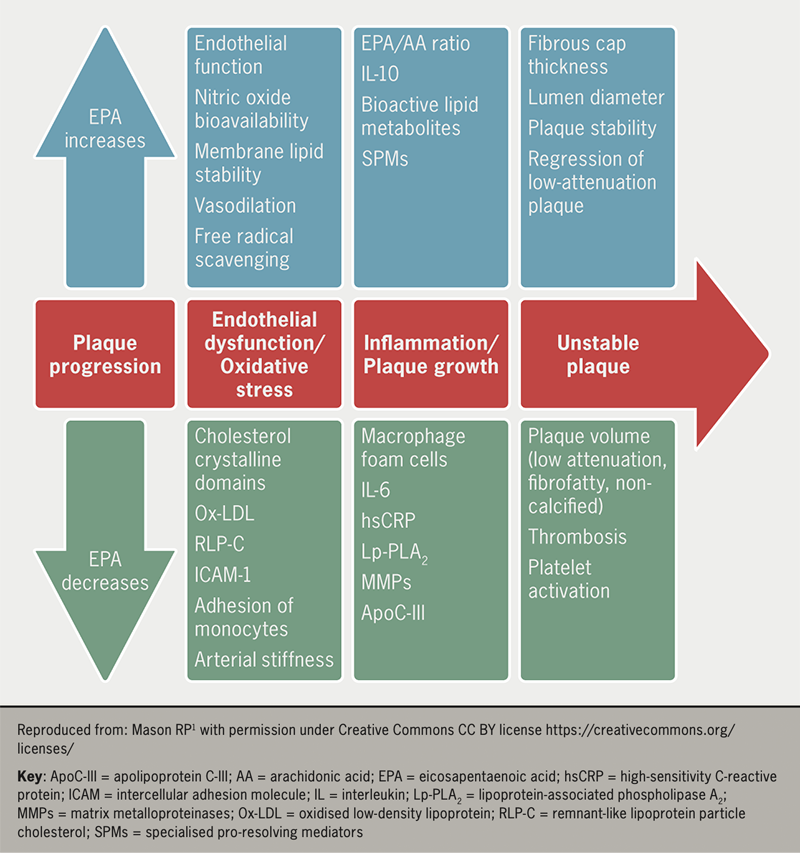
The consistent absence of a relationship between reductions in TG levels and CV outcomes raises questions about underlying mechanisms. Several reviews consistently reveal an absence of benefit in clinical trials wherein both EPA and DHA were tested in amounts of ~1 g/day. Several laboratory studies show a pleiotropic effect of fish oils that may have contributed to the reductions seen in CV events in some trials (figure 1). In REDUCE-IT, there was reduced lipoprotein-associated phospholipase A2, interleukin-6, high-sensitivity C-reactive protein, apolipoprotein C-III, and oxidised LDL-C concentrations compared with placebo controls.14,15

BJC Supplement 2 - Zaman - Figure 1. Atheroprotective effects of EPA during progression of arterial disease, including endothelial dysfunction, oxidative stress, inflammation and changes in plaque stability
Figure 1. Atheroprotective effects of EPA during progression of arterial disease, including endothelial dysfunction, oxidative stress, inflammation and changes in plaque stability

Experimental evidence of the pleiotropic effects16 of fish oil that may favourably modify atherothrombotic pathology include:

  • Anti-inflammatory. Studies show n-3 PUFAs reduce inflammation and suppress expressions of inflammatory cells and inflammatory cytokines (C-reactive protein, tumour necrosis factor, interferon and interleukins)16
  • Endothelial function. Trials show that fish oil consumption lowers circulating markers of endothelial dysfunction and increases flow-mediated vasodilation markers, which reflect improved endothelial function
  • Antithrombotic function. n-3 PUFAs affect platelet aggregation through several mechanisms, such as the generation of thromboxane A3 and prostacyclin, that have cardioprotective and antithrombotic effects in patients treated with EPA and DHA.

Clinical evidence from trials of n-3 PUFAs lend support to an atheroprotective effect of EPA and its bioactive lipid metabolites beyond TG reduction. The effect of IPE treatment on plaque development was studied in the Effect of VASCEPA® on Improving Coronary Atherosclerosis in People With High Triglycerides Taking Statin Therapy (EVAPORATE) trial.17 Following 18 months of treatment with 4 g/day of IPE in 80 patients with high TG levels and coronary artery disease, coronary computed tomography angiography revealed a relative reduction of 17% (p<0.01) in low-attenuation plaques. The importance of this finding relates to the strong predictive ability of low-attenuation atherosclerotic plaques for fatal or non-fatal MI. This modulatory, favourable effect on plaque composition may explain the CV risk reduction seen with high-dose EPA.18 Another study using intravascular ultrasound to image plaques in coronary arteries also demonstrated a significant reduction in plaque volume in the statin and EPA combination treatment group.19

Therefore, there is mounting evidence from laboratory and clinical investigations that provide insights into the CV benefits of IPE seen in REDUCE-IT and JELIS that are independent of TG reduction.5

Adverse effects of fish oil

The most commonly reported adverse effects are minor gastrointestinal upset, such as diarrhoea, nausea, dyspepsia and abdominal discomfort, which may limit use in patients with existing digestive disorders. There is a dose-dependent increased bleeding tendency with n-3 PUFAs that is not of clinical significance, even with concurrent antiplatelet therapy.20

Several trials report an increased risk of developing AF with high-dose n-3 PUFAs. REDUCE-IT showed a near 50% increase in hospitalisations for AF in the IPE arm, whilst the STRENGTH trial was stopped early, partly because of an increased risk of AF. Two meta-analyses, one of seven randomised control trials and the second of 38 randomised control trials, reported a 25% and 26% increase in new-onset AF, respectively, but no increase in ischaemic strokes.21,22 These findings were surprising, as earlier studies had suggested that n-3 PUFAs have anti-AF effects,23 which is concerning, given that AF is associated with an increased risk of morbidities (stroke and heart failure) and mortality. However, the results of a meta-analysis of trials of fish oils should be treated with caution as they may overestimate the risk of AF because of informative censoring i.e., if n-3 PUFAs reduce mortality or delay death, or the treated patients have more time alive to acquire AF than controls, which could potentially inflate the incidence of AF in the n-3 PUFA arm.24 For this reason, the real risk of AF from n-3 PUFA supplements remains unknown and further research is warranted. Until then, as with any intervention, clinicians should weigh the risk-benefit ratio when recommending n-3 PUFA supplements for primary or secondary prevention of CV disease.

Conclusion

Numerous trials of fish oils in patients with CV disease have failed to deliver a clear message unlike, for example, trials of statins in a similar patient cohort. Therefore, it is important to examine each trial on its own merit, rather than pooling results through meta-analyses.

Additionally, it is imperative to note that firstly, the fish oil preparations that were tested varied in composition. Secondly, in earlier studies, a majority of patients were not on a statin. Thirdly, the trial populations studied differed in their inclusion of primary and/or secondary risk patients and TG levels. Lastly, where used, the choice of placebos differed between trials.

A careful dissection of each trial suggests consistency in the ability of n-3 PUFAs to reduce TG levels but no correlation between TG reduction and CV events. To realise reductions in CV events, a high dose (2–4 g/day) of purified n-3 PUFAs is necessary to maintain high plasma EPA levels. The benefits of high-dose IPE, which include reduced revascularisation and favourable plaque modification, indicate that the mechanism is likely due to its pleiotropic actions that correlate with on-treatment EPA levels. Subgroup analyses show enhanced benefit in patients with diabetes and an eGFR <60 mL/min/1.73 m2.

Whilst increased rates of AF/flutter and bleeding were seen, these did not lead to strokes or fatalities. The impressive results from REDUCE-IT prompted an update to the European Society of Cardiology guidelines for dyslipidaemia to recommend IPE for patients with CV disease and high triglyceride levels in combination with a statin.25

Key messages

  • Triglycerides (TG) are associated with increased cardiovascular (CV) events; however, trials of effective TG reduction show no reduction in CV events
  • High-dose purified omega-3 polyunsaturated fatty acids (n-3 PUFAs) significantly reduce CV events and revascularisation episodes independent of their effect on TG reduction
  • The reduction in CV events is likely due to the pleiotropic effect of fish oils on modulating the atherosclerotic process

Conflicts of interest

AGZ has received lecture fees from Amarin. MO and HE: none declared.

Muntaser Omari
Medical Training Initiative Fellow

Holli Evans
Internal Medicine Trainee Year 2

Freeman Hospital, Newcastle upon Tyne, NE7 7DN

Azfar G Zaman
Consultant Interventional Cardiologist (Freeman Hospital, Newcastle), and Honorary Clinical Professor of Cardiology (Newcastle University)
Vascular Biology and Medicine, Newcastle University School of Medicine, Newcastle upon Tyne, NE2 4HH
([email protected])

Articles in this supplement

Introduction
Triglyceride-rich lipoproteins and their role in cardiovascular disease
REDUCE-IT: findings and implications for practice
Icosapent ethyl use in clinical practice: current and future directions

References

1. Mason RP, Eckel RH. Mechanistic insights from REDUCE-IT STRENGTHen the case against triglyceride lowering as a strategy for cardiovascular risk reduction. Am J Med 2021;134:1085–90. https://doi.org/10.1016/j.amjmed.2021.03.014

2. Yokoyama M, Origasa H, Matsuzaki M, et al. Effects of eicosapentaenoic acid on major coronary events in hypercholesterolaemic patients (JELIS): a randomised open-label, blinded endpoint analysis. Lancet 2007;369:1090–8. https://doi.org/10.1016/S0140-6736(07)60527-3

3. Bhatt DL, Steg PG, Miller M, et al.; REDUCE-IT Investigators. Cardiovascular risk reduction with icosapent ethyl for hypertriglyceridemia. N Engl J Med 2019;380:11–22. https://doi.org/10.1056/NEJMoa1812792

4. Bhatt DL, Steg PG, Miller M, et al.; REDUCE-IT Investigators. Effects of icosapent ethyl on total ischemic events: from REDUCE-IT. J Am Coll Cardiol 2019;73:2791–802. https://doi.org/10.1016/j.jacc.2019.02.032

5. Mason RP, Libby P, Bhatt DL. Emerging mechanisms of cardiovascular protection for the omega-3 fatty acid eicosapentaenoic acid. Arterioscler Thromb Vasc Biol 2020;40:1135–47. https://doi.org/10.1161/ATVBAHA.119.313286

6. Mason RP, Dawoud H, Jacob RF, Sherratt SCR, Malinski T. Eicosapentaenoic acid improves endothelial function and nitric oxide bioavailability in a manner that is enhanced in combination with a statin. Biomed Pharmacother 2018;103:1231–7. https://doi.org/10.1016/j.biopha.2018.04.118

7. Olshansky B, Chung MK, Budoff MJ, et al. Mineral oil: safety and use as placebo in REDUCE-IT and other clinical studies. Eur Heart J 2020;22(Suppl J):J34–48. https://doi.org/10.1093/eurheartj/suaa117

8. Pisaniello AD, Nicholls SJ, Ballantyne CM, Bhatt DL, Wong ND. Eicosapentaenoic acid: atheroprotective properties and the reduction of atherosclerotic cardiovascular disease events. EMJ 2020;5:29–36. https://www.emjreviews.com/cardiology/symposium/eicosapentaenoic-acid-atheroprotective-properties-and-the-reduction-of-atherosclerotic-cardiovascular-disease-events/

9. Food and Drug Administration. Endocrinologic and Metabolic Drugs Advisory Committee briefing document. Available at: http://epadruginitiative.com/files/FDA_Briefing_Document_for_ADCOM.pdf [accessed 31 March 2023].

10. Sharma G, Martin SS, Blumenthal RS. Effects of omega-3 fatty acids on major adverse cardiovascular events: what matters most: the drug, the dose, or the placebo? JAMA 2020;324:2262–4. https://doi.org/10.1001/jama.2020.22387

11. Peterson BE, Bhatt DL, Steg G, et al.; REDUCE-IT Investigators. Reduction in revascularization with icosapent ethyl insights from REDUCE-IT revascularization analyses. Circulation 2021;143:33–44. https://doi.org/10.1161/CIRCULATIONAHA.120.050276

12. American Diabetes Association. Substantial cardiovascular benefit from icosapent ethyl in patients with diabetes: REDUCE-IT DIABETES. 80th Scientific Session, June 12–16, 2020. Available at: https://plan.core-apps.com/tristar_ada20/abstract/2a6a17fd-a7a6-4bc9-8bf8-35847174c48d [accessed 31 March 2023].

13. Majithia A, Bhatt DL, Friedman AN, et al. Benefits of icosapent ethyl across the range of kidney function in patients with established cardiovascular disease or diabetes: REDUCE-IT RENAL. Circulation 2021;144:1750–9. https://doi.org/10.1161/CIRCULATIONAHA.121.055560

14. Bays HE, Ballantyne CM, Braeckman RA, Stirtan WG, Soni PN. Icosapent ethyl, a pure ethyl ester of eicosapentaenoic acid: effects on circulating markers of inflammation from the MARINE and ANCHOR studies. Am J Cardiovasc Drugs 2013;13:37–46. https://doi.org/10.1007/s40256-012-0002-3

15. Ballantyne CM, Bays HE, Braeckman RA, et al. Icosapent ethyl (eicosapentaenoic acid ethyl ester): effects on plasma apolipoprotein C-III levels in patients from the MARINE and ANCHOR studies. J Clin Lipidol 2016;10:635–45.e1. https://doi.org/10.1016/j.jacl.2016.02.008

16. Sherratt SCR, Libby P, Bhatt DL, Mason RP. A biological rationale for the disparate effects of omega-3 fatty acids on cardiovascular disease outcomes. Prostaglandins Leukot Essent Fatty Acids 2022;182:102450. https://doi.org/10.1016/j.plefa.2022.102450

17. Budoff MJ, Bhatt DL, Kinninger A, et al. Effect of icosapent ethyl on progression of coronary atherosclerosis in patients with elevated triglycerides on statin therapy: final results of the EVAPORATE trial. Eur Heart J 2020;41:3925–32. https://doi.org/10.1093/eurheartj/ehaa652

18. Williams MC, Moss AJ, Dweck M, et al. Coronary artery plaque characteristics associated with adverse outcomes in the SCOT-HEART study. J Am Coll Cardiol 2019;73:291–301. https://doi.org/10.1016/j.jacc.2018.10.066

19. Watanabe T, Ando K, Daidoji H, et al.; CHERRY study investigators. A randomized controlled trial of eicosapentaenoic acid in patients with coronary heart disease on statins. J Cardiol 2017;70:537–44. https://doi.org/10.1016/j.jjcc.2017.07.007

20. Bays HE. Safety considerations with omega-3 fatty acid therapy. Am J Cardiol 2007;99:35C−43C. https://doi.org/10.1016/j.amjcard.2006.11.020

21. Gencer B, Djousse L, Al-Ramady OT, Cook NR, Manson JE, Albert CM. Effect of long-term marine ɷ-3 fatty acids supplementation on the risk of atrial fibrillation in randomized controlled trials of cardiovascular outcomes: a systematic review and meta-analysis. Circulation 2021;144:1981–90. https://doi.org/10.1161/CIRCULATIONAHA.121.055654

22. Khan SU, Lone AN, Khan MS, et al. Effect of omega-3 fatty acids on cardiovascular outcomes: a systematic review and meta-analysis. EClinicalMedicine 2021;38:100997 https://doi.org/10.1016/j.eclinm.2021.100997

23. Calò L, Martino A, Tota C. The anti-arrhythmic effects of n-3 PUFAs. Int J Cardiol 2013;170(2 suppl 1):S21–7. https://doi.org/10.1016/j.ijcard.2013.06.043

24. Samuel M, Nattel S. Fish oil supplements may increase the risk for atrial fibrillation: what does this mean? Circulation 2021;144:1991–4. https://doi.org/10.1161/CIRCULATIONAHA.121.057464

25. Mach F, Baigent C, Catapano AL, et al.; ESC Scientific Document Group. 2019 ESC/EAS guidelines for the management of dyslipidaemias: lipid modification to reduce cardiovascular risk. Eur Heart J 2020;41:111–88. https://doi.org/10.1093/eurheartj/ehz455

Disclaimer:

Medical knowledge is constantly changing. As new information becomes available, changes in treatment, procedures, equipment and the use of drugs become necessary. The editors/authors/contributors and the publishers Medinews (Cardiology) Ltd have taken care to ensure that the information given in this text is accurate and up to date at the time of publication.

Readers are strongly advised to confirm that the information, especially with regard to drug usage, complies with the latest legislation and standards of practice. Medinews (Cardiology) Limited advises healthcare professionals to consult up-to-date Prescribing Information and the full Summary of Product Characteristics available from the manufacturers before prescribing any product. Medinews (Cardiology) Limited cannot accept responsibility for any errors in prescribing which may occur.

The opinions, data and statements that appear are those of the contributors. The publishers, editors, and members of the editorial board do not necessarily share the views expressed herein. Although every effort is made to ensure accuracy and avoid mistakes, no liability on the part of the publisher, editors, the editorial board or their agents or employees is accepted for the consequences of any inaccurate or misleading information.

© Medinews (Cardiology) Ltd 2023. All rights reserved. No part of this publication may be reproduced, stored in a retrieval system, or transmitted in any form or by any means, electronic, mechanical, photocopying, recording or otherwise, without the prior written permission of the publishers, Medinews (Cardiology) Ltd. It shall not, by way of trade or otherwise, be lent, re-sold, hired or otherwise circulated without the publisher’s prior consent.

REDUCE-IT: findings and implications for practice

Br J Cardiol 2023;30(suppl 2):S15–S18doi:10.5837/bjc.2023.s08 Leave a comment
Click any image to enlarge
Authors:
Sponsorship Statement: Amarin UK Limited has funded this supplement through an independent grant and has had no control or input into the education content of this activity. Editorial and content decisions were made solely by the BJC.

The Reduction of Cardiovascular Events with Icosapent Ethyl-Intervention Trial (REDUCE-IT) was a randomised, double-blind, placebo-controlled trial designed to test the hypothesis that patients with hypertriglyceridaemia and a prior history of cardiovascular (CV) disease or diabetes and multiple CV disease risk factors would benefit from icosapent ethyl (IPE), a highly purified (>96%) form of eicosapentaenoic acid (EPA). Participants (n=8,179) were assigned to IPE 2 g twice daily or matching placebo and monitored for a median period of 4.9 years. Overall, there was a 25% relative risk reduction and 4.8% (p<0.0001) absolute risk reduction in the primary composite end point (CV death, non-fatal myocardial infarction [MI], non-fatal stroke, coronary revascularisation or hospitalisation for unstable angina) in the IPE versus placebo group. Similarly, the key secondary composite end points of CV death, non-fatal MI and non-fatal stroke were reduced by 26% (p<0.0001) with an absolute between-group difference of 3.6%. A number of prespecified and/or post-hoc analyses have consistently demonstrated favourable results with IPE use. Considering the reductions in multiple CV events, REDUCE-IT was a landmark CV clinical trial that demonstrated the benefit of IPE in high-risk patients with hypertriglyceridaemia.

Introduction

Multiple epidemiologic studies have identified elevated levels of blood triglycerides (TGs) or the phenotypic state of hypertriglyceridaemia (HTG) as being associated with an elevated risk of cardiovascular (CV) disease.1 Among therapies demonstrating significant reduction in TGs – beyond fibrates, niacin and statins – are marine-derived omega-3 polyunsaturated fatty acids (n-3 PUFAs); they consist principally of eicosapentaenoic acid (EPA) and docosahexaenoic acid (DHA). Notably, the first Gruppo Italiano per lo Studio della Streptochiansi nell’Infarto (GISSI) supported a potential role of n-3 PUFAs in reducing death, non-fatal myocardial infarction (MI) and stroke – prior to studies conducted in the 1990s and the widespread use of statins.2 The Japan EPA Lipid Intervention Study (JELIS) was the first clinical trial to test purified EPA in patients with hyperlipidaemia, which demonstrated a 19% reduction in major CV events in Japanese patients with pre-existing coronary disease assigned to purified EPA and low-dose statin treatment (simvastatin or pravastatin) versus low-dose statin monotherapy.3 In a subgroup of patients with HTG (TG ≥1.7 mmol/L [≥150 mg/dL]) and low levels of high-density lipoprotein (HDL-C), a post-hoc analysis of JELIS found that EPA reduced the risk of CV disease by 53%.4 These impressive results provided the proof-of-concept foundation in support of a large clinical trial testing purified EPA in high-risk patients with HTG, thereby providing the rationale for the Reduction of Cardiovascular Events with Icosapent Ethyl – Intervention Trial (REDUCE-IT).5

REDUCE-IT

Icosapent ethyl (IPE)

REDUCE-IT was a randomised, double-blind, placebo-controlled trial in patients with HTG (fasting TG levels of 1.7–5.6 mmol/L [150–499 mg/dL]) with well-controlled low-density lipoprotein cholesterol (LDL-C) levels of 1.03–2.58 mmol/L (40–100 mg/dL) on stable statin ± ezetimibe therapy and other key inclusion criteria that consisted of 1) age ≥45 years with a prior history of CV disease (secondary prevention cohort) or 2) age ≥50 years with diabetes and at least one additional CV disease risk factor (primary prevention cohort). The CV disease risk factors were more advanced age (men ≥55 years and women ≥65 years), active cigarette smoking (or having quit within three months of study enrolment), hypertension (≥140 mmHg systolic or ≥90 mmHg diastolic), HDL-C ≤1.03 mmol/L (≤40 mg/dL) for men or ≤1.29 mmol/L (50 mg/dL) for women, high-sensitivity C-reactive protein (hsCRP) >3 mg/L, creatinine clearance between 30–60 mL/min/1.73 m2, retinopathy, albuminuria or an ankle brachial index (ABI) <0.9 without symptoms of intermittent claudication. A total of 8,179 patients were randomised to receive either icosapent ethyl (IPE) 2 g twice daily or matching placebo and monitored for a median period of 4.9 years.6 The primary end point was the first occurrence of a major adverse CV event (MACE) defined by five-point MACE (CV death, non-fatal MI, non-fatal stroke, coronary revascularisation or unstable angina necessitating hospitalisation) and the secondary end point was the first occurrence of a MACE defined by three-point MACE (CV death, non-fatal MI and non-fatal stroke). The effects on biomarkers between baseline and year 1 is shown in table 1. There were significant reductions in median TG levels (19.7%) in IPE-treated patients, as well as significant reductions in non-HDL-C, LDL-C, apolipoprotein B (ApoB) and hsCRP. Unsurprisingly, blood EPA levels also increased significantly (359%) between baseline and year 1 following IPE treatment.

Table 1. REDUCE-IT: effects on biomarkers between baseline and year 1

Icosapent ethyl
(n=4,089)
Median
Placebo
(n=4,090)
Median
Median between group difference at year 1
Biomarker Baseline Year 1 Baseline Year 1 Absolute change from baseline % Change from baseline % Change p-value
Triglycerides mmol/L (mg/dL) 2.45
(216.5)
1.98
(175.0)
2.44
(216.0)
2.50
(221.0)
−0.50
(−44.5)
−19.7 <0.0001
Non-HDL-C mmol/L
(mg/dL)
3.06
(118.0)
2.93
(113.0)
3.07
(118.5)
3.37
(130.0)
−0.40
(−15.5)
−13.1 <0.0001
LDL-C mmol/L
(mg/dL)
1.92
(74.0)
1.99
(77.0)
1.97
(76.0)
2.18
(84.0)
−0.13
(−5.0)
−6.6 <0.0001
HDL-C mmol/L
(mg/dL)
1.04
(40.0)
1.01
(39.0)
1.04
(40.0)
1.09
(42.0)
−0.06
(−2.5)
−6.3 <0.0001
ApoB g/L
(mg/dL)
0.82
(82.0)
0.80
(80.0)
0.83
(83.0)
0.89
(89.0)
−0.08
(−8.0)
−9.7 <0.0001
hsCRP mg/L 2.2 1.8 2.1 2.8 −0.9 −39.9 <0.0001
Log hsCRP mg/L 0.8 0.6 0.8 1.0 −0.4 −22.5 <0.0001
EPA μg/mL 26.1 144.0 26.1 23.3 +114.9 +358.8 <0.0001
Key: ApoB = apolipoprotein B; EPA = eicosapentaenoic acid; HDL-C = high-density lipoprotein cholesterol; hsCRP = high-sensitivity C-reactive protein; LDL-C = low-density lipoprotein cholesterol; REDUCE-IT = Reduction of Cardiovascular Events with Icosapent Ethyl – Intervention Trial

Overall, the results of REDUCE-IT showed that assignment to IPE produced a 25% relative risk reduction and 4.8% (p<0.0001) absolute risk reduction in the primary composite end point compared to placebo. Similarly, the key secondary composite end points were reduced by 26% (p<0.0001) with an absolute between-group difference of 3.6%. Individual secondary end points of fatal/non-fatal MI, fatal/non-fatal stroke and CV death were reduced by 31% (p<0.001), 28% (p=0.01) and 20% (p=0.03), respectively. The treatment was generally well tolerated. While there was a small increase in recurrent AF among IPE-treated patients, the clinical significance is unclear because incident stroke, the primary downstream complication of atrial fibrillation (AF), was reduced. Similarly, there was an increased risk of bleeding with IPE versus placebo (2.7% vs. 2.1%; p=0.06), although no differences were observed in gastrointestinal or central nervous system bleeding. Moreover, patients with a history of AF prior to study enrolment and during the study experienced consistent reductions in primary, key secondary and stroke end points if assigned to receive IPE.7 Taken together, REDUCE-IT provided the first demonstration that with a foundation of statin therapy, IPE effectively reduced CV events beyond statins in patients with HTG and CV disease or an elevated CV disease risk. To date, no other TG-lowering therapies (i.e., niacin, fibrates) have produced similar results.

Following the primary publication of REDUCE-IT, a number of additional analyses (pre-specified and/or post hoc) were performed and all demonstrated a consistent benefit of IPE use compared to placebo. They included a 30% reduction in total primary end point events,8 a 34% reduction in the need for initial coronary revascularisations and a 38% decrease in percutaneous coronary intervention (PCI) and coronary artery bypass grafting (CABG),9 29% reduction in primary and key secondary end points in patients with renal dysfunction as characterised by an estimated glomerular filtration rate (eGFR) <60 ml/min/1.73 m2.10 Additionally, there were reductions of 24%, 34%, 26% and 13% in the primary outcome events with a prior history of CABG, PCI, MI or heart failure, respectively,11–14 and a reduction of CV events in current and former cigarette smokers to similar levels observed in non-smokers.15 Table 2 illustrates event rates and hazard ratios (HRs) of primary and key secondary end points across the REDUCE-IT substudies published to date.

Table 2. Reduction in primary and key secondary end points in REDUCE-IT and associated substudies

Primary end point Key secondary end points
Placebo IPE HR p-value Placebo IPE HR p-value
N Event rate Event rate
REDUCE-IT 8,179 22% 17.2% 0.75 <0.001 14.8% 11.2% 0.74 <0.001
History of:
PCI 3,408 29.4% 20.8% 0.66 <0.001 17.4% 12.0% 0.66 <0.001
CABG 1,837 28.2% 22.0% 0.76 0.004 20.7% 14.7% 0.69 0.001
MI 3,693 26.1% 20.2% 0.74 <0.001 18.0% 13.3% 0.71 <0.001
HF 1,446 25.2% 22.8% 0.87 NS 19.5% 16.9% 0.85 NS
CKD 1,816 28.9% 21.8% 0.71 <0.001 22.5% 16.8% 0.71 0.001
Smoking* 4,913 30.2% 23.2% 0.77 <0.001 15.3% 12.0% 0.77 <0.001
*Smoking included current and former smokers
Key: CABG = coronary artery bypass grafting; CKD = chronic kidney disease (defined as glomerular filtration rate <60 ml/min/1.73 m2); HF = heart failure; HR = hazard ratio; IPE = icosapent ethyl; PCI = percutaneous coronary intervention; MI = myocardial infarction; NS = not significant; REDUCE-IT = Reduction of Cardiovascular Events with Icosapent Ethyl – Intervention Trial

Use of mineral oil as placebo

The selection of mineral oil as the placebo was based on the following considerations – firstly, mineral oil is similar in colour/translucency as IPE in contrast to other placebo fats, such as corn oil. Secondly, earlier studies evaluating IPE versus mineral oil demonstrated minimal effects on lipids, including LDL-C.16,17 Thirdly, the amount of mineral oil used in REDUCE-IT (4 mL) is considerably lower than the customary laxative dose of 15–30 mL. Despite concerns that mineral oil may have reduced the absorption of statins that, in turn, would seemingly have raised LDL-C and predicted increased CV disease risk, there was, in effect, no difference in CV disease risk among the placebo-treated patients who experienced or did not experience a rise in LDL-C levels.6 Finally, while small increases in inflammatory biomarkers were observed in placebo-treated patients, levels remained within normal limits, suggesting that the statistical increases were not clinically meaningful.18

Mechanism of action for the benefits observed in REDUCE-IT

Since TG lowering in IPE-treated patients was relatively modest in REDUCE-IT (19.7%) and neither the percent of TG reduction nor the on-treatment TG levels affected the IPE-mediated benefits observed,19 it is unlikely that TG lowering, per se, represented the primary mechanism of action for the CV event rate reductions observed in REDUCE-IT. Rather, TG lowering, in combination with changes in the levels of EPA and/or arachidonic acid – as reported in Greenlandic Eskimos where the combination of high levels of EPA, low levels of arachidonic acid and low TG levels have been associated with reduced CV disease risk – may explain these findings.20 Additional analyses of REDUCE-IT may shed further insight into this possibility.

Conclusion

REDUCE-IT was a landmark CV clinical trial that provided the first evidence that patients with HTG and CV disease risk benefit from IPE or highly purified EPA, despite modest TG-lowering effects. This benefit was observed across TG tertiles, thereby suggesting that other factors contributed to the CV disease risk reductions observed.

Key messages

  • Hypertriglyceridaemia is associated with an elevated risk of developing cardiovascular (CV) disease
  • The Reduction of Cardiovascular Events with Icosapent Ethyl–Intervention Trial (REDUCE-IT) was the first clinical trial to demonstrate reduction in CV events in hypertriglyceridaemic patients randomised to icosapent ethyl (IPE), a highly purified form of eicosapentaenoic acid (EPA)
  • The CV benefits observed in REDUCE-IT occurred beyond the triglyceride-lowering effects of IPE

Conflicts of interest

MM is a scientific advisor and consultant for Amarin Corp. He served on the Steering Committee for REDUCE-IT.

Michael Miller
Cardiologist and Professor of Medicine (Hospital of the University of Pennsylvania), and Chief of Medicine (Corporal Michael J Crescenz Veterans Affairs Medical Center)
Philadelphia, Pennsylvania, US
([email protected])

Articles in this supplement

Introduction
Triglyceride-rich lipoproteins and their role in cardiovascular disease
The evidence for fish oils and eicosapentaenoic acid in managing hypertriglyceridaemia
Icosapent ethyl use in clinical practice: current and future directions

References

1. Miller M, Stone NJ, Ballantyne C, et al.; American Heart Association Clinical Lipidology, Thrombosis, and Prevention Committee of the Council on Nutrition, Physical Activity, and Metabolism; Council on Arteriosclerosis, Thrombosis and Vascular Biology; Council on Cardiovascular Nursing; Council on the Kidney in Cardiovascular Disease. Triglycerides and cardiovascular disease: a scientific statement from the American Heart Association. Circulation 2011;123:2292–333. https://doi.org/10.1161/CIR.0b013e3182160726

2. Dietary supplementation with n-3 polyunsaturated fatty acids and vitamin E after myocardial infarction: results of the GISSI-Prevenzione trial. Gruppo Italiano per lo Studio della Sopravvivenza nell’Infarto miocardico. Lancet 1999;354:447–55. Erratum in: Lancet 2001;357:642. Erratum in: Lancet 2007;369:106.

3. Yokoyama M, Origasa H, Matsuzaki M, et al.; Japan EPA lipid intervention study (JELIS) Investigators. Effects of eicosapentaenoic acid on major coronary events in hypercholesterolaemic patients (JELIS): a randomised open-label, blinded endpoint analysis. Lancet 2007;369:1090–8. https://doi.org/10.1016/S0140-6736(07)60527-3

4. Saito Y, Yokoyama M, Origasa H, et al.; JELIS Investigators, Japan. Effects of EPA on coronary artery disease in hypercholesterolemic patients with multiple risk factors: sub-analysis of primary prevention cases from the Japan EPA Lipid Intervention Study (JELIS). Atherosclerosis 2008;200:135–40. https://doi.org/10.1016/j.atherosclerosis.2008.06.003

5. Bhatt DL, Steg PG, Brinton EA, et al.; REDUCE-IT Investigators. Rationale and design of REDUCE-IT: Reduction of Cardiovascular Events with Icosapent Ethyl-Intervention Trial. Clin Cardiol 2017;40:138–48. https://doi.org/10.1002/clc.22692

6. Bhatt DL, Steg PG, Miller M, et al.; REDUCE-IT Investigators. Cardiovascular risk reduction with icosapent ethyl for hypertriglyceridemia. N Engl J Med 2019;380:11–22. https://doi.org/10.1056/NEJMoa1812792

7. Olshansky B, Bhatt DL, Miller M, et al.; REDUCE-IT Investigators. Cardiovascular benefits of icosapent ethyl in patients with and without atrial fibrillation in REDUCE-IT. J Am Heart Assoc 2023;12:e026756. https://doi.org/10.1161/JAHA.121.026756

8. Bhatt DL, Steg PG, Miller M, et al.; REDUCE-IT Investigators. Effects of icosapent ethyl on total ischemic events: from REDUCE-IT. J Am Coll Cardiol 2019;73:2791–802. https://doi.org/10.1016/j.jacc.2019.02.032

9. Peterson BE, Bhatt DL, Steg PG, et al.; REDUCE-IT Investigators. Reduction in revascularization with icosapent ethyl: insights from REDUCE-IT revascularization analyses. Circulation 2021;143:33–44. https://doi.org/10.1161/CIRCULATIONAHA.120.050276

10. Majithia A, Bhatt DL, Friedman AN, et al. Benefits of icosapent ethyl across the range of kidney function in patients with established cardiovascular disease or diabetes: REDUCE-IT RENAL. Circulation 2021;144:1750–9. https://doi.org/10.1161/CIRCULATIONAHA.121.055560

11. Verma S, Bhatt DL, Steg PG, et al.; REDUCE-IT Investigators. Icosapent ethyl reduces ischemic events in patients with a history of previous coronary artery bypass grafting: REDUCE-IT CABG. Circulation 2021;144:1845–55. https://doi.org/10.1161/CIRCULATIONAHA.121.056290

12. Peterson BE, Bhatt DL, Steg PG, et al.; REDUCE-IT Investigators. Treatment with icosapent ethyl to reduce ischemic events in patients with prior percutaneous coronary intervention: Insights from REDUCE-IT PCI. J Am Heart Assoc 2022;11:e022937. https://doi.org/10.1161/JAHA.121.022937

13. Gaba P, Bhatt DL, Steg PG, et al.; REDUCE-IT Investigators. Prevention of cardiovascular events and mortality with icosapent ethyl in patients with prior myocardial infarction. J Am Coll Cardiol 2022;79:1660–71. https://doi.org/10.1016/j.jacc.2022.02.035

14. Selvaraj S, Bhatt DL, Steg PG, et al.; REDUCE-IT Investigators. Impact of icosapent ethyl on cardiovascular risk reduction in patients with heart failure in REDUCE-IT. J Am Heart Assoc 2022;11:e024999. https://doi.org/10.1161/JAHA.121.024999

15. Miller M, Bhatt DL, Steg PG, et al. Potential effects of icosapent ethyl on cardiovascular outcomes in cigarette smokers: REDUCE-IT smoking. Eur Heart J Cardiovasc Pharmacother 2023;9:129–37. https://doi.org/10.1093/ehjcvp/pvac045

16. Bays HE, Ballantyne CM, Kastelein JJ, Isaacsohn JL, Braeckman RA, Soni PN. Eicosapentaenoic acid ethyl ester (AMR101) therapy in patients with very high triglyceride levels (from the Multi-center, plAcebo-controlled, Randomized, double-blINd, 12-week study with an open-label Extension [MARINE] trial). Am J Cardiol 2011;108:682–90. https://doi.org/10.1016/j.amjcard.2011.04.015

17. Ballantyne CM, Bays HE, Kastelein JJ, Stein E, Isaacsohn JL, Braeckman RA, Soni PN. Efficacy and safety of eicosapentaenoic acid ethyl ester (AMR101) therapy in statin-treated patients with persistent high triglycerides (from the ANCHOR study). Am J Cardiol 2012;110:984–92. https://doi.org/10.1016/j.amjcard.2012.05.031

18. Ridker PM, Rifai N, MacFadyen J, et al. Effects of randomized treatment with icosapent ethyl and a mineral oil comparator on interleukin-1β, interleukin-6, C-reactive protein, oxidized low-density lipoprotein cholesterol, homocysteine, lipoprotein(a), and lipoprotein-associated phospholipase A2: A REDUCE-IT Biomarker Substudy. Circulation 2022;146:372–9. https://doi.org/10.1161/CIRCULATIONAHA.122.059410

19. Bhatt DL, Steg PG, Miller M, et al.; REDUCE-IT Investigators. Reduction in first and total ischemic events with icosapent ethyl across baseline triglyceride tertiles. J Am Coll Cardiol 2019;74:1159–61. https://doi.org/10.1016/j.jacc.2019.06.043

20. Dyerberg J, Bang HO, Stoffersen E, Moncada S, Vane JR. Eicosapentaenoic acid and prevention of thrombosis and atherosclerosis? Lancet 1978;2:117–9. https://doi.org/10.1016/s0140-6736(78)91505-2

Disclaimer:

Medical knowledge is constantly changing. As new information becomes available, changes in treatment, procedures, equipment and the use of drugs become necessary. The editors/authors/contributors and the publishers Medinews (Cardiology) Ltd have taken care to ensure that the information given in this text is accurate and up to date at the time of publication.

Readers are strongly advised to confirm that the information, especially with regard to drug usage, complies with the latest legislation and standards of practice. Medinews (Cardiology) Limited advises healthcare professionals to consult up-to-date Prescribing Information and the full Summary of Product Characteristics available from the manufacturers before prescribing any product. Medinews (Cardiology) Limited cannot accept responsibility for any errors in prescribing which may occur.

The opinions, data and statements that appear are those of the contributors. The publishers, editors, and members of the editorial board do not necessarily share the views expressed herein. Although every effort is made to ensure accuracy and avoid mistakes, no liability on the part of the publisher, editors, the editorial board or their agents or employees is accepted for the consequences of any inaccurate or misleading information.

© Medinews (Cardiology) Ltd 2023. All rights reserved. No part of this publication may be reproduced, stored in a retrieval system, or transmitted in any form or by any means, electronic, mechanical, photocopying, recording or otherwise, without the prior written permission of the publishers, Medinews (Cardiology) Ltd. It shall not, by way of trade or otherwise, be lent, re-sold, hired or otherwise circulated without the publisher’s prior consent.

Icosapent ethyl use in clinical practice: current and future directions

Br J Cardiol 2023;30(suppl 2):S19–S21doi:10.5837/bjc.2023.s09 Leave a comment
Click any image to enlarge
Authors:
Sponsorship Statement: Amarin UK Limited has funded this supplement through an independent grant and has had no control or input into the education content of this activity. Editorial and content decisions were made solely by the BJC.

Icosapent ethyl has been found to improve cardiovascular (CV) disease risk in high-risk patients when added to statin treatment; it has received regulatory clearance and recently, National Institute of Health and Care Excellence (NICE) approval1 to be used in the UK for selected patients with hypertriglyceridaemia. There are currently limited treatment options available to reduce the risk of CV events in people with controlled levels of low-density lipoprotein-cholesterol (on a statin) and raised triglyceride levels.

Introduction

Originally, icosapent ethyl (IPE) was developed as a treatment for hypertriglyceridaemia.2 However, in the Reduction of Cardiovascular Events With Icosapent Ethyl–Intervention Trial (REDUCE-IT),3 IPE significantly decreased the risk of ischaemic events (including cardiovascular [CV] death) by ~25% after a median follow-up of 4.9 years. The study included patients with fasting triglyceride (TG) levels of 1.7 to 5.6 mmol/L (150 to 499 mg/dL) and low-density lipoprotein cholesterol (LDL-C) levels of 1.1 to 2.6 mmol/L (41 to 100 mg/dL) who were on optimum statin treatment. Interestingly, this reduction in CV risk was independent of TG lowering. This opened up the discussion about the possible mechanisms behind the risk-lowering effects of the drug.3

Based on the results of REDUCE-IT, IPE is generally well tolerated, with a similar distribution of people between the two groups (IPE [81.8%] and mineral oil placebo [81.3%]) reporting adverse events, except in the case of atrial fibrillation and peripheral oedema, which was significantly higher in the IPE group than the placebo group. However, in the IPE group, there was a significant reduction in the rates of anaemia, diarrhoea and gastrointestinal adverse events.1,3

Icosapent ethyl use in clinical practice: current and future directions

IPE and cardiovascular risk reduction: what’s next?

IPE affects lipids by increasing the activity of lipoprotein lipase and decreasing liver lipogenesis.4 However, IPE also has well-known anti-inflammatory properties,5 and CV risk reduction in REDUCE-IT3 was more related to the reduction of anti-inflammatory markers rather than triglyceride lowering. Therefore, it is likely that IPE may be targeting the residual inflammatory risk, although this hypothesis needs to be proven by prospective studies that will evaluate the impact of IPE on coronary vessel inflammation. Importantly, the use of mineral oil as the placebo in REDUCE-IT and the increased event rate observed in the placebo group of the trial has raised the question of whether the observed benefit of IPE was enhanced by the potentially detrimental effect of the placebo treatment. This makes the mechanistic investigation of IPE’s CV effects even more imperative.

Cardiovascular inflammation assessment

Cardiovascular inflammation can be evaluated either by measuring less specific, but easy to apply, surrogate circulating biomarkers (e.g., high-sensitivity C-reactive protein [hsCRP]) or by using more sophisticated imaging methods like positron emission tomography or computed tomography.6 Indeed, a recently developed imaging method utilises attenuation indexing of the perivascular adipose tissue from coronary computed tomography angiography images, to generate a quantitative metric of coronary inflammation, the fat attenuation index (FAI)7,8 and the respective FAI Score.9 The FAI and FAI Score capture the residual inflammatory risk10 and can be used to monitor responsiveness of coronary inflammation (and the associated risk) to treatments.11 Upcoming clinical trials are expected to evaluate the responsiveness of coronary inflammation and the residual inflammatory risk to IPE treatment in the near future.

Expanding the indications of IPE use

Another open question that needs addressing, is that of the rather restrictive indications for the use of IPE in the UK. Indeed, the NICE guidance indicates the use of IPE only in patients with TG levels ≥1.7 mmol/L (≥150 mg/dL), who are taking statins and who have established CV disease.1 Although this target population comes from the inclusion criteria of REDUCE-IT, it does not take into account the fact that CV disease risk reduction in that trial was independent of the patients’ baseline TG levels.3 Opening the label to all patients presenting as high risk would be something worth considering as more data accumulate, in order to support the dissociation of IPE treatment from TG levels. In any case, adding IPE to optimum statin treatment (administered either based on high absolute risk or as treatment of hyperlipidaemia) should remain the preferred strategy. Another interesting population that may benefit from IPE, could be those intolerant to statins who are at high risk based on either conventional risk calculators (e.g., QRISK®3, the European Society of Cardiology Systematic COronary Risk Estimation [ESC-SCORE2]) or enhanced risk calculators that use imaging to also take into account the residual inflammatory risk (e.g., CaRi-HEARTTM).9 Patients with diabetes could also be further investigated as a target population of IPE treatment,12 even if their absolute 10-year risk is low; this proposal was introduced based on the sensitivity analyses of the diabetic population within REDUCE-IT.13

Furthermore, IPE was proven to significantly reduce ischaemic events in the subpopulation of REDUCE-IT with a history of myocardial infarction (MI).14 To further explore the potential of IPE in reducing the risk of recurrent MI or ischaemic events in this population, future clinical trials should focus more on secondary prevention. Moreover, clinical trials that would select patients based on either vascular inflammatory status (using the FAI) or based on IPE/EPA levels15,16 would be informative.

IPE and other diseases with indirect cardiovascular implications

Autoimmune diseases

Chronic inflammatory/autoimmune diseases lead to increased CV disease risk.17–19 Recent evidence suggests that EPA may prevent or even treat autoimmune diseases such as systemic lupus erythematosus (SLE), multiple sclerosis, rheumatoid arthritis (RA) or even type 1 diabetes,20 as discussed below:

  • SLE. Multiple studies on SLE have demonstrated that omega-3 polyunsaturated fatty acids (n-3 PUFA) treatment may affect disease activity, improve endothelial function, and supress systemic inflammation.21–26
  • Multiple sclerosis. In cases of multiple sclerosis, n-3 PUFAs were able to promote remyelination; these protective and repairing effects were specifically related to EPA-derived metabolites and directly impact oligodendrocytes and neurons.27
  • Rheumatoid arthritis. In vitro studies have shown that exposure of T-cells from patients with RA to EPA and DHA inhibits the release of pro-inflammatory cytokines from these cells; therefore, n-3 PUFAs could potentially modulate inflammation and play a role in the treatment of RA.28
  • Inflammatory bowel disease. Inflammatory bowel disease (IBD) is often related to an increased level of n-6 PUFAs and consequent decreases of n-3 PUFAs, specifically, EPA.29

Given the role of all these inflammatory diseases in CV risk,30 it appears logical to assume that EPA, and specifically IPE, may have a role to play in preventing CV events in patients with autoimmune or inflammatory diseases.

Conclusions

REDUCE-IT came to change the landscape of pharmacological reduction of CV disease risk; IPE appears to provide an effective solution to improve CV outcomes, in addition to statin treatment. Although currently restricted to use in those patients with hypertriglyceridaemia on statins, it is clear that the beneficial effects of IPE are not only limited to this population. Future mechanistic studies are anticipated, which should shed light onto the exact mechanisms behind the risk reduction caused by this drug and support its broader use in both primary and secondary prevention.

Key messages

  • Investigating the mechanisms by which icosapent ethyl (IPE) reduces cardiovascular risk may lead to its broader use in preventive cardiovascular medicine
  • Upcoming studies will explore the responsiveness of cardiovascular inflammation to IPE
  • As more data accumulates we will better understand the relationship of IPE to triglyceride levels

Conflicts of interest

CA is founder, shareholder and director of Caristo Diagnostics, a CT image analysis spinout company from the University of Oxford. CA has received honoraria from Amarin. CA is chair of the British Atherosclerosis Society. LV: none declared.

Lucrezia Volpe
Clinical Research Fellow at the University of Oxford

Charalambos Antoniades
BHF Chair of Cardiovascular Medicine at the University of Oxford

Acute Multidisciplinary Imaging and Interventional Centre, Division of Cardiovascular Medicine, University of Oxford, Level 6 West Wing, John Radcliffe Hospital, Headington, Oxford OX3 9DU
([email protected])

Articles in this supplement

Introduction
Triglyceride-rich lipoproteins and their role in cardiovascular disease
The evidence for fish oils and eicosapentaenoic acid in managing hypertriglyceridaemia
REDUCE-IT: findings and implications for practice

References

1. National Institute for Health and Care Excellence (NICE). Icosapent ethyl with statin therapy for reducing the risk of cardiovascular events in people with raised triglycerides. Technology appraisal guidance [TA805]. Published: 13 July 2022. Available at: https://www.nice.org.uk/guidance/ta805/chapter/3-Committee-discussion (accessed 23 March 2023).

2. Wang X, Verma S, Mason RP, Bhatt DL. The road to approval: a perspective on the role of icosapent ethyl in cardiovascular risk reduction. Curr Diab Rep 2020;20. Published online: 23 October 2020. https://doi.org/10.1007/s11892-020-01343-7

3. Bhatt DL, Steg PG, Miller M, et al.; REDUCE-IT Investigators. Cardiovascular risk reduction with icosapent ethyl for hypertriglyceridemia. N Engl J Med 2019;380:11–22. https://doi.org/10.1056/NEJMoa1812792

4. Ballantyne CM, Braeckman RA, Bays HE, et al. Effects of icosapent ethyl on lipoprotein particle concentration and size in statin-treated patients with persistent high triglycerides (the ANCHOR Study). J Clin Lipidol 2015;9:377–83. https://doi.org/10.1016/j.jacl.2014.11.009

5. Bays HE, Ballantyne CM, Braeckman RA, Stirtan WG, Soni PN. Icosapent ethyl, a pure ethyl ester of eicosapentaenoic acid: effects on circulating markers of inflammation from the MARINE and ANCHOR studies. Am J Cardiovasc Drugs 2013;13:37–46. https://doi.org/10.1007/s40256-012-0002-3

6. Slart RHJA, Glaudemans AWJM, Gheysens O, et al.; 4Is Cardiovascular Imaging: a joint initiative of the European Association of Cardiovascular Imaging (EACVI); European Association of Nuclear Medicine (EANM). Procedural recommendations of cardiac PET/CT imaging: standardization in inflammatory-, infective-, infiltrative-, and innervation (4Is)-related cardiovascular diseases: a joint collaboration of the EACVI and the EANM. Eur J Nucl Med Mol Imaging 2021;48:1016–39. https://doi.org/10.1007/s00259-020-05066-5

7. Antonopoulos AS, Sanna F, Sabharwal N, et al. Detecting human coronary inflammation by imaging perivascular fat. Sci Transl Med 2017;9:eaal2658. https://doi.org/10.1126/scitranslmed.aal2658

8. Oikonomou EK, Marwan M, Desai MY, et al. Non-invasive detection of coronary inflammation using computed tomography and prediction of residual cardiovascular risk (the CRISP CT study): a post-hoc analysis of prospective outcome data. Lancet 2018;392:929–39. https://doi.org/10.1016/S0140-6736(18)31114-0

9. Klüner LV, Oikonomou EK, Antoniades C. Assessing cardiovascular risk by using the fat attenuation index in coronary CT angiography. Radiol Cardiothorac Imaging 2021;3:e200563. https://doi.org/10.1148/ryct.2021200563

10. Antoniades C, Antonopoulos AS, Deanfield J. Imaging residual inflammatory cardiovascular risk. Eur Heart J 2020;41:748–58. https://doi.org/10.1093/eurheartj/ehz474

11. lnabawi YA, Oikonomou EK, Dey AK, et al. Association of biologic therapy with coronary inflammation in patients with psoriasis as assessed by perivascular fat attenuation index. JAMA Cardiol 2019;4:885–91. https://doi.org/10.1001/jamacardio.2019.2589

12. Nichols GA, Philip S, Reynolds K, Granowitz CB, Fazio S. Increased residual cardiovascular risk in patients with diabetes and high versus normal triglycerides despite statin-controlled LDL cholesterol. Diabetes Obes Metab 2019;21:366–71. https://doi.org/10.1111/dom.13537. Published online: 14 Oct 2018.

13. Brinton EA. Potential role for expanded use of icosapent ethyl in prevention of atherosclerotic cardiovascular disease in patients with diabetes. American College of Cardiology 2020. Available at: https://www.acc.org/latest-in-cardiology/articles/2020/05/18/08/26/potential-role-for-expanded-use-of-icosapent-ethyl (accessed 23 March 2023).

14. Gaba P, Bhatt DL, Steg PG, et al.; REDUCE-IT Investigators. Prevention of cardiovascular events and mortality with icosapent ethyl in patients with prior myocardial infarction. J Am Coll Cardiol 2022;79:1660–71. https://doi.org/10.1016/j.jacc.2022.02.035

15. Lázaro I, Rueda F, Cediel G, et al. Circulating omega-3 fatty acids and incident adverse events in patients with acute myocardial infarction. J Am Coll Cardiol 2020;76:2089–97. https://doi.org/10.1016/j.jacc.2020.08.073

16. Antiochos P, Ge Y. Effects of omega-3 fatty acids on ventricular remodeling and systemic inflammation after acute myocardial infarction. J Am Coll Cardiol 2021;77:1026. https://doi.org/10.1016/j.jacc.2020.11.071

17. Crowson CS, Liao KP, Davis JM 3rd, et al. Rheumatoid arthritis and cardiovascular disease. Am Heart J 2013;166:622–8. https://doi.org/10.1016/j.ahj.2013.07.010. Published online: 29 Aug 2013.

18. Tornvall P, Göransson A, Ekman J, Järnbert-Pettersson H. myocardial infarction in systemic lupus erythematosus: incidence and coronary angiography findings. Angiology 2021;72:459–64. https://doi.org/10.1177/0003319720985337. Published online: 8 Jan 2021.

19. Neimann AL, Shin DB, Wang X, Margolis DJ, Troxel AB, Gelfand JM. Prevalence of cardiovascular risk factors in patients with psoriasis. J Am Acad Dermatol 2006;55:829–35. https://doi.org/10.1016/j.jaad.2006.08.040. Published online: 26 Sep 2006.

20. Crupi R, Cuzzocrea S. Role of EPA in inflammation: mechanisms, effects, and clinical relevance. Biomolecules 2022;12:242. https://doi.org/10.3390/biom12020242

21. Arriens C, Hynan LS, Lerman RH, Karp DR, Mohan C. Placebo-controlled randomized clinical trial of fish oil’s impact on fatigue, quality of life, and disease activity in systemic lupus erythematosus. Nutr J 2015;14:82. https://doi.org/10.1186/s12937-015-0068-2

22. Elkan AC, Anania C, Gustafsson T, Jogestrand T, Hafström I, Frostegård J. Diet and fatty acid pattern among patients with SLE: associations with disease activity, blood lipids and atherosclerosis. Lupus 2012;21:1405–11. https://doi.org/10.1177/0961203312458471. Published online: 28 Aug 2012.

23. Duffy EM, Meenagh GK, McMillan SA, Strain JJ, Hannigan BM, Bell AL. The clinical effect of dietary supplementation with omega-3 fish oils and/or copper in systemic lupus erythematosus. J Rheumatol 2004;31:1551–6.

24. Das UN. Beneficial effect of eicosapentaenoic and docosahexaenoic acids in the management of systemic lupus erythematosus and its relationship to the cytokine network. Prostaglandins Leukot Essent Fatty Acids 1994;51:207–13. https://doi.org/10.1016/0952-3278(94)90136-8

25. Westberg G, Tarkowski A. Effect of MaxEPA in patients with SLE. A double-blind, crossover study. Scand J Rheumatol 1990;19:137–43. https://doi.org/10.3109/03009749009102117

26. Clark WF, Parbtani A, Huff MW, Reid B, Holub BJ, Falardeau P. Omega-3 fatty acid dietary supplementation in systemic lupus erythematosus. Kidney Int 1989;36:653-60. https://doi.org/10.1038/ki.1989.242

27. Li X, Bi X, Wang S, Zhang Z, Li F, Zhao AZ. Therapeutic potential of ω-3 polyunsaturated fatty acids in human autoimmune diseases. Front Immunol 2019;10:2241. https://doi.org/10.3389/fimmu.2019.02241

28. Kremer JM, Lawrence DA, Jubiz W, et al. Dietary fish oil and olive oil supplementation in patients with rheumatoid arthritis. Clinical and immunologic effects. Arthritis Rheum 1990;33:810–20. https://doi.org/10.1002/art.1780330607

29. Marton LT, Goulart RA, Carvalho ACA, Barbalho SM. Omega fatty acids and inflammatory bowel diseases: an overview. Int J Mol Sci 2019;20:4851. https://doi.org/10.3390/ijms20194851

30. Roifman I, Beck PL, Anderson TJ, Eisenberg MJ, Genest J. Chronic inflammatory diseases and cardiovascular risk: a systematic review. Can J Cardiol 2011;27:174–82. https://doi.org/10.1016/j.cjca.2010.12.040

Disclaimer:

Medical knowledge is constantly changing. As new information becomes available, changes in treatment, procedures, equipment and the use of drugs become necessary. The editors/authors/contributors and the publishers Medinews (Cardiology) Ltd have taken care to ensure that the information given in this text is accurate and up to date at the time of publication.

Readers are strongly advised to confirm that the information, especially with regard to drug usage, complies with the latest legislation and standards of practice. Medinews (Cardiology) Limited advises healthcare professionals to consult up-to-date Prescribing Information and the full Summary of Product Characteristics available from the manufacturers before prescribing any product. Medinews (Cardiology) Limited cannot accept responsibility for any errors in prescribing which may occur.

The opinions, data and statements that appear are those of the contributors. The publishers, editors, and members of the editorial board do not necessarily share the views expressed herein. Although every effort is made to ensure accuracy and avoid mistakes, no liability on the part of the publisher, editors, the editorial board or their agents or employees is accepted for the consequences of any inaccurate or misleading information.

© Medinews (Cardiology) Ltd 2023. All rights reserved. No part of this publication may be reproduced, stored in a retrieval system, or transmitted in any form or by any means, electronic, mechanical, photocopying, recording or otherwise, without the prior written permission of the publishers, Medinews (Cardiology) Ltd. It shall not, by way of trade or otherwise, be lent, re-sold, hired or otherwise circulated without the publisher’s prior consent.

Reasons and resolutions for gender inequality among cardiologists and cardiology trainees

Br J Cardiol 2023;30:51–5doi:10.5837/bjc.2023.013 Leave a comment
Click any image to enlarge
Authors:
First published online 2nd May 2023

Women represented 29% of cardiology trainees and 16% of consultants in the UK in 2021. While the numbers of women in cardiology have increased over the last 20 years, these proportions remain among the lowest in comparison with other medical specialties. This essay aims to explore the contributing factors behind, and plans to reduce, gender disparity in cardiology.

PubMed was searched using keywords such as ‘gender’, ‘inequality’, ‘women’, ‘training’ and ‘cardiology’. Retrieved studies were screened for themes contributing towards, and strategies to overcome, gender inequality within cardiology.

Reasons for gender inequality included poor perceptions of cardiology as a female-friendly specialty, experiences of gender-based discrimination, inflexible working hours, poor work–life balance, and lack of female role models. Recommended resolutions should target these themes; increase opportunities for flexible working hours, enforce a discrimination-free workplace culture, and encourage mentoring relationships between female senior and junior doctors. Improving the experience of the existing female workforce in cardiology will have a knock-on effect on the perceptions of trainees rotating through departments, in addition to initiatives promoting cardiology as a female-friendly specialty.

In conclusion, promoting gender equality within cardiology remains an ongoing challenge. Nationwide efforts to increase retention and improve perceptions should target issues highlighted by the voices of women.

Introduction

Miss Portwood, Medical Student

In 2021, doctors identifying as women represented 16% of consultants and 29% of cardiology trainees in the UK.1 While the number of women training in cardiology has increased from 17% since 2003,2 cardiology remains an outlier among medical specialties. Women have outnumbered men entering medical school since 1997,3 39% of medical consultants are women, and gender representation in trainees of most other medical specialties is approaching parity.1 It has been proposed that cardiology will ‘catch up’ with other specialties, however, only 27% of female medical graduates declared an interest in cardiology in 2015 compared with 29% of those in 2005 and 2008–9.4

The underrepresentation of women in cardiology is a worldwide issue. Women represented only 15% of cardiology trainees in an analysis reporting on North America, the UK, and Australasia, while women represented 43% overall in all internal medicine programmes.5 This trend is exacerbated in subspecialty selection. In comparison with their colleagues who identify as men, significantly fewer women trainees prefer interventional cardiology (29% vs. 43%) and electrophysiology (6% vs. 17%).6 Women are underrepresented in leadership roles and in academia within cardiology worldwide.7,8 Women represented only 10% of authorship in pivotal efficacy trials of novel cardiovascular drugs approved by the Food and Drug Administration (FDA) between 2008 and 2020,9 and are underrepresented on committees responsible for producing current treatment guidelines in cardiology.10

In 2005, a working group of the (then) Cardiac Society concluded that increasing representation of women in cardiology was required ‘to maintain high standards of cardiological practice and research in this country’.2 Emerging evidence suggests that physician gender impacts patient treatment,11 and that concordance of physician–patient gender can improve patient outcomes in cardiology.12 Representation of women in those leading the design of clinical trials and treatment guidelines should be part of the solution in dissipating systemic bias in medicine that has historically overlooked female patients. Looking to the future, diversity in medicine has been proven to improve innovation.13 Therefore, it remains important to strive for increased representation of women in cardiology.

This essay aims to analyse the contributing factors behind gender disparity in cardiology through a rapid review of the current literature, highlighting barriers provided by the voices of women. This essay will explore strategies to overcome these barriers to inform future recommendations to increase the representation of women in cardiology.

Method

PubMed was searched for studies published between 1 January 1980 and 27 October 2022, using the key words:

  • Women OR Female OR Gender OR Sex OR Pregnancy
  • Training OR Trainee OR Physician OR Doctor OR Professional OR Medical Student OR Consultant
  • Inequality OR Inequity OR Representation OR Gap OR Barriers OR Perception OR Sexism
  • Cardiology OR Cardiologist.

There were no restrictions on country, language, or methodology.

Inclusion criteria were study design (surveys, interviews), population (doctors or medical student cohorts including women) and perceptions of cardiology (represented as quantitative data in agreement with pre-defined statements or as free-text quotations). Study characteristics and data on perceptions of barriers to a career in cardiology were extracted. Where available, data stratified by perceptions of women versus men were extracted. A thematic analysis was compiled. An exploration of strategies to overcome the barriers identified by the thematic analysis will be included in the discussion of this essay.

Results

The literature search yielded 1,336 citations, of which 17 studies including 9,335 participants were eligible for inclusion in the thematic analysis. Sixteen studies gathered perceptions using online surveys, while one used one-to-one semi-structured interviews. Studies were conducted between 2007 and 2021, from countries spanning the UK, North America, Latin America, Australia, New Zealand, and Japan. This reflects the length of time of cardiology’s status as an outlier in physician diversity and the international scale of the issue. One study specifically addressed interventional cardiology. Study characteristics are available on file.14-30

Thematic analysis identified four recurring themes that constitute barriers to women in cardiology:

  1. Culture of cardiology
  2. Work–life balance and dual-responsibility
  3. Mentorship
  4. Lack of opportunity.

Data from included studies on these four themes are presented in a table available on file. Any miscellaneous barriers are presented under ‘Other’. The culture of cardiology and work–life balance/dual-responsibility are identified more frequently as barriers. However, it is difficult to interpret with certainty that these themes are the most pervasive barriers due to the differences in study aims, which may dictate the nature of questions directed towards participants.

Culture of cardiology

Twelve studies reported data on perceptions of the culture of cardiology, including experiences of gender-related bias, discrimination, and sexual harassment. Cardiology was described as a ‘men’s club’, and women were consistently more likely to experience gender-related bias, discrimination, and harassment compared with men. A participant from Banks et al.14 pointed out that effective systems are not in place to report and prevent misogyny in cardiology. Women identified the culture of cardiology as a barrier that makes training more difficult, or as a main deterrent in choosing to pursue cardiology.

Work–life balance and dual-responsibility

Thirteen studies reported data on perceptions of work–life balance and the impact of managing dual-responsibilities in cardiology. Dual-responsibility includes managing maternity, childcare and elderly care responsibilities, which women are more likely to take on than men. Cardiology is a competitive specialty with high expectations of trainees, who often take on out-of-training fellowships and postgraduate degrees to stand out. These factors contribute towards long and unpredictable working hours. From the included studies, women in cardiology were less likely to be married and have children than their male colleagues. If they did have children, they were more likely to have sole child-bearing responsibilities and to have delayed their career progression for children. Women were more likely to identify poor work–life balance as a significant deterrent from pursuing cardiology.

Mentorship

Five studies highlighted the value of mentorship from fellow women or female role models. Cardiology has historically been a male-dominated field. Women stated that seeing female role models in the field assured them that ‘it could be done’. An absence of women trainees in cardiology reinforced the perception that cardiology did not foster a culture acceptable to women.

Lack of opportunity

Seven studies reported data on the lack of opportunity as a barrier to women in cardiology. Women were more likely to agree that they had been excluded from opportunities based on their gender compared with their male colleagues. Women were more likely to agree that they felt excluded from research projects, and more likely to be dissatisfied with support from seniors in academia. In one study, 29% of female respondents identified a lack of opportunity as the main reason for not pursuing interventional cardiology.

Other

Several studies identified barriers such as perceptions of cardiology as not a ‘female-friendly’ specialty, a lack of interest in prioritising diversity when enrolling cardiology fellows, and increased levels of career dissatisfaction in women compared with men in cardiology. Concerns about radiation exposure, particularly with regards to childbearing plans, were the most prominent ‘miscellaneous’ themes.

Discussion

While the numbers of women in cardiology have increased over the past two decades, it is imperative to renew efforts to address the barriers highlighted by the thematic analysis. It is important to recognise that the effect of barriers to women in cardiology is compound. Most studies highlighted work–life balance as a barrier, however, this does not appear such a significant barrier in other specialties where high levels of out-of-hours work are expected, such as obstetrics and gynaecology, where women represent 80% of trainees in the UK as of 2018.31 This suggests that women consider multiple barriers when choosing not to pursue cardiology. Additionally, barriers are often interlinked. A lack of opportunity for women in cardiology may be linked with gender-related bias and discrimination. For example, an analysis of national health research grants in Canada found that gender gaps in grant funding are attributable to less favourable assessments of women as lead investigators, not the quality of their research proposal.32 Therefore, a multi-faceted approach addressing each of the themes raised by women is required to continue making progress. Strategies targeting each of the themes to promote and retain women in cardiology are summarised in figure 1.

Portwood - Figure 1. Barriers to women in cardiology and targeted strategies to overcome barriers
Figure 1. Barriers to women in cardiology and targeted strategies to overcome barriers

Transforming the culture of cardiology

It should not be acceptable that women are experiencing gender-based bias, discrimination, and harassment in cardiology departments in 2022. Improving the culture of cardiology so that it is more receptive to diversity should be a priority. This will improve the experience of cardiology trainees, as well as junior doctors and medical students rotating through cardiology departments prior to specialisation. Implicit bias training for senior committees responsible for selecting trainees,33 and artificial intelligence algorithms to pre-screen applications to overcome bias have been explored with success.34 Banks et al.14 highlighted systemic issues that prevent women from reporting instances of discrimination or harassment. Therefore, it is important to ensure that Trusts have clearly sign-posted, anonymous, and effective channels to report such instances. Support for dealing with discrimination and harassment should be obviously available, particularly as junior doctors rotate between Trusts frequently and may not be familiar with new policies.

A work–life balance for all

Work–life balance should be improved for cardiologists of any gender. In 2020, Health Education England (HEE) rolled out a scheme to enable trainees from any specialty to work less than full time (LTFT) for personal choice.35 It is the joint responsibility of seniors and fellow trainees to destigmatise working LTFT for all genders and for any reason. Currently, only 10% of consultants in cardiology work LTFT.1 Trusts should focus on increasing the number of flexible consultant and training posts to enable women with child-caring responsibilities to pursue competitive careers in cardiology. The COVID-19 pandemic has demonstrated that it is possible for a portion of doctors’ workload to be completed effectively remotely.14 This flexibility should be retained as we move out of the pandemic. The National Health Service (NHS) offers 52 weeks of maternity leave, which is significantly longer than maternity leave offered in the US. However, clear infrastructure to support women returning to work following maternity leave should be implemented in all cardiology departments, including support such as the opportunity for staggered return to work, supervision sessions from seniors to explore worries and establish personal return plans, and provisions for mothers to continue breastfeeding, if required. Women also highlighted exposure to radiation during childbearing years as a barrier to working in cardiology. Cardiology departments should ensure that doctors are aware of their radiation exposure policies. Finally, it is important to promote a culture where women feel empowered to utilise the options of LTFT work, maternity support, and radiation protection without retribution.

Reasons and resolutions for gender inequality among cardiologists and cardiology trainees

Opportunities for all

Creating a culture that prioritises equality, inclusion, and belonging is central to promoting opportunities for women. Departments should focus on reducing implicit bias in appointing roles/awards, and offering support for women applying to opportunities, for example, through mentorship. Opportunities to foster a successful career in cardiology should be available for, or include, women from medical school through to senior doctors.

Representing and supporting success in cardiology

Surveys highlighted a desire for mentorship and increased visibility of women in cardiology to provide individual support as they navigate their careers, and to provide reassurance that it is possible for women to navigate the culture of cardiology, work–life balance, and benefit from opportunity. Mentorship should be promoted at the level of the department, regionally, and by societies. Levels of mentorship are particularly important to promote continuity of contact with rotating UK trainees.

Conclusion

Promoting gender equality within cardiology remains an ongoing challenge. Cumulatively, targeting these themes highlighted by the voices of women should improve the experiences of existing women in cardiology and perceptions of those yet to specialise. We should aspire to a culture of inclusivity and equality, where doctors of all genders can pursue a successful career in cardiology with flexible hours, equal access to opportunity, and support from those around them. Actioning strategies to increase diversity requires honest introspection and commitment to progress by cardiology departments worldwide.

Key messages

  • Women remain underrepresented in cardiology, representing 29% of cardiology trainees and 16% of consultants in the UK in 2021
  • Barriers to women pursuing a career in cardiology include the culture of cardiology, poor work–life balance, limited mentorship and representation, and a lack of opportunity
  • Strategies to promote the representation of women in cardiology must target each of these barriers in concert

Conflicts of interest

None declared.

Funding

None.

Study approval

None required.

Editors’ note

This essay was submitted for, and the winner of, the British Cardiovascular Society Women in Cardiology Medical Student Essay Competition 2022. Supplementary files are available from the author on request.

References

1. Census of consultant physicians and higher specialty trainees in the UK. Working differently in the shadow of COVID-19: the UK 2021 census of consultant, higher specialty trainee and SAS physicians. London: Royal College of Physicians, 2022. Available from: https://www.rcplondon.ac.uk/projects/outputs/working-differently-shadow-covid-19-uk-2021-census-consultant-higher-specialty [accessed 31 October 2022].

2. Timmis AD, Baker C, Banerjee S et al. Women in UK cardiology: report of a Working Group of the British Cardiac Society. Heart 2005;91:283. https://doi.org/10.1136/hrt.2004.047340

3. Moberly T. Number of women entering medical school rises after decade of decline. BMJ 2018;360:k254. https://doi.org/10.1136/bmj.k254

4. Lambert TW, Smith F, Goldacre MJ. Career specialty choices of UK medical graduates of 2015 compared with earlier cohorts: questionnaire surveys. Postgrad Med J 2018;94:191–7. https://doi.org/10.1136/postgradmedj-2017-135309

5. Zaman S, Shaw E, Ellenberger K et al. Representation of women in internal medicine specialties in North America, the United Kingdom, and Australasia: cardiology’s outlier status and the importance of diversity. Am J Cardiol 2022;185:122–8. https://doi.org/10.1016/j.amjcard.2022.08.030

6. Sinclair HC, Joshi A, Allen C et al. Women in cardiology: the British Junior Cardiologists’ Association identifies challenges. Eur Heart J 2019;40:227–31. https://doi.org/10.1093/eurheartj/ehy828

7. Borrelli N, Brida M, Cader A et al. Women leaders in cardiology. Contemporary profile of the WHO European region. Eur Heart J Open 2021;1:oeab008. https://doi.org/10.1093/ehjopen/oeab008

8. Ouyang D, Sing D, Shah S et al. Sex disparities in authorship order of cardiology scientific publications. Circ Cardiovasc Qual Outcomes 2018;11:e005040. https://doi.org/10.1161/CIRCOUTCOMES.118.005040

9. Shahid I, Khan MS, Sohail A et al. Evaluation of representation of women as authors in pivotal trials supporting US Food and Drug Administration approval of novel cardiovascular drugs. JAMA Netw Open 2022;5:E220035. https://doi.org/10.1001/jamanetworkopen.2022.0035

10. Sardar P, Kundu A, Poppas A, Abbott JD. Representation of women in American College of Cardiology/American Heart Association guideline writing committees. J Am Coll Cardiol 2018;72:464–6. https://doi.org/10.1016/j.jacc.2018.06.011

11. Bertakis KD. The influence of gender on the doctor-patient interaction. Patient Educ Couns 2009;76:356–60. https://doi.org/10.1016/j.pec.2009.07.022

12. Lau ES, Hayes SN, Volgman AS, Lindley K, Pepine CJ, Wood MJ. Does patient-physician gender concordance influence patient perceptions or outcomes? J Am Coll Cardiol 2021;77:1135–8. https://doi.org/10.1016/j.jacc.2020.12.031

13. O’Brien KR, Scheffer M, van Nes EH, van der Lee R. How to break the cycle of low workforce diversity: a model for change. PLoS One 2015;10:e0133208. https://doi.org/10.1371/journal.pone.0133208

14. Banks L, Randhawa VK, Caterini J et al. Sex, gender, and equity in cardiovascular medicine, surgery, and science in Canada: challenges, successes, and opportunities for change. CJC Open 2020;2:522. https://doi.org/10.1016/j.cjco.2020.06.016

15. Biondi-Zoccai G, Cerrato E, Peruzzi M et al. An international survey on taking up a career in cardiovascular research: opportunities and biases toward would-be physician-scientists. PLoS One 2015;10:e0131900. https://doi.org/10.1371/journal.pone.0131900

16. Capranzano P, Kunadian V, Mauri J et al. Motivations for and barriers to choosing an interventional cardiology career path: results from the EAPCI Women Committee worldwide survey. EuroIntervention 2016;12:53–9. https://doi.org/10.4244/EIJY15M07_03

17. Crowley AL, Damp J, Sulistio MS et al. Perceptions on diversity in cardiology: a survey of cardiology fellowship training program directors. J Am Heart Assoc 2020;9:e017196. https://doi.org/10.1161/JAHA.120.017196

18. Dettmer S, Wenzel A, Trenkwalder T, Tiefenbacher C, Regitz-Zagrosek V. Gender and career in cardiology – a cross-sectional study. Herz 2021;46:150–7. https://doi.org/10.1007/s00059-021-05027-0

19. Douglas PS, Rzeszut AK, Noel Bairey Merz C et al. Career preferences and perceptions of cardiology among US internal medicine trainees: factors influencing cardiology career choice. JAMA Cardiol 2018;3:682–91. https://doi.org/10.1001/jamacardio.2018.1279

20. Jaijee SK, Kamau-Mitchell C, Mikhail GW, Hendry C. Sexism experienced by consultant cardiologists in the United Kingdom. Heart 2021;107:895–901. https://doi.org/10.1136/heartjnl-2020-317837

21. Kurdi H, Morgan H, Williams C. Women not in cardiology: where are we going wrong? A survey of the perceptions and barriers to training. Br J Cardiol 2020;27:119–23. https://doi.org/10.5837/bjc.2020.030

22. Lewis SJ, Mehta LS, Douglas PS et al. Changes in the professional lives of cardiologists over 2 decades. J Am Coll Cardiol 2017;69:452–62. https://doi.org/10.1016/j.jacc.2016.11.027

23. Raymundo-Martínez GIM, Alanis-Estrada GP, Araiza-Garaygordobil D et al. Gender differences in cardiology professionals: a Latin American survey. Arch Cardiol Mex 2022;92:11. https://doi.org/10.24875/ACM.200001591

24. Ruzycki SM, McFadden C, Jenkins J, Kuriachan V, Keir M. Experiences and impacts of harassment and discrimination among women in cardiac medicine and surgery: a single-center qualitative study. CJC Open 2022;4:676–84. https://doi.org/10.1016/j.cjco.2022.04.003

25. Sarma AA, Nkonde-Price C, Gulati M, Duvernoy CS, Lewis SJ, Wood MJ. Cardiovascular medicine and society: the pregnant cardiologist. J Am Coll Cardiol 2017;69:92–101. https://doi.org/10.1016/j.jacc.2016.09.978

26. Tsukada YT, Tokita M, Kato K et al. Solutions for retention of female cardiologists: from the survey of gender differences in the work and life of cardiologists. Circ J 2009;73:2076–83. https://doi.org/10.1253/circj.CJ-09-0206

27. Castles AV, Burgess S, Robledo K et al. Work-life balance: a comparison of women in cardiology and other specialties. Open Heart 2021;8:e001678. https://doi.org/10.1136/openhrt-2021-001678

28. Wong B, Brennan A, James S et al. A report from the Irish women in cardiology survey, exploring Europe’s largest gender gap in cardiology. Eur Heart J Open 2022;2:oeac033. https://doi.org/10.1093/ehjopen/oeac033

29. Yong CM, Abnousi F, Rzeszut AK et al. Sex differences in the pursuit of interventional cardiology as a subspecialty among cardiovascular fellows-in-training. JACC Cardiovasc Interv 2019;12:219–28. https://doi.org/10.1016/j.jcin.2018.09.036

30. York M, Douglas PS, Damp JB et al. Professional preferences and perceptions of cardiology among internal medicine residents: temporal trends over the past decade. JAMA Cardiol 2022;12:e223485. https://doi.org/10.1001/jamacardio.2022.3485

31. Royal College of Obstetricians and Gynaecologists. O&G Workforce Report 2018. London: RCOG, 2018. Available from: https://www.rcog.org.uk/media/k2xd2qyo/rcog-og-workforce-report-2018.pdf

32. Witteman HO, Hendricks M, Straus S, Tannenbaum C. Are gender gaps due to evaluations of the applicant or the science? A natural experiment at a national funding agency. Lancet 2019;393:531–40. https://doi.org/10.1016/S0140-6736(18)32611-4

33. Gonzaga AMR, Appiah-Pippim J, Onumah CM, Yialamas MA. A framework for inclusive graduate medical education recruitment strategies: meeting the ACGME standard for a diverse and inclusive workforce. Acad Med 2020;95:710–16. https://doi.org/10.1097/ACM.0000000000003073

34. Lau CO, Johnson AB, Nolder AR, King D, Strub GM. A novel algorithm to reduce bias and improve the quality and diversity of residency interviewees. Laryngoscope Investig Otolaryngol 2022;7:1367–75. https://doi.org/10.1002/lio2.908

35. Health Education England. Delivering greater flexibility. Available at: https://www.hee.nhs.uk/our-work/doctors-training/delivering-greater-flexibility [accessed 2 November 2022].

Assessing opinion on lower LDL-cholesterol lowering, and the role of newer lipid-reducing treatment options

Br J Cardiol 2023;30:69doi:10.5837/bjc.2023.014 Leave a comment
Click any image to enlarge
Authors:
First published online 2nd May 2023

While statins are the gold standard for lipid-lowering therapies, newer therapies, such as PCSK9 inhibitors, have also demonstrated low-density lipoprotein cholesterol (LDL-C) reduction, but with a similar or better safety profile. Conflicting guidance has contributed to a low uptake. More up-to-date, evidence-led guidance supports greater use of newer therapies, particularly in combination with statins, to reduce LDL-C to levels shown to be effective in trials. The aim of this study was to determine how such guidance can be implemented more effectively in the UK.

Using a modified Delphi approach, a panel of healthcare professionals with an interest in the management of dyslipidaemia developed 27 statements across four key themes. These were used to form an online survey that was distributed to healthcare professionals working in cardiovascular care across the UK. Stopping criteria included 100 responses received, a seven-month window for response (September 2021 to March 2022), and 90% of statements passing the predefined consensus threshold of 75%.

A total of 109 responses were analysed with 23 statements achieving consensus (four statements <75%). Variance was observed across respondent role, and by UK region. From the high degree of consensus, seven recommendations were established as to how evidence-based guidance can be delivered, including a call for personalised therapy strategies and simplification of LDL-C goals, which should be achieved within as short a time as possible.

Introduction

Statins are the gold-standard lipid-lowering therapy based on their efficacy in reducing serum low-density lipoprotein cholesterol (LDL-C) and general tolerability.1 While statins have an extensive body of evidence that have shown them to reduce the risk of cardiovascular (CV) events,2,3 there are concerns around side effects. An increase in cases of treatment-induced comorbidities, such as new-onset diabetes mellitus (NODM) has been observed.4-6 When combined with patient and media concerns, this has led to a reported 50% drop-out rate within 12 months.1

In response, other LDL-C lowering medications have been developed. Proprotein convertase subtilisin-kexin type 9 inhibitors (PCSK9i), such as alirocumab and evolocumab, reduce LDL-C by approximately 60%, reaching maximal reduction within four weeks at a similar safety profile to other interventions.7 PCSK9i have also been shown to reduce inflammation and progression of atherosclerotic disease.8

Despite inclusion in the Lipid Management – Rapid Uptake Product programme,7 the number of prescriptions issued for PCSK9i remains far below projected levels (figure 1).

Connolly - Figure 1. Observed use and range of expected use of proprotein convertase subtilisin-kexin type 9 inhibitors (PCSK9i, combined alirocumab and evolocumab) in primary and secondary care prescribing from April 2018 to March 2019
Figure 1. Observed use and range of expected use of proprotein convertase subtilisin-kexin type 9 inhibitors (PCSK9i, combined alirocumab and evolocumab) in primary and secondary care prescribing from April 2018 to March 2019

The National Institute for Health and Care Excellence (NICE) recommends starting PCSK9i for patients with LDL-C concentrations persistently above 4.0/3.5 mmol/L, despite maximally tolerated lipid-lowering therapy (including statins, high-intensity statins, and combination therapy with ezetimibe).10 This is problematic as NICE guidelines for cardiovascular disease (CVD) specify a 40% reduction in serum non-high density lipoprotein cholesterol (non-HDL-C, defined as total cholesterol minus HDL-C) as a treatment goal rather than specifying goals based on particular LDL-C levels.11 In contrast, European Society of Cardiology (ESC) guidelines focus on LDL-C as a marker for CVD risk.12

These issues are further compounded through familial hypercholesterolaemia (FH). While a relatively common genetic disorder with a prevalence of between one in 250 and one in 500,13,14 with an estimated 120,000–260,000 individuals affected in the UK,13,14 85% are unaware they have the condition.13 As a result, untreated men are at a 50% risk of a coronary event before 50 years of age, with women at a 30% risk before 60 years.15

Though evidence suggests PCSK9i are effective in getting FH patients to target,16 the current NICE guidelines state that PCSK9i can only be prescribed in those individuals with uncontrolled LDL-C or do not achieve a 50% reduction from baseline.17 NICE further recommends starting FH patients without CVD and at high/very high risk of CVD only if LDL-C concentration is persistently above 5.0/3.5 mmol/L, respectively.10

Confusion regarding lipid parameters to measure (LDL-C vs. non-HDL-C) may result in hesitancy among National Health Service (NHS) prescribers to escalate treatment (e.g. to include PCSK9i and bempedoic acid, along with novel treatments such as inclisiran), resulting in UK patients receiving suboptimal treatment.

Given these points, the objective of this project was to determine the strength of opinion held by UK healthcare professionals (HCPs) involved in lipid care as to how evidence-based guidance can be implemented, and what role newer therapies can have in delivering these guidelines.

Method

A panel of experts in the management of CVD from across the UK convened in July 2021 to discuss current challenges in LDL-C reduction and how additional treatment options could be utilised to achieve these goals. Using a modified Delphi methodology guided by an independent facilitator, the panellists identified four main topics of focus:

  1. Why do we need to address current practice in lipid management?
  2. Defining a treatment target
  3. Best practice principles
  4. Considerations (targets) for other populations.

These topics were each discussed further, from which 27 statements were developed for wider testing using an online questionnaire using Microsoft Forms. The questionnaire was distributed through a convenience sampling18 method to HCPs working within CVD care across the UK. No incentive was provided to any responder. Stopping criteria were defined as a seven-month time period to collect responses (September 2021 to March 2022), more than 90% of statements exceeding the threshold established for consensus, and a minimum target of 100 responses within the pre-defined time period.

The threshold for consensus agreement was defined as 75%. This was further defined as ‘high’ at ≥75% and ‘very high’ at ≥90%. Respondents were offered a four-point Likert scale (‘strongly disagree’, ‘tend to disagree’, ‘tend to agree’, and ‘strongly agree’) to indicate their corresponding level of agreement with each statement. The questionnaire also captured some demographic data for further analyses, including country of work, role, and time in role.

Completed, anonymised surveys were collected and analysed by an independent facilitator to produce an arithmetic agreement score for each statement. This information was then reviewed by the panellists and recommendations made accordingly.

As this study only sought the anonymous opinions of HCPs and no patient-specific data were captured, ethical approval was not required.

Results

A total of 109 responses from across the UK were received between September 2021 and March 2022 (figure 2).

Connolly - Figure 2. Respondent numbers by role
Figure 2. Respondent numbers by role

From this first round, 14/27 statements attained very high agreement (≥90%), 9/27 attained high agreement (<90% and ≥75%), and 4/27 statements did not reach the threshold for consensus (<75%).

Given the high level of agreement displayed to the statements, and that the stopping criteria had been met, it was decided not to undertake a second round of testing.

The defined consensus statements and corresponding levels of agreement from 109 responses are shown in table 1.

Table 1. Defined consensus statements and corresponding levels of agreement from 109 responses

No. Statement Score %
Topic 1: Why do we need to address current practice in lipid management?
1 The lifetime burden of LDL-C has significant healthcare consequences 100
2 Current risk calculations are underestimating the lifetime burden of LDL-C and its consequences 94
3 There is conflicting guidance on appropriate lipid targets with NICE versus ESC 97
4 Conflicting guidance on appropriate lipid targets in the UK leads to suboptimal health outcomes 90
5 More intensive lipid management in the UK will lead to improved patient outcomes 94
6 An evidence-based, population-based approach to target setting should be the goal in the UK 89
7 The European Society of Cardiology (ESC) 2019 targets should be adopted in the UK 88
Topic 2: Defining a treatment target
8 The use of non-HDL-C targets in the UK leads to confusion in the application of clinical guidelines 82
9 Non-HDL-C should no longer be used in routine clinical practice in the UK 69
10 An LDL-C target should be the standard of measurement in future UK guidance 80
11 It is important to get all patients with high LDL-C to the treatment target as quickly as possible 87
12 LDL-C targets should apply equally irrespective of the lipid-lowering therapy being used 93
13 For patients with high CV risk, the absolute treatment target for LDL-C should be 1.8 mmol/L and a ≥50% reduction from baseline LDL-C 93
14 For patients with very-high CV risk, the absolute treatment target for LDL-C should be 1.4 mmol/L and a ≥50% reduction from baseline LDL-C 89
15 Secondary prevention ASCVD patients should be initiated on a combination of a high-dose statin and ezetimibe following their initial ASCVD event 72
16 After treatments to reduce LDL-C have been initiated, treatment should be reviewed (and optimised) within 3 months 96
17 The addition of PCSK9 inhibitors to lipid management improves patient outcomes 95
18 PCSK9 inhibitors should be prescribed in primary care 73
19 In my practice there are barriers to prescribing PCSK9 inhibitors 74
20 The current NICE guidance for the use of PCSK9 inhibitors is too restrictive 84
Topic 3: Best practice principles
21 Combination therapy for lipid management should be recommended, as is the case for hypertension and diabetes 95
22 If patients fit the NICE criteria for a PCSK9 inhibitor, they should be initiated on this option rather than bempedoic acid (± ezetimibe) 87
Topic 4: Considerations (targets) for other populations
23 The nocebo effect should be considered before patients are considered statin intolerant (the nocebo effect is the opposite of the placebo effect. It describes a situation where a negative outcome occurs due to a belief that the intervention will cause harm) 94
24 Other lipid-lowering therapies should always be considered in patients for whom statins cannot be used 99
25 Other lipid-lowering treatment options should always be considered in patients that experience side effects 96
26 Patients with known CVD, type 2 diabetes, very high levels of individual risk factors and chronic kidney disease (CKD) are at high or very-high total cardiovascular risk and need active management of all risk factors 100
27 Patients with known CVD, type 1 diabetes with microalbuminuria are at high or very-high total cardiovascular risk and need active management of all risk factors 100
Key: ASCVD = atheroasclerotic cardiovascular disease; CV = cardiovascular; CVD = cardiovascular disease; ESC = European Society of Cardiology; LDL-C = low-density lipoprotein cholesterol; NICE = National Institute for Health and Care Excellence; non-HDL-C = non-high-density lipoprotein cholesterol; PCSK9 = proprotein convertase subtilisin-kexin type 9

Discussion

Research-led guidelines with a focus to personalised treatment goals should become the gold standard within the UK

All respondents recognised that the concept of lifetime burden of LDL-C in predisposing an individual to atheromatous CVD is currently underappreciated (S1, 100%). This has potential implications for patients, as delaying treatment through a misconception of lipids being a ‘slow-burning’ condition will lead to adverse consequences for the individual if their lipids are left unoptimised. This is compounded by the lack of provision to support personalised LDL-C goals within NICE guidelines,11 contrary to recent research findings.19

Given the levels of agreement in statements 3 and 7 (97% and 88%), it is clear that HCPs recognise that the ESC guidelines are more appropriate to manage individual patient needs. This is in part due to the stratification of targets according to risk category alongside the personalisation of LDL-C goals for each patient.12,20 As these guidelines are also frequently updated in line with current research, the authors suggest that ESC guidelines should be adopted within the UK.

Focus on LDL-C rather than non-HDL-C

The use of non-HDL-C goals within the UK exacerbate confusion around patient lipid goals. A subanalysis of agreement against respondent role demonstrates the differences in opinion between lipid specialists and other roles, with the other specialist disciplines demonstrating a marked lower level of agreement to statement 9. Education for all HCPs should, therefore, be improved to reduce variation in understanding of lipid goals. However, current NICE guidance uses both non-HDL-C and LDL-C goals,11,21 the agreement for statement 10 demonstrates that HCPs recognise that LDL-C should be the standard unit of measurement in future guidance, as currently supported by the ESC.12

The urgency of treatment is as important as the treatment used

The lifetime burden of LDL-C is recognised as a key issue (S1, 100%). What is evident is that the respondents are aware of the need to lower LDL-C as fast as possible (S11, 87%). This result corresponds to the accepted viewpoint in the field that ‘lower is better’ as it has been shown that a 1.0 mmol/L reduction in LDL-C corresponds to a 22% reduction in relative risk.16,22

Statin monotherapy is no longer the only treatment for lipid management

Since the introduction of statins, there are now five main available therapies for use, with different combinations and approaches available. It is important that these options are employed in an evidence-based approach.

While respondents agreed that there should be one LDL-C target regardless of the therapy used (S12, 93%), care should be taken to employ the most efficacious combinations. Recent data show that a combination of statins and bempedoic acid is only 16% more effective in reducing LDL-C compared with other methods.23 Within high-risk groups, statin monotherapy is often less effective, warranting combination therapy use, a position supported by recent research,18,20,24 to the point of starting those most at risk immediately on statin/non-statin combination therapy.20

Furthermore, respondents agree it is vital to ensure that treatments are reviewed and optimised within three months to ensure an individual meets their LDL-C goals (S16, 96%).

Combination therapies are key to getting patients to achieve goals

Respondents indicated very strong agreement (S21, 95%) that combination therapies20 should be used to achieve goals, as is the case for hypertension and glucose lowering in type 2 diabetes mellitus.

As a first step, widening the usage of PCSK9i would be the logical approach. The utility of PCSK9i is recognised by HCPs as shown by the degree of support for statements 17, 20, and 22 (95%, 84%, and 87%, respectively). However, as NICE guidance around their use is recognised to be too restrictive (S20, 84%), the authors suggest that addressing the barriers inhibiting their use would increase uptake. Potential barriers may include:

  • a lack of a multi-disciplinary approach
  • confusion around goals for implementation and requirements to measure LDL-C
  • a lack of experience and knowledge of PCSK9i
  • a need to improve patient knowledge and engagement of this class of treatment.

The authors also welcome the population health agreement between the NHS in England and the manufacturer of inclisiran to provide a further tool for the treatment of CVD, but suggest similar options for the other patented treatments so more combination therapies can be delivered.

Recommendations

Based on the levels of agreement, the authors offer the following set of recommendations:

  1. Tools for managing the lifetime risk of LDL-C should be better implemented in clinical practice.
  2. Lipid-lowering therapies should always be personalised, optimised within as short a time period as possible, and all patients should be brought to their goals as quickly as possible.
  3. Goals for lipid-lowering therapies should be the same across all therapeutic classes and reflect the evidence-based LDL-C goals set by the ESC guidelines (1.4–3.0 mmol/L for very-high-risk to low-risk patients).
  4. LDL-C should be the standard unit of measurement in future UK guidance.
  5. Use of combination therapy should become normal, and treatments made widely accessible.
  6. Awareness around the advantages of PCSK9i monoclonal antibodies, inclisiran and bempedoic acid should be raised for both appropriate patients and for HCPs.
  7. There needs to be recognition of intensive treatment options for high-risk populations.

In summary, two key drivers to improve the lipid care pathways for patients emerge. The first is that NICE guidelines should follow the standards set by the evidence-led ESC. The second is that there needs to be investment in education and support around the early implementation of newer therapies, such as PCSK9i, to ensure that patients goals are optimised swiftly. Achieving these key drivers for patients throughout the UK will improve patient quality of life (QoL) and CVD outcomes.

The results represent the opinions of a representative sample of practising professionals within the field, in response to questions generated by a panel of experts, and provide a reflective grounding towards the current state of CVD care as practised in primary and secondary care settings.

As a consensus study, the data represent a qualitative sample, and the evidence tier may reduce the impact of the findings. As convenience sampling methods were used, the results may have been affected by motivation bias, however, this was mitigated by the seven-month time frame within the study design. As most results originated from England, this may have weighted the results.

Key messages

  • Despite up-to-date, evidence-led guidance supporting greater use of newer therapies in combination with statins to reduce low-density lipoprotein cholesterol (LDL-C), uptake in the UK is low
  • Using a modified Delphi-based methodology, a panel of 109 UK lipid specialists were sampled to determine how evidence-based guidance can be better implemented in the UK through a 27 statement Delphi consensus survey
  • A set of seven recommendations were derived from the results to inform future guideline development, including a call for personalised therapy strategies and simplification of LDL-C goals, which should be achieved within as short a time period as possible

Conflicts of interest

DLC has received research funding or advisory board/speaker fees from Amgen, AstraZeneca, Bayer, BMS, Boehringer Ingelheim, Daiichii Sankyo, Novartis, Novo Nordisk, Pfizer, Sanofi-Aventis. SCB has received personal fees from AstraZeneca, Boehringer Ingelheim, Eli Lilly, Merck Sharp & Dohme, Novo Nordisk, and Sanofi-Aventis. SCB is a shareholder in Glycosmedia. KF has received funding from Amgen, Novartis and Daiichi-Sankyo. AZ has received funding from Amgen, Sanofi, Amarin, Novartis, AstraZeneca and Pfizer. NEC: none declared.

Funding

The study was initiated and funded by Amgen Ltd. All authors received funding from Amgen Ltd. while attending meetings when undertaking this study. Amgen Ltd. commissioned Triducive Partners Limited to facilitate the project and analyse the responses to the consensus statements in line with the Delphi methodology.

Acknowledgements

The authors wish to thank Tim Warren and Thomas Scoble from Triducive Partners Limited for their support in analysing the results, writing the manuscript, and reviewing the final draft.

Study approval

As this study only sought the anonymous opinions of HCPs and no patient-specific data were captured, ethical approval was not required.

References

1. Kasichayanula S, Grover A, Emery M et al. Clinical pharmacokinetics and pharmacodynamics of evolocumab, a PCSK9 inhibitor. Clin Pharmacokinet 2018;57:769–79. https://doi.org/10.1007/s40262-017-0620-7

2. Cholesterol Treatment Trialists’ Collaboration. Efficacy and safety of LDL-lowering therapy among men and women: meta-analysis of individual data from 174,000 participants in 27 randomised trials. Lancet 2015;385:1397–405. https://doi.org/10.1016/S0140-6736(14)61368-4

3. Koskinas KC, Siontis GCM, Piccolo R et al. Effect of statins and non-statin LDL-lowering medications on cardiovascular outcomes in secondary prevention: a meta-analysis of randomized trials. Eur Heart J 2018;39:1172–80. https://doi.org/10.1093/eurheartj/ehx566

4. Adhyaru BB, Jacobson TA. Safety and efficacy of statin therapy. Nat Rev Cardiol 2018;15:757–69. https://doi.org/10.1038/s41569-018-0098-5

5. Chrysant SG. New onset diabetes mellitus induced by statins: current evidence. Postgrad Med 2017;129:430–5. https://doi.org/10.1080/00325481.2017.1292107

6. Keni R, Sekhar A, Gourishetti K et al. Role of statins in new-onset diabetes mellitus: the underlying cause, mechanisms involved, and strategies to combat. Curr Drug Targets 2021;22:1121–8. https://doi.org/10.2174/1389450122666210120125945

7. NHS England. NHS Accelerated Access Collaborative. Lipid management – rapid uptake product. Available at: https://www.england.nhs.uk/aac/what-we-do/what-innovations-do-we-support/rapid-uptake-products/lipid-management/ [accessed 18 May 2022].

8. Ding Z., Pothineni NV, Goel A et al. PCSK9 and inflammation: role of shear stress, pro-inflammatory cytokines, and LOX-1. Cardiovasc Res 2020;116:908–15. https://doi.org/10.1093/cvr/cvz313

9. NHS Digital. NICE technology appraisals in the NHS in England (innovation scorecard) to March 2019. Estimates report. Available at: https://digital.nhs.uk/data-and-information/publications/statistical/nice-technology-appraisals-in-the-nhs-in-england-innovation-scorecard/to-march-2019/2.-estimates-report#primary-hypercholesterolaemia-and-mixed-dyslipidaemia [accessed 18 May 2022].

10. National Institute for Health and Care Excellence. Evolocumab for treating primary hypercholesterolaemia and mixed dyslipidaemia. TA394. London: NICE, 2016. Available from: https://www.nice.org.uk/guidance/ta394

11. National Institute for Health and Care Excellence. Cardiovascular disease: risk assessment and reduction, including lipid modification. CG181. London: NICE, 2016. Available from: https://www.nice.org.uk/guidance/cg181/

12. Mach F, Baigent C, Catapano A et al. 2019 ESC/EAS guidelines for the management of dyslipidaemias: lipid modification to reduce cardiovascular risk. Eur Heart J 2019;41:111–88. https://doi.org/10.1093/eurheartj/ehz455

13. Public Health England. Familial hypercholesterolaemia: implementing a systems approach to detection and management. London: Public Health England, 2018. Available from: https://assets.publishing.service.gov.uk/government/uploads/system/uploads/attachment_data/file/731873/familial_hypercholesterolaemia_implementation_guide.pdf

14. NHS England. Briefing: familial hypercholesterolaemia in England. London: NHS England, 2013. Available from: https://www.england.nhs.uk/wp-content/uploads/2013/11/fh_eEngland-briefing11_2013.pdf

15. Youngblom E, Pariani M, Knowles JW. Familial hypercholesterolemia. In: Adam MP, Ardinger HH, Pagon RA et al. (eds). GeneReviews. Seattle (WA): University of Washington, Seattle, 1993–2022. Available from: https://www.ncbi.nlm.nih.gov/books/NBK174884/

16. Rosenson RS, Hegele RA, Fazio S, Cannon CP. The evolving future of PCSK9 inhibitors. J Am Coll Cardiol 2018;72:314–29. https://doi.org/10.1016/j.jacc.2018.04.054

17. National Institute for Health and Care Excellence. Familial hypercholesterolaemia: identification and management. CG71. London: NICE, 2019. Available from: https://www.nice.org.uk/guidance/cg71

18. Battaglia M. Convenience sampling. In: Lavrakas PJ (ed). Encyclopedia of Survey Research Methods. Sage Publications, Inc., 2008; pp. 149. https://doi.org/10.4135/9781412963947.n105

19. De Backer G, Jankowski P, Kotseva K et al. Management of dyslipidaemia in patients with coronary heart disease: results from the ESC-EORP EUROASPIRE V survey in 27 countries. Atherosclerosis 2019;285:135–46. https://doi.org/10.1016/j.atherosclerosis.2019.03.014

20. Ray K, Reeskamp L, Laufs U et al. Combination lipid-lowering therapy as first-line strategy in very high-risk patients. Eur Heart J 2021;43:830–3. https://doi.org/10.1093/eurheartj/ehab718

21. Khatib R, Neely D; on behalf of the Accelarated Access Collaborative Clinical Subgroup. Summary of national guidance for lipid management for primary and secondary prevention of CVD. London: NHS England, November 2022. Available from: https://www.england.nhs.uk/aac/publication/summary-of-national-guidance-for-lipid-management/

22. Packard C, Chapman MJ, Sibartie M, Laufs U, Masana L. Intensive low-density lipoprotein cholesterol lowering in cardiovascular disease prevention: opportunities and challenges. Heart 2021;107:1369–75. https://doi.org/10.1136/heartjnl-2020-318760

23. Ray K, Bays H, Catapano A et al. Safety and efficacy of bempedoic acid to reduce LDL cholesterol. N Engl J Med 2019;380:1022–32. https://doi.org/10.1056/NEJMoa1803917

24. Ray K, Molemans B, Schoonen W et al. EU-wide cross-sectional observational study of lipid-modifying therapy use in secondary and primary care: the DA VINCI study. Eur J Prev Cardiol 2020;28:1279–89. https://doi.org/10.1093/eurjpc/zwaa047

Concurrent left ventricular and left anterior coronary artery thrombus: is COVID-19 an innocent bystander?

Br J Cardiol 2023;30:79–80doi:10.5837/bjc.2023.015 Leave a comment
Click any image to enlarge
Authors:
First published online 2nd May 2023

We present the angiographic findings of a case of myocardial infarction associated with COVID-19 with a heavy burden of thrombus, despite only minor obstructive coronary disease.

Introduction

Myocarditis is a known complication of COVID-19, however, recently concerns have been raised regarding myocardial injury in the presence of a substantial coronary thrombus burden, in combination with atherosclerotic plaque.1,2 Widespread community transmission of COVID-19 has led to some presentations of myocardial infarction associated with active COVID-19 infection.1 We present the angiographic findings of such a case with a heavy burden of thrombus, despite only minor obstructive coronary disease.

Case presentation

A 36-year-old man was admitted to a local hospital with respiratory failure secondary to COVID-19 pneumonia. Initial management included oxygen, dexamethasone and baricitinib. On day 14 of illness the patient developed severe chest pain associated with widespread dynamic ST-segment elevation and an elevated high-sensitivity troponin I (17,516 ng/L). He was transferred to a tertiary centre for cardiac evaluation. Cardiovascular risk factors included treated hypertension and obesity (body mass index [BMI] 34 kg/m2). Blood pressure was 145/95 mmHg and heart rate was 90 bpm in sinus rhythm. There were bilateral crackles in lower lung fields and 3 L/min of O2 therapy was required to maintain arterial saturations >90%. The initial clinical impression was myocarditis, however, given the severity of the patient’s chest pain, clinical suspicion for an acute coronary syndrome remained high. Coronary angiogram revealed a large filling defect (figure 1A) at the mid-vessel of the left anterior descending (LAD) at the first diagonal bifurcation. Dual antiplatelet therapy, heparin and tirofiban infusion were commenced. Furthermore, echocardiography performed the same day revealed a reduced left ventricular ejection fraction (LVEF) secondary to LAD territory hypokinesis and an associated apical left ventricular thrombus measuring 2.2 × 1.3 cm (figure 1B). Once established on anticoagulation the patient experienced no further chest pain and was discharged home on warfarin. The left ventricular thrombus was monitored for resolution in the community with serial transthoracic echocardiograms and the LVEF improved with guideline-directed heart failure therapy.

Somma - Figure 1. A. Posteroanterior (PA) cranial projection of the left coronary system. Filling defect (see arrow) at the mid-vessel of the left anterior descending (LAD) at the first diagonal bifurcation (likely thrombus). B. An off-axis apical four-chamber view. There was severe apical akinesis with an estimated left ventricular (LV) ejection fraction of 45%. There is an echogenic mass (see arrow) seen in the LV apex measuring 2.2 × 1.3 cm, suggestive of thrombus
Figure 1. A. Posteroanterior (PA) cranial projection of the left coronary system. Filling defect (see arrow) at the mid-vessel of the left anterior descending (LAD) at the first diagonal bifurcation (likely thrombus). B. An off-axis apical four-chamber view. There was severe apical akinesis with an estimated left ventricular (LV) ejection fraction of 45%. There is an echogenic mass (see arrow) seen in the LV apex measuring 2.2 × 1.3 cm, suggestive of thrombus

Discussion

The association between COVID-19 infection and acute coronary syndrome remains uncertain. Bangalore and colleagues presented findings of huge thrombosis with both obstructive and non-obstructive atherosclerotic plaque among patients with COVID-19 infection early in the pandemic.3 The cytokine storm triggered by a SARS-CoV-2 infection can be thrombogenic, resulting from increased platelet activation and decreased fibrinolysis.4,5 This process may further complicate the evaluation, and acute coronary syndrome does need consideration, even in those patients at a younger age with minimal risk factors.

Conclusion

Pericarditis or myopericarditis is a common complication during COVID-19 infection and can be difficult to distinguish from acute coronary syndrome. Unique factors in this case included: age, minimal cardiovascular risk factors, burden and location of coronary artery thrombus. These factors are atypical for a clinical presentation or angiographic findings of acute coronary syndrome and favour COVID-19 as a potential contributing factor. The implication of this includes consideration of standard protocol, particularly regarding anticoagulation and timing of invasive diagnostic assessment, as well as reinforcing the importance of registries to quantify the extent of the problem.

Key messages

  • Pericarditis or myopericarditis is often the leading differential diagnosis in patients with diffuse ST-elevation, and chest pain during a COVID-19 infection
  • Given the potentially pro-thrombotic state, proximal coronary artery lesions should be considered, as demonstrated by this case

Conflicts of interest

None declared.

Funding

None.

Patient consent

Appropriate written informed consent for interventional procedure and subsequent publication of patient’s clinical and angiographic imaging details was obtained. All attempts were made to ensure patient confidentiality was maintained.

Supplementary material

Videos are available from the author on request:

Video 1. Diagnostic angiogram (posteroanterior cranial view) showing a lesion in left anterior descending.

Video 2. Transthoracic echocardiography (off-axis apical four-chamber view) showing an echogenic density in the apex of the left ventricle.

References

1. Dominguez-Erquicia P, Dobarro D, Raposeiras-Roubín S, Bastos-Fernandez G, Iñiguez-Romo A. Multivessel coronary thrombosis in a patient with COVID-19 pneumonia. Eur Heart J 2020;41:2132. https://doi.org/10.1093/eurheartj/ehaa393

2. Tedeschi D, Rizzi A, Biscaglia S, Tumscitz C. Acute myocardial infarction and large coronary thrombosis in a patient with COVID‐19. Catheter Cardiovasc Interv 2021;97:272–7. https://doi.org/10.1002/ccd.29179

3. Bangalore S, Sharma A, Slotwiner A et al. ST-segment elevation in patients with Covid-19 – a case series. N Engl J Med 2020;382:2478–80. https://doi.org/10.1056/NEJMc2009020

4. Loo J, Spittle DA, Newnham M. COVID-19, immunothrombosis and venous thromboembolism: biological mechanisms. Thorax 2021;76:412–20. https://doi.org/10.1136/thoraxjnl-2020-216243

5. Kermani-Alghoraishi M. A review of coronary artery thrombosis: a new challenging finding in COVID-19 patients and ST-elevation myocardial infarction. Curr Probl Cardiol 2021;46:100744. https://doi.org/10.1016/j.cpcardiol.2020.100744

Improving access to echocardiography for the detection and follow-up of heart valve disease in the UK

Br J Cardiol 2023;30:43–4doi:10.5837/bjc.2023.010 Leave a comment
Click any image to enlarge
Authors:

Access to echocardiography represents the main current barrier to early detection of heart valve disease in the UK. One-third to two-thirds of outpatient echocardiography requests are made to investigate a murmur,1–3 and almost a fifth of cases have moderate or severe heart valve disease.3 In early 2022, 155,000 people were waiting for outpatient echocardiography in the UK,4 with up to 91,450 likely to have a murmur, and up to 16,461 likely to have moderate or severe heart valve disease. Delayed diagnosis causes delay in management with potential negative consequences on patient outcome. Consequently, the British Heart Valve Society (BHVS) recommends easy access to echocardiography for patients with suspected heart valve disease; it also recommends that echocardiography departments have a system of alerts for detected significant heart valve disease.

Dr Garbi (President, British Heart Valve Society, and Consultant Cardiologist)

Capacity increase is imminently needed to meet the current demand. An immediate increase in workforce to increase capacity is not realistic, because of the national shortage of cardiac physiologists and the time needed to appropriately train them in echocardiography. Thus, the BHVS proposed an increase in capacity by reducing the time used for echocardiography when a comprehensive study is not needed. The complete description of the proposal is available on the BHVS website (https://bhvs.org/bhvs-management-of-echocardiography-requests/).

The BHVS proposes the use of four levels of echocardiography: basic (level 1), focused, minimum standard, and disease specific. To ensure safety and efficiency of the assessment, all four levels of echocardiography, including basic and focused, should be performed by accredited and highly experienced echocardiographers. A basic (level 1) study may suffice to exclude pathology, or it may confirm the need for further assessment. A focused study may be used to answer a clinical question; it may be used to detect change in patients having had previous minimum-standard studies, for example in specialist valve clinics at follow-up. A minimum-standard study is needed if the basic study identifies pathology. Additional disease-specific image acquisition and measurements may be needed to complement a minimum-standard study for the assessment of heart valve disease.

Furthermore, the BHVS proposes triage of outpatient and inpatient echocardiography requests, prioritising studies in need of urgent assessment, assigning the appropriate level of echocardiography, and rejecting requests that lack a robust indication.

Outpatient echocardiography requests

Improving access to echocardiography for the detection and follow-up of heart valve disease in the UK

Outpatient echocardiography requests for murmurs should be prioritised depending on the severity of symptoms. In case of critical symptoms, like syncope or chest pain on exertion, and new-onset or severe breathlessness, an urgent appointment should be offered, ideally in a specialist heart valve clinic within two weeks. All symptomatic patients should be assessed as soon as possible, not later than in six weeks. In the absence of symptoms, murmurs should be assessed with an initial basic (level 1) study, to detect or exclude pathology.

Echocardiography is not indicated in native heart valve disease at shorter than recommended intervals in the absence of clinical change. It is also not indicated for regular follow-up of mechanical valves, or biological valves, for at least five years following implantation.

In a specialist valve clinic providing one-stop echocardiography, focused echocardiography can be used to answer a specific clinical question posed by the cardiologist. For example, focused echocardiography may be used to detect haemodynamic consequences of asymptomatic severe heart valve disease that indicate intervention, such as, for instance, a drop in ejection fraction of the left ventricle. Focused echocardiography may also be used to detect progression of previously mild heart valve disease in need of a minimum-standard study.

Minimum-standard echocardiography is indicated for the serial assessment of significant heart valve disease at a guideline-compliant time interval; the study can be complemented by disease-specific echocardiography.

Inpatient echocardiography requests

For inpatient requests, the need for echocardiography, the level of echocardiography and the clinical urgency, should be discussed with the clinician in charge, and with the patient. For example, echocardiography is needed if infective endocarditis is likely based on clinical presentation and blood test results, but it should not be used indiscriminately as a fever screen.

Basic (level 1) echocardiography can be used for the assessment of an incidental murmur in patients admitted for a different indication; this study may have to be performed as an emergency, if diagnosis of heart valve disease could change management. Basic (level 1) echocardiography can be used to detect or exclude pericardial effusion following valve intervention and to inform the need for life-saving treatment in the acutely unwell patient.

Focused echocardiography can be performed before discharge after cardiac surgery to detect pathology in need of immediate management, for example pericardial effusion, left ventricular dysfunction or dysfunction of a prosthetic valve.

Minimum-standard echocardiography is always needed in patients with known heart valve disease admitted with heart failure, and in patients with a murmur developed after acute myocardial infarction. Urgent echocardiography is recommended in unexplained heart failure or cardiogenic shock, to exclude heart valve disease as a cause.

Conflicts of interest

The author of this editorial is the current President of the British Heart Valve Society.

Funding

None.

References

1. Chambers JB, Kabir S, Cajeat E. The detection of heart disease by open access echocardiography: a retrospective analysis. Br J Gen Prac 2014;64:86–7. https://doi.org/10.3399/bjgp14X677167

2. Van Heur LMSG, Baur LHB, Tent M et al. Evaluation of an open access echocardiography service in the Netherlands: a mixed methods study of indications, outcomes, patient management and trends. BMC Health Serv Res 2010;10:37. https://doi.org/10.1186/1472-6963-10-37

3. Mahadevan VS, Earley M, McClements B. Open access echocardiography has diagnostic yield similar to outpatient echocardiography and is highly rated by general practitioners and patients. Int J Cardiol 2005;99:389–93. https://doi.org/10.1016/j.ijcard.2004.01.042

4. Punshon G, Leary A. A survey of the echocardiography workforce in the UK. London: British Society of Echocardiography, February 2022. Available from: https://www.bsecho.org/common/Uploaded%20files/Membership/Workforce/BSE-Echocardiography-workforce-survey-report.pdf

The role of vitamin D/calmodulin/calcium signalling/ACE2 pathway in COVID-19

Br J Cardiol 2023;30:75–6doi:10.5837/bjc.2023.011 Leave a comment
Click any image to enlarge
Authors:

There has been suggestion that vitamin D may play a role in protection against severe infection with COVID-19. In this article a potential mechanism involving angiotensin-converting enzyme 2 (ACE2) is proposed.

Introduction

Retrospective studies revealed that vitamin D may protect against severe COVID-19 disease,1,2 and some pilot studies suggest that it even improves prognosis.3,4 The two most widely accepted theories are the vitamin D modulation of immunity and the renin–angiotensin system.5,6 So far, the mechanism of the benefit of vitamin D in COVID-19 remains unknown.

Role of ACE2

Angiotensin-converting enzyme 2 (ACE2) converts angiotensin (Ang) II to Ang(1–7) and Ang I to Ang(1–9) (figure 1).7 Ang(1–7) has a very short half-life (<9 seconds), and the release of a soluble catalytic ectodomain of ACE2 (ceACE2) from the vascular endothelium may serve to alter systemic Ang(1–7) concentrations and the relative peripheral balance of ACE2/ACE.8 Ang(1–7) acts on the Mas receptor to provide beneficial cardiovascular effects. Thus, the ACE2/Ang(1–7)/Mas axis exerts cardiovascular protection by providing antifibrotic, antihypertrophic, antithrombotic, and vasodilator effects. In contrast, the ACE/Ang II/Ang II receptor axis exerts the opposite effects.9 Many studies revealed that high levels of soluble ceACE2 plasma activity is a predictor of adverse cardiovascular events, cardiovascular mortality, and all-cause mortality,10,11 and also could be a potential biomarker of cardiac remodelling.11

Garcia-Escobar - Figure 1. Angiotensin-converting enzyme 2 (ACE2) is >300 times more effective converting Angiotensin II to Angiotensin(1–7) than Angiotensin I to Angiotensin(1–9), and in the presence of neutral endopeptidase (NEP), can convert Angiotensin I to Angiotensin(1–7)
Figure 1. Angiotensin-converting enzyme 2 (ACE2) is >300 times more effective converting Angiotensin II to Angiotensin(1–7) than Angiotensin I to Angiotensin(1–9), and in the presence of neutral endopeptidase (NEP), can convert Angiotensin I to Angiotensin(1–7)

Role of vitamin D

ACE2 can be present in two forms: the first as a transmembrane cell-associated form of ACE2, with mRNA cell expression present in the cardiovascular system, kidney, intestine, and alveolar type II cells,7,12 and the second as a soluble ceACE2 that is present in plasma and other body fluids.11,13 The catalytic active ectodomain of ACE2 is located in the extracellular region and undergoes shedding that results in a soluble ceACE2 form;11,13 this process is via a disintegrin and metalloproteinase domain-containing protein 17 (ADAM17),14 which is a protease up-regulated in heart failure (HF).8 In addition, the calcium signalling pathway is involved in the ceACE2 shedding process regulated by calmodulin (CaM),13 which is a ubiquitous calcium-binding protein. Basic science studies show that 1,25(OH)2D (vitamin D) enhances CaM function by increasing the ability of the vesicles to accumulate calcium.15 Moreover, a preclinical study proved that vitamin D produced slight increases in levels of soluble ceACE2 plasma activity.16

Implications for COVID-19

Many studies have proved that the administration of transgenic forms of soluble ceACE2 exerts an effect on the ACE2/Ang(1–7) axis modulation in HF, cardiovascular disease, and lung injury.9,17,18 Given that the catalytic ectodomain of ACE2 is an essential entry receptor for SARS-CoV2 infection, in vitro studies demonstrated that the administration of transgenic forms of soluble ceACE2 inhibits cell entry and replication of SARS-COV-2.19,20 Therefore, the administration of transgenic forms of soluble ceACE2, or enhancing the shedding of soluble ceACE2 with vitamin D, could be a potential therapy for inhibiting cell entry and replication of SARS-COV-2. Furthermore, vitamin D receptor is highly expressed in the cuboidal alveolar type II cells of the lung and preclinical studies revealed that overexpression of vitamin D receptor exerts anti-inflammatory effects in the lung, which decreases the storm of cytokines and chemokines.21 In this regard, meta-analysis (n=75541) reported that vitamin D supplementation was safe and had a small risk reduction of acute respiratory infections compared with placebo, protection was associated with administration of daily doses of 400–1000 IU for up 12 months and patients with severe vitamin D deficiency may experience the greatest benefit.22 Of note, to correct vitamin D deficiency in severely sick patients requires higher doses than usual, probably related to impaired hepatic conversion of vitamin D into 25-dihydroxyvitamin D.23 Thus, calcifediol may have some advantages over the native vitamin D, as it has a more reliable intestinal absorption (close to 100%) and can rapidly restore serum concentrations of 25-dihydroxyvitamin D, as it does not require hepatic 25-hydroxylation. Moreover, impaired hepatic 25-hydroxylation due to affected CYP2R1 expression has been demonstrated in preclinical models of obesity, diabetes and glucocorticoid excess.23 In this regard, some studies have showed that the administration of calcifediol (25-hydroxyvitamin D3) helps in the speedy recovery from early-stage mild to moderate symptoms of COVID-19,24 as well as reduced need for ICU treatment of patients requiring hospitalisation due to proven COVID-19.25 In addition, the studies demonstrated that the administration of calcifediol at high doses (0.532 mg on day 1, 0.266 mg on day 3 and 7, and then weekly until discharge) was well tolerated and without significant adverse effects.25

Hence, vitamin D replacement is a feasible and harmless adjuvant treatment for COVID-19, especially in those with vitamin D deficiency. Nevertheless, more clinical trials are required to confirm the therapeutic role of vitamin D in COVID-19.

Key messages

  • Retrospective studies suggest that vitamin D may protect against severe COVID-19 disease
  • The catalytic ectodomain (ce) of angiotensin-converting enzyme 2 (ACE2) is an essential entry receptor for SARS-CoV2 infection
  • ACE2 can be present in two forms: a transmembrane cell-associated form of ACE2 and as a soluble ceACE2 that is present in plasma and other body fluids
  • Vitamin D can enhance the shedding of soluble ceACE2 via the calmodulin calcium-signalling pathway
  • Vitamin D could be a potential therapy for inhibiting cell entry and replication of SARS-COV-2

Conflicts of interest

None declared.

Funding

None.

References

1. Angelidi AM, Belanger MJ, Lorinsky MK et al. Vitamin D status is associated with in-hospital mortality and mechanical ventilation: a cohort of COVID-19 hospitalized patients. Mayo Clin Proc 2021;96:875–86. https://doi.org/10.1016/j.mayocp.2021.01.001

2. Infante M, Buoso A, Pieri M et al. Low vitamin D status at admission as a risk factor for poor survival in hospitalized patients with COVID-19: an Italian retrospective study. J Am Coll Nutr 2021;41:250–65. https://doi.org/10.1080/07315724.2021.1877580

3. Entrenas Castillo M, Entrenas Costa LM, Vaquero Barrios JM et al. Effect of calcifediol treatment and best available therapy versus best available therapy on intensive care unit admission and mortality among patients hospitalized for COVID-19: a pilot randomized clinical study. J Steroid Biochem Mol Biol 2020;203:105751. https://doi.org/10.1016/j.jsbmb.2020.105751

4. Annweiler G, Corvaisier M, Gautier J et al. Vitamin D supplementation associated to better survival in hospitalized frail elderly COVID-19 patients: the GERIA-COVID quasi-experimental study. Nutrients 2020;12:3377. https://doi.org/10.3390/nu12113377

5. Aygun H. Vitamin D can reduce severity in COVID-19 through regulation of PD-L1. Naunyn Schmiedebergs Arch Pharmacol 2022;395:487–94. https://doi.org/10.1007/s00210-022-02210-w

6. Ahmed F. A network-based analysis reveals the mechanism underlying vitamin D in suppressing cytokine storm and virus in SARS-CoV-2 infection. Front Immunol 2020;11:590459. https://doi.org/10.3389/fimmu.2020.590459

7. Donoghue M, Hsieh F, Baronas E et al. A novel angiotensin-converting enzyme-related carboxypeptidase (ACE2) converts angiotensin I to angiotensin 1-9. Circ Res 2000;87:E1–E9. https://doi.org/10.1161/01.RES.87.5.e1

8. Epelman S, Tang WH, Chen SY, Van Lente F, Francis GS, Sen S. Detection of soluble angiotensin-converting enzyme 2 in heart failure: insights into the endogenous counter-regulatory pathway of the renin-angiotensin-aldosterone system. J Am Coll Cardiol 2008;52:750–4. https://doi.org/10.1016/j.jacc.2008.02.088

9. Patel VB, Zhong JC, Grant MB, Oudit GY. Role of the ACE2/angiotensin 1-7 axis of the renin-angiotensin system in heart failure. Circ Res 2016;118:1313–26. https://doi.org/10.1161/CIRCRESAHA.116.307708

10. Narula S, Yusuf S, Chong M et al. Plasma ACE2 and risk of death or cardiometabolic diseases: a case-cohort analysis. Lancet 2020;396:968–76. https://doi.org/10.1016/S0140-6736(20)31964-4

11. García-Escobar A, Jiménez-Valero S, Galeote G, Jurado-Román A, García-Rodríguez J, Moreno R. The soluble catalytic ectodomain of ACE2 a biomarker of cardiac remodelling: new insights for heart failure and COVID19. Heart Fail Rev 2021;26:961–71. https://doi.org/10.1007/s10741-020-10066-6

12. Qian Z, Travanty EA, Oko L et al. Innate immune response of human alveolar type II cells infected with severe acute respiratory syndrome-coronavirus. Am J Respir Cell Mol Biol 2013;48:742–8. https://doi.org/10.1165/rcmb.2012-0339OC

13. García-Escobar A, Vera-Vera S, Jurado-Román A, Jiménez-Valero S, Galeote G, Moreno R. Calcium signaling pathway is involved in the shedding of ACE2 catalytic ectodomain: new insights for clinical and therapeutic applications of ACE2 for COVID-19. Biomolecules 2022;12:76. https://doi.org/10.3390/biom12010076

14. Iwata M, Silva Enciso JE, Greenberg BH. Selective and specific regulation of ectodomain shedding of angiotensin-converting enzyme 2 by tumor necrosis factor alpha-converting enzyme. Am J Physiol Cell Physiol 2009;297:C1318–C1329. https://doi.org/10.1152/ajpcell.00036.2009

15. Bikle DD, Munson S. 1,25-Dihydroxyvitamin D increases calmodulin binding to specific proteins in the chick duodenal brush border membrane. J Clin Invest 1985;76:2312–16. https://doi.org/10.1172/JCI112241

16. Bártová E, Legartová S, Krejčí J, Arcidiacono OA. Cell differentiation and aging accompanied by depletion of the ACE2 protein. Aging (Albany NY) 2020;12:22495–508. https://doi.org/10.18632/aging.202221

17. Khan A, Benthin C, Zeno B et al. A pilot clinical trial of recombinant human angiotensin-converting enzyme 2 in acute respiratory distress syndrome. Crit Care 2017;21:234. https://doi.org/10.1186/s13054-017-1823-x

18. Basu R, Poglitsch M, Yogasundaram H, Thomas J, Rowe BH, Oudit GY. Roles of angiotensin peptides and recombinant human ACE2 in heart failure. J Am Coll Cardiol 2017;69:805–19. https://doi.org/10.1016/j.jacc.2016.11.064

19. Monteil V, Kwon H, Prado P et al. Inhibition of SARS-CoV-2 infections in engineered human tissues using clinical-grade soluble human ACE2. Cell 2020;181:905.e7–913.e7. https://doi.org/10.1016/j.cell.2020.04.004

20. Tada T, Fan C, Chen JS et al. An ACE2 microbody containing a single immunoglobulin Fc domain is a potent inhibitor of SARS-CoV-2. Cell Rep 2020;33:108528. https://doi.org/10.1016/j.celrep.2020.108528

21. Ishii M, Yamaguchi Y, Isumi K et al. Transgenic Mice Overexpressing Vitamin D Receptor (VDR) Show Anti-Inflammatory Effects in Lung Tissues. Inflammation 2017;40:2012–19. https://doi.org/10.1007/s10753-017-0641-2

22. Jolliffe DA, Camargo CA Jr, Sluyter JD et al. Vitamin D supplementation to prevent acute respiratory infections: a systematic review and meta-analysis of aggregate data from randomised controlled trials. Lancet Diabetes Endocrinol 2021;9:276–292. https://doi.org/10.1016/S2213-8587(21)00051-6

23. Bouillon R, Bikle D. Vitamin D Metabolism Revised: Fall of Dogmas. J Bone Miner Res 2019;34:1985–1992. https://doi.org/10.1002/jbmr.3884

24. Khan A, Iqtadar S, Mumtaz SU et al. Oral Co-Supplementation of Curcumin, Quercetin, and Vitamin D3 as an Adjuvant Therapy for Mild to Moderate Symptoms of COVID-19-Results From a Pilot Open-Label, Randomized Controlled Trial. Front Pharmacol 2022;13:898062. https://doi.org/10.3389/fphar.2022.898062

25. Entrenas Castillo M, Entrenas Costa LM, Vaquero Barrios JM et al. “Effect of calcifediol treatment and best available therapy versus best available therapy on intensive care unit admission and mortality among patients hospitalized for COVID-19: A pilot randomized clinical study”. J Steroid Biochem Mol Biol 2020;203:105751. https://doi.org/10.1016/j.jsbmb.2020.105751

Diagnosis and acute management of type A aortic dissection

Br J Cardiol 2023;30:62–8doi:10.5837/bjc.2023.012 Leave a comment
Click any image to enlarge
Authors:

Acute type A aortic dissection is a devastating aortic disease associated with significant morbidity and mortality. Clinicians should maintain a high degree of suspicion in patients presenting with sudden-onset chest pain, although the diagnosis may be confounded by the broad spectrum of attendant symptoms and signs. Accurate and timely identification of the acute dissection is of paramount importance to ensure suitable patients are referred promptly for definitive surgical management. This review focuses on the diagnosis of acute type A aortic dissection and discusses the haematological tests, and electrocardiographic, echocardiographic and radiological investigations necessary to confirm the diagnosis and assess for associated complications. The acute medical management of patients with acute type A dissection is also reviewed.

Introduction

The acute aortic syndrome refers to a spectrum of potentially life-threatening emergencies encompassing intra-mural haematoma, penetrating aortic ulcer and acute aortic dissection, each with different pathophysiological mechanisms.1 Of these, acute dissections comprise 85–95% of acute aortic syndrome, with an annual incidence of 3–4 per 100,000 in the UK and US.2 According to the Stanford classification, type A aortic dissection (ATAD) involves the aorta proximal to the left subclavian artery origin, whereas type B dissections occur distal to this landmark. The disruption of aortic wall integrity in ATAD with proximal extension may cause tamponade from intra-pericardial aortic rupture, myocardial infarction from coronary ostial involvement or acute aortic valve regurgitation, while antegrade propagation may lead to end-organ malperfusion, including stroke, renal failure, and ischaemia of the spine, viscera and lower limbs.3 ATAD, consequently, carries a significantly worse prognosis, with an untreated mortality rate of 1% per hour in the first 48 hours after symptom onset, and necessitates urgent or emergency surgical management, whereas uncomplicated acute type B dissection is generally managed conservatively.4 Accurate and timely diagnosis and prompt referral for definitive specialist intervention are, therefore, essential. This review focuses on the diagnosis and peri-operative medical treatment of ATAD.

Clinical presentation and diagnosis

Table 1. Presenting signs and symptoms in acute type A aortic dissection5

Sign/symptom Frequency
Chest pain 80%
Back pain 40%
Aortic regurgitation 40–75%
Cardiac tamponade <20%
Myocardial ischaemia or infarction 10–15%
Heart failure <10%
Pleural effusion 15%
Syncope 15%
Stroke <10%
Spinal cord injury <1%
Mesenteric ischaemia <5%
Acute renal failure <20%
Lower limb ischaemia <10%

Patients with ATAD can demonstrate a variety of signs and symptoms (table 1),5 which may confound diagnosis, and the broad differential diagnosis can involve multiple organ systems (table 2).6 Sudden-onset, severe chest pain is the presenting symptom in more than 90% of ATAD cases, often radiating to the neck or interscapular region, although pain may be absent in 10%.3,7 Depending on the location and extent of the dissection, malperfusion may manifest with acute stroke or paraplegia, abdominal pain, renal impairment or lower limb ischaemia.3,4 Patients with ATAD are usually normotensive or hypotensive on presentation with associated tachycardia. Hypotension may indicate aortic rupture, cardiac tamponade, myocardial infarction or heart failure from acute aortic regurgitation.8 Syncope occurs in 9–15% of patients with ATAD and may be attributed to cardiac causes including aortic rupture or cardiac tamponade, or arise following cerebral vessel obstruction or central baroreceptor stimulation.5,8 Physical examination may reveal a widened pulse pressure or murmur in aortic insufficiency,3 and pulse deficits may develop in up to 30% of patients with ATAD. Sympathetic left pleural effusions are common in ATAD, although acute haemothorax implies imminent aortic rupture.8

Table 2. The differential diagnosis of acute type A aortic dissection6

Cardiac Gastrointestinal Musculoskeletal Neurological Pulmonary
Myocardial infarction Oesophageal spasm Rib fracture Cerebrovascular accident Pulmonary embolus
Pericarditis Peptic ulcer disease Muscular strain Seizures Pneumothorax
Angina Biliary tract disease Disc disease Spinal cord compression Pleuritis
Aortic aneurysm Pancreatitis Syncope

Clearly, these varying presenting signs and symptoms warrant a high index of clinical suspicion. To enhance diagnosis in the most vulnerable individuals, the American Heart Association (AHA) has devised a risk assessment tool to determine the probability of acute aortic syndrome, incorporating high-risk features across three categories of predisposing conditions, pain characteristics and examination findings.9 High-risk predisposing conditions include Marfan syndrome, recent aortic manipulation, or known thoracic aneurysm. High-risk pain features include abrupt onset of ripping, tearing or stabbing pain in the chest, back or abdomen. High-risk examination features are pulse or blood pressure discrepancy, neurological deficit, new aortic regurgitation murmur, and hypotension or shock. The presence of ≥1 high-risk feature in the absence of both electrocardiogram (ECG) changes of myocardial infarction and history or examination findings strongly suggestive of an alternative diagnosis should prompt urgent aortic imaging.

Following confirmation of a diagnosis of ATAD, the next step involves delineation of the proximal and distal extents of the dissection, and defining the involvement of the coronary arteries, aortic valve, supra-aortic vessels and other major branches of the descending aorta, to select the appropriate treatment strategy.

Haematological studies

Patients admitted to hospital with suspected ATAD should undergo baseline haematological investigations to confirm the diagnosis and analyse the severity of any associated complications.5,9 A full blood count for haemoglobin levels is useful for assessing bleeding and pre-operative anaemia, alongside the white cell count as a marker for infection and inflammation in systemic inflammatory response syndrome (SIRS). Pro-calcitonin can help to differentiate between SIRS and sepsis where the white cell count is significantly elevated, and C-reactive protein (CRP) levels can help to gauge the inflammatory response to acute dissection. Baseline creatinine measurements can identify pre-existing or evolving renal failure and should be correlated with imaging findings where renal involvement is suspected, particularly in anticipation of prolonged durations of cardiopulmonary bypass, which may exert a deleterious effect on renal function. Similarly, liver function tests to determine aspartate transaminase and alanine aminotransferase levels can distinguish patients with liver ischaemia or pre-existing liver dysfunction, which in turn may influence liver synthetic function and induce troublesome post-operative coagulopathy. Elevated creatinine kinase may suggest reperfusion injury or rhabdomyolysis. Substantial increases in troponin I or T may reflect underlying myocardial ischaemia or infarction due to coronary ostial involvement. However, care should be taken in the interpretation of troponin levels since injudicious anticoagulation for a primary acute coronary syndrome in the setting of unrecognised ATAD will compound surgical bleeding risks. Elevated D-dimers should increase clinical suspicion of ATAD, since levels are usually very high following ATAD, although their diagnostic utility is greatest during the first hour.10 A D-dimer level >500 ng/ml has been validated as being highly sensitive for acute dissection (~97%, negative predictive value 96%) although quite non-specific (56%, positive predictive value 60%).11–13 However, D-dimer is not a specific biomarker for ATAD, and may be raised in other conditions presenting with chest pain, such as myocardial infarction with mural thrombus or pulmonary embolism.14 D-dimer levels also decline with time, reducing its applicability as a screening tool in patients with delayed presentation. Furthermore, even the most sensitive D-dimer assay cannot be utilised to adequately exclude acute dissection in high-risk populations, and both the American and European guidelines do not recommend D-dimer screening across all patients with suspected ATAD.5,9 Arterial blood gas sampling with lactate measurement can provide useful information on metabolic function and oxygenation, and is particularly valuable in detecting bowel ischaemia.

A variety of other plasma biomarkers are currently being evaluated as a non-invasive and rapid tool to discriminate patients with ATAD. Substances released during vascular injury from the vascular endothelial or smooth muscle cells (smooth muscle myosin), the vascular interstitium (calponin, matrix metalloproteinase 8), the aortic elastic laminae (soluble elastin fragments), or during inflammation (tenascin-C) or thrombosis (D-dimers), in addition to circulating microRNAs and the novel biomarker ST2, have demonstrated promise in preliminary research.11,15–20 However, these biomarkers have not yet emerged in routine clinical application to screen individuals with suspected ATAD, owing to their lack of prospective evaluation in large, randomised trials.

Electrocardiography

As a common investigation in the emergency department, the main utility of ECG in ATAD is to eliminate other conditions causing chest pain, principally myocardial infarction, in patients at lower risk of ATAD.21 Importantly, if a myocardial infarction is being considered in a patient with risk factors for ATAD, then additional diagnostic studies should be undertaken specifically to exclude ATAD before thrombolytic or anticoagulant therapy is administered. Ischaemic changes on ECG due to coronary ostial involvement are only reported to occur in around 20% of patients.3 The ECG in ATAD can yield an assortment of findings, and in the International Registry of Aortic Dissection (IRAD) analysis, ECG changes in patients with ascending aortic dissection included non-specific ST-segment and T-wave changes (42%), ischaemic changes (15%), acute myocardial infarction (5%) or was normal (31%).22 However, ECG cannot be used to exclude aortic dissection,23 and a normal ECG should not delay cross-sectional imaging in patients in whom there is a high index of suspicion for ATAD.

Chest radiography

Chest X-ray (CXR) is a commonly performed and inexpensive initial investigation, which can suggest ATAD, although a normal CXR is not sufficient to exclude ATAD and should not delay definitive imaging.22 The CXR is abnormal in 60–90% of patients with ATAD, with the classical feature being a widened mediastinum, which is observed in 50% of acute dissections,24 but may be absent in 20–28%.25 Additional CXR findings include cardiomegaly, pleural effusion, haemothorax, irregular aortic contour, double aortic shadow, displacement of aortic knob calcification, thickening of the aortic wall beyond intimal calcification, and apical cap.3,23–26 Since 10–20% of patients with ATAD will have an unremarkable CXR, additional imaging should be performed in all patients. The sensitivity and specificity of CXR for acute dissection is reported at 64% and 67%, respectively, although sensitivity is lower for proximal aortic involvement, meaning that CXR has limited benefit in the diagnosis of ATAD.7 It is also advisable that the CXR is omitted in unstable patients to expedite treatment.27

Diagnostic imaging

Computed tomography (CT), magnetic resonance imaging (MRI), trans-oesophageal echocardiography (TOE) and aortography represent the standard diagnostic imaging modalities for ATAD in the contemporary era, and generally demonstrate high sensitivity and specificity. These imaging techniques are compared in table 3.28,29 The specific choice of imaging depends on institutional availability and radiological expertise, while considering the patient’s clinical status.30 Patients with suspected ATAD require imaging of the entire aorta, while echocardiography is first line for unstable patients for whom transfer to the radiology suite is inappropriate. Contemporary CT scanning has largely superseded invasive aortography, once the gold standard for diagnosis of acute aortic syndrome, for the visualisation of thoracic and abdominal aortic side branches.

Table 3. Comparison of the imaging modalities used in the diagnosis of acute type A aortic dissection28,29

Imaging modality Strengths Weaknesses
Radiography
  • Frequent first-line investigation
  • Quick
  • Low radiation exposure
  • Low sensitivity and specificity
  • Poor for evaluating extent of dissection
Echocardiography
  • Available at the bedside and intra-operatively
  • Assesses cardiac and valve function
  • Operator dependent
  • Requires sedation and endo-tracheal intubation
  • Limited views of aortic arch, distal ascending aorta, and abdominal aorta
Computed tomography (CT)
  • Quick
  • Excellent definition of anatomy, dissection extent, detection of complications, predictors of progression and end-organ ischaemia
  • Exposure to ionising radiation
  • Exposure to contrast agents
  • Not available at bedside
Magnetic resonance imaging (MRI)
  • Good definition of anatomy, dissection extent, detection of complications and end-organ ischaemia
  • Long scan times
  • Not appropriate for unstable patients
  • Limited availability
Aortography
  • Detects localised tears and assesses branch vessels
  • Useful for endovascular stenting and fenestration procedures
  • Invasive
  • Exposure to contrast agents

The ideal imaging modality in ATAD should first, confirm or disprove the presence of acute dissection, second, determine whether the dissection involves the ascending aorta in isolation or extends to the aortic arch and descending aorta, and third, demonstrate the extent of the dissection, the site(s) of the entry and re-entry tears, presence of false lumen thrombus and aortic branch vessel involvement.27,31

Computed tomography

Multi-detector CT angiography is recommended in European guidelines as a primary investigation in patients with suspected ATAD,27 and is the most common initial diagnostic imaging worldwide for acute dissection.32 CT possesses an average sensitivity of 95% and specificity 87–100%.33 The diagnosis of ATAD with CT is based upon evidence of an intimal flap separating the true and false lumens,33 while indirect CT signs of aortic dissection include true lumen compression by the false lumen, displacement of intimal calcification, and widening of the aortic lumen.34 Besides establishing the presence of dissection, CT scanning permits open surgical or endovascular planning by accurately delineating the proximal and distal extents of the dissection, demonstrating malperfusion, determining involvement of aortic side branches and the sizes of the true and false lumens, and prognostication by assessment of aortic dimensions.3,4,30 CT can additionally reveal thrombus volume, aortic calcification patterns, and peri-aortic, pericardial and pleural fluid collections.35 CT scanning in the emergency setting of acute dissection confers particular advantages, since it is readily available at most institutions, is performed with shorter image acquisition and processing times to facilitate triage decisions, and is familiar to physicians and surgeons. Recent ‘triple rule-out’ CT protocols can differentiate between acute aortic syndrome, pulmonary embolism and coronary artery disease in patients presenting with acute chest pain to the emergency department.36,37 However, CT is limited by the use of iodinated contrast agents, which can provoke allergic reactions or exacerbate renal dysfunction in the peri-operative period, exposure to ionising radiation, particularly in younger patients, and inability to characterise aortic valve function.4 Another drawback of CT in ATAD is that the intimal tear is recognised in <75% of cases, while the entry site is rarely identified.38 An important consideration is that CT angiography should be performed with ECG-gated protocols to improve temporal resolution and, thereby, avoid aortic root and ascending aortic pulsation artefacts, which hinder precise evaluation of the dissected aorta and, especially, the coronary ostia.35,39 These often generate diagnostic uncertainties that translate into delays in patient care. CT scanning in patients with ATAD should include examination of both the thoracic and abdominal aorta down to the iliac arteries because of the likelihood of distal extension, to plan subsequent endovascular procedures and to gauge vessel diameter for peripheral cannulation for cardiopulmonary bypass.24,28,40

Magnetic resonance imaging

MRI achieves the greatest sensitivity and specificity in the characterisation of ATAD of all the imaging modalities.3,26 The modality affords comprehensive structural evaluation of the dissected aorta, while providing valuable information on cardiac and aortic valve function, and quantifying flow patterns in the true and false lumens.3 Sophisticated black blood and bright blood techniques, combined with gadolinium-contrast aortography, provide high-resolution detail from which the location of the intimal tear, presence of aortic wall thrombus and branch vessel involvement can be determined.26 In the IRAD database, only 0.7% of ATAD were diagnosed with MRI.22 MRI is rarely utilised as the initial imaging modality in ATAD since it is not readily available, particularly out-of-hours, and compatibility issues exist in patients with implanted metal devices. Additionally, the extended scan durations and space restrictions within the MRI suite pose difficulties with patient monitoring, and mean that MRI cannot be applied in haemodynamically unstable patients, or may be poorly tolerated in awake patients with uncontrolled pain or claustrophobia.3 In contrast, MRI may be of greater benefit in the longer-term surveillance of patients following dissection surgery, since the gadolinium-based contrast agents used are less nephrotoxic than those used in CT scanning, and also because the risks of ionising radiation are eliminated.4

Echocardiography

TOE represents a high sensitivity (almost 100%) and specificity (95%) adjunct in the diagnosis of ATAD,34 often at the bedside in unstable patients unfit for transfer for cross-sectional imaging. The echocardiographic diagnosis of ATAD is confirmed by identification of a mobile echogenic membrane separating the true and false aortic lumens,3 with the true lumen demonstrating systolic expansion and diastolic collapse. TOE provides rapid, real-time functional data on aortic valve and ventricular function, and accurately evaluates coronary ostial involvement in ATAD.3 Although perhaps less available than CT, operator-dependent and necessitating sedation, TOE is portable and can be used intra-operatively to guide endovascular interventions within the desired aortic lumen, but cannot visualise the sub-diaphragmatic aorta.35 Oesophageal disease is a relative contraindication. Acoustic blind spots at the distal ascending aorta and arch restrict TOE evaluation of these aortic segments.26,28 Trans-thoracic echocardiography (TTE) can immediately provide essential information on aortic valve structure and function, biventricular function and even proximal aortic dissection, necessary to guide surgical decision-making in time-critical scenarios.41 Although TTE can also detect complications of ATAD, including aortic regurgitation, pericardial effusion, cardiac tamponade and regional wall motion abnormalities, its low accuracy implies that a negative study does not exclude ATAD.42

Aortography

Retrograde aortography was historically the modality of choice in the diagnosis of ATAD, with 86–88% sensitivity and 75–94% specificity,24,26,33 but has been largely succeeded by CT, MRI and TOE. It can evaluate aortic valve competence, ventricular function, coronary artery dissection, native coronary disease, the location of the dissection and arch vessel involvement.3,26,34 Diagnosis of ATAD is based on the appearance of two aortic lumens or the presence of an intimal flap.34 Features on aortography consistent with ATAD include splitting or distortion of the contrast column, flow alterations, stasis and non-filling of major vessels.24 The major disadvantages associated with aortography are its invasive nature, expense, the use of contrast agents, the risk of aortic injury from manipulation of stiff catheters within a potentially dissected aorta, time delays in organising a team for the procedure, and the availability of skilled personnel to perform and interpret the study.3 Aortography is, however, still used concomitant to endovascular intervention for malperfusion or coronary angiography in selected cases suspected to have significant coronary artery disease.5

Initial medical management of ATAD

Once the diagnosis of ATAD has been confirmed, medical management is directed at limiting extension of the dissection and propagation of the false lumen, controlling pain, managing hypotension and determining which patients are surgical candidates.3,5,9 A multi-disciplinary team approach, involving skilled nursing care, emergency care physicians, cardiologists, cardiac and vascular surgeons, interventional radiologists and intensivists, is of paramount importance. A proposed algorithm for the management of suspected acute aortic dissection is shown in figure 1.43

Mariscalco - Figure 1. Proposed algorithm for management of suspected acute aortic dissection
Figure 1. Proposed algorithm for management of suspected acute aortic dissection43

Patients with confirmed ATAD require adequate oxygenation and ventilation, and monitoring of respiratory, cardiovascular and neurological function. Within the emergency department, two large-bore intravenous lines should be placed for intravascular volume resuscitation and monitoring of heart rate and rhythm with ECG, invasive blood pressure monitoring via arterial line and urine output should be instituted. This usually mandates escalation to coronary care, high dependency or intensive care settings. Close attention should be paid to the avoidance of hypertension and tachycardia alongside maintenance of end-organ perfusion, as indicated by urine output, peripheral vascular condition and neurological state.4

Pharmacological management

Pharmacological therapy aims to reduce aortic wall stress by aggressively moderating systolic blood pressure, and decreasing shear stress on the dissected aortic segments with anti-impulse therapy to reduce the rate of left ventricular pressure development (dP/dT), thereby lowering the risk of aortic rupture.3,5,9,26,35,44,45 Beta blockers may be commenced to achieve a target heart rate of 60–80 bpm and a systolic blood pressure of approximately 100–120 mmHg, while ensuring sufficient coronary, cerebral and renal perfusion.4,5,9,26,33,44,45 A first-line agent is intravenous labetalol offering combined alpha- and beta-blockade with resultant control of blood pressure and heart rate. In patients with asthma, chronic obstructive pulmonary disease or congestive cardiac failure in whom beta-blockade is contraindicated, a trial of esmolol may be reasonable, owing to its short half-life.9,33 Non-dihydropyridine calcium-channel blockers, such as verapamil and diltiazem, are alternative antihypertensives that may be employed in patients with beta blocker intolerance.33,41 Multiple antihypertensive agents, and vasodilators, such as sodium nitroprusside, alongside beta blockers, may be necessary to rapidly achieve adequate blood pressure control.35 Isolated vasodilator use is not recommended because they may cause reflex tachycardia, and their augmentation of left ventricular ejection force translates into increased aortic wall stress.33 Effective pain management is another important aspect of peri-operative care in patients with ATAD, to counteract pain-related sympathetic stimulation, which can lead to surges in blood pressure and heart rate.9 Intravenous opiate analgesia and anxiolytics may be beneficial.

Management of hypotension

Clinicians should be attentive towards normo- or hypotensive patients on admission, in whom intravascular volume loss from bleeding into the false lumen, pericardium, mediastinum or pleura must be investigated. Other mechanisms for hypotension in ATAD include severe aortic regurgitation, true lumen compression by an expanding false lumen and acute myocardial infarction. Indeed, there is only a limited scope for medical management in dissection-related hypotension, since these conditions require immediate operative intervention. Initially, fluid resuscitation titrated to the blood pressure response should be observed. The introduction of vasopressors or inotropic agents may temporise the situation by maintaining perfusion pressures, but may contribute to false lumen enlargement and aortic wall shear stress, respectively.9 Pre-operative pericardiocentesis to treat cardiac tamponade may be detrimental, since it may reduce intra-pericardial pressure and, thus, accelerate bleeding,46 although guidelines advise that limited pericardiocentesis to maintain acceptable perfusion pressure is appropriate in patients with cardiac tamponade who cannot survive until surgery.9 Patients admitted with, or developing, haemodynamic instability require prompt intubation and mechanical ventilation, and confirmatory imaging to determine the underlying dissection-related mechanism.

Transfer for surgical management

ATAD can be a rapidly fatal disease. Concurrent with medical stabilisation, consultation with cardiac surgery should, therefore, be initiated from the onset of diagnosis of ATAD to minimise delays in transfer to an aortic centre for definitive operative management. Surgical indications and optimal timing require careful consideration of the patient’s pre-operative comorbidities and risk profile, the location and severity of aortic involvement in ATAD, the presence of complications, and the patient’s current clinical condition.47

Conclusion

ATAD is a complex surgical emergency presenting with a diverse range of signs and symptoms. The broad differential diagnosis combined with its potentially life-threatening nature warrants a high index of clinical suspicion. Prompt diagnosis is essential to determine the location and extent of the dissection, the presence of complications, and to inform decision-making for patient selection for definitive surgery. Once the diagnosis is established, medical management entails close monitoring, conscientious haemodynamic management with anti-impulse therapy and preparation for surgical intervention where appropriate.

Key messages

  • Acute type A dissection (ATAD) is a potentially life-threatening condition with high mortality necessitating prompt and accurate diagnosis
  • ATAD should be suspected in patients presenting with sudden-onset, severe chest pain
  • Diagnosis is based mainly on radiological and echocardiographic investigations, although other electrocardiography and haematological tests play a role
  • Acute medical management of ATAD includes cardiovascular monitoring, pharmacological anti-impulse therapy to reduce systolic blood pressure and aortic wall shear stress, management of pain and hypotension, and preparation for surgical intervention

Conflicts of interest

None declared.

Funding

None.

References

1. Vilacosta I, Román JA. Acute aortic syndrome. Heart 2001;85:365–8. https://doi.org/10.1136/heart.85.4.365

2. Olsson C, Thelin S, Stahle E et al. Thoracic aortic aneurysm and dissection: increasing prevalence and improved outcomes reported in a nationwide population-based study of more than 14 000 cases from 1987 to 2002. Circulation 2006;114:2611–18. https://doi.org/10.1161/CIRCULATIONAHA.106.630400

3. Hines G, Dracea C, Katz DS. Diagnosis and management of acute type A aortic dissection. Cardiol Rev 2011;19:226–32. https://doi.org/10.1097/CRD.0b013e3182203ed9

4. Thrumurthy SG, Karthikesalingam A, Patterson BO et al. The diagnosis and management of aortic dissection. BMJ 2011;344:d8290. https://doi.org/10.1136/bmj.d8290

5. Erbel R, Aboyans V, Boileau C et al. 2014 ESC guidelines on the diagnosis and treatment of aortic diseases: document covering acute and chronic aortic diseases of the thoracic and abdominal aorta of the adult. The Task Force for the Diagnosis and Treatment of Aortic Diseases of the European Society of Cardiology (ESC). Eur Heart J 2014;35:2873–926. https://doi.org/10.1093/eurheartj/ehu281

6. Patel PD, Arora RR. Pathophysiology, diagnosis, and management of aortic dissection. Ther Adv Cardiovasc Dis 2008;2:439–68. https://doi.org/10.1177/1753944708090830

7. Von Kodolitsch Y, Schwartz AG, Nienaber CA. Clinical prediction of acute aortic dissection. Arch Intern Med 2000;160:2977–82. https://doi.org/10.1001/archinte.160.19.2977

8. Braverman AC. Acute aortic dissection: clinical update. Circulation 2010;122:184–8. https://doi.org/10.1161/CIRCULATIONAHA.110.958975

9. Hiratzka LF, Bakris GL, Beckman JA et al. 2010 ACCF/AHA/AATS/ACR/ASA/SCA/SCAI/SIR/STS/SVM guidelines for the diagnosis and management of patients with thoracic aortic disease: a report of the American College of Cardiology Foundation/American Heart Association Task Force on Practice Guidelines, American Association for Thoracic Surgery, American College of Radiology, American Stroke Association, Society of Cardiovascular Anesthesiologists, Society for Cardiovascular Angiography and Interventions, Society of Interventional Radiology, Society of Thoracic Surgeons, and Society for Vascular Medicine. Circulation 2010;121:e266–e369. https://doi.org/10.1161/CIR.0b013e3181d4739e

10. Rogers AM, Hermann LK, Booher AM et al. Sensitivity of the aortic dissection detection risk score, a novel guideline-based tool for identification of acute aortic dissection at initial presentation: results from the international registry of acute aortic dissection. Circulation 2011;123:2213–18. https://doi.org/10.1161/CIRCULATIONAHA.110.988568

11. Suzuki T, Distante A, Zizza A et al. Diagnosis of acute aortic dissection by D-dimer: the International Registry of Acute Aortic Dissection Substudy on Biomarkers (IRAD-Bio) experience. Circulation 2009;119:2702–07. https://doi.org/10.1161/CIRCULATIONAHA.108.833004

12. Marill KA. Serum D-dimer is a sensitive test for the detection of acute aortic dissection: a pooled meta-analysis. J Emerg Med 2008;34:367–76. https://doi.org/10.1016/j.jemermed.2007.06.030

13. Sodeck G, Domanovits H, Schillinger M et al. D-dimer in ruling out acute aortic dissection: a systematic review and prospective cohort study. Eur Heart J 2007;28:3067–75. https://doi.org/10.1093/eurheartj/ehm484

14. Bossone E, LaBounty TM, Eagle KA. Acute aortic syndromes: diagnosis and management, an update. Eur Heart J 2018;39:739d–749d. https://doi.org/10.1093/eurheartj/ehx319

15. Sutherland A, Escano K, Coon TP. D-dimer as the sole screening test for acute aortic dissection: a review of the literature. Ann Emerg Med 2008;52:339–43. https://doi.org/10.1016/j.annemergmed.2007.12.026

16. Suzuki T, Distante A, Zizza A et al. Preliminary experience with the smooth muscle troponin-like protein, calponin, as a novel biomarker for diagnosing acute aortic dissection. Eur Heart J 2008;29:1439–45. https://doi.org/10.1093/eurheartj/ehn162

17. Giachino F, Loiacono M, Lucchiari M et al. Rule out of acute aortic dissection with plasma matrix metalloproteinase 8 in the emergency department. Crit Care 2013;17:R33. https://doi.org/10.1186/cc12536

18. Nozato T, Sato A, Hirose S et al. Preliminary study of serum tenascin-C levels as a diagnostic or prognostic biomarker of type B acute aortic dissection. Int J Cardiol 2013;168:4267–9. https://doi.org/10.1016/j.ijcard.2013.04.211

19. Dong J, Bao J, Feng R et al. Circulating microRNAs: a novel potential biomarker for diagnosing acute aortic dissection. Sci Rep 2017;7:12784. https://doi.org/10.1038/s41598-017-13104-w

20. Wang Y, Tan X, Gao H et al. Magnitude of soluble ST2 as a novel biomarker for acute aortic dissection. Circulation 2018;137:259–69. https://doi.org/10.1161/CIRCULATIONAHA.117.030469

21. Mussa FF, Horton JD, Moridzadeh R et al. Acute aortic dissection and intramural hematoma: a systematic review. JAMA 2016;316:754–63. https://doi.org/10.1001/jama.2016.10026

22. Hagan PG, Nienaber CA, Isselbacher EM et al. The International Registry of Acute Aortic Dissection (IRAD): new insights into an old disease. JAMA 2000;283:897–903. https://doi.org/10.1001/jama.283.7.897

23. Tsai TT, Trimarchi S, Nienaber CA. Acute aortic dissection: perspectives from the International Registry of Acute Aortic Dissection (IRAD). Eur J Vasc Endovasc Surg 2009;37:149–59. https://doi.org/10.1016/j.ejvs.2008.11.032

24. Khan IA, Nair CK. Clinical, diagnostic, and management perspectives of aortic dissection. Chest 2002;122:311–28. https://doi.org/10.1378/chest.122.1.311

25. Harris KM, Stauss CE, Eagle KA et al. Correlates of delayed recognition and treatment of acute type A aortic dissection. Circulation 2011;124:1911–18. https://doi.org/10.1161/CIRCULATIONAHA.110.006320

26. Dieter RS, Kalya A, Pacanowski JP Jr et al. Acute aortic syndromes: aortic dissections, penetrating aortic ulcers and intramural aortic hematomas. Expert Rev Cardiovasc Ther 2005;3:423–31. https://doi.org/10.1586/14779072.3.3.423

27. Erbel R, Alfonso F, Boileau C et al. Diagnosis and management of aortic dissection. Eur Heart J 2001;22:1642–81. https://doi.org/10.1053/euhj.2001.2782

28. Baliyan V, Parakh A, Prabhakar AM et al. Acute aortic syndromes and aortic emergencies. Cardiovasc Diag Ther 2018;8(suppl 1):S82–S96. https://doi.org/10.21037/cdt.2018.03.02

29. DeAnda A, Cayne NS. Acute aortic syndromes. Hosp Pract (1995) 2013;41:34–44. https://doi.org/10.3810/hp.2013.04.1024

30. Bossone E, Suzuki T, Eagle KA et al. Diagnosis of acute aortic syndromes: imaging and beyond. Herz 2013;38:269–76. https://doi.org/10.1007/s00059-012-3710-1

31. Mitsis A, Yuan X, Akin I et al. Acute aortic syndromes. Medicine 2018;46:560–5. https://doi.org/10.1016/j.mpmed.2018.06.011

32. Moore AG, Eagle KA, Bruckman D et al. Choice of computed tomography, transesophageal echocardiography, magnetic resonance imaging, and aortography in acute aortic dissection: International Registry of Acute Aortic Dissection (IRAD). Am J Cardiol 2002;89:1235–8. https://doi.org/10.1016/S0002-9149(02)02316-0

33. Tsai TT, Nienaber CA, Eagle KA. Acute aortic syndromes. Circulation 2005;112:3802–13. https://doi.org/10.1161/CIRCULATIONAHA.105.534198

34. Mukerjee D, Eagle KA. Aortic dissection – an update. Curr Prob Cardiol 2005;30:287–325. https://doi.org/10.1016/j.cpcardiol.2005.01.002

35. Nordon IM, Hinchcliffe RJ, Loftus IM et al. Management of acute aortic syndrome and chronic aortic dissection. Cardiovasc Intervent Radiol 2011;34:890–902. https://doi.org/10.1007/s00270-010-0028-3

36. Rogers IS, Banerji D, Siegel EL et al. Usefulness of comprehensive cardiothoracic computed tomography in the evaluation of acute undifferentiated chest discomfort in the emergency department (CAPTURE). Am J Cardiol 2011;107:643–50. https://doi.org/10.1016/j.amjcard.2010.10.039

37. Maroules CD, Blaha MJ, El-Haddad MA et al. Establishing a successful coronary CT angiography program in the emergency department: official writing of the Fellow and Resident Leaders of the Society of Cardiovascular Computed Tomography (FiRST). J Cardiovasc Comput Tomogr 2013;7:150–6. https://doi.org/10.1016/j.jcct.2013.05.001

38. Vasile N, Mathieu D, Keita K et al. Computed tomography of thoracic aortic dissection: accuracy and pitfalls. J Comput Assist Tomogr 1986;10:211–15. https://doi.org/10.1097/00004728-198603000-00008

39. Clough RE, Nienaber CA. Management of acute aortic syndrome. Nat Rev Cardiol 2015;12:103–14. https://doi.org/10.1038/nrcardio.2014.203

40. Cigarroa JE, Isselbacher EM, DeSanctis RW et al. Diagnostic imaging in the evaluation of suspected aortic dissection. Old standards and new directions. N Engl J Med 1993;328:35–43. https://doi.org/10.1056/NEJM199301073280107

41. Bossone E, LaBounty TM, Eagle JA. Acute aortic syndromes: diagnosis and management, and management, an update. Eur Heart J 2018;39:739d–749d. https://doi.org/10.1093/eurheartj/ehx319

42. Goldstein SA, Evangelista A, Abbara S et al. Multimodality imaging of diseases of the thoracic aortic in adults: from the American Society of Echocardiography and the European Association of Cardiovascular Imaging: endorsed by the Society of Cardiovascular Computed Tomography and Society for Cardiovascular Magnetic Resonance. J Am Soc Echocardiogr 2015;28:119–82. https://doi.org/10.1016/j.echo.2014.11.015

43. Golledge J, Eagle KA. Acute aortic dissection. Lancet 2008;372:55–66. https://doi.org/10.1016/S0140-6736(08)60994-0

44. Morris JH, Mix D, Cameron SJ. Acute aortic syndromes: update in current medical management. Curr Treat Options Cardiovasc Med 2017;19:29. https://doi.org/10.1007/s11936-017-0523-9

45. Nienaber CA, Powell JT. Management of acute aortic syndromes. Eur Heart J 2012;33:26b–35b. https://doi.org/10.1093/eurheartj/ehr186

46. Isselbacher EM, Cigarroa JE, Eagle KA. Cardiac tamponade complicating proximal aortic dissection: is pericardiocentesis harmful? Circulation 1994;90:2375–8. https://doi.org/10.1161/01.CIR.90.5.2375

47. Bonaca MP, O’Gara PT. Diagnosis and management of acute aortic syndromes: dissection, intramural haematoma, and penetrating aortic ulcer. Curr Cardiol Rep 2014;15:536. https://doi.org/10.1007/s11886-014-0536-x