Fighting failure: reducing heart failure mortality by 25% over the next 25 years

Br J Cardiol 2023;30:45–50 Leave a comment
Click any image to enlarge
Authors:

To mark the 25th anniversary of the British Society of Heart Failure (BSH), the focus of its recent annual meeting was an aim to reduce heart failure mortality by 25% in the next 25 years. The meeting was held at the QEII Conference Centre, London, on 1st–2nd December 2022. Dr Sarah Birkhoelzer reports its highlights.

Preparing for the next 25 years

The British Society for Heart Failure (BSH)

Opening the meeting, BSH Chair Professor Roy Gardner (University of Glasgow) spoke about the BSH‘s aim to reduce HF mortality by 25% in 25 years, which would need the bringing together of all stakeholders to improve:

  1. Prevention strategies
  2. Identifying those at risk
  3. Early accurate diagnosis
  4. Appropriate treatment

In his speech, he encouraged us to be more ambitious for further progress, to raise awareness of HF, and to educate more widely to achieve further progress and benefit more patients.

25 Fellows for 25 years

Table 1. The new British Society for Heart Failure Fellows

John Baxter, Sunderland
Lynda Blue (posthumous), Glasgow
John Burn, Newcastle
John Cleland, Glasgow
Andrew Clark, Hull
Peter Cowburn, Southampton
Martin Cowie, Cambridge
Henry Dargie, Glasgow
Sian Harding, London
Suzanna Hardman, London
Tiny Jaarsma, Linköping, Sweden
Paul Kalra, Portsmouth
Annie MacCallum , Gloucestershire
Theresa McDonagh, London
John McMurray, Glasgow
Jayne Masters, Southampton
Yvonne Millerick, Glasgow
Jayan Parameshwar, Cambridge
Milton Packer, Dallas, USA
Marc Pfeffer, Boston, USA
Philip Poole-Wilson (posthumous), London
Iain Squire, Leicester
Karl Swedberg, Gothenberg, Sweden
Jackie Taylor, Glasgow
Simon Williams, Manchester

In celebration of the BSH’s 25th anniversary, the Society announced 25 fellowships at the meeting. The fellowships are the first to be awarded by the society and were given to 25 individuals (see table 1) who have made a significant contribution to HF in the past 25 years or more.

Best of British clinical trials

DELIVER

Professor John McMurray (University of Glasgow) presented the DELIVER (Dapagliflozin Evaluation to Improve the LIVEs of Patients with Preserved Ejection Fraction Heart Failure) trial.1 The trial reported that dapagliflozin in ambulant patients with HF and an ejection fraction (EF) >40% met its primary end point of lowering cardiovascular death or worsening HF in preserved HF.

IRONMAN

In the IRONMAN trial,2 Professor Paul Kalra (Portsmouth Hospitals NHS Trust) displayed the benefits of intravenous ferric carboxymaltose in patients with HF and an EF <45% and iron deficiency. Intravenous iron improves patients’ wellbeing and reduces the risk of HF hospitalisation by 18% compared with usual care (p = 0.07). Although the primary end point was not met, the results are supportive of iron repletion in this HF population. Reassuringly, there is no excess risk of hospitalisation due to infection or cardiovascular death.3

REVIVED

Chair of the BSH Committee Dr Mark Petrie (University of Glasgow) presented the REVIVED (Percutaneous Revascularisation for Ischemic Left Ventricular Dysfunction) trial at the meeting. REVIVED showed that percutaneous coronary intervention (PCI) in patients with HF with reduced ejection fraction (HFrEF) did not reduce the incidence of all-cause death or hospitalisation for heart failure.

BSH 25th Annual Meeting Awards

The BSH awards are always popular sessions at the meeting and this year’s winners are shown in table 2.

Table 2. British Society of Heart Failure 25th Annual Meeting Awards

Lynda Blue Award Yvonne Millerick, Glasgow Caledonian University
Research Fellowships 2023–2025 Elton Lue, Castle Hill Hospital, Hull
Matthew Sadler, King’s College Hospital, London
Early Investigator Award 2023–2025 Patients referred by non-cardiology physicians show higher mortality in real world heart failure data analysis
Alicja Jasinska-Piadlo, Craigavon Area Hospital

HF with preserved ejection fraction (HFpEF)

What is HFpEF and what are we doing about it?

The incidence of HF is increasing. HFpEF has a global prevalence of 2% and it is estimated it will increase by 50% by 2035 in ageing populations.4 BSH committee member and General Practitioner with Special Interest (GPSI) Dr Rushabh Shah (Nottingham) highlighted the key challenges of HFpEF summarised in table 3.

Table 3. Key challenges in HFpEF

Patients
  • Present with non-specific signs and symptoms
  • Have multiple comorbidities
Medical
  • Complexity of diagnosis
  • Lack of focus on teaching at undergraduate and postgraduate level
  • Lack of resources and commissioning of services for HFpEF
Research
  • Lack of specific treatment (until recently)
  • Primary care patients
  • Gold standard for diagnosis is right heart catheter (PAWP >15 mmHG)
Key: HFpEF = heart failure with preserved ejection fraction; PAWP = pulmonary artery wedge pressure

Diagnosis is complex, and it is based on functional and structural changes, demonstrated by imaging, and elevation of the biomarker brain natriuretic peptide (BNP). New scoring systems have been developed to support the diagnosis, e.g. the H2FPEF score,5 which include such parameters as body mass index (BMI), blood pressure, presence of atrial fibrillation and/or pulmonary hypertension, age, and evidence of raised filling pressure. To add to the challenges of the diagnosis of HFpEF, there is no consensus of what the cut off for BNP should be, and multiple variables can alter BNP levels (see table 4).

Table 4. Alterations of BNP levels

Falsely low BNP levels Falsely high BNP levels
Obesity >70 years of age
African or African Caribbean origin Patients with:
LVH, ischaemia, atrial fibrillation, RV overload, diabetes, renal dysfunction, COPD
Ongoing therapy with diuretic, ACEi, ARB, beta blocker and MRA
Key: ACEi = angiotensin-converting enzyme inhibitors; ARB = angiotensin-receptor blockers; BNP = brain natriuretic peptide; COPD = chronic obstructive pulmonary disease; LVH = left ventricular hypertrophy; MRA = mineralocorticoid receptor antagonist; RV = right ventricle

Dr Rosita Zakeri (King’s College Hospital, London) raised the importance of the first step in HFpEF management which is to confirm the diagnosis and consider HFpEF mimics such as cardiac amyloidosis, hypertrophic cardiomyopathy, and pericardial disease. For patients with HF with mildly reduced ejection fraction (HFmrEF) with an EF of 40–49%, it is recommended to consider core therapy recommended in HFrEF. In those patients with improvement ejection fraction (HFimpEF), therapies for HFrEF should be continued despite normalised EF.

In HFpEF the treatment of comorbidities like arterial hypertension, atrial fibrillation, coronary artery disease, and obesity is a cornerstone of care. Dr Zakeri highlighted the importance of avoiding oral nitrates6 and beta blockers.7 Nitrates might cause harm due to a reduction in activity levels and in decreasing quality of life in patients with HFpEF.6 Beta blockers should be used cautiously in patients with HFpEF, poor exercise capacity and chronotropic incompetence.7 Finally, she raised the importance of a multidisciplinary HF team and specialist follow-up within two weeks of discharge.8

Should we treat everybody with HFpEF the same?

Table 5. HFpEF Registry recruitment criteria

Inclusion criteria Exclusion criteria
  • Diagnosis of HFpEF by a heart failure specialist
  • Natriuretic peptide levels measured
  • LVEF <40%
Key: HFpEF = heart failure with preserved ejection fraction; LVEF = left ventricular ejection fraction

BSH committee member Professor Chris Miller (University of Manchester) presented the vision of the UK HFpEF Registry to the meeting. The Registry allows HFpEF to be reclassified into a more distinct diagnosis and to evaluate how different patient groups respond differently to treatment. It will offer precise risk stratification and can be used as a platform for trials. Patients recruited to the registry (see table 5) will have the following procedures: medical history, physical status including Rockwood Frailty Scale, blood sampling (including genetics, metabolomics, proteomics), Minnesota Living with Heart Failure (MLHF) questionnaire, six-minute walk test, standardise echo protocol and cardiac magnetic resonance scan. The aim is to recruit 10.000 patients and follow them up for 10 years.

Heart failure pathways

Can a digital HF pathway improve care?

Table 6. Heart failure (HF) pathway

7 high level steps of the pathway

  1. Detection
  2. Diagnosis
  3. Management and treatment of acute HF
  4. Management and treatment of chronic HF
  5. Discharge and follow up
  6. Palliative and psychological care
  7. Digital solutions to complement standards of care and treatment

Specialists recognise that there is a need for a HF pathway with more detail, support and direction for health care professionals who come in to contact with patients with HF (table 6). Professor Alan Gillies (AGLC Ltd.) and Steve Callaghan (EQE Health) supported a team of clinicians to develop a digital and easily accessible pathway based on clinical guidelines. The digital pathway team encourages all healthcare professionals to use the pathway and provide feedback on it to improve it for all users and ultimately improve patient care. The vision is for this to become an instrument of change that will reduce variation, allow more people to be diagnosed in primary care, and improve outcomes for patients. http://bsh.pathway.org.uk

How can we keep HF patients at home?

HF hospitalisation costs are the biggest HF-related costs.9 Each year there are around 200,000 new diagnoses of HF, a quarter of which are re-admitted within 30 days of discharge. Professor Nick Linker (National Clinical Director for Heart Disease; and consultant cardiologist) presented the three pillars of the ‘managingHF@home’ programme:

  • remote support and monitoring
  • a personalised care approach
  • an Integrated care approach sharing information across organisational boundaries.

The programme aims to support and empower patients to manage their HF at home, helping them to prevent deterioration, as well as saving costs due to the reduction in the number and time of appointments, less appointments with general practitioners, and reduced emergency admissions. The virtual ward model can be a cornerstone to keep HF patients at home and is a safe and efficient alternative to inpatient care. There is a national aim to offer 40–50 virtual beds per 100,000 population to reduce HF length of stay and improve patients’ care and wellbeing.

Patient initiated follow up (PIFU)

To help empower the patient post-discharge, the HF team from Shrewsbury and Telford Hospital presented their PIFU pathway. This allows patients to have more control, clarifies how and why to contact, as well as what happens when patients contact the HF team. If a patient had not contacted the team within 12 months, a multidisciplinary team discussion would take place to decide on the future.

Katy Horton-Fawkes (NHS England & Improvement) highlighted the three key people needed to implement a PIFU pathway:

  • the lead HF clinician
  • lead HF nurse
  • an operation manager.

The BSH patient representative voiced the polarised view of patients on the PIFU pathway. Their key concern is the selection of the right patients and the potential mental strain on them managing their own healthcare with a long-term condition. There is apprehension about being discharged many patients had poor experience in primary care.

Metabolic Renal Cardiac Clinics

Table 7. Multidisciplinary team members of the Metabolic Renal Cardiac Clinic

  1. Nephrologist
  2. Diabetic consultant
  3. Diabetes specialist nurse
  4. Heart failure multidisciplinary team including frailty and palliative care
  5. Consultant nurse with specialist interest in frailty
  6. Research fellow and nurse

With his vision of managing multiple long-term conditions across specialties (table 7), Professor Philip Kalra (Salford Royal NHS Foundation Trust) implemented a Metabolic Renal Cardiac Clinic covering kidney disease, diabetes, cardiac disease, and frailty. Over the proceeding eight months, 50% of patients seen in the clinic had HF, 50% had hypertension, 74% diabetes, 85% CKD stage 3–4.

The clinic is divided into three sections covering:

  1. Virtual primary care monitoring, which enables digital population management and target intervention.
  2. Virtual multi-specialty team, which allows digitally multimorbidity management within and across community services.
  3. Multimorbidity MDT clinic in person with specialist care delivered in a traditional way.

Patient advisors present on the panel highlighted that the key benefits of Metabolic Renal Cardiac Clinics included bringing patients to the centre of the care, reducing hospital outpatient appointments and reducing burden on the patient. Lastly, these clinics change the landscape of medical education for both the healthcare professionals working closely across specialist areas but also for the patient.

Xenotransplantation: a new era of medicine?

BSH 2022

In the Journal of the American College of Cardiology (JACC) HF lecture, Professor Christopher McGregor (Director of Cardiac Xenotransplantation and Professor of Cardiac Surgery, University Hospital London) outlined the progress and challenges of xenotransplantation. He presented xenotransplantation as an opportunity to overcome the tremendous human and financial cost of the current organ transplant shortfall with over 100,000 patients on the transplant list. He proposed that xenotransplantation provided a solution to current ethical problems in allotransplantation including: human organs, exploitation of the poor to organ donate, the forced organ donation from the execution of religious political prisoners in China, the controversies in living donation and non-brain-dead donation. He said cardiac xenotransplantation cannot reach patients without commercial funding. A healthy partnership between clinical investigators and companies is essential to start a new era for patients with end-stage HF.

Digital health and the Aintree HF passport

Table 8. Key features of the Aintree Heart Failure passport

Status check asking for symptoms
Weight check with graphic representation about weight change
Medication tracker
Mood check
Appointments
Emergency contacts including telephone number for local HF team

The use of digital technologies has been accelerated by the COVID-19 pandemic. Digital HF Research Fellow Dr Debar Rasoul (Liverpool) presented the Aintree Heart Failure passport that can be used on patients’ smartphones (table 8). The app offers a user profile with specifics about their disease and the ability to share this information with other healthcare professionals. It also offers educational media content and links to charities such as Pumping Marvellous which offer self-help and community health support groups. Results from the feasibility study have shown that self-care mobile apps for people with acute decompensated HF can lead to improved medication adherence and self-care behaviour, as well as reduced 30-day readmission.

Practical advice for living with HF

How can we treat persistent breathlessness?

Table 9. Treatment options for persistent breathlessness

Evidence based-complex interventions
Pulmonary rehabilitation12,13
Cardiac rehabilitation14
Generic rehabilitation15
Breathlessness intervention services16
Simple intervention
Hand-held fan17
Opioids18 if severely breathless
No evidence/no benefit
No evidence for or against benzodiazepines
No benefits for sertraline18

Professor Miriam Johnson (Hull York Medical School) a trailblazer for HF palliative care, offered insights into how HF specialists can support patients with persistent breathlessness. (table 9) Persistent breathlessness is a disabling breathlessness that persists despite optimal treatment of the underlying pathophysiology.10 Breathlessness has a negative impact on mental and physical health, quality of life and health service utilisation.11 She highlighted how the spiral of breathlessness including dysfunctional breathing, fear and anxiety of breathlessness, and deconditioning, results in further reduced activity and social interaction.

Teaching self-management to HF patients

Table 10. Benefits and barriers of self-care in heart failure

Benefits of self-care
  • improve quality of life
  • reduce mortality rates
  • reduce readmission rates
Barriers that contribute to insufficient care
  • barriers in communication
  • misconceptions
  • lack of knowledge

Deputy Chair of the BSH Nurse Forum Delyth Rucarean (Swansea Bay University Health Board) presented three different concepts of self-care management.19

  1. Self-care maintenance: taking medication as prescribed, physical activity and adhering to a healthy lifestyle
  2. Self-care monitoring: regular weighing
  3. Self-care management: changing diuretic dose in response to symptoms.

She encourages healthcare professionals to have conversations about food supplements which might contain high sodium and potassium levels, smoking, alcohol consumption and exercise. In addition, the audience was reminded to have a dialogue about adherence to medication, mental health, and sexual activity. Benefits and barriers to self-care in HF are shown in table 10.

The next 25 years

Table 11. What do the next 25 years in heart failure hold?

What might the future hold?
Polypill for HF
Metabolic manipulation & Q10
Acute pulmonary oedema trials
Platform trials
Diagnostic improvements in DCM
Advanced heart failure care and xenotransplantation
AI will advance cardiac imaging
What should we abandon?
Revascularisation in HF
Heart failure with normal ejection fraction
Stem cells in left ventricular ejection fraction
Key: DCM = dilated cardiomyopathy; HF = heart failure

In his keynote lecture ‘The next 25 years: what are we looking forward to?’, Research Committee member Professor Andrew Clark (Hull York Medical School) presented his view of the future of HF (table 11).

He raised the question about whether drug trials needed to change their focus on how drugs are given to looking at, for example, polypills for HF. He also spoke about the potential metabolic manipulation holds including inhibition of beta-oxidation (ranolazine, trimetazidine), inhibition of carnitine palmitoyl-transferase (CPT 1/2 inhibitors perhexiline, etomoxir), and insulin sensitisation with metformin. Will the coenzyme Q10 be used soon, he asked, after it has shown improved symptoms and reduced major adverse cardiovascular events?20 He anticipates platform trials, such as the Recovery trial, will be set up to study possible treatments with diuretics.

He is convinced that we will abandon revascularisation and the implantation of primary prevention implantable cardioverter defibrillators (ICDs) in HF but will continue to use secondary prevention ICDs.

He also said that we will need to better understand the aetiology of dilated cardiomyopathy (DCM) in individual patients in the future, which might be due to infection, cardiac auto-antibodies, or genetic abnormalities. This means, we should expect that the treatment for DCM is likely to differ from the standard pillars of HF care.

He anticipated that there is no limit to individual human ingenuity for the treatment of advanced HF. To solve the problem of organ shortages, advances will be made in the development of total artificial hearts, left ventricular assist devices (LVADs) without external drivelines, and xenotransplantation.

He also predicts that artificial intelligence will advance cardiac imaging, will help in phenotyping HF and support the management of patient remotely. As people are living longer, he reminded us that HF is part of an ageing population. Reassuringly, however, he said although HF incidence is rising, all-cause mortality is falling.

Sarah Birkhoelzer
Clinical Research Fellow
Oxford Centre for Clinical Magnetic Resonance Research, University of Oxford
[email protected]

References

1. Solomon SD, McMurray J, Claggett B, et al. Dapagliflozin in heart failure with mildly reduced or preserved ejection fraction. N Engl J Med 2022;387:1089–98. https://doi.org/10.1056/NEJMoa2206286

2. Kalra PR, Cleland J, Petrie MC, Thomson EA, et al. Intravenous ferric derisomaltose in patients with heart failure and iron deficiency in the UK (IRONMAN): an investigator-initiated p, randomised, open-label, blinded-endpoint trial. Lancet 2022; published online November 5th 2022. https://doi.org/10.1016/S0140-6736(22)02083-9

3. Altmann U, Böger CA, Farkas S, et al. Effects of reduced kidney function because of living kidney donation on left ventricular mass. Hypertension 2017;69:297–303. https://doi.org/10.1161/HYPERTENSIONAHA.116.08175

4. Lin Y, Fu S, Yao Y, Li Y, Shao Y, Luo L. Heart failure with preserved ejection fraction based on aging and comorbidities. J Transl Med 2021;19:291. https://doi.org/10.1186/s12967-021-02935-x

5. Reddy YN, Carter RE, Obokata M, Redfield MM, Borlaug BA. A simple, evidence-based approach to help guide diagnosis of heart failure with preserved ejection fraction. Circulation 2018;138:861–70. https://doi.org/10.1161/CIRCULATIONAHA.118.034646

6. Wagdy K, Hassan M. EAT HFpEF: Organic nitrates fail to deliver. Global Cardiology Science & Practice 2016;1:e201601. https://doi.org/10.21542/gcsp.2016.1

7. Palau P, Seller J, Domínguez E, et al. Effect of β-blocker withdrawal on functional capacity in heart failure and preserved ejection fraction. J Am Coll Cardiol 2021;78:2042–56. https://doi.org/10.1016/j.jacc.2021.08.073

8. BSH position statement on heart failure with preserved ejection fraction. Br J Cardiol 2022;29(2). https://bjcardio.co.uk/2022/05/bsh-position-statement-on-heart-failure-with-preserved-ejection-fraction/ [last accessed 21st February 2023]

9. Urbich M, Globe, G, Pantiri, K et al. A systematic review of medical costs associated with heart failure in the USA (2014–2020). PharmacoEconomics 2020;38:1219–36. https://doi.org/10.1007/s40273-020-00952-0

10. Johnson MJ, Yorke J, Hansen-Flaschen J, et al. Towards an expert consensus to delineate a clinical syndrome of chronic breathlessness. Eur Resp J 2017;49(5):1602277. https://doi.org/10.1183/13993003.02277-2016

11. Currow DC, Chang S, Ekström M, et al. Health service utilisation associated with chronic breathlessness: random population sample. Eur Resp J Open Research 2021;7(4):00415–2021. https://doi.org/10.1183/23120541.00415-202

12. McCarthy B, Casey D, Devane D, et al. Pulmonary rehabilitation for chronic obstructive pulmonary disease. Cochrane Database Syst Rev 2015;2:CD003793. https://doi.org/10.1002/14651858.CD003793.pub3

13. Spruit MA, Singh SJ, Garvey C, et al. An official American Thoracic Society/European Respiratory Society statement: key concepts and advances in pulmonary rehabilitation. Amer J Resp Crit Care Med 2013;188(8):e13–64. https://doi.org/10.1164/rccm.201309-1634ST

14. O’Connor CM, Dunne MW, Pfeffer MA, et al. Azithromycin for the secondary prevention of coronary heart disease events: the WIZARD study: a randomized controlled trial. JAMA 2003;290:1459–66. https://doi.org/10.1001/jama.290.11.1459

15. Evans RA, Singh SJ, Collier R, Loke I, et al. Generic, symptom based, exercise rehabilitation; integrating patients with COPD and heart failure. Resp Med 2010;104:1473–81. https://doi.org/10.1016/j.rmed.2010.04.024

16. Brighton LJ, Miller S, Farquhar M, et al. Holistic services for people with advanced disease and chronic breathlessness: a systematic review and meta-analysis. Thorax 2019;74:270–81. https://doi.org/10.1136/thoraxjnl-2018-211589

17. Luckett T, Phillips J, Johnson MJ, et al. Contributions of a hand-held fan to self-management of chronic breathlessness. Eur Resp J 2017;50(2):1700262. https://doi.org/10.1183/13993003.00262-2017

18. Ekström M, Bajwah S, Bland JM, et al. One evidence base; three stories: do opioids relieve chronic breathlessness? Thorax 2018;73:88–90. https://doi.org/10.1136/thoraxjnl-2016-209868

19. Jaarsma T, Hill L, Bayes‐Genis A, et al. Self‐care of heart failure patients: practical management recommendations from the Heart Failure Association of the European Society of Cardiology. Eur J Heart Fail 2021;23:157–74. https://doi.org/10.1002/ejhf.2008

20. Mortensen SA, Rosenfeldt F, Kumar A, et al. The effect of coenzyme Q10 on morbidity and mortality in chronic heart failure: results from Q-SYMBIO: a randomized double-blind trial. JACC Heart Fail 2014;2:641–9. https://doi.org/10.1016/j.jchf.2014.06.008

Introduction: overcoming barriers to treating severe aortic stenosis

Br J Cardiol 2023;30(suppl 1):S3–S4doi:10.5837/bjc.2023.s01 Leave a comment
Click any image to enlarge
Authors:
Sponsorship Statement:

This supplement is fully funded by Edwards Lifesciences SA. The content of the papers in the supplement represent the contributors’ views and not necessarily those of Edwards Lifesciences, and are the sole responsibility of the authors.
Job code: PP–EU-5649

Severe aortic stenosis (AS) has a poor prognosis1 – the five-year mortality rate, estimated at 67%, is worse than in many cancers and even higher (94%) in those who do not undergo intervention.1–3 However, while patients with suspected cancer may expect to enter a treatment pathway within two weeks of presentation, those with severe AS may wait many months before diagnosis, assessment or treatment.

Several factors contribute to this treatment delay, including low patient awareness of the symptoms of valve disease, poor detection rates within primary care, limited access to diagnostic echocardiography, delayed referral to Heart Valve Clinics or Heart Centres, and prolonged waiting lists for surgical or transcatheter intervention.4–7 The prevalence of AS increases with age and typical symptoms, including breathlessness and dizziness, may be misinterpreted by primary care physicians as general signs of ageing rather than red flags for severe AS.6,8 In Europe, many patients are unable to access regular stethoscope checks to identify severe AS before the onset of symptoms, while one in five patients with symptomatic severe AS who require treatment according to guideline recommendations do not proceed to intervention (with adverse impact on survival).5,7,9 High-quality echocardiographic imaging, an essential tool for accurate diagnosis of severe AS,10 may not be accessible in all regions and is applied to varying standards.

Solutions to these issues are key to establishing more effective treatment pathways for patients with AS. In this supplement, three articles describe the evolution and development of an optimised transcatheter aortic valve implantation (TAVI) treatment pathway, whose use in eligible patients would overcome many of these barriers. In each, the authors highlight practical steps that can be incorporated into local healthcare systems to facilitate and accelerate treatment.

In the first article, Eltchaninoff et al. describe the epidemiology of AS and evolution of its treatment, from surgical aortic valve replacement (SAVR) established in the 1960s to minimally invasive surgery and TAVI. While SAVR remains the treatment of choice for most low-risk, younger patients, TAVI has revolutionised AS treatment in those ineligible for surgery or at high surgical risk, and its use has recently expanded to intermediate- and lower-risk patients.9,11,12

In the second article, Delgado et al. emphasise how education to increase awareness of AS symptoms and improve auscultation competencies within primary care can facilitate early detection and referral. A standardised network, comprising primary care physicians, Heart Valve Clinics, Heart Centres and the Heart Team, is also described, which can be leveraged to improve all aspects of the treatment pathway. Clear communication between referring physicians, the Heart Valve Clinic and Heart Valve Centre is key to success of the pathway, particularly in relation to diagnosis, imaging, treatment outcomes and patient monitoring.

Finally, Durand et al. discuss the practical steps required to set up a successful TAVI programme that aims to resolve AS safely and efficiently whilst enabling rapid patient discharge and minimising complication rates. In support of this approach, the authors describe evidence-based best practice that can be incorporated into local healthcare systems to improve clinical outcomes.13

Together, these articles provide physicians and healthcare workers with a practical blueprint for the development of a successful TAVI programme that can provide timely, effective and safe resolution of severe AS based on established systems and, most importantly, be adapted according to the circumstances of the local healthcare environment.

Conflicts of interest

BP has received unrestricted institutional educational and research grants from Edwards Lifesciences, and speaker/consultancy fees from Abbott, Anteris, Edwards Lifesciences, Medtronic, and Microport.

Bernard Prendergast
Guest editor
[email protected]

Definition of terms used in supplement

Heart Valve Centres are centres of excellence in the treatment of valvular heart disease (VHD) that deliver optimal quality of care which is patient-centred.

Heart Valve Clinic is a dedicated and structured outpatient clinic (Chambers 2017) aiming to provide standardised care based on guidelines. Its main function is to confirm and refine the diagnosis of heart valve disease, follow patients and determine the correct timing of referral to the appropriate Heart Team. The Heart Valve Clinic is the centre of a valve disease network initiating and coordinating care between cardiac centres, referring hospitals and the community.

Heart Team is a specialised care team in VHD that includes interventional cardiologists, cardiothoracic surgeons, imaging specialists, anesthesiologists, cardiac cath lab staff, and a cardiologist. Together, the specialised Heart Team will conduct a comprehensive evaluation to determine which procedure is the most appropriate for the patient. Nursing personnel with expertise in the care of VHD patients are an important asset to the Heart Team.

Articles in this supplement

The past, present and future of aortic stenosis treatment
A standardised network to improve the detection and referral of patients with aortic stenosis
Ensuring continuous and sustainable access to aortic stenosis treatment

References

1. Strange G, Stewart S, Celermajer D et al. Poor long-term survival in patients with moderate aortic stenosis. J Am Coll Cardiol 2019;74:1851–63. https://doi.org/10.1016/j.jacc.2019.08.004

2. Nuffield Trust. Cancer survival rates. Available at: https://www.nuffieldtrust.org.uk/resource/cancer-survival-rates (last accessed 28 September 2022).

3. Kapadia SR, Leon MB, Makkar RR et al. 5-year outcomes of transcatheter aortic valve replacement compared with standard treatment for patients with inoperable aortic stenosis (PARTNER I™): A randomised controlled trial. Lancet 2015;385:2485–91. https://doi.org/10.1016/S0140-6736(15)60290-2

4. d’Arcy JL, Coffey S, Loudon MA et al. Large-scale community echocardiographic screening reveals a major burden of undiagnosed valvular heart disease in older people: The OxVALVE Population Cohort Study. Eur Heart J 2016;37:3515–22. https://doi.org/10.1093/eurheartj/ehw229

5. Eugène M, Duchnowski P, Prendergast B et al. Contemporary management of severe symptomatic aortic stenosis. J Am Coll Cardiol 2021;78:2131–43. https://doi.org/10.1016/j.jacc.2021.09.864

6. Thoenes M, Bramlage P, Zamorano P et al. Patient screening for early detection of aortic stenosis (AS) – Review of current practice and future perspectives. J Thorac Dis 2018;10:5584–94. https://doi.org/10.21037/jtd.2018.09.02

7. Gaede L, Aarberge L, Brandon Bravo Bruinsma G et al. Heart Valve Disease Awareness Survey 2017: What did we achieve since 2015? Clin Res Cardiol 2019;108:61–7. https://doi.org/10.1007/s00392-018-1312-5

8. Coffey S, Roberts-Thomson R, Brown A et al. Global epidemiology of valvular heart disease. Nat Rev Cardiol 2021;18:853–64. https://doi.org/10.1038/s41569-021-00570-z

9. Vahanian A, Beyersdorf F, Praz F et al. 2021 ESC/EACTS Guidelines for the management of valvular heart disease. Eur Heart J 2022;43:561–632. https://doi.org/10.1093/eurheartj/ehab395

10. Baumgartner H, Hung J, Bermejo J et al. Recommendations on the Echocardiographic Assessment of Aortic Valve Stenosis: A Focused Update from the European Association of Cardiovascular Imaging and the American Society of Echocardiography. J Am Soc Echocardiogr 2017;30:372–92. https://doi.org/10.1016/j.echo.2017.02.009

11. Carroll JD, Mack MJ, Vemulapalli S et al. STS-ACC TVT Registry of Transcatheter Aortic Valve Replacement. J Am Coll Cardiol 2020;76:2492–516. https://doi.org/10.1016/j.jacc.2020.09.595

12. Auffret V, Lefevre T, Van Belle E et al. Temporal trends in transcatheter aortic valve replacement in France: FRANCE 2 to FRANCE TAVI. J Am Coll Cardiol 2017;70:42–55. https://doi.org/10.1016/j.jacc.2017.04.053

13. McCalmont G, Durand E, Lauck S et al. Setting a benchmark for resource utilization and quality of care in patients undergoing transcatheter aortic valve implantation in Europe—rationale and design of the international BENCHMARK registry. Clin Cardiol 2021;44:1344–53. https://doi.org/10.1002/clc.23711

Disclaimer:

Medical knowledge is constantly changing. As new information becomes available, changes in treatment, procedures, equipment and the use of drugs become necessary. The editors/authors/contributors and the publishers Medinews (Cardiology) Ltd have taken care to ensure that the information given in this text is accurate and up to date at the time of publication.

Readers are strongly advised to confirm that the information, especially with regard to drug usage, complies with the latest legislation and standards of practice. Medinews (Cardiology) Limited advises healthcare professionals to consult up-to-date Prescribing Information and the full Summary of Product Characteristics available from the manufacturers before prescribing any product. Medinews (Cardiology) Limited cannot accept responsibility for any errors in prescribing which may occur.

The opinions, data and statements that appear are those of the contributors. The publishers, editors, and members of the editorial board do not necessarily share the views expressed herein. Although every effort is made to ensure accuracy and avoid mistakes, no liability on the part of the publisher, editors, the editorial board or their agents or employees is accepted for the consequences of any inaccurate or misleading information.

© Medinews (Cardiology) Ltd 2023. All rights reserved. No part of this publication may be reproduced, stored in a retrieval system, or transmitted in any form or by any means, electronic, mechanical, photocopying, recording or otherwise, without the prior written permission of the publishers, Medinews (Cardiology) Ltd. It shall not, by way of trade or otherwise, be lent, re-sold, hired or otherwise circulated without the publisher’s prior consent.

The past, present and future of aortic stenosis treatment

Br J Cardiol 2023;30(suppl 1):S5–S11doi:10.5837/bjc.2023.s02 Leave a comment
Click any image to enlarge
Authors:
Sponsorship Statement:

This supplement is fully funded by Edwards Lifesciences SA. The content of the papers in the supplement represent the contributors’ views and not necessarily those of Edwards Lifesciences, and are the sole responsibility of the authors.
Job code: PP–EU-5649

Aortic stenosis (AS) is characterised by progressive narrowing of the aortic valve,1 which may be clinically silent or associated with a range of symptoms caused by reduced cardiac output, including shortness of breath, angina and syncope.2 Symptomatic patients are typically elderly with multiple comorbidities.3 Severity of AS is determined using an integrated approach based on echocardiographic parameters, valve morphology, blood pressure and symptoms.4 Both moderate and severe AS are associated with poor prognosis, but whereas severe AS is an indication for prompt intervention,5 the benefits of intervention in moderate AS remain under investigation.6,7 Herein, we describe the epidemiology of AS, the history and evolution of AS treatments, and key studies that have driven the development of AS treatment guidelines.

Epidemiology of AS

Prevalence

The past, present and future of aortic stenosis treatment

AS is the most common valvular heart disease, accounting for 43% of valvular pathologies.1 Most cases are caused by calcification of the aortic valve leading to valve degeneration (termed calcified aortic valve disease [CAVD]), but rheumatic disease and congenital defects may also be responsible.8,9

The number of CAVD cases worldwide has been estimated at 9.4–12.6 million,9,10 although this may be an underestimate since many cases are asymptomatic and remain undiagnosed.11 Prevalence is particularly high in regions with a high sociodemographic index, including Europe, North America and Australasia, possibly reflecting regional variations in access to diagnostic echocardiography or associated risk factors.9,12

The prevalence of AS increases with age,2,10 rising from <50 cases per 100,000 in those aged 25–49 years to >1,300 cases per 100,000 in those aged 80 years or over.10 In Europe, the estimated number of patients with AS is <1 million in the population aged 20–64 years and almost 2.5 million in those >85 years of age.2 Ageing populations worldwide have led to a dramatic increase in the global prevalence of AS, from 46 cases per 100,000 people in 1990 to 116 cases per 100,000 in 2019.12 These numbers are expected to rise even further over the next 20–30 years, with estimates in some regions predicting a doubling before 2050.11

Mortality

The increasing worldwide prevalence of AS is reflected in an increasing number of associated deaths, which rose by 138% between 1990 and 2019.10 In line with prevalence data, age-standardised mortality associated with AS is highest in Western Europe, North America, southern Latin America and Australasia.9,10 Mortality is proportionate to disease severity – data from an Australian registry indicate five-year mortality rates in moderate and severe AS of 56% and 67%, respectively.5 Furthermore, the large iENHANCED-AS study found that even patients with mild AS have increased risk of 10-year mortality, regardless of age, gender or comorbidities, with adjusted mortality risks similar to those in moderate and severe AS.13 Importantly, patients with severe symptomatic AS who remain untreated have a particularly poor prognosis; in the Placement of Aortic Transcatheter Valve (PARTNER™) I trial, those considered unsuitable for surgery who did not undergo transcatheter intervention had a five-year mortality rate of 94%, highlighting the importance of effective and timely treatment.14

Evolution of AS treatment

Open-heart surgery for AS was introduced in 1913 and the first surgical aortic valve replacement (SAVR) was performed in the early 1960s using a caged-ball prosthesis.15 Since then, surgery has evolved towards less invasive approaches (mini-sternotomy or mini-thoracotomy) to reduce tissue trauma, and the use of minimal extracorporeal circulation to reduce the inflammatory effects of cardiopulmonary bypass.16 Until the 1980s, SAVR was the only effective treatment option for patients with AS, although many of those aged >70 years were considered too high risk for surgery and remained untreated.17 In 1985, Alain Cribier developed the first catheter-based approach for the treatment of AS – balloon aortic valvuloplasty – which provided early improvement in quality of life for inoperable patients but was steadily discarded due to high rates of restenosis.17,18 Following successful development in pre-clinical models, the first-in-human transcatheter aortic valve implantation (TAVI) was conducted by Cribier and colleagues in Rouen in 2002.19 Despite early pushback from the medical community, this ground-breaking achievement triggered a revolution in the treatment of AS and opened the field for transcatheter treatment of mitral, tricuspid and pulmonary valve disease.

The 20-year period since the first TAVI procedure has witnessed growing adoption of the technique, driven by technological and procedural advances (figures 1–2).20 Successive bioengineering improvements have resulted in progressively smaller delivery systems and increasingly sophisticated devices, including balloon-expanding, self-expanding and mechanically expanded bioprostheses.20 National and international TAVI registries established to monitor outcomes in different regions (including TVT,20 OCEAN-TAVI,21 the UK TAVI registry,22 the Asia Pacific TAVI registry,23 GARY,24 FRANCE 2 and FRANCE TAVI,25) have highlighted significant changes in patient baseline characteristics, treatment approaches, and procedural and clinical outcomes.20,25

Prendergast - Figure 1. Developments in TAVI leading to improved outcomes for patients<sup>20,24,26</sup>
Figure 1. Developments in TAVI leading to improved outcomes for patients20,24,26
Prendergast - Figure 2. The growing impact of TAVI on the treatment of AS.20 A: TAVI volume; B: TAVI by risk group; C: mortality rate
Figure 2. The growing impact of TAVI on the treatment of AS.20 A: TAVI volume; B: TAVI by risk group; C: mortality rate

Procedures were initially focused on inoperable or high-risk patients, but have expanded to intermediate- and low-risk patients over time.20,25 Transfemoral access now predominates and use of conscious sedation with local anaesthesia has become increasingly common.20,25 Growing operator experience and procedural simplification combined with emphasis on pre-procedural imaging and planning have reduced vascular access site complications, paravalvular aortic regurgitation and the need for blood transfusion.20,25 This has been accompanied by a shorter duration of hospitalisation (with resulting cost benefits) and significant improvements in 30-day all-cause mortality.20,25

The evolution of AS treatment guidelines

Treatment guidelines for AS are constantly evolving, driven by accumulating clinical experience, the availability of new treatment options, and a growing body of clinical evidence.

The latest iteration of the European Society of Cardiology and European Association for Cardio-Thoracic Surgery (ESC/EACTS) guidelines for the management of valvular heart disease includes expanded indications for earlier intervention in patients with asymptomatic AS and a number of revised Class I recommendations regarding the mode of intervention in AS (primarily based on age thresholds rather than surgical risk scores) (figure 3).26 Importantly, the guidelines emphasise the critical role of the Heart Team in coordinating AS treatment and the importance of including the values and preferences of the patient (and their family) in the decision-making process.26

Prendergast - Figure 3. ESC/EACTS guidelines for the treatment of AS26
Figure 3. ESC/EACTS guidelines for the treatment of AS26 (click to enlarge)

In the ESC/EACTS guidelines, SAVR is recommended for younger patients with severe AS who are low risk for surgery (<75 years and STS-PROM/EuroSCORE II <4%) and those who are operable and unsuitable for transfemoral TAVI, whereas TAVI is recommended for older patients (≥75 years) or those who are high risk (STS-PROM/EuroSCORE II >8%) or unsuitable for surgery. For remaining patients, SAVR or TAVI should be considered according to individual clinical, anatomical and procedural characteristics.26 These recommendations are based on a series of pivotal randomised studies involving patients in different surgical risk categories. See the following link (https://academic.oup.com/view-large/figure/364291378/ehab395f4.jpeg).

The PARTNER I™ and CoreValve US Pivotal High Risk trials laid foundations for the use of TAVI in high-risk and inoperable patients with severe AS. PARTNER I™ demonstrated similar rates of mortality at one, two, three and five-year follow-up after TAVI or SAVR in high-risk patients,27 and a substantial reduction in five-year mortality after TAVI compared with medical treatment (with or without balloon aortic valvuloplasty) in inoperable patients.14 Similarly, the CoreValve US Pivotal High Risk Trial demonstrated no difference in mid-term survival in high-risk patients randomised to TAVI or SAVR.28

Data from the PARTNER IIA™ and SURTAVI (Surgical Replacement and Transcatheter Aortic Valve Implantation) trials then drove expansion of TAVI to intermediate-risk patients with equivalent rates of the primary composite end point of death from any cause or disabling stroke at two-year follow-up.29,30 More recently, the PARTNER 3™ and Evolut Low Risk (Evolut Surgical Replacement and Transcatheter Aortic Valve Implantation in Low Risk Patients) trials have supported further expansion of the use of TAVI to low-risk patients. In the PARTNER 3™ trial, TAVI demonstrated superiority over SAVR with regards to the primary end point of death, stroke or re-hospitalisation at one- and two-year follow-up, accompanied by shorter hospital stay and improved quality of life at 30 days.31,32 Meanwhile, the Evolut Low Risk Trial demonstrated non-inferiority of TAVI compared with SAVR in relation to death or disabling stroke at two years.33

Most randomised trials comparing TAVI or SAVR in specific surgical risk groups have been conducted in Europe or the US. However, there are marked geographic variations in life expectancy around the world and treatment decisions should take account of differing age thresholds for SAVR and TAVI, according to regional patient characteristics.

The American Heart Association/American College of Cardiology (AHA/ACC) guidelines recommend use of a mechanical prosthesis in patients aged <50 years at the time of SAVR and either a mechanical or bioprosthetic valve for patients aged 50–65 years (or with a life expectancy >20 years).34 TAVI is recommended in those aged >80 years (or with a life expectancy <10 years). Patients aged 65–80 years remain eligible for either TAVI or SAVR, the choice being determined by anticipated longevity, valve durability and shared decision-making with the patient.34 (https://www.ahajournals.org/doi/10.1161/CIR.0000000000000923#d1e4306)

In Japan (where life expectancy is three to five years longer than in Europe and the USA), guidelines recommend composite clinical assessment rather than a specific age threshold to determine the choice between TAVI or SAVR, but suggest use of TAVI in patients aged ≥80 years and SAVR in those aged <75 years.8

Although surgery remains the gold-standard treatment in younger, low-risk patients, the use of TAVI is also expanding in this cohort.19,26,34 Primary barriers to wider adoption in younger patients include concerns over long-term durability of transcatheter valves, paravalvular leak, permanent pacing and need for re-operation. Latest data from the PARTNER™ study indicate stable valve haemodynamics five years after TAVI,27 whilst eight-year data from the NOTION (Nordic Aortic Valve Intervention) study (using different definitions of durability) demonstrate lower risk of structural valve deterioration and equivalent risk of bioprosthetic valve failure in low-risk patients treated with TAVI or SAVR.35

These findings may allay some concerns over valve durability following TAVI, but the extended lifespan of younger patients means that longer term follow-up data are still required. Paravalvular leak is consistently higher after TAVI than after SAVR in clinical trials,27,36 although rates of paravalvular leak comparable with surgery have been achieved with balloon-expandable TAVI valves.32 Similarly, pacemaker implantation rates are often worse following TAVI compared with SAVR,33 but appear similar with balloon-expandable TAVI valves.27,32 Pacemaker implantation rates are typically low in younger patients,32 but long-term implications for patients with extended life expectancy are unknown. Finally, the question of a second (or even third) procedure is a significant consideration for younger patients. While TAVI in TAVI procedures are feasible, this is still an emerging field with limited outcome data, and surgical explantation of TAVI bioprostheses is not always straightforward due to neo endothelialisation between the device and native tissue.37–40

COVID-19 as a potential catalyst of TAVI adoption

During the COVID-19 pandemic, cardiac surgery was limited to emergency cases in many institutions to reduce COVID-19 infection among patients and healthcare workers, and to protect hospital resources.41 Since continued access to treatment is crucial in severe AS, diversion of management from SAVR to TAVI was recommended for many patients.42 For those eligible, TAVI demonstrated several advantages over SAVR in this setting, including reduced need for mechanical ventilation and intensive care, shorter length of hospital stay, and reduced risk of COVID-19 infection for patients and healthcare workers.42 Furthermore, since extended waiting times can impact on clinical outcomes,43 significant streamlining of the TAVI pathway was achieved in many centres, allowing treatment of additional patients throughout the pandemic (see article by Durand E et al. later in this supplement on pages S17–S23). Despite the intense pressure on healthcare resources during this period, use of TAVI was associated with no excess complications or mortality and no significant time delays for treatment.44,45

Conclusion

Since its introduction for the treatment of patients with inoperable AS, use of TAVI has steadily expanded to high-, intermediate- and low-risk patient populations, driven by a wealth of clinical evidence.27,28,30,32,33 Developments in TAVI over the last 10 years have resulted in improved patient survival and lower complication rates that match or improve on those seen with SAVR, and reduced impact on hospital resources.20,27,32 Based on Class IA evidence, the ESC/EACTS guidelines recommend transfemoral TAVI as the standard of care for patients aged 75 years and over.26 These guidelines emphasise the need for a Heart Team assessment to balance clinical evidence and patient perspectives to ensure an individualised treatment choice.26

Ongoing studies provide the opportunity for generation of complementary long-term evidence. In the PARTNER 3™ trial, for example, data are beginning to accumulate for low-risk patients who were aged <75 years at the time of intervention, and these studies will provide long-term follow-up to determine whether initially positive outcomes after TAVI are sustained in patients with longer life expectancy.32 Other studies to evaluate whether early intervention using TAVI is beneficial in asymptomatic patients with moderate or severe AS are approaching completion,6,7,46 whilst the utility of alternative access routes is being explored in high-risk patients who are unsuitable for transfemoral TAVI and currently require surgery.47

While the full potential of TAVI in different patient populations is still emerging, its introduction 20 years ago has already revolutionised the treatment of AS and paved the way for transcatheter treatment of mitral, tricuspid and pulmonary valve disease. Remaining barriers to the wider treatment of AS include physician and patient awareness, variations in access to diagnostic imaging, and the reluctance of general practitioners, general physicians and cardiologists to refer patients based on perceived futility of treatment. Educational efforts and greater collaboration between Heart Teams, physicians and elderly care specialists are now required to ensure timely referral and prompt Heart Team assessment of all patients with severe AS to enable appropriate intervention that will enhance their quality of life and overall survival.

Key messages

  • Aortic stenosis (AS) is the most common valvular heart disease and mortality is high if left untreated
  • Transcatheter aortic valve implantation (TAVI) has revolutionised the treatment of AS and benefits a broad patient population encompassing high-, intermediate- and low-risk patients
  • Current treatment guidelines endorse the role of TAVI in the treatment of AS and place increasing emphasis on individualised treatment by balancing clinical evidence with patient preference
  • More data are required to better understand the full potential of TAVI in different patient populations, particularly younger, lower risk patients and those with asymptomatic severe AS

Conflicts of interest

HE has received speaker/consultancy fees from Edwards Lifesciences. CL has received unrestricted institutional educational and research grants from Edwards Lifesciences, and speaker/consultancy fees from Abbott and Edwards Lifesciences. BP has received unrestricted institutional educational and research grants from Edwards Lifesciences, and speaker/consultancy fees from Abbott, Anteris, Edwards Lifesciences and Medtronic.

Funding

HE has received two grants from the French Government, managed by the National Research Agency (ANR) under the program ‘Investissements d’avenir’ with the reference ANR-16-RHUS-0003, and from the GCS G4 (FHU CARNAVAL).

Hélène Eltchaninoff
Interventional Cardiologist and Head of Department
Normandie Univ, UNIROUEN, U1096, CHU Rouen, Department of Cardiology, F-76000 Rouen, France

Clinton Lloyd
Consultant Cardiac Surgeon
Department of Cardiothoracic Surgery, University Hospitals Plymouth NHS Trust, Plymouth, Devon, PL6 8DH

Bernard Prendergast
Chair of Cardiology, Cleveland Clinic London; Consultant Cardiologist, St Thomas’ Hospital, London
St Thomas’ Hospital, Westminster Bridge Road, London, SE1 7EH, UK

Correspondence to:
[email protected]

Articles in this supplement

Introduction: overcoming barriers to treating severe aortic stenosis
A standardised network to improve the detection and referral of patients with aortic stenosis
Ensuring continuous and sustainable access to aortic stenosis treatment

References

1. Iung B, Vahanian A. Epidemiology of acquired valvular heart disease. Can J Cardiol 2014;30:962–70. https://doi.org/10.1016/j.cjca.2014.03.022

2. Thoenes M, Bramlage P, Zamorano P et al. Patient screening for early detection of aortic stenosis (AS)-review of current practice and future perspectives. J Thorac Dis 2018;10:5584–94. https://doi.org/10.21037/jtd.2018.09.02

3. Frey N, Steeds RP, Rudolph TK et al. Symptoms, disease severity and treatment of adults with a new diagnosis of severe aortic stenosis. Heart 2019;105:1709–16. https://doi.org/10.1136/heartjnl-2019-314940

4. Baumgartner HC, Hung JC-C, Bermejo J et al. Recommendations on the echocardiographic assessment of aortic valve stenosis: A focused update from the European Association of Cardiovascular Imaging and the American Society of Echocardiography. Eur Heart J Cardiovasc Imaging 2017;18:254–75. https://doi.org/10.1093/ehjci/jew335

5. Strange G, Stewart S, Celermajer D et al. Poor long-term survival in patients with moderate aortic stenosis. J Am Coll Cardiol 2019;74:1851–63. https://doi.org/10.1016/j.jacc.2019.08.004

6. ClinicalTrials.gov. Transcatheter aortic valve replacement to UNload the left ventricle in patients with ADvanced heart failure (TAVR UNLOAD). Available at: https://clinicaltrials.gov/ct2/show/NCT02661451 [Accessed 06/04/2022].

7. ClinicalTrials.gov. Management of moderate aortic stenosis by clinical surveillance or TAVR (PROGRESS). Available at: https://clinicaltrials.gov/ct2/show/NCT04889872 [Accessed 06/04/2022].

8. Izumi C, Eishi K, Ashihara K et al. JCS/JSCS/JATS/JSVS 2020 guidelines on the management of valvular heart disease. Circ J 2020;84:2037–119. https://doi.org/10.1253/circj.CJ-20-0135

9. Yadgir S, Johnson CO, Aboyans V et al. Global, regional, and national burden of calcific aortic valve and degenerative mitral valve diseases, 1990–2017. Circulation 2020;141:1670–80. https://doi.org/10.1161/CIRCULATIONAHA.119.043391

10. Coffey S, Roberts-Thomson R, Brown A et al. Global epidemiology of valvular heart disease. Nat Rev Cardiol 2021;18:853–64. https://doi.org/10.1038/s41569-021-00570-z

11. d’Arcy JL, Coffey S, Loudon MA et al. Large-scale community echocardiographic screening reveals a major burden of undiagnosed valvular heart disease in older people: The OxVALVE Population Cohort Study. Eur Heart J 2016;37:3515–22. https://doi.org/10.1093/eurheartj/ehw229

12. Roth GA, Mensah GA, Johnson CO et al. Global burden of cardiovascular diseases and risk factors, 1990–2019: Update from the GBD 2019 study. J Am Coll Cardiol 2020;76:2982–3021. https://doi.org/10.1016/j.jacc.2020.11.010

13. Strom JB, Playford D, Stewart S, et al. Increasing risk of mortality across the spectrum of aortic stenosis is independent of comorbidity and treatment: An international, parallel cohort study of 248,464 patients. PLoS ONE 2022;17(7):e0268580. https://doi.org/10.1371/journal.pone.0268580

14. Kapadia SR, Leon MB, Makkar RR et al. 5-year outcomes of transcatheter aortic valve replacement compared with standard treatment for patients with inoperable aortic stenosis (PARTNER I™): A randomised controlled trial. Lancet 2015;385:2485–91. https://doi.org/10.1016/S0140-6736(15)60290-2

15. Vaslef SN, Roberts WC. Early descriptions of aortic valve stenosis. Am Heart J 1993;125:1465–74. https://doi.org/10.1016/0002-8703(93)91036-e

16. Rajakaruna C, Webb G, Bennett M et al. Minimal access aortic valve replacement using minimal extracorporeal circuit. Asian Cardiovasc Thorac Ann 2012;20:358–60. https://doi.org/10.1177/0218492312440891

17. Cribier AG. The odyssey of TAVR from concept to clinical reality. Tex Heart Inst J 2014;41:125–30. https://doi.org/10.14503/THIJ-14-4137

18. Cribier A, Savin T, Saoudi N et al. Percutaneous transluminal valvuloplasty of acquired aortic stenosis in elderly patients: An alternative to valve replacement? Lancet 1986;1:63–7. https://doi.org/10.1016/s0140-6736(86)90716-6

19. Cribier A, Eltchaninoff H, Bash A et al. Percutaneous transcatheter implantation of an aortic valve prosthesis for calcific aortic stenosis: First human case description. Circulation 2002;106:3006–8. https://doi.org/10.1161/01.cir.0000047200.36165.b8

20. Carroll JD, Mack MJ, Vemulapalli S et al. STS-ACC TVT registry of transcatheter aortic valve replacement. J Am Coll Cardiol 2020;76:2492–516. https://doi.org/10.1016/j.jacc.2020.09.595

21. Enta Y, Miyasaka M, Taguri M et al. Patients’ characteristics and mortality in urgent/emergent/salvage transcatheter aortic valve replacement: Insight from the OCEAN-TAVI registry. Open Heart 2020;7: e001467. https://doi.org/10.1136/openhrt-2020-001467

22. Ludman PF. UK TAVI registry. Heart 2019;105:s2–s5. https://doi.org/10.1136/heartjnl-2018-313510

23. Tay E, Khaing T, Yin W et al. Asia Pacific TAVI registry (an APSIC initiative): Initial report of early outcomes: Asia Pacific TAVI registry. AsiaIntervention 2021;7:54–9. https://doi.org/10.4244/aij-d-18-00053

24. Hamm CW, Beyersdorf F. GARY-the largest registry of aortic stenosis treatment worldwide. Eur Heart J 2020;41:733–5. https://doi.org/10.1093/eurheartj/ehaa048

25. Auffret V, Lefevre T, Van Belle E et al. Temporal trends in transcatheter aortic valve replacement in France: FRANCE 2 to FRANCE TAVI. J Am Coll Cardiol 2017;70:42–55. https://doi.org/10.1016/j.jacc.2017.04.053

26. Vahanian A, Beyersdorf F, Praz F et al. 2021 ESC/EACTS guidelines for the management of valvular heart disease. Eur Heart J 2022;43:561–632. https://doi.org/10.1093/eurheartj/ehab395

27. Mack MJ, Leon MB, Smith CR et al. 5-year outcomes of transcatheter aortic valve replacement or surgical aortic valve replacement for high surgical risk patients with aortic stenosis (PARTNER I™): A randomised controlled trial. Lancet 2015;385:2477–84. https://doi.org/10.1016/S0140-6736(15)60308-7

28. Gleason TG, Reardon MJ, Popma JJ et al. 5-year outcomes of self-expanding transcatheter versus surgical aortic valve replacement in high-risk patients. J Am Coll Cardiol 2018;72:2687–96. https://doi.org/10.1016/j.jacc.2018.08.2146

29. Leon MB, Smith CR, Mack MJ et al. Transcatheter or surgical aortic-valve replacement in intermediate-risk patients. N Engl J Med 2016;374:1609–20. https://doi.org/10.1056/NEJMoa1514616

30. Reardon MJ, Van Mieghem NM, Popma JJ. Surgical or transcatheter aortic-valve replacement. N Engl J Med 2017;377:197–8. https://doi.org/10.1056/NEJMc1706234

31. Leon MB, Mack MJ, Hahn RT et al. Outcomes 2 years after transcatheter aortic valve replacement in patients at low surgical risk. J Am Coll Cardiol 2021;77:1149–61. https://doi.org/10.1016/j.jacc.2020.12.052

32. Mack MJ, Leon MB, Thourani VH et al. Transcatheter aortic-valve replacement with a balloon-expandable valve in low-risk patients. N Engl J Med 2019;380:1695–705. https://doi.org/10.1056/NEJMoa1814052

33. Popma JJ, Deeb GM, Yakubov SJ et al. Transcatheter aortic-valve replacement with a self-expanding valve in low-risk patients. N Engl J Med 2019;380:1706–15. https://doi.org/10.1056/NEJMoa1816885

34. Otto CM, Nishimura RA, Bonow RO et al. 2020 ACC/AHA guideline for the management of patients with valvular heart disease: A report of the American College of Cardiology/American Heart Association joint committee on clinical practice guidelines. Circulation 2021;143:e72–e227. https://doi.org/10.1161/CIR.0000000000000923

35. Jorgensen TH, Thyregod HGH, Ihlemann N et al. Eight-year outcomes for patients with aortic valve stenosis at low surgical risk randomized to transcatheter vs. surgical aortic valve replacement. Eur Heart J 2021;42:2912–9. https://doi.org/10.1093/eurheartj/ehab375

36. Lerakis S, Hayek SS, Douglas PS. Paravalvular aortic leak after transcatheter aortic valve replacement: current knowledge. Circulation 2013;127:397–407. https://doi.org/10.1161/CIRCULATIONAHA.112.142000

37. Fukuhara S, Brescia AA, Deeb GM. Surgical explantation of transcatheter aortic bioprostheses: An analysis from the Society of Thoracic Surgeons database. Circulation 2020;142:2285–7. https://doi.org/10.1161/circulationaha.120.050499

38. Fukuhara S, Nguyen CTN, Yang B et al. Surgical explantation of transcatheter aortic bioprostheses: Balloon vs self-expandable devices. Ann Thorac Surg 2022;113:138–45. https://doi.org/10.1016/j.athoracsur.2021.01.041

39. Vrachatis DA, Vavuranakis M, Tsoukala S et al. TAVI: Valve in valve. A new field for structuralists? Literature review. Hellenic J Cardiol 2020;61:148–53. https://doi.org/10.1016/j.hjc.2019.10.016

40. Makkar R. Outcomes of repeat transcatheter aortic valve replacement with balloon expandable SAPIEN 3™/SAPIEN 3 Ultra™ valve. TCT, 2021, Orlando, USA.

41. NHS England. Speciality guides for patient management during the coronavirus pandemic, NHS England and NHS Improvement. Clinical guide for the management of cardiology patients during the coronavirus pandemic. 2020. Available at: https://www.england.nhs.uk/coronavirus/wp-content/uploads/sites/52/2020/03/specialty-guide-cardiolgy-coronavirus-v1-20-march.pdf [Accessed 06/04/22 2022].

42. Khialani B, MacCarthy P. Transcatheter management of severe aortic stenosis during the COVID-19 pandemic. Heart 2020;106:1183–90. https://doi.org/10.1136/heartjnl-2020-317221

43. Roule V, Rebouh I, Lemaitre A et al. Impact of wait times on late postprocedural mortality after successful transcatheter aortic valve replacement. Sci Rep 2022;12:5967. https://doi.org/10.1038/s41598-022-09995-z

44. Joseph J, Kotronias RA, Estrin-Serlui T et al. Safety and operational efficiency of restructuring and redeploying a transcatheter aortic valve replacement service during the COVID-19 pandemic: The Oxford experience. Cardiovasc Revasc Med 2021;31:26–31. https://doi.org/10.1016/j.carrev.2020.12.002

45. Tan J, Teoh K, Ivanova J et al. The impact of the COVID-19 pandemic on transcatheter aortic valve implantation (TAVI) services in the United Kingdom: A tertiary centre experience. Heart 2021;107:A13–4. https://doi.org/10.1136/heartjnl-2021-BCS.17

46. ClinicalTrials.gov. EARLY TAVR: Evaluation of TAVR compared to surveillance for patients with asymptomatic severe aortic stenosis Available at: https://clinicaltrials.gov/ct2/show/NCT03042104 [Accessed 06/04/2022].

47. Eng MH, Qintar M, Apostolou D, O’Neill WW. Alternative access for transcatheter aortic valve replacement: a comprehensive review. Interv Cardiol Clin 2021;10:505–17. https://doi.org/10.1016/j.iccl.2021.06.001

Disclaimer:

Medical knowledge is constantly changing. As new information becomes available, changes in treatment, procedures, equipment and the use of drugs become necessary. The editors/authors/contributors and the publishers Medinews (Cardiology) Ltd have taken care to ensure that the information given in this text is accurate and up to date at the time of publication.

Readers are strongly advised to confirm that the information, especially with regard to drug usage, complies with the latest legislation and standards of practice. Medinews (Cardiology) Limited advises healthcare professionals to consult up-to-date Prescribing Information and the full Summary of Product Characteristics available from the manufacturers before prescribing any product. Medinews (Cardiology) Limited cannot accept responsibility for any errors in prescribing which may occur.

The opinions, data and statements that appear are those of the contributors. The publishers, editors, and members of the editorial board do not necessarily share the views expressed herein. Although every effort is made to ensure accuracy and avoid mistakes, no liability on the part of the publisher, editors, the editorial board or their agents or employees is accepted for the consequences of any inaccurate or misleading information.

© Medinews (Cardiology) Ltd 2023. All rights reserved. No part of this publication may be reproduced, stored in a retrieval system, or transmitted in any form or by any means, electronic, mechanical, photocopying, recording or otherwise, without the prior written permission of the publishers, Medinews (Cardiology) Ltd. It shall not, by way of trade or otherwise, be lent, re-sold, hired or otherwise circulated without the publisher’s prior consent.

A standardised network to improve the detection and referral of patients with aortic stenosis

Br J Cardiol 2023;30(suppl 1):S12–S17doi:10.5837/bjc.2023.s03 Leave a comment
Click any image to enlarge
Authors:
Sponsorship Statement:

This supplement is fully funded by Edwards Lifesciences SA. The content of the papers in the supplement represent the contributors’ views and not necessarily those of Edwards Lifesciences, and are the sole responsibility of the authors.
Job code: PP–EU-5649

Transcatheter aortic valve implantation (TAVI) has revolutionised the treatment of severe aortic stenosis (AS) over the last 15 years, improving patient survival regardless of operative risk and reducing treatment burden.1–4

Management aims in severe AS are promptly delivering appropriate treatment for patients and optimising survival and quality of life outcomes. Despite adequate treatment options, many patients with severe symptomatic AS remain undiagnosed or suffer delays in referral for treatment, resulting in poor outcomes.5,6

Access-to-care challenges during the COVID-19 pandemic have exacerbated these issues but have also highlighted the importance of developing a streamlined, consistent and robust treatment pathway for patients with severe AS.7

This article discusses how to break down the barriers to successful detection, referral and treatment of patients with severe AS, and provides strategies to achieve a streamlined patient pathway based on a standardised network of healthcare professionals.

AS awareness and detection

A standardised network to improve the detection and referral of patients with aortic stenosis

Low detection rates of valvular heart disease (VHD) and AS are widespread, as many patients are diagnosed only when symptoms occur.5,8 The OxVALVE study (https://academic.oup.com/eurheartj/article/37/47/3515/2844994) showed that 51% of the population aged 65 years and older have undiagnosed VHD, and 1.3% have undiagnosed AS.5 Among the general population, a lack of awareness exists of AS and its symptoms. In a European survey of over 12,000 people aged 60 years and over, only a fifth were aware of VHD, and less than 4% could provide an accurate description of AS.9 National campaigns are recommended to raise public awareness of VHD symptoms and encourage people to contact their primary care physicians.10,11

Specific education and training are also required within the primary care setting to improve recognition of severe AS ‘red flag’ symptoms.10 Common symptoms of severe AS, including breathlessness and fatigue, are often regarded as general signs of ageing and may not be recognised as early warning signs for severe AS in elderly patients.

Most Europeans aged 60 or older report not receiving a regular stethoscope check from their general practitioner (GP), despite their wish for VHD to be part of their standard health checks.9 Improving auscultation delivery and competencies should be a key goal of educational initiatives within primary care. Active community screening using auscultation combined with new technologies, such as digital stethoscopes or hand-held ultrasound systems, can effectively detect patients with AS earlier in their disease. In the UK, screening elderly patients for AS during routine vaccination visits is feasible using inexpensive and straightforward diagnostic measures, including heart auscultation and hand-held two-dimensional (2D) ultrasound systems, which could identify an estimated 130,000 cases of moderate AS annually.12 In Spain, hand-held 2D cardiac ultrasonography can be helpful for rapidly identifying patients with echocardiographic abnormalities and those who may not require further echocardiographic follow-up.13 Notably, this technology can be used by family doctors or specialist-trained clinical scientists in a community setting.13,14

Diagnosis and referral

Access to high-quality echocardiographic imaging is essential for the early diagnosis of severe AS, reducing waiting lists, and allowing prioritisation of patients. A broad base of echocardiography resources is necessary to support the AS patient pathway. The Global Heart Hub recommends using data-based workforce planning to increase the number of physicians and physiologists performing quality echocardiography.10

Echocardiography should be used as early as possible to confirm an AS diagnosis and evaluate disease severity. It should be offered to symptomatic patients within two weeks of referral and to asymptomatic patients within six weeks.10,11

Various locations can potentially provide echocardiography, including community settings, referring hospitals, Heart Valve Clinics, or Heart Valve Centres. It is vital that consistent imaging standards and reporting are met to ensure correct AS grading and to avoid the need for repeat testing. The British Society of Echocardiography has outlined the minimum dataset required for transthoracic echocardiography assessment and reporting in patients with suspected AS (see table 1).15,16 According to these guidelines, echocardiographic images should be stored in a file identified by the patient’s name, hospital number and date of birth, and include details of the echocardiographer.16 Importantly, echocardiographic images should be available for all key stakeholders to avoid repetition, facilitate rapid decision-making, and allow feedback for referring physicians as part of their training.

Table 1. Proposed minimum requirements for echocardiographic assessment and reporting for patients with suspected aortic stenosis (AS)15,16

Echocardiographic views to be obtained
  • Parasternal long axis (including tilted right ventricular inflow and outflow)
  • Parasternal short axis (including aortic valve, mitral valve, left ventricle: base, mid, apex; right ventricular inflow and outflow)
  • Apical four chamber (modified for right ventricle and optimised for left atrium volume measure)
  • Apical five chamber
  • Apical two chamber (optimised for left atrium volume measure)
  • Apical three chamber
  • Subcostal (including cardiac chambers and inter atrial septum, inferior vena cava, hepatic vein and abdominal aorta)
  • Suprasternal
Minimum dataset
Demographics

  • Height, weight, body surface area, blood pressure, heart rate and rhythm
Aortic valve morphology

  • Tricuspid/bicuspid/unicuspid
  • Severity and extent of calcification
Left ventricular outflow tract

  • Dimensions and velocity time integral
  • Report any change on left ventricular outflow tract from previous echo study
AS severity

  • Aortic valve Vmax; mean gradient: include window from which maximal values were obtained
  • Change in aortic valve Vmax from previous echo study
  • Aortic valve area
  • Doppler velocity index
  • Description of severity (mild/moderate/severe/very severe)
Aortic regurgitation

  • Presence and severity
Aorta

  • Size
Additional prognostic markers

  • Left ventricular ejection fraction
  • Global longitudinal strain
  • Indexed left ventricular mass
  • Pulmonary artery systolic pressure
  • Significant mitral and/or tricuspid valve disease
Table compiled from data in Ring L et al.15 and Robinson S et al.16 with permission under Creative Commons CC BY license https://creativecommons.org/licenses/

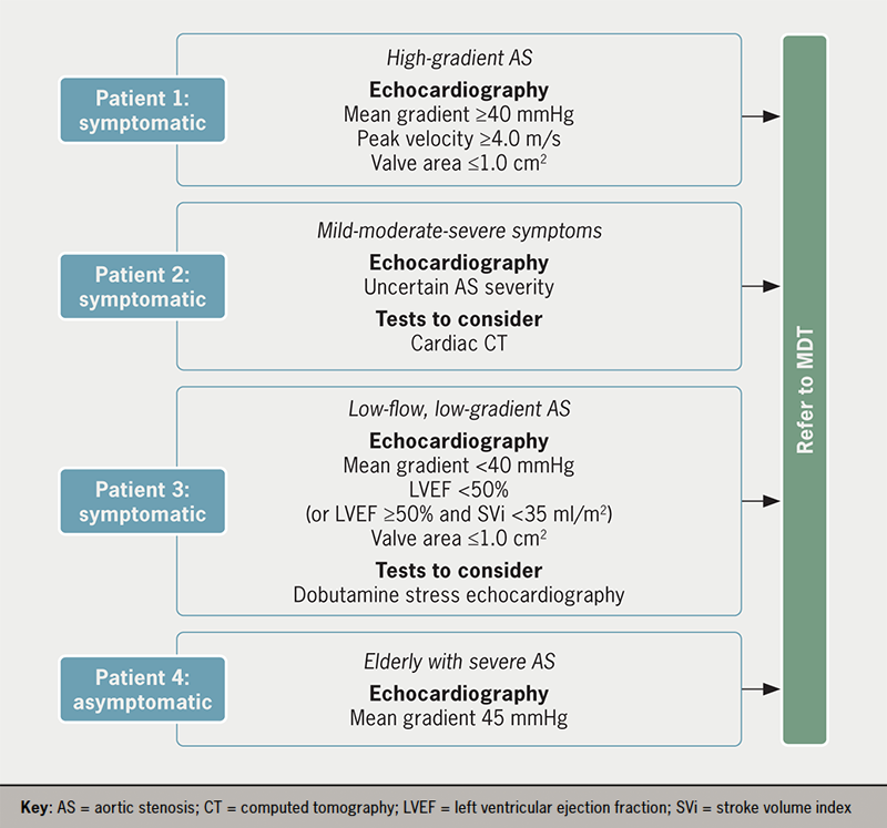
Once the minimum dataset for echocardiographic imaging is obtained alongside any additional diagnostic tests, the patient can be referred to the Heart Team for a treatment decision (figure 1). Typical profiles of patients referred to the Heart Team are outlined in figure 2.

Ruparelia - Figure 1. Decision tree for referral of AS patients to the Heart Team
Figure 1. Decision tree for referral of AS patients to the Heart Team
Ruparelia - Figure 2. Typical profiles of patients referred to the Heart Team
Figure 2. Typical profiles of patients referred to the Heart Team

Even when patients are diagnosed with severe AS, many do not go on to receive appropriate treatment. A European survey demonstrated that one in five patients with severe symptomatic AS did not proceed to any intervention, despite its Class I recommendation in the latest European Society of Cardiology/European Association of Cardio-Thoracic Surgery (ESC/EACTS) guidelines (https://academic.oup.com/eurheartj/article/43/7/561/6358470?login=false#364291358).6,17 The most common reasons for non-referral for aortic valve intervention were the short life expectancy or the physician’s consideration of the patient as inoperable or high risk.18 Substantial advances in TAVI technology, operator (and institutional) experience and meticulous procedure planning, with systematic use of CT scanning, have led to significant improvements in outcomes and, therefore, prognosis and quality of life for elderly patients, including those with comorbidities.2

Educational initiatives should be developed for primary care to facilitate patient referral. Decision support tools and workflows, for example, can tackle hesitancy in referring patients early after symptom onset or when symptoms are not severe. Untreated, symptomatic, severe AS has a poor prognosis and low survival rate,19 and a recent study demonstrated that even asymptomatic patients benefit from early treatment instead of conservative care.20 It is thus paramount to ensure physicians are aware of the benefits of treating severe AS in elderly patients and those with comorbidities.

Establishing a standardised network for management of AS

An integrated network ideally provides the management and treatment of patients with severe AS, comprising:21

  • Primary care physicians and hospital referral centres involved in early diagnosis and referral of patients
  • Heart Valve Clinics involved in early detection, referral, patient monitoring and network support, as required
  • Heart Centres, responsible for delivering treatment via the specialist Heart Team.

As discussed below, optimising how these functions operate within the network is key to delivering timely and effective patient care. The facilities and personnel at individual referral centres, Heart Valve Clinics and Heart Valve Centres vary considerably. Therefore, it is crucial to tailor the treatment pathway, ensuring the appropriate local infrastructure and resources are available for timely referral.

Role of the Heart Valve Clinic

Heart Valve Clinics are specialist, multidisciplinary outpatient clinics providing centralised management to patients with VHD.22,23 While some are located in large Heart Centres, they can also be in district hospitals or community settings, providing support to patients and primary care physicians at a more local level. They have arisen in response to the rapidly growing numbers of patients with VHD and an increased need for local specialist knowledge to ensure patients are diagnosed and referred for treatment as quickly as possible.

Key roles served by Heart Valve Clinics include:21

  • Initiation and coordination of care between the community, referring hospitals and heart centres throughout the patient pathway, including post-discharge
  • Support for detection and diagnosis of VHD and specialist imaging services
  • Education of major stakeholders, such as patients and healthcare professionals, both before and after treatment
  • Involvement with Heart Teams.

Heart Valve Clinics are at the centre of the VHD network and improve efficiency by tackling the significant variability in referral habits among general cardiologists.24,25 Monitoring patients via the Heart Valve Clinic helps detect symptoms earlier and at less severe stages.25 This is especially important for patients with severe AS, as the severity of preoperative symptoms is a prognostic indicator for postoperative survival.25

Reducing redundancy and repetition during diagnosis is essential for a streamlined patient pathway. Heart Valve Clinics can provide a one-stop service for diagnostics and testing, including specialist support for echocardiographic imaging.21,24 To achieve consistency, these clinics should also provide education and training for clinicians in primary care settings and investigate opportunities for delivering echocardiography in community and hospital settings.24

Heart Valve Clinics are also responsible for providing dedicated care throughout the patient pathway, including delivering a comprehensive and individualised management plan for the patient.24 Ideally, patients should have a single point of contact within the clinic, such as a nurse practitioner, who can coordinate care for patients as they progress along the treatment pathway.26 This is particularly beneficial if the patient has an early stage disease and requires monitoring before referring for intervention. In addition, the Heart Valve Clinic must monitor patients and manage their care post-discharge to identify and address potential complications appropriately.

Significantly, Heart Valve Clinics not only benefit patient care but also have the potential to reduce overall costs in the treatment of AS by reducing follow-up costs for patients before and after intervention.27

Role of Heart Valve Centres and the Heart Team

Heart Valve Centres provide multidisciplinary treatment decision-making and treatment. These centres must have demonstrable experience in treating VHD, including sufficient volume of procedures (e.g. a minimum of 100 TAVI procedures per year and 200 surgical procedures per year in France) and transparency of efficacy and safety outcomes to encourage patient flow.28 The recommended components of a Heart Valve Centre are set out in the latest ESC/EACTS guidelines for the management of VHD (https://academic.oup.com/eurheartj/article/43/7/561/6358470?login=false#364291358), and include:17

  • A multidisciplinary Heart Team
  • Provision of the entire spectrum of surgical and transcatheter heart valve procedures
  • Multimodality imaging capabilities
  • A Heart Valve Clinic for outpatient and follow-up management
  • Review procedures for evaluation of outcomes and quality of care
  • Educational programmes targeting patients, primary care, operators, diagnostic and interventional imagers and referring cardiologists.

The multidisciplinary Heart Team is the central treatment decision-making body within the network, comprising a clinical cardiologist, interventional cardiologist, cardiac surgeon, imaging specialist with expertise in interventional imaging, cardiovascular anaesthesiologist, and additional specialists.17,29 With its combined expertise, the Heart Team facilitates a balanced assessment of patients, even where supportive data are limited, and allows for effective allocation of resources to maximise patient benefit.17,29

The Heart Team should discuss all patients referred for treatment at multidisciplinary team (MDT) meetings. However, treatment of standard cases may be aligned to pre-agreed internal protocols depending on the availability of local resources. In the COVID-19 era, Heart Team meetings are usually virtual, allowing referring physicians to dial in and present patients directly to the Heart Team. A single point of entry to the Heart Team should be established to reduce referral barriers while ensuring the team functions efficiently. This contact can provide primary care physicians with a clear pathway for referral of patients to the Heart Team and ensure all appropriate data requirements are in place. This can also aid ongoing education and training of referring teams.

To optimise patient outcomes, treatment needs to be delivered at the appropriate time for the patient. Patients with early-stage disease, for example, may not be considered for treatment by the MDT and may be monitored by the Heart Valve Clinic until treatment is deemed appropriate and the patient is referred back to the MDT.14 For patients identified by the MDT as requiring treatment, the management pathway should be individualised, considering patients’ preferences, as highlighted in ESC/EACTS treatment guidelines (https://academic.oup.com/eurheartj/article/43/7/561/6358470?login=false#364291358).17 Treatment must also be considered in the context of patients’ expected future requirements. In the case of TAVI, procedural access, valve durability, coronary access and the potential need for permanent pacemaker implantation or a future transcatheter heart valve (THV)-in-THV procedure must be considered before treatment decisions are taken.30 Similar considerations apply when the Heart Team opts for surgical aortic valve replacement, where prosthesis choice and positioning should take into account available durability data for that specific valve and the feasibility of a potential THV-in-valve procedure.30

Clear communication between referring physicians, the Heart Valve Clinic, and the Heart Valve Centre throughout the treatment pathway is essential. The key stakeholders should share information on diagnostics, imaging, treatment outcomes, and pre-and post-procedural monitoring. This ensures that patients are optimally managed throughout the process and allows feedback and training from specialists to referring physicians/centres to encourage future timely referrals. A ‘Contact Coordinator’ could be assigned to ensure these communication pathways are accessible and manageable for all and appropriate to the local infrastructure and resources.

Conclusion

With an ever-increasing burden of AS among the population, the issues of low rates of AS detection and referral of patients to specialist centres need to be addressed as soon as possible to ensure early, effective treatment. This will require a concerted effort to reshape existing treatment pathways for AS based on current best practices and in line with ESC/EACTS treatment guidelines (https://academic.oup.com/eurheartj/article/43/7/561/6358470?login=false#364291358).

Promoting informed health and care decisions among healthcare professionals, patient organisations, and researchers can facilitate rapid diagnosis and treatment for patients with AS.10 Key targets for this initiative are to ensure:

  • Greater awareness of AS symptoms among the public and primary care professionals through public awareness campaigns and educational initiatives for primary care
  • Introduction of quality standards for diagnostics and detection
  • Clear structures and communication pathways for rapid referral of patients to Heart Teams
  • A standardised network of healthcare professionals to optimise the patient pathway according to local infrastructure and resources, with Heart Valve Clinics at the centre of the pathway.

Key messages

  • Despite effective treatment options, many patients with severe aortic stenosis (AS) remain undiagnosed or suffer delays in referral for treatment, leading to poor outcomes
  • Increasing awareness of early AS symptoms among the public and healthcare professionals, and introducing routine auscultation in primary care, can improve its early detection
  • Offering prompt echocardiography facilitates early diagnosis and referral, but it should meet quality standards to avoid misdiagnosis and the need for repetition
  • The optimisation of patient detection, referral and treatment of severe AS requires a streamlined patient pathway based on a standardised network of healthcare professionals, and ensuring there are appropriate local infrastructure and resources needed to accomplish this objective

Conflicts of interest

VD has received speaker fees from Abbott Vascular, Edwards Lifesciences, GE Healthcare, Medtronic, and Novartis, and consultancy fees from Novo Nordisk. PP has received funding from Edwards Lifesciences, Medtronic, Pi-Cardia, and Cardiac Phoenix for echocardiography core laboratory analyses and research studies in the field of transcatheter valve therapies, for which he received no personal compensation; he has received lecturer fees from Edwards Lifesciences and Medtronic. FS has participated in advisory boards, and received lecturer fees from Abbott Vascular, Edwards Lifesciences and Medtronic. NR: none declared.

Funding

PP holds the Canada Research Chair in Valvular Heart Disease, supported by the Canadian Institutes of Health Research (Ottawa, Ontario, Canada).

Victoria Delgado
Cardiologist and Assistant Professor of Cardiology
Heart Institute, Department of Cardiology, University Hospital Germans Trias i Pujol, 08916 Badalona, Barcelona, Spain

Philippe Pibarot
Head of Cardiology Research and Canada Research Chair in Valvular Heart Disease
Québec Heart and Lung Institute, Laval University, Québec, Canada, G1V 4G5

Neil Ruparelia
Consultant Cardiologist
Hammersmith Hospital, Imperial College Healthcare NHS Trust, DuCane Road, W12 0HS, London, UK

Francesco Saia
Interventional Cardiologist
Cardio-Thoracic-Vascular Department, IRCCS University Hospital of Bologna, Policlinico S. Orsola (Pav. 23), Via Massarenti, 9 – 410138 Bologna, Italy

Correspondence to:
[email protected]

Articles in this supplement

Introduction: overcoming barriers to treating severe aortic stenosis
The past, present and future of aortic stenosis treatment
Ensuring continuous and sustainable access to aortic stenosis treatment

References

1. Auffret V, Lefevre T, Van Belle E et al. Temporal trends in transcatheter aortic valve replacement in France: FRANCE 2 to FRANCE TAVI. J Am Coll Cardiol 2017;70:42–55. https://doi.org/10.1016/j.jacc.2017.04.053

2. Carroll JD, Mack MJ, Vemulapalli S et al. STS-ACC TVT registry of transcatheter aortic valve replacement. J Am Coll Cardiol 2020;76:2492–516. https://doi.org/10.1016/j.jacc.2020.09.595

3. Mack MJ, Leon MB, Smith CR et al. 5-year outcomes of transcatheter aortic valve replacement or surgical aortic valve replacement for high surgical risk patients with aortic stenosis (PARTNER I™): A randomised controlled trial. Lancet 2015;385:2477–84. https://doi.org/10.1016/S0140-6736(15)60308-7

4. Siontis GCM, Overtchouk P, Cahill TJ et al. Transcatheter aortic valve implantation vs. surgical aortic valve replacement for treatment of symptomatic severe aortic stenosis: an updated meta-analysis. Eur Heart J 2019;40:3143–53. https://doi.org/10.1093/eurheartj/ehz275

5. d’Arcy JL, Coffey S, Loudon MA et al. Large-scale community echocardiographic screening reveals a major burden of undiagnosed valvular heart disease in older people: The OxVALVE Population Cohort Study. Eur Heart J 2016;37:3515–22. https://doi.org/10.1093/eurheartj/ehw229

6. Eugene M, Duchnowski P, Prendergast B et al. Contemporary management of severe symptomatic aortic stenosis. J Am Coll Cardiol 2021;78:2131–43. https://doi.org/10.1016/j.jacc.2021.09.864

7. Joseph J, Kotronias RA, Estrin-Serlui T et al. Safety and operational efficiency of restructuring and redeploying a transcatheter aortic valve replacement service during the COVID-19 pandemic: The Oxford experience. Cardiovasc Revasc Med 2021;31:26–31. https://doi.org/10.1016/j.carrev.2020.12.002

8. Thoenes M, Bramlage P, Zamorano P et al. Patient screening for early detection of aortic stenosis (AS)-review of current practice and future perspectives. J Thorac Dis 2018;10:5584–94. https://doi.org/10.21037/jtd.2018.09.02

9. Gaede L, Aarberge L, Brandon Bravo Bruinsma G et al. Heart valve disease awareness survey 2017: What did we achieve since 2015? Clin Res Cardiol 2019;108:61–7. https://doi.org/10.1007/s00392-018-1312-5

10. Wait S, Krishnaswamy P, Borregaard B et al. Heart valve disease: Working together to create a better patient journey. 2020. Available at: https://globalhearthub.org/wp-content/uploads/2021/11/HVD_report-final-2021.pdf [last accessed 05/05/2022].

11. Pibarot P, Lauck S, Morris T et al. Patient care journey for patients with heart valve disease. Can J Cardiol 2022;38:1296–9. https://doi.org/10.1016/j.cjca.2022.02.025

12. Steeds RP, Potter A, Mangat N et al. Community-based aortic stenosis detection: clinical and echocardiographic screening during influenza vaccination. Open Heart 2021;8:e001640. https://doi.org/10.1136/openhrt-2021-001640

13. Evangelista A, Galuppo V, Mendez J et al. Hand-held cardiac ultrasound screening performed by family doctors with remote expert support interpretation. Heart 2016;102:376–82. https://doi.org/10.1136/heartjnl-2015-308421

14. Draper J, Subbiah S, Bailey R et al. Murmur clinic: validation of a new model for detecting heart valve disease. Heart 2019;105:56–9. https://doi.org/10.1136/heartjnl-2018-313393

15. Ring L, Shah BN, Bhattacharyya S et al. Echocardiographic assessment of aortic stenosis: a practical guideline from the British Society of Echocardiography. Echo Res Pract 2021;8:G19–G59. https://doi.org/10.1530/ERP-20-0035

16. Robinson S, Rana B, Oxborough D et al. A practical guideline for performing a comprehensive transthoracic echocardiogram in adults: the British Society of Echocardiography minimum dataset. Echo Res Pract 2020;7:G59–G93. https://doi.org/10.1530/ERP-20-0026

17. Vahanian A, Beyersdorf F, Praz F et al. 2021 ESC/EACTS guidelines for the management of valvular heart disease. Eur Heart J 2022;43:561–632. https://doi.org/10.1093/eurheartj/ehab395

18. Asteggiano R, Bramlage P, Richter DJ. European Society of Cardiology Council for Cardiology Practice worldwide survey of transcatheter aortic valve implantation beliefs and practices. Eur J Prev Cardiol 2018;25:608–17. https://doi.org/10.1177/2047487318760029

19. Taniguchi T, Morimoto T, Takeji Y et al. Contemporary issues in severe aortic stenosis: Review of current and future strategies from the Contemporary Outcomes after Surgery and Medical Treatment in Patients with Severe Aortic Stenosis registry. Heart 2020;106:802–9. https://doi.org/10.1136/heartjnl-2019-315672

20. Kang DH, Park SJ, Lee SA et al. Early surgery or conservative care for asymptomatic aortic stenosis. N Engl J Med 2020;382:111–19. https://doi.org/10.1056/NEJMoa1912846

21. Chambers JB, Lancellotti P. Heart valve clinics, centers, and networks. Cardiol Clin 2020;38:65–74. https://doi.org/10.1016/j.ccl.2019.09.006

22. Chambers JB, Prendergast B, Iung B et al. Standards defining a ‘Heart Valve Centre’: ESC Working Group on Valvular Heart Disease and European Association for Cardiothoracic Surgery Viewpoint. Eur Heart J 2017;38:2177–83. https://doi.org/10.1093/eurheartj/ehx370

23. Lancellotti P, Rosenhek R, Pibarot P et al. ESC Working Group on Valvular Heart Disease position paper–heart valve clinics: organization, structure, and experiences. Eur Heart J 2013;34:1597–606. https://doi.org/10.1093/eurheartj/ehs443

24. Chambers JB. Specialist valve clinic: Why, who and how? Heart 2019;105:1913–20. https://doi.org/10.1136/heartjnl-2019-315203

25. Zilberszac R, Lancellotti P, Gilon D et al. Role of a heart valve clinic programme in the management of patients with aortic stenosis. Eur Heart J Cardiovasc Imaging 2017;18:138–44. https://doi.org/10.1093/ehjci/jew133

26. Lauck S, Forman J, Borregaard B et al. Facilitating transcatheter aortic valve implantation in the era of COVID-19: Recommendations for programmes. Eur J Cardiovasc Nurs 2020;19:537–44. https://doi.org/10.1177/1474515120934057

27. Ionescu A, McKenzie C, Chambers JB. Are valve clinics a sound investment for the health service? A cost-effectiveness model and an automated tool for cost estimation. Open Heart 2015;2:e000275. https://doi.org/10.1136/openhrt-2015-000275

28. Haute Autorité Santé. Critères d’éligibilité des centres implantant des TAVIs. 2020. Available at: https://www.has-sante.fr/upload/docs/application/pdf/2020-11/rapport_tavis.pdf [last accessed 13/05/2022].

29. Nerla R, Prendergast BD, Castriota F. Optimal structure of TAVI heart centres in 2018. EuroIntervention 2018;14:AB11–AB8. https://doi.org/10.4244/EIJ-D-18-00656

30. Tarantini G, Nai Fovino L. Lifetime strategy of patients with aortic stenosis: The first cut is the deepest. JACC Cardiovasc Interv 2021;14:1727–30. https://doi.org/10.1016/j.jcin.2021.06.029

Disclaimer:

Medical knowledge is constantly changing. As new information becomes available, changes in treatment, procedures, equipment and the use of drugs become necessary. The editors/authors/contributors and the publishers Medinews (Cardiology) Ltd have taken care to ensure that the information given in this text is accurate and up to date at the time of publication.

Readers are strongly advised to confirm that the information, especially with regard to drug usage, complies with the latest legislation and standards of practice. Medinews (Cardiology) Limited advises healthcare professionals to consult up-to-date Prescribing Information and the full Summary of Product Characteristics available from the manufacturers before prescribing any product. Medinews (Cardiology) Limited cannot accept responsibility for any errors in prescribing which may occur.

The opinions, data and statements that appear are those of the contributors. The publishers, editors, and members of the editorial board do not necessarily share the views expressed herein. Although every effort is made to ensure accuracy and avoid mistakes, no liability on the part of the publisher, editors, the editorial board or their agents or employees is accepted for the consequences of any inaccurate or misleading information.

© Medinews (Cardiology) Ltd 2023. All rights reserved. No part of this publication may be reproduced, stored in a retrieval system, or transmitted in any form or by any means, electronic, mechanical, photocopying, recording or otherwise, without the prior written permission of the publishers, Medinews (Cardiology) Ltd. It shall not, by way of trade or otherwise, be lent, re-sold, hired or otherwise circulated without the publisher’s prior consent.

Ensuring continuous and sustainable access to aortic stenosis treatment

Br J Cardiol 2023;30(suppl 1):S18–S24doi:10.5837/bjc.2023.s04 Leave a comment
Click any image to enlarge
Authors:
Sponsorship Statement:

This supplement is fully funded by Edwards Lifesciences SA. The content of the papers in the supplement represent the contributors’ views and not necessarily those of Edwards Lifesciences, and are the sole responsibility of the authors.
Job code: PP–EU-5649

The incidence of aortic stenosis (AS) worldwide is expected to increase steeply over the next three decades, mainly driven by an ageing population.1,2 In the face of this emerging epidemic, the demand for AS treatment resources is increasing, including the provision of surgical aortic valve replacement (SAVR) and transcatheter aortic valve implantation (TAVI). The COVID-19 pandemic added challenges for patients with AS, who cannot delay their treatment without impacting clinical outcomes.3 In this setting, TAVI is recognised as an effective and efficient solution for treating AS in eligible patients, offering no significant delays in treatment (depending on local resources), no excess complications or mortality, and a shorter hospital length-of-stay compared to treatment before COVID 19.4,5

A successful TAVI programme aims to resolve AS safely and efficiently, enabling the patient to be discharged home rapidly without sustaining in-hospital complications and thus improving outcomes. This requires individualised treatment decisions based on patient presentation and preferences, and available local resources. Data from clinical trials and real-world studies have driven improvements in the care coordination for patients undergoing TAVI, transforming patient pathways toward the TAVI programme goals.6–12 Based on these data, a streamlined TAVI pathway aimed at safe and early discharge of patients is recommended to optimise outcomes and improve access to care.

The evidence supporting early discharge after TAVI

Ensuring continuous and sustainable access to aortic stenosis treatment

Patients discharged early from hospital generally have a reduced risk of the physical and functional consequences associated with immobilisation and longer hospital stay, including hospital-acquired infections, functional dependency and cognitive decline.13,14 Elderly and frail patients are at risk of hospital-associated complications and are most likely to benefit from a shorter hospital stay.7,14

In the context of TAVI, the feasibility and safety of early discharge are well established.6–10,15 As early as 2015, early discharge was shown to be feasible and safe in approximately 80% of patients.6,7 In the North American 3M TAVR (The Vancouver 3M [Multidisciplinary, Multimodality But Minimalist] Clinical Pathway Facilitates Safe Next-Day Discharge Home at Low-, Medium-, High-Volume Transfemoral Transcatheter Aortic Valve Replacement Centres) study, 89.5% of patients were safely discharged home within 48 hours of a TAVI procedure, and over 80% within 24 hours.8 (https://www.jacc.org/doi/10.1016/j.jcin.2018.12.020) A Belgian study of matched patient outcomes before and after the transition to a fast-track (two to four days length of stay) TAVI pathway found no differences in all-cause mortality, risk of new permanent pacemaker implantation, or rehospitalisation rates at three months.15 In a meta analysis of eight studies involving 1,775 patients undergoing TAVI, Kotronias et al. found that patients were less likely to be re-admitted after early (up to three days) versus standard discharge.10

The most appropriate early discharge target for a fast-track TAVI pathway is yet to be defined. Timing is heavily dependent on local resources, institutional protocols and healthcare systems, all of which differ between countries.16 In this article, we discuss the adoption of a 48-hour target for early discharge to reflect an achievable goal across diverse regions and healthcare systems; we acknowledge that studies have demonstrated the feasibility of next day discharge following TAVI,8,17 and even same-day discharge for highly selected patients to mitigate the competing demands of the COVID-19 pandemic on health resources.18

Achieving safe, early discharge

While the benefits of early discharge after TAVI are known, the processes required to achieve this target are less well established, and many centres struggle to reach a discharge goal within 48 hours of admission. In the European FAST-TAVI (Feasibility and Safety of Early Discharge After Transfemoral Transcatheter Aortic Valve Implantation) trial, logistical reasons contributed to a delay in hospital discharge for over a third of patients.9 (https://eurointervention.pcronline.com/article/optimizing-patient-discharge-management-after-transfemoral-transcatheter-aortic-valve-implantation-the-multicentre-european-fast-tavi-trial) To achieve consistent early discharge, all aspects of care must be scrutinised to identify and address the inefficiencies and barriers within the local systems and protocols.

The international, multicentre BENCHMARK registry has recently been developed to assess the implementation of evidence-based quality of care measures to facilitate early discharge following TAVI without impacting patient safety.19 The eight best practices outlined in the BENCHMARK registry19 (see figure 1) include:

  • education of the patient/family on early discharge expectations
  • education and alignment of the internal team
  • determination of an anticipated discharge date at admission
  • post-procedural imaging to confirm appropriate management of complications
  • early mobilisation four to six hours after the intervention
  • early and consistent assessment of the need for a pacemaker
  • daily patient visits by the implanter and criteria based discharge.
Lauck - Figure 1. Best practices to achieve a streamlined patient pathway to facilitate safe discharge home within 48 hours after transcatheter aortic valve implantation (TAVI)19
Figure 1. Best practices to achieve a streamlined patient pathway to facilitate safe discharge home within 48 hours after transcatheter aortic valve implantation (TAVI)19 (click to enlarge)

Role of the TAVI coordinator

Implementing a streamlined TAVI patient pathway requires engagement and buy-in from all major stakeholders, including the TAVI Heart Team (e.g. the interventional cardiologist, cardiac surgeon, anaesthetist, TAVI nurse, procedure room team, cardiac care team, post-procedural nurses), other hospital staff (e.g. cardiac programme administration, care coordinators), and patients and their families. The availability of a designated TAVI coordinator improves the pathway efficiency and increases patient satisfaction.20 The coordinator should be the single point of contact for patients and their relatives from referral to follow-up. The TAVI coordinator is responsible for ensuring timely and appropriate communication, not only within and between the hospital teams but also with stakeholders outside the hospital (e.g. general cardiologist, referring physician); and for systematically assessing patients’ health status and urgency for intervention when waiting for TAVI. This essential role has been endorsed by international guidelines to ensure processes of care, early discharge planning and follow-up to address patient and programme needs.12

Specialised cardiovascular nurses are considered the most appropriate choice for the role of TAVI coordinator based on their clinical and programme development expertise.11,21 In addition to their competencies in the assessment of patients’ evolving clinical presentation and individualised needs, nurses offer leadership in streamlining patient pathways, advocating for resources and monitoring patient access and quality of care.21

According to current guidelines, shared decision-making (SDM) should be at the centre of treatment decisions in AS, with the consideration of patients’ preferences and priorities to achieve an individualised treatment approach and higher patient satisfaction.22 Given the complexity of treatment decisions and organisational constraints, physicians and the TAVI multidisciplinary team may find SDM challenging in the context of severe AS, and its application is not routine in older patients.23,24 In spite of these perceived barriers, SDM minimally extends initial outpatient appointments and leads to higher-quality treatment decisions and patient satisfaction. Therefore, TAVI teams should promote patient participation in SDM to ensure high-quality care according to the principles set out in the treatment guidelines.23

Peri-procedural optimisation

Patients entering a fast-track TAVI pathway should ideally be admitted on the day of the procedure to avoid an unnecessary overnight stay and mitigate the risks of bedrest and in-hospital complications.12 Most patients are suitable for early discharge;8 several preprocedural factors have been identified as predictors of successful TAVI and/or a short hospital length of stay, including good patient mobility,25,26 a pre-existing pacemaker and absence of acute kidney injury before TAVI.6 It is important, however, to identify patients for whom rapid discharge may not be appropriate or feasible. This may include high-risk patients who cannot receive care according to the standardised TAVI clinical pathway for various reasons, including clinical presentation, peri- or post-procedural complications, or lack of social support.6,27 The assessment of frailty using established tools provides additional information to assess individuals’ functional status and appropriate timing for safe discharge. However, baseline frailty scores alone are not a reliable predictor for early or late discharge.

When preparing for early discharge, clear communication with patients, family members and caregivers is key to promote consistent information and encourage patients and their families to prepare a plan for returning home. Pre-operative consultations should focus on explaining the entire treatment pathway to patients, highlighting the benefits of early discharge and beginning the process of discharge planning. A clear target of the expected discharge day based on local practice (e.g. next-day, post-procedure day 2) should be emphasised by every care provider at every time point of interactions with patients and their families. The TAVI coordinator is ideally positioned to ensure consistency and repetition of strategies for supporting patient motivation and participation for early discharge planning.

The adoption of a minimalist TAVI procedure is well established,28 and is well suited to achieve early discharge. Invasive lines should be avoided or minimised, including not using a central venous catheter/arterial monitoring and/or urinary catheter. Local anaesthesia may offer several advantages over general anaesthesia, including a shorter procedure time, decreased requirements for critical care and a shorter length of stay, the avoidance of delirium or functional decline, reduced need for inotropic support, lower 30-day mortality and reduced costs.29–32 Peri-procedure teams should be ready to manage potential peri-procedure complications, such as severe haemodynamic instability or transitioning to general anaesthesia in the event of emergencies.12 Ultrasound-guided vascular access may reduce the risk of vascular access complications during procedures,33 and hence should be favoured to facilitate predictable access site haemostasis and early mobilisation.

Post-procedural optimisation

An effective post-procedure protocol was developed in the 3M TAVR study comprising three parallel activities: patient monitoring, reconditioning and discharge planning (figure 2).34

Lauck - Figure 2. Post-procedure protocol based on the 3M TAVR study
Figure 2. Post-procedure protocol based on the 3M TAVR study

Following the procedure, patients should be assessed regularly for vital signs, neurological status, cardiac rhythm, vascular access haemostasis and pain. Most TAVI patients do not experience significant pain; discomfort and mild pain can be treated with simple analgesic agents. Importantly, sedatives or opioids should not be used in order to avoid delirium. Transthoracic echocardiography (TTE) should be conducted to examine device position, paravalvular leak (PVL) and biventricular function, and laboratory tests for haemoglobin, complete blood counts and renal function should be conducted.34 The standardised scheduling and review of post-procedure TTE can prevent delays to early discharge.

Early mobilisation is a critical component of a fast-track TAVI programme. The post-procedure protocol developed in the 3M TAVR study includes an effective nurse-led protocol for standardised mobilisation four to six hours after TAVI. This evidence-based nursing care standard can be adapted according to individual hospital resources. Before mobilisation, any invasive lines need to be removed at the earliest time point. Progressive mobilisation can then begin, as detailed in figure 3, provided there are no vascular or other complications. A target of walking twice (e.g. to the toilet) on postoperative day 0 facilitates early reconditioning and the avoidance of complications. Mobilisation must be tailored to individual patients and care should be taken to ensure that any aids typically used by the patient, such as a walking stick, walker or spectacles, are in place. Appropriate hydration and encouragement to resume regular meals are important to facilitate a return to baseline health status.34

Lauck - Figure 3. Early mobilisation protocol11,34
Figure 3. Early mobilisation protocol11,34

Prespecified criteria should be defined and met before a patient can be safely discharged home (table 1). The specified list of discharge criteria should be agreed upon within the institution based on local resources and should include the availability of post-discharge support for the patient.

Table 1. Proposed discharge criteria (based on European FAST-TAVI and 3M TAVR studies)9,34

Discharge criteria
Multidisciplinary consensus of readiness for discharge and physician’s order considerations may include:

  • New York Heart Association functional class ≤II
  • No cardiac ischaemia-associated chest pain
  • No stroke/transient ischaemic attack
  • No untreated major arrhythmias
  • Absence of persistent (>3 hrs) intraventricular conduction delays
  • No paravalvular leak with aortic regurgitation less than moderate
  • No signs or symptoms of complications
  • No fever during the last 24 hours and no signs of an infectious cause
  • No unresolved type 3 acute kidney injury
  • No red blood cell transfusion during the preceding 72 hours
  • Stable haemoglobin
  • No haemodynamic instability
  • Absence of laboratory contraindications (i.e., clinically important change in Hgb and eGFR)
Return to baseline mobilisation
Confirmed availability of family member for 24 hours to remain with patient
Discharge teaching completed and confirmed follow-up plan

Successful completion of the streamlined clinical pathway is dependent on good communication, continuity of care, and management of expectations, with nurses playing a crucial role in facilitating post-procedural pathways. Good communication with patients and their families is required to ensure patients are discharged to a safe setting with appropriate care. Communication beyond discharge is also important to ensure the patient continues to be engaged with their rehabilitation. In this respect, the TAVI coordinator may contact the patient by telephone on the day after discharge and at longer follow-up to assess symptoms, recovery and health status, and may assist in ensuring post-procedural blood tests are offered in some places to reduce the risks associated with delayed complications.12 Cardiac rehabilitation services are available in some areas to support patients after discharge through regular communications, home exercise programmes and group classes. These services provide patients with support and advice on lifestyle changes and how to improve risk factors following TAVI.

Educational tools to support a fast-track TAVI pathway

Educational tools, such as information leaflets (e.g. Your TAVI [https://www.rbht.nhs.uk/sites/nhs/files/PILs/TAVI.pdf]) and videos (e.g. ‘Explain my procedure’ https://www.explainmyprocedure.com/procedure/transcatheter-aortic-valve-implantation-tavi-english/ [a multi-language tool]) can help patients to understand and engage with the patient pathway. Educational tools should also be developed to support hospital teams. These may include decision trees, flowcharts and checklists to facilitate passage along the patient pathway, the management of complications, criteria for early discharge and scenarios for individual patient groups. Hospital-led education of referring colleagues is also required so that appropriate patients can gain timely access to a TAVI programme.

Cost benefits of a fast-track TAVI pathway

The study of cost savings and effectiveness of fast-track TAVI highlights opportunities for significant savings related to short hospital admission, avoidance of critical care resources, and mitigation of risk of complications.35 For intermediate-risk patients in the PARTNER II™ (Placement of Aortic Transcatheter Valves II – XT Intermediate) trial, reductions in hospital length-of-stay in patients treated with the latest balloon-expandable TAVI valve led to a $4,000 cost-saving compared with SAVR.35 A fast-track TAVI pathway, focusing on early discharge, would be expected to reduce costs even further. Indeed, a US cost analysis comparing standard TF-TAVI with a minimalist approach that reduced hospital length-of-stay by two days found an almost $10,000 cost-saving with the fast-track pathway.36 Economic analyses based on the PARTNER 3™ (Safety and Effectiveness of the SAPIEN 3™ Transcatheter Heart Valve in Low Risk Patients with Aortic Stenosis) trial have also shown cost-savings with TAVI versus SAVR in low-risk patients.37,38 Together, these studies suggest that a TAVI pathway focusing on the mitigation of in-hospital risks and early discharge would reap benefits not only from a patient’s perspective but also for resource use.

Conclusion

Care goals for elderly, fragile patients requiring treatment for AS are consistent across institutions. However, resources differ substantially between regions, so local practices must be adapted accordingly. There have been several drivers for a systematic, standardised approach to AS treatment. First, the number of patients requiring treatment is increasing dramatically, driven by an ageing population, a greater appreciation of the burden of asymptomatic severe AS, and expansion of TAVI into lower-risk groups. Second, the COVID-19 pandemic has emphasised the need for a more streamlined pathway to shorten hospital stays and reduce hospital-based complications. Upfront investment in a well informed, structured and standardised approach can lead to earlier discharge, better patient outcomes and fewer complications. With careful attendance to documented best practices and full support from the entire TAVI team, centres can move towards an optimised patient pathway that achieves consistently safe, early discharge for patients. This approach is effective from a patient care perspective and can lead to substantial cost savings and resource efficiencies.

Key messages

  • A successful transcatheter aortic valve implantation (TAVI) programme aims to resolve aortic stenosis with minimal short- and long-term complications, enabling patients to be discharged efficiently and safely
  • Early reconditioning and safe discharge of eligible patients after TAVI reduces the risk of hospital-associated complications without increasing the risk of mortality, new permanent pacemaker implantation or re-hospitalisation
  • The adoption of evidence-based best practices and the full support from the multidisciplinary TAVI team enable centres to consistently achieve earlier discharge, better patient outcomes and fewer complications, alongside substantial cost savings and resource efficiencies to improve access to care

Conflicts of interest

ED, SL and DF are consultants for Edwards Lifesciences. JR has received consultancy fees from Edwards Lifesciences.

Funding

Professor Eric Durand has received a grant from the French Government managed by the National Research Agency (ANR) under the program ‘Investissements d’avenir’ (reference ANR-16-RHUS-0003) and is also supported by a grant from the GCS G4 as part of the FHU-CARNAVAL, labeled by AVIESAN.

Eric Durand
Professor of Cardiology
Normandie University, UNIROUEN, U1096, CHU Rouen, Department of Cardiology, FHU CARNAVAL, F-76000 Rouen, France

Sandra Lauck
Clinician Scientist , St Paul’s Hospital, and Associate Professor, St Paul’s Hospital Professorship in Cardiovascular Nursing
University of British Columbia, Vancouver, Canada

Derk Frank
Professor of Cardiology and Director
Internal Medicine III at UKSH, Arnold-Heller-Straße 3, 24105 Kiel, Germany, and German Centre of Cardiovascular Research (DZHK), partner site Hamburg, Lübeck, Kiel, Germany

John Rawlins
Consultant Interventional Cardiologist
Coronary Research Group, University Hospital Southampton NHS Foundation Trust, Southampton, SO16 6YD, UK

Correspondence to:
[email protected]

Articles in this supplement

Introduction: overcoming barriers to treating severe aortic stenosis
The past, present and future of aortic stenosis treatment
A standardised network to improve the detection and referral of patients with aortic stenosis

Further information: cost-benefit analyses of TAVI

Some information on the cost-benefit analyses of TAVI can be found in the following links:

Gilard M, Eltchaninoff H, Iung B et al. Transcatheter aortic valve implantation procedure compared with surgery in patients with severe aortic stenosis at low risk or surgical mortality in France. Value Health 2022;25:605–13. https://doi.org/10.1016/j.jval.2021.10.003
https://www.sciencedirect.com/science/article/pii/S1098301521017861

Hospital Healthcare Europe. Economic implications and cost effectiveness of TAVI. https://hospitalhealthcare.com/edwards/economic-implications-and-cost-effectiveness-of-tavi/ (last accessed 10th November 2022)

Lorenzoni V, Barbieri G, Saia F et al. The cost-effectiveness of transcatheter aortic valve implantation: exploring the Italian National Health System perspective and different patient risk groups. Eur J Health Econ 2021;22:1349–63. https://doi.org/10.1007/s10198-021-01314-z
https://link.springer.com/article/10.1007/s10198-021-01314-z

Mennini FS, Meucci F, Pesarini G et al. Cost-effectiveness of transcatheter aortic valve implantation versus surgical aortic valve replacement in low surgical risk aortic stenosis patients. Int J Cardiol 2022;357:26–32. https://doi.org/10.1016/j.ijcard.2022.03.034
https://www.sciencedirect.com/science/article/pii/S016752732200393X

Rodríguez JMV, Bermúdez EP, Zamorano JL et al. Cost-effectiveness of SAPIEN 3™ transcatheter aortic valve implantation in low surgical mortality risk patients in Spain. REC Interv Cardiol 2022; published online 19th October 2022. https://doi.org/10.24875/RECICE.M22000340
https://recintervcardiol.org/en/original-article/cost-effectiveness-of-sapien-3-transcatheter-aortic-valve-implantation-in-low-surgical-mortality-risk-patients-in-spain

References

1. Roth GA, Mensah GA, Johnson CO et al. Global burden of cardiovascular diseases and risk factors, 1990–2019: Update from the GBD 2019 study. J Am Coll Cardiol 2020;76:2982–3021. https://doi.org/10.1016/j.jacc.2020.11.010

2. d’Arcy JL, Coffey S, Loudon MA et al. Large-scale community echocardiographic screening reveals a major burden of undiagnosed valvular heart disease in older people: The OxVALVE Population Cohort Study. Eur Heart J 2016;37:3515–22. https://doi.org/10.1093/eurheartj/ehw229

3. Roule V, Rebouh I, Lemaitre A et al. Impact of wait times on late postprocedural mortality after successful transcatheter aortic valve replacement. Sci Rep 2022;12:5967. https://doi.org/10.1038/s41598-022-09995-z

4. Joseph J, Kotronias RA, Estrin-Serlui T et al. Safety and operational efficiency of restructuring and redeploying a transcatheter aortic valve replacement service during the COVID-19 pandemic: The Oxford experience. Cardiovasc Revasc Med 2021;31:26–31. https://doi.org/10.1016/j.carrev.2020.12.002

5. Tan J, Teoh K, Ivanova J et al. The impact of the COVID-19 pandemic on transcatheter aortic valve implantation (TAVI) services in the United Kingdom: A tertiary centre experience. Heart 2021;107:A13–A14. https://doi.org/10.1136/heartjnl-2021-BCS.17

6. Durand E, Eltchaninoff H, Canville A et al. Feasibility and safety of early discharge after transfemoral transcatheter aortic valve implantation with the Edwards SAPIEN-XT™ prosthesis. Am J Cardiol 2015;115:1116–22. https://doi.org/10.1016/j.amjcard.2015.01.546

7. Serletis-Bizios A, Durand E, Cellier G et al. A prospective analysis of early discharge after transfemoral transcatheter aortic valve implantation. Am J Cardiol 2016;118:866–72. https://doi.org/10.1016/j.amjcard.2016.06.035

8. Wood DA, Lauck SB, Cairns JA et al. The Vancouver 3M (Multidisciplinary, Multimodality, But Minimalist) clinical pathway facilitates safe next-day discharge home at low-, medium-, and high-volume transfemoral transcatheter aortic valve replacement centers: The 3M TAVR study. JACC Cardiovasc Interv 2019;12:459–69. https://doi.org/10.1016/j.jcin.2018.12.020

9. Barbanti M, van Mourik MS, Spence MS et al. Optimising patient discharge management after transfemoral transcatheter aortic valve implantation: The multicentre European FAST-TAVI trial. EuroIntervention 2019;15:147–54. https://doi.org/10.4244/EIJ-D-18-01197

10. Kotronias RA, Teitelbaum M, Webb JG et al. Early versus standard discharge after transcatheter aortic valve replacement: A systematic review and meta-analysis. JACC Cardiovasc Interv 2018;11:1759–71. https://doi.org/10.1016/j.jcin.2018.04.042

11. Lauck SB, Wood DA, Baumbusch J et al. Vancouver Transcatheter Aortic Valve Replacement clinical pathway: Minimalist approach, standardized care, and discharge criteria to reduce length of stay. Circ Cardiovasc Qual Outcomes 2016;9:312–21. https://doi.org/10.1161/CIRCOUTCOMES.115.002541

12. Lauck S, Forman J, Borregaard B et al. Facilitating transcatheter aortic valve implantation in the era of COVID-19: Recommendations for programmes. Eur J Cardiovasc Nurs 2020;19:537–44. https://doi.org/10.1177/1474515120934057

13. Tirado-Conte G, Freitas-Ferraz AB, Nombela-Franco L et al. Incidence, causes, and impact of in-hospital infections after transcatheter aortic valve implantation. Am J Cardiol 2016;118:403–9. https://doi.org/10.1016/j.amjcard.2016.05.012

14. Calero-García MJ, Ortega AR, Navarro E et al. Relationship between hospitalization and functional and cognitive impairment in hospitalized older adults patients. Aging Ment Health 2017;21:1164–70. https://doi.org/10.1080/13607863.2016.1220917

15. Baekke PS, Jørgensen TH, Søndergaard L. Impact of early hospital discharge on clinical outcomes after transcatheter aortic valve implantation. Catheter Cardiovasc Interv 2021;98:E282–E90. https://doi.org/10.1002/ccd.29403

16. Lutz M, Messika-Zeitoun D, Rudolph TK et al. Differences in the presentation and management of patients with severe aortic stenosis in different European centres. Open Heart 2020;7(2)e001345. https://doi.org/10.1136/openhrt-2020-001345

17. Gupta R, Mahajan S, Mehta A et al. Next-day discharge vs early discharge after transcatheter aortic valve replacement: Systematic review and meta-analysis. Curr Probl Cardiol 2021;47:100998. https://doi.org/10.1016/j.cpcardiol.2021.100998

18. Pop AM, Barker M, Hickman L et al. Same day discharge during the COVID-19 pandemic in highly selected transcatheter aortic valve replacement patients. Struct Heart 2021;5:596–604. https://doi.org/10.1080/24748706.2021.1988780

19. McCalmont G, Durand E, Lauck S et al. Setting a benchmark for resource utilization and quality of care in patients undergoing transcatheter aortic valve implantation in Europe – Rationale and design of the international BENCHMARK registry. Clin Cardiol 2021;44:1344–53. https://doi.org/10.1002/clc.23711

20. Bohmann K, Burgdorf C, Zeus T et al. The COORDINATE Pilot Study: Impact of a transcatheter aortic valve coordinator program on hospital and patient outcomes. J Clin Med 2022;11:1205. https://doi.org/10.3390/jcm11051205

21. Lauck SB, McGladrey J, Lawlor C et al. Nursing leadership of the transcatheter aortic valve implantation Heart Team: Supporting innovation, excellence, and sustainability. Healthc Manage Forum 2016;29:126–30. https://doi.org/10.1177/0840470416632004

22. Vahanian A, Beyersdorf F, Praz F et al. 2021 ESC/EACTS guidelines for the management of valvular heart disease. Eur Heart J 2022;43:561–632. https://doi.org/10.1093/eurheartj/ehab395

23. van Beek-Peeters J, van Noort EHM, Faes MC et al. Shared decision making in older patients with symptomatic severe aortic stenosis: A systematic review. Heart 2020;106:647–55. https://doi.org/10.1136/heartjnl-2019-316055

24. van Beek-Peeters J, van der Meer JBL, Faes MC et al. Professionals’ views on shared decision-making in severe aortic stenosis. Heart 2022;108:558–64. https://doi.org/10.1136/heartjnl-2021-320194

25. Cockburn J, Singh MS, Rafi NH et al. Poor mobility predicts adverse outcome better than other frailty indices in patients undergoing transcatheter aortic valve implantation. Catheter Cardiovasc Interv 2015;86:1271–7. https://doi.org/10.1002/ccd.25991

26. Amin R, Arunothayaraj S, Kirtchuk D et al. Mobility aids predict mortality after transcatheter aortic valve implantation. Catheter Cardiovasc Interv 2022;99:e31–e37. https://doi.org/10.1002/ccd.29981

27. Durand E, Avinée G, Gillibert A et al. Analysis of length of stay after transfemoral transcatheter aortic valve replacement: Results from the FRANCE TAVI registry. Clin Res Cardiol 2021;110:40–9. https://doi.org/10.1007/s00392-020-01647-4

28. Sawan MA, Calhoun AE, Grubb KJ et al. Update on Minimalist TAVR Care Pathways: Approaches to Care in 2022. Curr Cardiol Rep 2022;24:1179–87. https://doi.org/10.1007/s11886-022-01737-x

29. Villablanca PA, Mohananey D, Nikolic K et al. Comparison of local versus general anesthesia in patients undergoing transcatheter aortic valve replacement: A meta-analysis. Catheter Cardiovasc Interv 2018;91:330–42. https://doi.org/10.1002/ccd.27207

30. Lauck S, Wood D, Sathananthan J et al. Anesthesia for TAVR patients: Should we focus on goals of care? Structural Heart 2020;4:310–11. https://doi.org/10.1080/24748706.2020.1774950

31. Herrmann HC, Cohen DJ, Hahn RT et al. Utilization, costs, and outcomes of conscious sedation versus general anesthesia for transcatheter aortic valve replacement. Circ Cardiovasc Interv 2021;14:e010310. https://doi.org/10.1161/CIRCINTERVENTIONS.120.010310

32. Husser O, Fujita B, Hengstenberg C et al. Conscious sedation versus general anesthesia in transcatheter aortic valve replacement: The German Aortic Valve Registry. JACC Cardiovasc Interv 2018;11:567–78. https://doi.org/10.1016/j.jcin.2017.12.019

33. Kotronias RA, Scarsini R, De Maria GL et al. Ultrasound guided vascular access site management and left ventricular pacing are associated with improved outcomes in contemporary transcatheter aortic valve replacement: Insights from the OxTAVI registry. Catheter Cardiovasc Interv 2020;96:432–9. https://doi.org/10.1002/ccd.28578

34. Lauck SB, Sathananthan J, Park J et al. Post-procedure protocol to facilitate next-day discharge: Results of the multidisciplinary, multimodality but minimalist TAVR study. Catheter Cardiovasc Interv 2020;96:450–8. https://doi.org/10.1002/ccd.28617

35. Baron SJ, Wang K, House JA et al. Cost-effectiveness of transcatheter versus surgical aortic valve replacement in patients with severe aortic stenosis at intermediate risk. Circulation 2019;139:877–88. https://doi.org/10.1161/CIRCULATIONAHA.118.035236

36. Babaliaros V, Devireddy C, Lerakis S et al. Comparison of transfemoral transcatheter aortic valve replacement performed in the catheterization laboratory (minimalist approach) versus hybrid operating room (standard approach): Outcomes and cost analysis. JACC Cardiovasc Interv 2014;7:898–904. https://doi.org/10.1016/j.jcin.2014.04.005

37. Santé HAd. SAPIEN 3™ modèle 9600 TFX bioprothese valvulaire aortique avec systeme de mise en place Edwards COMMANDER™. 2021. Available at: https://www.has-sante.fr/jcms/p_3244168/en/sapien-3-modele-9600-tfx-bioprothese-valvulaire-aortique-avec-systeme-de-mise-en-place-edwards-commander [last accessed 4 May 2022].

38. Health NIoP. Transcatheter aortic valve implantation (TAVI) versus surgical aortic valve replacement (SAVR) for patients with severe aortic stenosis and low surgical risk and across surgical risk groups: A health technology assessment. 2021. Available at: https://www.fhi.no/en/publ/2021/TAVI-vs-SAVR-for-patients-with-severe-aortic-stenosis-and-low-surgical-risk-and-across-surgical-risk-groups/ [last accessed 31 May 2022].

Disclaimer:

Medical knowledge is constantly changing. As new information becomes available, changes in treatment, procedures, equipment and the use of drugs become necessary. The editors/authors/contributors and the publishers Medinews (Cardiology) Ltd have taken care to ensure that the information given in this text is accurate and up to date at the time of publication.

Readers are strongly advised to confirm that the information, especially with regard to drug usage, complies with the latest legislation and standards of practice. Medinews (Cardiology) Limited advises healthcare professionals to consult up-to-date Prescribing Information and the full Summary of Product Characteristics available from the manufacturers before prescribing any product. Medinews (Cardiology) Limited cannot accept responsibility for any errors in prescribing which may occur.

The opinions, data and statements that appear are those of the contributors. The publishers, editors, and members of the editorial board do not necessarily share the views expressed herein. Although every effort is made to ensure accuracy and avoid mistakes, no liability on the part of the publisher, editors, the editorial board or their agents or employees is accepted for the consequences of any inaccurate or misleading information.

© Medinews (Cardiology) Ltd 2023. All rights reserved. No part of this publication may be reproduced, stored in a retrieval system, or transmitted in any form or by any means, electronic, mechanical, photocopying, recording or otherwise, without the prior written permission of the publishers, Medinews (Cardiology) Ltd. It shall not, by way of trade or otherwise, be lent, re-sold, hired or otherwise circulated without the publisher’s prior consent.

Introduction to the three-part series on aortic dissection

Br J Cardiol 2023;30:5–6doi:10.5837/bjc.2023.008 Leave a comment
Click any image to enlarge
Authors:
First published online 7th March 2023

Aortic dissection is often thought of as a rare condition with a poor prognosis and to be the provenance of a few medical specialists. Beyond this misconception, there are further challenges; half of the people who suffer an acute aortic dissection die before reaching hospital,1 and the number of sufferers is set to double by 2050.1 As many people suffer an aortic dissection each year as are diagnosed with a brain tumour,2 and 7% of people who have an out-of-hospital cardiac arrest do so due to an aortic dissection.3

While treatment of patients with an acute dissection is usually undertaken by a few medical specialists, a wide range of healthcare professionals may interact with patients with an aortic dissection. As well as cardiac and vascular surgeons, pre-hospital clinicians, emergency medicine, acute medicine, cardiology and general practice are all likely to encounter patients with aortic dissection.

Education and pathway improvement are key elements of improving outcomes for patients with aortic dissection. It is a great pleasure to share three articles as a partnership between The Aortic Dissection Charitable Trust and British Journal of Cardiology, and in collaboration with the UK-Aortic Society.

The first article is written by Dr Karen Booth who is a Consultant Cardiac Surgeon at Freeman Hospital, Newcastle upon Tyne, and focuses on the epidemiology, pathophysiology and natural history of acute aortic dissection.

The Aortic Dissection Charitable Trust

Aortic Dissection Charitable Trust

Before 2021 no charity existed in the UK and Ireland to shine a spotlight on aortic dissection and shape a safer future for people impacted by the condition. The Aortic Dissection Charitable Trust (www.tadct.org, @aorticdissectCT) is the UK and Ireland charity uniting patients, families and the medical community in a shared goal of improving diagnosis, increasing survival and reducing disability due to aortic dissection.

Our ambition is to bring consistency of treatment across the whole patient pathway through:

  • Increased access to education for medical professionals and patients in the UK and Ireland.
  • Working with those responsible for healthcare policy in the UK and Ireland to ensure that there is consistency in the provision of diagnosis for acute aortic dissection, specialised follow-up for survivors and access to clinical genetics for relatives.
  • Supporting and funding research into the detection, prevention, treatment and cure for aortic dissection.

UK-Aortic Society

UK Aortic Society

UK-Aortic Society (www.uk-as.org) is a collaboration of Cardiac and Vascular Surgeons, Interventional Radiologists, and Allied Health Professionals. Our overarching strategy is to advance patient care and experience in aortic interventions throughout the UK. We strongly promote education. The Society strives to improve all aspects of treatment of aortic diseases in the UK including service delivery, quality, governance, research, education and innovation.

Conflicts of interest

None declared.

Funding

None.

Editors’ note

The first article of the aortic dissection series can be found at https://doi.org/10.5837/bjc.2023.009.

References

1. Howard DP, Banerjee A, Fairhead JF, Perkins J, Silver LE, Rothwell PM; Oxford Vascular Study. Population-based study of incidence and outcome of acute aortic dissection and premorbid risk factor control: 10-year results from the Oxford Vascular Study. Circulation 2013;127:2031–7. https://doi.org/10.1161/CIRCULATIONAHA.112.000483

2. NHS Digital. Cancer registration statistics, England. London: Office for National Statistics. Available at: https://digital.nhs.uk/data-and-information/publications/statistical/cancer-registration-statistics [accessed 22 August 2022].

3. Melo RGE, Machado C, Caldeira D et al. Incidence of acute aortic dissections in patients with out of hospital cardiac arrest: a systematic review and meta-analysis of observational studies. Int J Cardiol Heart Vasc 2021;38:100934. https://doi.org/10.1016/j.ijcha.2021.100934

Acute aortic dissection (AAD) – a lethal disease: the epidemiology, pathophysiology and natural history

Br J Cardiol 2023;30:16–20doi:10.5837/bjc.2023.009 Leave a comment
Click any image to enlarge
Authors:
First published online 7th March 2023

Aortic dissection is a life-threatening condition that is often under-recognised. In the first in a series of articles about the condition, the epidemiology, pathology, classification and clinical presentation of aortic dissection are discussed.

Epidemiology

King George II

“On 25 October 1760 George II, then 76, rose at his normal hour of 6 AM, called as usual for his chocolate, and repaired to the closet-stool. The German valet de chambre heard a noise, memorably described as ‘louder than the royal wind’, and then a groan; he ran in and found the King lying on the floor, having cut his face in falling. Mr Andrews, surgeon of the household, was called and bled his Majesty but in vain, as no sign of life was observed from the time of his fall. At necropsy the next day Dr Nicholls, physician to his late Majesty, found the pericardium distended with a pint of coagulated blood, probably from an orifice in the right ventricle, and a transverse fissure on the inner side of the ascending aorta 3.75 cm long, through which blood had recently passed in its external coat to form a raised ecchymosis, this appearance being interpreted as an incipient aneurysm of the aorta.”

Taken from King George II Biography – Facts, Childhood, Family Life & Achievements (thefamouspeople.com)

Booth - Figure 1. Intra-operative picture of an aortic dissection, once the aorta has been opened, surgeon’s view
Figure 1. Intra-operative picture of an aortic dissection, once the aorta has been opened, surgeon’s view

Aortic dissection was described historically as far back as the time of King George II (box),1 and is now understood better in modern times. It is rare in medicine that a new disease is discovered, more that over time we come to understand its pathology and natural history better. An aortic dissection is a catastrophic event inside the biggest blood vessel inside the body (figure 1). A tear on the innermost lining of the vessel, which creates false channels, enlargement of the size of the vessel and rupture, as well as lack of blood to some of the most vital organs inside the body, it gets little ‘press’ compared with other diseases such as cancer and heart disease. The incidence of aortic dissection is rarely published. Due to how diseases and deaths are recorded we have information on what happens to patients who are treated via hospital admission, but those who die in the community without a post-mortem often go unrecorded. As many of half of all who suffer an aortic dissection, die before making it to a hospital.

For this article, to establish the recorded incidence of aortic dissection in the UK, the lead author reviewed Office for National Statistics (ONS) data for 1998–2019.2 ONS data use the International Classification of Diseases (ICD) to classify cause of death. This means, the number of death registrations where the underlying cause was aortic aneurysm and dissection, listed by sex and five-year age group in England and Wales could be reviewed. The ONS data for the population of the UK in mid-2020 was 67 million, of which 59,597,300 people resided in England and Wales. There were 4,106 deaths registered as aortic aneurysm and dissection (0.007%), which is equivalent to seven per 100,000 population. The average age was over 60 years and the majority were male. We, therefore, can infer that in a city or region of the UK with a population of 2 million people, roughly 140 people per year will die from this disease. Many more will have been diagnosed and survived.

Throughout the world, other studies of large registries of patients suffering aortic dissection, such as the International Registry of Aortic Dissection (IRAD) paper of 1998, study, not incidence, but symptoms, initial investigation, management, and outcomes.3 This means any patient admitted was reviewed and included in this prospective study. As of 31 December 1998, 464 patients had been enrolled. Two thirds of those patients were male. The mean age of all patients was 63.1 years (95% confidence interval [CI] 61.8 to 64.4 years). Type A dissection was identified in 62.3% of patients. Patients with type B dissection were, on average, older (p<0.001). A history of cardiac surgery was present in 83 patients (17.9%). Iatrogenic dissection was reported in 20 patients (4.3%). Sixty per cent of patients initially presented to an outside hospital and were referred to IRAD centres for continued management. A history of hypertension was elicited in 72.1% of all patients. Marfan syndrome was present in 4.9% of all patients (mean age 36 years; range 13–52 years). Surgical management occurred in 72% and 28% were not offered suitable intervention due to comorbidity, intramural haematoma or death prior to intervention.

From this we can ascertain that dissection is generally a disease of older age, mostly caused by hypertension, and is more common in men.

To really understand epidemiology, published small population-based studies are limited, but are combined in table 1.4–15 Large national registries, such as those in Japan or Spain, provide substantial data on the management of patients with acute aortic dissection (AAD), they can only display a partial view of the whole natural history, because of the incomplete inclusion of patients deceased prior to medical assistance and diagnosis. Yet, population-based studies including death cases provide a better estimation of the actual epidemiology of AAD.4–15

Table 1. The incidence and outcomes of acute aortic dissection (AAD) in different population-based studies4–15

Study Melvinsdottir et al. Howard et al. Smedberg et al. Yeh et al. Lee et al. Dinh et al. Yamaguchi et al.
Study period 1992–2013 2002–2012 2002–2016 2005–2012 2005–2016 2017–2018 2016–2018
Population Iceland Oxfordshire, UK Sweden Taiwan Korea NSW, Australia Miyazaki, Japan
Demographic Nationwide Regional Nationwide Nationwide Nationwide Regional Regional
Number with AAD 153 52 8,057 9,092 18,565 273 79
Mean age, years 66.9 72 68 64.4 67 76
Incidence per 105 inhabitants 2.53 6 7.2 5.6 3.76 3.47 17.6
Deaths included Yes Yes Yes No No No Yes
Emergency surgery, % 43.7 36.5 32 38.3 51 30
30-day mortality 45.2 55.8 23 17.7 10.8 35.5 74.5

In the papers referenced in table 1, Yamaguchi and colleagues report an AAD incidence as high as 17.6 per 100,000 inhabitants in a population-based study conducted from 2016 to 2018 in the Miyazaki prefecture in Japan. Although the study period was relatively short (three years), and the geographical area relatively limited, the age-adjusted design had the potential to provide representative data about AAD incidence and mortality in that country. In contrast to previous studies, the authors used post-mortem non-injected computed tomography (CT) to clarify the causes of deaths, as a less complex alternative to full autopsy.

Interestingly, almost half of patients included (46.8%) were dead at hospital arrival. The autopsy rate has continuously decreased, and varies widely between countries and hospitals. Two previous Japanese studies have validated the use of non-injected CT scan in such a setting. While non-injected CT scan may miss limited aortic lesions (e.g. penetrating ulcer), it is implausible that acute aortic syndromes with limited lesions lead to patient death without extensive dissection and/or rupture.

Such a high AAD incidence, reported by Yamaguchi et al., which represents almost twice the value in previous reports, could be mostly explained by an older mean age of patients (76 years) in the country with the highest life-expectancy, and the fact that almost all patients (96.5%) with cardiopulmonary arrest at arrival underwent CT investigation. Regarding outcome, Yamaguchi et al. reported an overall AAD 30-day mortality rate as high as 57%: only one patient out of four survived in case of type A AAD while one out of four died in the presence of type B AAD. Overall, the age-adjusted 30-day mortality of AAD per 100,000 inhabitants was 9.9, a rate much higher than that reported in previous studies, that could again be explained by a better capture of pre-hospital deaths. For instance, 60% of type A AAD were in cardiac arrest at hospital arrival.

Although the interval from the symptom onset to arrival at hospital was short, the outcome of AAD was poor. The high incidence and mortality of such a life-threatening condition with limited management alternatives, underline the importance of prevention. Long history of uncontrolled or undiagnosed hypertension and the presence of aortic aneurysm represent the main risk factors associated with AAD, followed by bicuspid aortic valve and genetic connective tissue disorders.

Pathology and natural history

AAD is one of the most devastating cardiovascular diseases, with a high risk of death. The aorta is made up of four main layers, and some describe it as similar to an onion, in which layers can be peeled away. This is useful when we think of the pathophysiology of aortic dissection in which the innermost layer, the intima, is damaged and then stripped away in a catastrophic event (dissection), making a single tube of aorta into a tube with two separate lumens. The typical presentation is characterised by acute onset of severe pain. However, clinical manifestations are diverse, and what were previously considered to be classic symptoms and signs are often absent. Therefore, a high clinical index of suspicion is necessary. With so many other causes of chest pain more prevalent in the mind of clinicians, it is so important that we ‘Think Aorta’.

Diagrammatic representation of aortic dissection is seen in figure 2. A tear in the aortic intima leads to redirection of the blood flow into the media, creating two tubes within one. The inner true lumen (TL) and an outer false lumen (FL), separated by the dissection flap. The tear is caused by cystic medial necrosis or by progression of an intramural haematoma (IMH) or a penetrating aortic ulcer (PAU). The intimal tear usually occurs in the ascending aorta, 2 to 3 cm above the aortic valve, but can also occur within the aortic root, the arch or the distal thoracic aorta (DTA). The dissection may then extend antegradely for a variable distance, and may compromise the perfusion of the organs supplied by the side branches of the aorta, although both lumens can communicate distally through the presence of fenestrations. It can also extend retrogradely, towards the aortic valve and the coronary ostia, causing aortic regurgitation and myocardial ischaemia, respectively. In short, the complications of the dissection, tamponade, heart attack, stroke, abdominal organ and leg ischaemia are the complications that cause the death of the patient.

Booth - Figure 2. Acute aortic syndrome in diagram view, short-axis view of the aorta
Figure 2. Acute aortic syndrome in diagram view, short-axis view of the aorta

Classification

Classification systems for aortic dissection (AD) provide important guides to clinical decision-making. The oldest systems were anatomic or temporal (figure 3). The DeBakey classification system of Types I, II, IIIa, and IIIb were published in 1965, followed by the simpler Stanford classification (Type A and B) in 1970, based on the involvement of the ascending aorta. Dissection was classified as acute (<14 days), subacute (15 to 92 days), or chronic (>90 days), depending on the timing of onset of symptoms.

Booth - Figure 3. Common classification systems used in aortic dissection
Figure 3. Common classification systems used in aortic dissection

Clinical presentation

High index of suspicion is usually required for the diagnosis, as the presentation might be initially similar to more frequent conditions, such as a heart attack (acute coronary syndrome) or pulmonary embolism. The most frequent symptom is severe chest and/or back pain, described as an abrupt, tearing or ripping sensation; it could migrate from its point of origin, following the dissection path as it extends through the aorta. Abdominal pain can also be a presenting symptom, but less frequently. If the dissection affects the competency of the aortic valve, the patient will present with signs and symptoms of acute heart failure, due to the poorly tolerated acute overload of a nondilated left ventricle. The clinical presentation of acute aortic syndrome (AAS), including AD, ranges from incidental detection to acute onset with chest pain, and symptoms of progressive perfusion deficits and end-organ ischaemia. The classical presentation is described as acute, severe, tearing chest pain (TAAD), or back pain (TBAD), but this can also be described as a sharp or stabbing pain. A high index of suspicion and early imaging is critical for diagnosis (figure 4), especially because TAAD is associated with an hourly mortality rate of 1%.

Booth - Figure 4. Computed tomography (CT) images of type A aortic dissection. From left to right: type A aortic dissection axial view, type A aortic dissection coronal view, type A aortic dissection sagittal view. All demonstrate the creation of a new false lumen within the vessel as a result of the dissection
Figure 4. Computed tomography (CT) images of type A aortic dissection. From left to right: type A aortic dissection axial view, type A aortic dissection coronal view, type A aortic dissection sagittal view. All demonstrate the creation of a new false lumen within the vessel as a result of the dissection

The dissection can rupture into the pericardium and progress into cardiac tamponade, presenting with hypotension or even sudden death. Depending on the progression of the dissection compromising the blood supply to different aortic branches, either by dissection of the branch itself or by mechanical compression induced by the FL, the following complications might occur:

  • Stroke related to malperfusion to the brain, due to involvement of the supra-aortic trunks.
  • Mesenteric ischaemia, which is usually insidious and might be diagnosed too late when the bowel is already not viable. High suspicion and identification via increasing lactate levels are paramount for its diagnosis.
  • Acute renal failure, due to hypoperfusion or prolonged hypotension, is identifiable by monitoring of urine output and serial creatinine.
  • Lower limb ischaemia or spinal cord ischaemia, presenting with acute paraplegia.

Risk factors

AAD and broader AAS are linked to conditions with increased wall stress (e.g. uncontrolled hypertension, phaeochromocytoma, cocaine or other stimulant use, weightlifting, trauma with deceleration or torsional injury, and aortic coarctation) and conditions with connective tissue abnormalities (e.g. Marfan, Loeys–Dietz, Ehlers–Danlos type 4, Turner syndrome, familial thoracic aortic aneurysm in dissection syndrome, bicuspid aortic valve associated with NOTCH, and other aortopathies).

Screening and prevention

Screening for congenital aortopathies is based on the identification of specific genotypes known to predispose patients to dissection. Identification of a specific gene influences the size at which repair is considered (4.5 cm for Marfan, 4.2 cm for Loeys-Dietz, 4.5 cm for Moyamoya, and 5 cm for Ehlers–Danlos), as well as the type of repair.

Summary

The incidence of aortic dissection in the UK is 7–10 per 100,000 population, meaning that in an area with a population of two million people, over 140 will suffer from this disease annually and half will die before making it to hospital. A life-threatening and time-dependent condition, clinicians must ‘Think Aorta’ to diagnose it, as the most common symptom is chest pain, often attributable to other heart conditions. It is commonly seen in those in their seventh decade of life, in males, and appears in younger patients with congenital aortopathies, and needs a high index of suspicion. The pain is the tearing of the inside of the innermost layer of the aorta, the aortic intima, and has a characteristic appearance on CT scan (like a picture of a tennis ball), which is the easiest method to diagnose it.

Key messages

  • Aortic dissection occurs at a rate of roughly 7 per 100,000 population and only half of those who present with it make it to hospital
  • Aortic dissection is a life-threatening condition recognised by a strong history of severe chest or back pain
  • The computed tomography (CT) findings for diagnosis are a classic image of two lumens created in the aorta, which normally only has one

Conflicts of interest

None declared.

Funding

None.

Editors’ note

See also the editorial from this issue, which can be found at https://doi.org/10.5837/bjc.2023.008.

References

1. Trench CC. George II. London: Allen Lane, 1973;pp. 298.

2. Office for National Statistics. Number of death registrations where the underlying cause was aortic aneurysm and dissection by sex and five year age group, England and Wales: 1998 to 2019. London: ONS, 28 October 2020. Available from: https://www.ons.gov.uk/peoplepopulationandcommunity/birthsdeathsandmarriages/deaths/adhocs/12426numberofdeathregistrationswheretheunderlyingcausewasaorticaneurysmanddissectionbysexandfiveyearagegroupenglandandwales1998to2019

3. Hagan PG, Nienaber CA, Isselbacher EM et al. The International Registry of Acute Aortic Dissection (IRAD): new insights into an old disease. JAMA 2000;283:897–903. https://doi.org/10.1001/jama.283.7.897

4. Pacini D, Di Marco L, Fortuna D et al. Acute aortic dissection: epidemiology and outcomes. Int J Cardiol 2013;167:2806–12. https://doi.org/10.1016/j.ijcard.2012.07.008

5. Melvinsdottir IH, Lund SH, Agnarsson BA, Sigvaldason K, Gudbjartsson T, Geirsson A. The incidence and mortality of acute thoracic aortic dissection: results from a whole nation study. Eur J Cardiothorac Surg 2016;50:1111–17. https://doi.org/10.1093/ejcts/ezw235

6. Howard DP, Banerjee A, Fairhead JF, Perkins J, Silver LE, Rothwell PM; Oxford Vascular Study. Population-based study of incidence and outcome of acute aortic dissection and premorbid risk factor control: 10-year results from the Oxford Vascular Study. Circulation 2013;127:2031–7. https://doi.org/10.1161/CIRCULATIONAHA.112.000483

7. Smedberg C, Steuer J, Leander K, Hultgren R. Sex differences and temporal trends in aortic dissection: a population-based study of incidence, treatment strategies, and outcome in Swedish patients during 15 years. Eur Heart J 2020;41:2430–8. https://doi.org/10.1093/eurheartj/ehaa446

8. Yeh TY, Chen CY, Huang JW, Chiu CC, Lai WT, Huang YB. Epidemiology and medication utilization pattern of aortic dissection in Taiwan: a population-based study. Medicine (Baltimore) 2015;94:e1522. https://doi.org/10.1097/01.md.0000481359.77220.0b

9. Lee JH, Cho Y, Cho YH et al. Incidence and mortality rates of thoracic aortic dissection in Korea – inferred from the nationwide health insurance claims. J Korean Med Sci 2020;35:e360. https://doi.org/10.3346/jkms.2020.35.e360

10. Dinh MM, Bein KJ, Delaney J, Berendsen Russell S, Royle T. Incidence and outcomes of aortic dissection for emergency departments in New South Wales, Australia 2017–2018: a data linkage study. Emerg Med Australas 2020;32:599–603. https://doi.org/10.1111/1742-6723.13472

11. Yamaguchi T, Nakai M, Sumita Y et al. Current status of the management and outcomes of acute aortic dissection in Japan: analyses of nationwide Japanese registry of all cardiac and vascular diseases – diagnostic procedure combination data. Eur Heart J Acute Cardiovasc Care 2020;9(suppl 3):S21–S31. https://doi.org/10.1177/2048872619872847

12. Evangelista A, Rabasa JM, Mosquera VX et al. Diagnosis, management and mortality in acute aortic syndrome: results of the Spanish Registry of Acute Aortic Syndrome (RESA-II). Eur Heart J Acute Cardiovasc Care 2018;7:602–08. https://doi.org/10.1177/2048872616682343

13. Yamaguchi T, Nakai M, Yano T et al. Population-based incidence and outcomes of acute aortic dissection in Japan. Eur Heart J Acute Cardiovasc Care 2021;10:701–09. https://doi.org/10.1093/ehjacc/zuab031

14. Acosta S, Gottsäter A. Stable population-based incidence of acute type A and B aortic dissection. Scand Cardiovasc J 2019;53:274–9. https://doi.org/10.1080/14017431.2019.1642509

15. Tanaka Y, Sakata K, Sakurai Y et al. Prevalence of type A acute aortic dissection in patients with out-of-hospital cardiopulmonary arrest. Am J Cardiol 2016;117:1826–30. https://doi.org/10.1016/j.amjcard.2016.03.015

SGLT2 inhibitors in CKD and HFpEF: two new large trials and two new meta-analyses

Br J Cardiol 2023;30:7–9doi:10.5837/bjc.2023.003 Leave a comment
Click any image to enlarge
Authors:
First published online 21st February 2023

Chronic kidney disease (CKD) and heart failure with preserved ejection fraction (HFpEF) commonly co-exist. Sodium-glucose co-transporter 2 (SGLT2) inhibitors have recently emerged as key disease-modifying therapies for both conditions. In the second half of 2022, EMPA-KIDNEY (Empagliflozin in Patients with Chronic Kidney Disease) and DELIVER (Dapagliflozin Evaluation to Improve the LIVEs of Patients With PReserved Ejection Fraction Heart Failure) – two large placebo-controlled trials conducted in these populations – published their main results and expanded the evidence base in patients with and without diabetes. About one-half of each of the trials’ respective populations did not have diabetes at recruitment.1,2 Importantly, EMPA-KIDNEY represents patients with low levels of kidney function: mean estimated glomerular filtration rate (eGFR) of 37 ± 14 ml/min/1.73 m2. Both trials’ main reports were accompanied by meta-analyses in The Lancet, ensuring the new results could be reviewed in the context of the totality of evidence.

Vaduganathan et al. aggregated results from five heart failure trials,3 and the Nuffield Department of Population Health Renal Studies Group with the SGLT2 inhibitor Meta-Analysis Cardio-Renal Trialists’ Consortium combined standardised data from 13 large placebo-controlled SGLT2 inhibitor trials from three different patient populations. It included results from trials studying 42,568 patients with type 2 diabetes at high risk of atherosclerotic cardiovascular disease, 21,974 patients in heart failure trials, and 25,898 patients in CKD trials.4

Across the 13 trials, the risk of the composite of hospitalisation for heart failure or cardiovascular death was reduced by almost one-quarter, with consistent effects in patients with and without diabetes, and across the three different trial populations.4 In the meta-analysis restricted to the heart failure trials, there were broadly consistent proportional benefits across the full range of left ventricular ejection fractions studied, and across the wide range of other subgroups.3 SGLT2 inhibitors are the first clearly effective disease-modifying therapy for HFpEF, following large trials of renin–angiotensin system (RAS) inhibitors, angiotensin-receptor/neprilysin inhibitors and mineralocorticoid receptor antagonists (MRA), which have not met their primary efficacy end points. Furthermore, evidence supporting important reductions in the risk of heart failure events when using SGLT2 inhibitors to treat acute decompensated heart failure has recently emerged from a 530-participant trial.5

The main focus of the 6,609-participant EMPA-KIDNEY trial was to establish the effect of SGLT2 inhibition on kidney disease progression. The pre-specified composite primary outcome was cardiovascular death or kidney disease progression (itself defined as initiation of maintenance dialysis or receipt of a kidney transplant, a sustained decline in eGFR to less than 10 ml/min/1.73 m2 or by at least 40% from the randomisation value, or death from kidney failure).2 Empagliflozin reduced the risk of this composite primary outcome by 28%, including a clear 27% reduction in the risk of a composite of cardiovascular death or the need to start maintenance dialysis or receive a kidney transplant.2 Cardiovascular event rates were lower than expected, and consequently 888 of the 990 participants with a primary outcome experienced kidney disease progression.2 EMPA-KIDNEY recruited 2,282 participants with an eGFR less than 30 ml/min/1.73 m2 and demonstrated remarkably consistent relative-risk reductions for its primary outcome across the full range of eGFR down to (and below) an eGFR of 20 ml/min/1.73 m2.2 Proportional effects were also similar in patients with or without a history of prior cardiovascular disease (27% of participants reported such disease at recruitment).2 When the kidney outcomes from the 13 trials were standardised to the same definitions, kidney benefits were consistent among patients with and without diabetes, and irrespective of underlying primary kidney diagnosis.4

The kidney benefits of SGLT2 inhibitors extend beyond CKD progression. Acute kidney injury (AKI) is common in patients with heart failure and in patients with CKD, and was initially considered a potential safety concern due to the natriuretic and osmotic diuretic effects of SGLT2 inhibition. However, AKI has emerged as a treatment benefit. Across all 13 trials, risk of AKI was reduced by nearly one-quarter.4 In other meta-analyses, risk of serious hyperkalaemia has been shown to be reduced by about 16%.6 SGLT2 inhibitors may, therefore, facilitate adherence to RAS inhibitors and MRA, which can cause AKI and hyperkalaemia. In EMPA-KIDNEY, the first protocol-specified recheck of kidney function was two months after initiation of empagliflozin. A routine check of eGFR (or potassium) shortly after initiation (as is common when prescribing RAS inhibitors) is not considered routinely necessary for SGLT2 inhibitors.

In EMPA-KIDNEY, empagliflozin also reduced the risk of total all-cause hospitalisations by 14% (hazard ratio [HR] 0.86, 95% confidence interval [CI] 0.78 to 0.95) – an effect that was not specific to any particular reason for hospitalisation, and was apparently irrespective of prior history of cardiovascular disease or kidney disease characteristics. SGLT2 inhibitors were also well tolerated. Ketoacidosis was a rare event in the trials despite a doubling of its risk with SGLT2 inhibitors.4 There are unanswered questions on the effects of SGLT2 inhibitors on kidney and cardiovascular outcomes in patients with type 1 diabetes, in whom the absolute risk of ketoacidosis is much higher than in patients with type 2 diabetes. There has been only a single starvation ketoacidosis event reported in a patient without diabetes during ~30,000 years of trial participant follow-up.4 The hypothesis that SGLT2 inhibitors increase amputations, raised by the CANVAS Program (CANagliflozin cardioVascular Assessment Study), has not been confirmed in the other 12 large trials.4 Similarly, the totality of evidence provides reassurances for those concerned about risk of urinary tract infection (UTI). The vast majority of UTIs in patients on an SGLT2 inhibitor in the trials were not caused by the resultant glycosuria (relative risk for UTI 1.08, 95%CI 1.02 to 1.15).4

Conclusion

In conclusion, the results from two new trials and two new meta-analyses conclusively demonstrate the cardio-protective and kidney-protective effects of SGLT2 inhibitors across the breadth of heart failure and CKD populations studied, with absolute benefits convincingly outweighing the potential harms (figure 1).4 The two new trials cement SGLT2 inhibitors as foundational therapy for both conditions, and provide clinicians with the evidence that SGLT2 inhibitors can be prescribed at low levels of eGFR, and without routinely needing additional blood monitoring following initiation.

Mayne - Figure 1. Results from two new trials and two new meta-analyses conclusively demonstrate the cardio-protective and kidney-protective effects of sodium-glucose co-transporter 2 (SGLT2) inhibitors across the breadth of heart failure and chronic kidney disease populations studied, with absolute benefits convincingly outweighing the potential harms
Figure 1. Results from two new trials and two new meta-analyses conclusively demonstrate the cardio-protective and kidney-protective effects of sodium-glucose co-transporter 2 (SGLT2) inhibitors across the breadth of heart failure and chronic kidney disease populations studied, with absolute benefits convincingly outweighing the potential harms

Conflicts of interest

CTSU at the University of Oxford has a staff policy of not accepting honoraria or consultancy fees (see https://www.ctsu.ox.ac.uk/about/ctsu_honoraria_25june14-1.pdf). The authors report funds paid to their institution for the EMPA-KIDNEY trial from Boehringer Ingelheim and for the ASCEND PLUS trial from Novo Nordisk.

Funding

Funding is provided to CTSU by the UK Medical Research Council (MRC) (MC_UU_00017/3), the British Heart Foundation and Health Data Research (UK). WGH was funded by an MRC–Kidney Research UK Professor David Kerr Clinician Scientist Award (MR/R007764/1).

Data access and rights retention statement

The data presented within this manuscript are available in previously published reports. For the purpose of open access, the authors have applied a Creative Commons Attribution (CC BY) licence to any Author Accepted Manuscript version arising.

References

1. Solomon SD, McMurray JJV, Claggett B et al. Dapagliflozin in heart failure with mildly reduced or preserved ejection fraction. N Engl J Med 2022;387:1089–98. https://doi.org/10.1056/NEJMoa2206286

2. Herrington WG, Staplin N, Wanner C et al. Empagliflozin in patients with chronic kidney disease. N Engl J Med 2023;388:117–27. https://doi.org/10.1056/NEJMoa2204233

3. Vaduganathan M, Docherty KF, Claggett BL et al. SGLT-2 inhibitors in patients with heart failure: a comprehensive meta-analysis of five randomised controlled trials. Lancet 2022;400:757–67. https://doi.org/10.1016/S0140-6736(22)01429-5

4. The Nuffield Department of Population Health Renal Studies Group and the SGLT2 inhibitor Meta-Analysis Cardio-Renal Trialists’ Consortium. Impact of diabetes on the effects of sodium glucose co-transporter-2 inhibitors on kidney outcomes: collaborative meta-analysis of large placebo-controlled trials. Lancet 2022;400:1788–801. https://doi.org/10.1016/S0140-6736(22)02074-8

5. Voors AA, Angermann CE, Teerlink JR et al. The SGLT2 inhibitor empagliflozin in patients hospitalized for acute heart failure: a multinational randomized trial. Nat Med 2022;28:568–74. https://doi.org/10.1038/s41591-021-01659-1

6. Neuen BL, Oshima M, Agarwal R et al. Sodium-glucose cotransporter 2 inhibitors and risk of hyperkalemia in people with type 2 diabetes: a meta-analysis of individual participant data from randomized, controlled trials. Circulation 2022;145:1460–70. https://doi.org/10.1161/CIRCULATIONAHA.121.057736

Antibiotic prophylaxis for patients at risk of infective endocarditis: an increasing evidence base?

Br J Cardiol 2023;30:26–30doi:10.5837/bjc.2023.006 Leave a comment
Click any image to enlarge
Authors:
First published online 21st February 2023

Around 100 years ago, the first link between infective endocarditis (IE) and dental procedures was hypothesised; shortly after, physicians began to use antibiotics in an effort to reduce the risk of developing IE. Whether invasive dental procedures are linked to the development of IE, and antibiotic prophylaxis (AP) is effective, have since remained topics of controversy. This controversy, in large part, has been due to the lack of prospective randomised clinical trial data. From this suboptimal position, guideline committees representing different societies and countries have struggled to reach an optimal position on whether AP use is needed for invasive dental procedures (or other procedures) and in whom. We present the findings from an investigation involving a large US patient database, published earlier this year, by Thornhill and colleagues. The work featured the use of both a cohort and case-crossover design and demonstrated there was a significant temporal association between invasive dental procedures and development of IE in high-IE-risk patients. Furthermore, the study showed that AP use was associated with a reduced risk of IE. Additional data, also published this year, from a separate study using nationwide hospital admissions data from England by Thornhill’s group, showed that certain dental and non-dental procedures were significantly associated with the subsequent development of IE. Two other investigations have reported similar concerns for non-dental invasive procedures and risk of IE. Collectively, the results of this work support a re-evaluation of the current position taken by the National Institute for Health and Care Excellence (NICE) and other organisations that are responsible for publishing practice guidelines.

Introduction

Infective endocarditis (IE) is a devastating syndrome with a high in-hospital and one-year mortality.1,2 Frequently, valve replacement is required, and inpatient stays are prolonged. Moreover, the incidence of IE is increasing in the UK and across Europe.3,4 There is likely no one cause for this increase, and it probably represents the convergence of multiple factors. These include an ageing population, increasing rates of diabetes mellitus, rising rates of medical intervention, and, possibly, a reduction in the provision of antibiotic prophylaxis in the setting of invasive procedures.

Links between dentistry and the development of IE stretch back almost 100 years. Lewis and Grant were the first to suggest that bacteria released into the bloodstream during dental procedures might cause IE in 1923.5 Okell and Elliott,6 in 1935, noted that most patients had oral viridans group streptococci detectable in their bloodstream following dental extraction, and directly linked this to the aetiology of IE. In 1941, the first recommendations for antibiotic prophylaxis (AP) were produced, using sulfanilamide, by Thomas et al.7 In 1955, the first national guidelines with recommended protocols for AP and invasive procedures to prevent IE were published in America, and included in a revised statement on the prevention of rheumatic fever, originally published in 1953 by the American Heart Association (AHA).8

Ten years later, the AHA published its first document solely focused on the prevention of IE, and also recognised the importance of both dental and non-dental procedures.9 There were further iterations, with refinements and expansion in those recommended to receive prophylaxis and which ‘at-risk’ procedures should be considered, culminating in the AHA 1997 guidelines.10-13 These guidelines recommended prophylaxis for specific dental, respiratory, gastrointestinal and genitourinary tract procedures in those at moderate risk (e.g. native valve disease) and high risk (e.g. a prosthetic heart valve).

Guideline evolution

In the UK and Europe, similar guidelines were developed over time. The British Society of Antimicrobial Chemotherapy (BSAC) produced its first recommendation in 1982.14 The European Society of Cardiology (ESC) produced its first consensus document in 1995.15 These paralleled the recommendations of the AHA guidelines. In 2004, the ESC, British Cardiac Society (BCS) and Royal College of Physicians (RCP) of London came together to produce a comprehensive set of guidelines. This too recommended prophylaxis to patients with a wide variety of cardiac conditions for a wide variety of procedures,16 similar to the AHA 1997 guidelines.

But the tide was turning; David Durack published an editorial in 1998 which had a considerable impact.17 There was concern about the development of antimicrobial resistance and the recognition that only a small proportion of cases were likely to be prevented by AP. Between 2007 and 2009, guidelines from the AHA, ESC and National Institute for Health and Care Excellence (NICE) drastically scaled back recommendations for AP.

The AHA was first in 2007.18 They restricted recommendations for AP to those undergoing dental procedures only, and only in those patients deemed to be at high risk of an adverse outcome related to IE. The ESC produced very similar guidance in 2009.19 NICE went further, however, in 2008, effectively banning AP in the UK.20

We documented this impact, and found a drastic fall in prescribing of AP and a concomitant acceleration in the rate of increase of IE cases.21 Although there was a temporal association between the changes, it did not confirm causation, and in 2015 NICE reiterated its guidance that “antibiotic prophylaxis against infective endocarditis is not recommended for people undergoing dental procedures”. Nonetheless, that study and other pressures led NICE to change their wording in 2016. They added the word ‘routinely’, so the guidance became “antibiotic prophylaxis against infective endocarditis is not routinely recommended for people undergoing dental procedures”.20

Table 1. Patients at high risk who should be considered for antibiotic prophylaxis (from Scottish Dental Clinical Effectiveness Programme – SDCEP)*

Patients with any prosthetic valve, including a transcatheter valve, or those in whom any prosthetic material was used for cardiac valve repair
Patients with a previous episode of infective endocarditis
Patients with congenital heart disease (CHD):

  • Any type of cyanotic CHD
  • Any type of CHD repaired with a prosthetic material, whether placed surgically or by percutaneous techniques, up to 6 months after the procedure, or lifelong if a residual shunt or valvular regurgitation remains
*The American Heart Association (AHA) includes “Cardiac transplant recipients who develop cardiac valvulopathy” as a fourth category.

This, however, caused further confusion for dentists and cardiologists. To try and address this confusion the Scottish Dental Clinical Effectiveness Programme (SDCEP) produced advice (endorsed by NICE) on how to implement the NICE guidelines, stating that “The vast majority of patients at increased risk of IE will not be prescribed antibiotic prophylaxis. However, for a very small number of patients (table 1), it may be prudent to consider antibiotic prophylaxis (non-routine management) in consultation with the patient and their cardiologist or cardiac surgeon”.22 Dentists were advised to consult with the patient’s cardiologist or cardiac surgeon to determine if they should be considered for AP before invasive dental procedures. In patients for whom cardiologists recommend consideration of AP, SDCEP advises dentists they must “discuss the potential benefits and risks of prophylaxis for invasive dental procedures with the patient to allow them to make an informed decision about whether prophylaxis is right for them.” Unfortunately, the data to inform such discussions has been lacking. The NICE/SDCEP advice, therefore, still lacks the preciseness and clarity of the ESC and AHA guidance.

Medico-legal consequences

From a medico-legal perspective, when the NICE guidelines were introduced in March 2008, the position in relation to AP to prevent IE was clear in the UK. AP was no longer recommended. This meant that practitioners could, in theory, be at risk if they prescribed AP and an adverse drug event occurred. Indeed, dental defence unions withdrew cover for such an event very rapidly after the introduction of the guidance.

In light of the Montgomery versus Lanarkshire Health Board ruling,23 and the SDCEP implementation advice, as well as current AHA and ESC recommendations, it is now essential that patients at high risk are told about the potential risks and benefits of AP prior to embarking on an invasive dental procedure, and not to do so potentially opens the practitioner up to legal challenge if they develop an adverse drug reaction or they develop IE.

The long-term limitation that guideline writers have had to accept is the lack of evidence. There has never been a randomised placebo-controlled clinical trial defining the efficacy and safety of AP. Due to the large number of patients that would have to be enrolled, because of the rarity of IE, coupled with the enormous financial cost and ethical concerns about withholding or prescribing placebo AP to patients at high IE risk, it is unlikely that there will ever be such a trial. Therefore, the guideline writers have had to balance estimates of the risks to patients and society of giving AP against the risks of developing IE and its complications.

Recent publications

Our group, led by Professor Thornhill, recently published several studies that may shed light on this ongoing conundrum. The first paper was published in the Journal of the American College of Cardiology in September 2022.24 We combined US data on patients with employer-provided medical, dental and prescription benefits cover to examine admissions with IE, invasive dental procedures, and the administration of AP in almost 8 million people. It is important to highlight that we did not have access to medical records to manually confirm the accuracy of each record or the underlying microbiology. It was also retrospective and non-randomised. We undertook both cohort and case-crossover studies. As expected, the risk of developing IE in those at high risk was significantly greater than in those at low/unknown risk (467.6 per million procedures vs. 3.8 per million procedures). We demonstrated an association between invasive dental procedures and the development of IE; it was strongest for dental extractions and oral surgical procedures. We also found an association between AP and a reduced risk of IE (figure 1). We noted that, despite AHA guideline recommendations that patients at high risk should receive AP before invasive dental procedures, we could only find evidence of AP preceding an invasive dental procedure in 33% of cases. Similarly, low compliance has also been reported in other countries where AP is recommended, for example, in France.25 We tried to repeat the analysis with UK data but could not because of limitations in collecting dental records nationally.26

Dayer - Figure 1. Incidence of infective endocarditis (IE) in high-risk individuals after invasive dental procedures with and without antibiotic prophylaxis (AP)
Figure 1. Incidence of infective endocarditis (IE) in high-risk individuals after invasive dental procedures with and without antibiotic prophylaxis (AP)

This study supports current ESC and AHA guidance, and contradicts the current NICE guideline recommendations, by providing evidence to support the recommendation that those at high IE risk should receive AP before invasive dental procedures. It also provides data on the risk of IE associated with invasive dental procedures and the potential benefit provided by AP for those at high IE risk that, along with earlier data on the risks of AP,27 can be used to inform the type of discussions with patients advised by SDCEP. It also highlights the need for improved education of dentists, and communication between patients, dentists, cardiologists, and cardiothoracic surgeons.

The second study looked at invasive procedures more broadly; recall that AP was recommended for several invasive medical, as well as dental procedures, before 2007. Studies by Janszky et al.28 and Mohee et al.29 have previously identified several invasive medical procedures as being associated with the development of IE. We used English hospital admissions data to identify patients admitted with IE and found an association between specific procedures and the subsequent development of IE.30 As expected, dental extractions and cardiac implantable electronic device implantation were associated with the development of IE. However, there were also associations with other procedures, particularly upper and lower gastrointestinal endoscopy and bronchoscopy. Findings from the study suggest that the restriction and focus of AP only on dental procedures may have been premature. Again, this study suffers from the same limitations associated with using administrative data as did our previous study.

Future developments

So what further evidence might become available? Professor Bruno Hoen is leading a study in France – PROPHETS (Effectiveness of antibiotic PROPHylaxis of infective Endocarditis before invasive dental procedures in high-risk patienTS) – a randomised registry-based trial (like TASTE31).32 However, numerous methodological assumptions will have to be met, particularly the effectiveness of a dental education package, if this study is to achieve its aims and produce a definitive result. This study builds on work by Tubiana et al.,25 which highlighted that only around one-half of patients eligible for AP in France received it. They leveraged that difference and showed a statistically significant association between invasive dental procedures and oral streptococcal IE in patients with prosthetic heart valves, but the authors noted that their study lacked sufficient statistical power to demonstrate if AP was effective in reducing this risk.25

Although some guideline committees may be reluctant to change their guidance, these studies provide previously missing evidence of the link between invasive dental procedures and the development of IE, and the effect of AP in reducing the risk of IE following invasive dental procedures in those at the highest risk. In the final analysis, all agree, however, that maintaining excellent oral hygiene and reducing the risk of infection at the time of procedures by careful preparation and procedural techniques that may involve AP (for example, recommended for device implantation procedures in the UK)33 is critical in preventing IE.

The recent data demonstrating a significant association between invasive dental procedures (particularly dental extractions and oral surgery procedures) and IE, and the data showing that AP significantly reduced the incidence of IE for patients at high IE risk undergoing these procedures, suggest it is time NICE consider revising its guidelines, in light of the new evidence, to better align with ESC and AHA guidelines recommending AP for those at highest IE-risk. Whether NICE will consider new guidance remains to be seen.

Key messages

  • New data support the hypothesis that there is a link between certain invasive procedures and the development of infective endocarditis (IE) in those at high risk of IE
  • New data have also linked the use of antibiotic prophylaxis and a reduced risk of developing IE following invasive dental procedures in those at high risk of IE
  • Prospectively gathered randomised trial data to confirm these observations remains elusive

Conflicts of interest

MJD reports payment for expert testimony from Bevan Brittan, honoraria for presentations and support for attending meetings from Biotronik. MT reports other grant support from the National Institutes for Health (USA) and Delta Dental of Michigan Research and Data Institute’s Research Committee and Renaissance Health Service Corporation (USA). LMB reports consulting for Boston Scientific and Roivant Sciences, and royalty payments from UpToDate, Inc.

Funding

None.

References

1. Ostergaard L, Voldstedlund M, Bruun NE et al. Temporal changes, patient characteristics, and mortality, according to microbiological cause of infective endocarditis: a nationwide study. J Am Heart Assoc 2022;11:e025801. https://doi.org/10.1161/JAHA.122.025801

2. Jensen AD, Ostergaard L, Petersen JK et al. Temporal trends of mortality in patients with infective endocarditis: a nationwide study. Eur Heart J Qual Care Clin Outcomes 2022;online first. https://doi.org/10.1093/ehjqcco/qcac011

3. Thornhill MH, Dayer MJ, Nicholl J, Prendergast BD, Lockhart PB, Baddour LM. An alarming rise in incidence of infective endocarditis in England since 2009: why? Lancet 2020;395:1325–7. https://doi.org/10.1016/S0140-6736(20)30530-4

4. Talha KM, Baddour LM, Thornhill MH et al. Escalating incidence of infective endocarditis in Europe in the 21st century. Open Heart 2021;8:e001846. https://doi.org/10.1136/openhrt-2021-001846

5. Lewis T, Grant RT. Observations relating to subacute infective endocarditis. Heart 1923;10:21–99.

6. Okell CC, Elliott SD. Bacteraemia and oral sepsis with special reference to the aetiology of subacute endocarditis. Lancet 1935;226:869–72. https://doi.org/10.1016/S0140-6736(00)47788-3

7. Thomas CB, France R, Reichsman F. Prophylactic use of sulphanilamide in patients susceptible to rheumatic fever. JAMA 1941;116:516–60. https://doi.org/10.1001/jama.1941.02820070001001

8. Jones TD, Baumgartner L, Bellows MT et al.; Committee on Prevention of Rheumatic Fever and Bacterial Endocarditis, American Heart Association. Prevention of rheumatic fever and bacterial endocarditis through control of streptococcal infections. Circulation 1955;11:317–20.

9. Wannamaker LW, Denny FW, Diehl A et al.; Committee on Prevention of Rheumatic Fever and Bacterial Endocarditis, American Heart Association. Prevention of bacterial endocarditis. Circulation 1965;31:953–4. https://doi.org/10.1161/01.CIR.31.6.953

10. Dajani AS, Taubert KA, Wilson W et al. Prevention of bacterial endocarditis. Recommendations by the American Heart Association. JAMA 1997;277:1794–801. https://doi.org/10.1001/jama.1997.03540460058033

11. Dajani AS, Taubert KA, Wilson W et al. Prevention of bacterial endocarditis. Recommendations by the American Heart Association. Circulation 1997;96:358–66. https://doi.org/10.1161/01.CIR.96.1.358

12. Dajani AS, Taubert KA, Wilson W et al. Prevention of bacterial endocarditis: recommendations by the American Heart Association. J Am Dent Assoc 1997;128:1142–51. https://doi.org/10.14219/jada.archive.1997.0375

13. Dajani AS, Taubert KA, Wilson W et al. Prevention of bacterial endocarditis: recommendations by the American Heart Association. Clin Infect Dis 1997;25:1448–58. https://doi.org/10.1086/516156

14. Report of a working party of the British Society for Antimicrobial Chemotherapy. The antibiotic prophylaxis of infective endocarditis. Lancet 1982;2:1323–6. https://doi.org/10.1016/S0140-6736(82)91523-9

15. Leport C, Horstkotte D, Burckhardt D. Antibiotic prophylaxis for infective endocarditis from an international group of experts towards a European consensus. Group of Experts of the International Society for Chemotherapy. Eur Heart J 1995;16(suppl B):126–31. https://doi.org/10.1093/eurheartj/16.suppl_B.126

16. Horstkotte D, Follath F, Gutschik E et al. Guidelines on prevention, diagnosis and treatment of infective endocarditis executive summary: the task force on infective endocarditis of the European Society of Cardiology. Eur Heart J 2004;25:267–76. https://doi.org/10.1016/j.ehj.2003.11.008

17. Durack DT. Antibiotics for prevention of endocarditis during dentistry: time to scale back? Ann Intern Med 1998;129:829–31. https://doi.org/10.7326/0003-4819-129-10-199811150-00015

18. Wilson W, Taubert KA, Gewitz M et al. Prevention of infective endocarditis: guidelines from the American Heart Association. A guideline from the American Heart Association Rheumatic Fever, Endocarditis, and Kawasaki Disease Committee, Council on Cardiovascular Disease in the Young, and the Council on Clinical Cardiology, Council on Cardiovascular Surgery and Anesthesia, and the Quality of Care and Outcomes Research Interdisciplinary Working Group. Circulation 2007;116:1736–54. https://doi.org/10.1161/CIRCULATIONAHA.106.183095

19. Habib G, Hoen B, Tornos P et al. Guidelines on the prevention, diagnosis, and treatment of infective endocarditis (new version 2009): the Task Force on the Prevention, Diagnosis, and Treatment of Infective Endocarditis of the European Society of Cardiology (ESC). Endorsed by the European Society of Clinical Microbiology and Infectious Diseases (ESCMID) and the International Society of Chemotherapy (ISC) for Infection and Cancer. Eur Heart J 2009;30:2369–413.

20. National Institute for Health and Care Excellence. Prophylaxis against infective endocarditis: antimicrobial prophylaxis against infective endocarditis in adults and children undergoing interventional procedures. CG64. London: NICE, 17 March 2008. Last updated: 08 July 2016. Available from: https://www.nice.org.uk/guidance/cg64

21. Dayer MJ, Jones S, Prendergast B, Baddour LM, Lockhart PB, Thornhill MH. Incidence of infective endocarditis in England, 2000–13: a secular trend, interrupted time-series analysis. Lancet 2015;385:1219–28. https://doi.org/10.1016/S0140-6736(14)62007-9

22. Scottish Dental Clinical Effectiveness Programme. Antibiotic prophylaxis against infective endocarditis. Implementation advice. Dundee: SDCEP, 2018. Available from: https://www.sdcep.org.uk/published-guidance/antibiotic-prophylaxis/

23. The Supreme Court. Montgomery (Appellant) v Lanarkshire Health Board (Respondent) (Scotland). 2015. Available from: https://www.supremecourt.uk/cases/uksc-2013-0136.html [accessed 18 October 2022].

24. Thornhill MH, Gibson TB, Yoon F et al. Antibiotic prophylaxis against infective endocarditis before invasive dental procedures. J Am Coll Cardiol 2022;80:1029–41. https://doi.org/10.1016/j.jacc.2022.06.030

25. Tubiana S, Blotiere PO, Hoen B et al. Dental procedures, antibiotic prophylaxis, and endocarditis among people with prosthetic heart valves: nationwide population based cohort and a case crossover study. BMJ 2017;358:j3776. https://doi.org/10.1136/bmj.j3776

26. Thornhill MH, Crum A, Rex S et al. Infective endocarditis following invasive dental procedures: IDEA case-crossover study. Health Technol Assess 2022;26:1–86. https://doi.org/10.3310/NEZW6709

27. Thornhill MH, Jones S, Prendergast B et al. Quantifying infective endocarditis risk in patients with predisposing cardiac conditions. Eur Heart J 2018;39:586–95. https://doi.org/10.1093/eurheartj/ehx655

28. Janszky I, Gemes K, Ahnve S, Asgeirsson H, Moller J. Invasive procedures associated with the development of infective endocarditis. J Am Coll Cardiol 2018;71:2744–52. https://doi.org/10.1016/j.jacc.2018.03.532

29. Mohee AR, West R, Baig W, Eardley I, Sandoe JA. A case-control study: are urological procedures risk factors for the development of infective endocarditis? BJU Int 2014;114:118–24. https://doi.org/10.1111/bju.12550

30. Thornhill MH, Crum A, Campbell R et al. Temporal association between invasive procedures and infective endocarditis. Heart 2022;online first. https://doi.org/10.1136/heartjnl-2022-321519

31. Frobert O, Lagerqvist B, Olivecrona GK et al. Thrombus aspiration during ST-segment elevation myocardial infarction. N Engl J Med 2013;369:1587–97. https://doi.org/10.1056/NEJMoa1308789

32. Hoen B. Antibiotic prophylaxis of IE: when will we have a clinical trial to clarify our doubts? Presentation at International Society for Cardiovascular Infectious Diseases 16th symposium, Barcelona, June 2022. Available from: https://iscvid2022.com/images/site/ponencias/19/8.30%20-%208.50%20-%20Hoen%20-%20When%20will%20we%20have%20a%20clinical%20trial,%20v4,%20220618.Encrypted.pdf

33. Sandoe JA, Barlow G, Chambers JB et al. Guidelines for the diagnosis, prevention and management of implantable cardiac electronic device infection. Report of a joint Working Party project on behalf of the British Society for Antimicrobial Chemotherapy (BSAC, host organization), British Heart Rhythm Society (BHRS), British Cardiovascular Society (BCS), British Heart Valve Society (BHVS) and British Society for Echocardiography (BSE). J Antimicrob Chemother 2015;70:325–59. https://doi.org/10.1093/jac/dku383

Cardiac sarcoidosis: the role of cardiac MRI and 18F-FDG-PET/CT in the diagnosis and treatment follow-up

Br J Cardiol 2023;30:35–8doi:10.5837/bjc.2023.007 Leave a comment
Click any image to enlarge
Authors:
First published online 21st February 2023

Sarcoidosis is a multi-factorial inflammatory disease characterised by the formation of non-caseating granulomas in the affected organs. Cardiac involvement can be the first, and occasionally the only, manifestation of sarcoidosis. The prevalence of cardiac sarcoidosis (CS) is higher than previously suspected. CS is associated with increased morbidity and mortality. Thus, early diagnosis is critical to introducing immunosuppressive therapy that could prevent an adverse outcome. Endomyocardial biopsy (EMB) has limited utility in the diagnostic pathway of patients with suspected CS. As a result, advanced imaging modalities, i.e. cardiac magnetic resonance imaging (MRI) and positron emission tomography with 18F-Fluorodeoxyglucose/computed tomography scan (18F-FDG-PET/CT), have emerged as alternative tools for diagnosing CS and might be considered the new ‘gold standard’. This focused review will discuss the epidemiology and pathology of CS, when to suspect and evaluate CS, highlight the complementary roles of cardiac MRI and 18F-FDG-PET/CT, and their diagnostic and prognostic values in CS, in the current content of guidelines for the diagnostic workflow of CS.

Introduction

Cardiac sarcoidosis (CS) is associated with increased morbidity and mortality.1 Thus, early diagnosis is crucial to introducing immunosuppressive therapy that could prevent an adverse outcome.2 This focused review will discuss the pathology of CS, when to suspect and evaluate CS, and highlight the roles of advanced imaging modalities, i.e. cardiac magnetic resonance imaging (MRI) and positron emission tomography (PET) with 18F-Fluorodeoxyglucose/computed tomography (CT) scan (18F-FDG-PET/CT), and their diagnostic and prognostic values in CS in the current content of guidelines for the diagnostic workflow of CS.3

Epidemiology and clinical presentation

CS occurs in less than 5% of patients with clinically manifested pulmonary/systemic sarcoidosis.4 However, 27% of autopsied sarcoidosis patients from the US had cardiac involvement.5 The prevalence was as high as 39% in sarcoidosis patients with symptoms (palpitations, pre-syncope, or syncope) or abnormal results (electrocardiogram [ECG], Holter monitoring, and transthoracic echocardiography [TTE]) when studied with cardiac MRI or PET.6

Cardiac involvement may range from silent myocardial granulomas to symptomatic conduction disturbances, ventricular arrhythmias, progressive heart failure and sudden cardiac death (SCD).7

Histopathology

The histological hallmark of sarcoidosis is the formation of non-caseating epithelioid granulomas in the affected organs.8 Sarcoid granuloma consists of a central core surrounded by mainly CD4+ T lymphocytes. The core includes macrophages and multi-nucleate giant cells, which are fused macrophages surrounded by large macrophages called epithelioid cells.9

At an early stage, lymphocyte numbers are noticeable, but the numbers decline with disease progression. The granuloma/fibrosis preferentially affects the sub-epicardial portion of the left ventricular (LV) free wall, followed by the basal interventricular septum and the right ventricle.10 Autopsy of SCD cases due to undiagnosed CS showed the co-presence of dense fibrosis and lymphocytic infiltration in most cases.11

Diagnostic approach

AlHayja - Figure 1. An algorithm to screen patients with extracardiac sarcoidosis for cardiac involvement
Figure 1. An algorithm to screen patients with extracardiac sarcoidosis for cardiac involvement

The overall diagnostic yield of endomyocardial biopsy (EMB) is low.12 Also, EMB is not suitable for therapy monitoring.13 As a result, cardiac MRI and 18F-FDG-PET/CT have emerged as alternative tools for diagnosing CS, and might be considered the new ‘gold standard’.

It is advisable to screen all patients with extracardiac sarcoidosis with an ECG14 and TTE with longitudinal strain analysis15 (figure 1). Conduction abnormalities are associated with increased SCD.16 In addition, patients with abnormal ECG or cardiac symptoms (e.g. palpitations, pre-syncope, syncope) should receive further diagnostic testing, i.e. serum N-terminal of the prohormone brain natriuretic peptide (NT-proBNP),17 high-sensitivity cardiac troponin T (hs-cTnT),18 ambulatory ECG monitoring,19 cardiac MRI,20 and 18F-FDG-PET/CT21 (figure 2). Limited data suggest that early immunosuppressive treatment (<1 month from diagnosis) could be associated with a better outcome.22 Therefore, an early investigational work-up is warranted.

TTE features suggestive of CS include regional wall motion abnormality, wall aneurysm, basal septum wall thinning, reduced left ventricular ejection fraction (LVEF) <50%, and abnormal longitudinal strain.19 In addition, the TTE sensitivity and specificity for the diagnosis of CS were 10–47% and 82–92%, respectively.3 Thus, the primary role of TTE is to determine and follow LV function.3

AlHayja - Figure 2. An algorithm for diagnosing clinically suspected cardiac sarcoidosis (CS)
Figure 2. An algorithm for diagnosing clinically suspected cardiac sarcoidosis (CS)

Cardiac MRI (CMR)

The main strength of cardiac MRI in diagnosing CS is identifying patchy foci of late gadolinium enhancement (LGE) in the myocardium.23 The distribution of the LGE foci is usually sub-epicardial or mid-myocardial (non-ischaemic pattern) and along the right ventricular insertion points.24 In addition, the presence of LGE is associated with increased all-cause mortality and ventricular arrhythmia in CS.25

Gadolinium is an extracellular contrast agent with a rapid washout from normal areas of the normal myocardium. However, scar or extracellular expansion due to inflammation can expand the extracellular space and result in a slower washout of gadolinium, leading to increased T1-signal enhancement. A common error is that LGE always means an irreversible scar.12 Therefore, LGE alone is insufficient to distinguish between active and non-active disease (figure 3).26 T2-weighted imaging can detect oedema/inflammation but suffers from low sensitivity due to low signal-to-noise ratio and artefacts.3 The overall sensitivity and specificity of CMR-LGE for diagnosing CS were 93% and 85%, respectively.27

AlHayja - Figure 3. A 51-year-old man with cardiac and pulmonary sarcoidosis. Holter ECG revealed non-sustained ventricular tachycardia (VT) (not shown). An invasive coronary angiogram showed non-obstructive coronary arteries (not shown). Images A (apical four-chamber) and B (apical short-axis) represent cardiac magnetic resonance imaging (MRI) with a non-ischaemic distribution pattern of late gadolinium enhancement (LGE) in the apical inferoseptal and inferior segments (yellow arrows). Images C, D, and E represent the <sup>18</sup>F-fluorodeoxyglucose positron emission tomography/computed tomography (<sup>18</sup>F-FDG-PET/CT) imaging with focal <sup>18</sup>F-FDG uptake in the apical region of the myocardium and mediastinal lymph nodes (yellow arrows). Subsequently, he underwent endobronchial ultrasound-guided transbronchial needle aspiration (EBUS-TBNA) of the mediastinal lymph nodes, and the histopathology showed non-caseating granulomas. Images F, G, and H represent <sup>18</sup>F-FDG-PET/CT imaging after six months of immunosuppressive treatment with prednisolone and methotrexate with complete resolution of the focal <sup>18</sup>F-FDG uptake in the apical region of the myocardium and mediastinal lymph nodes
Figure 3. A 51-year-old man with cardiac and pulmonary sarcoidosis. Holter ECG revealed non-sustained ventricular tachycardia (VT) (not shown). An invasive coronary angiogram showed non-obstructive coronary arteries (not shown). Images A (apical four-chamber) and B (apical short-axis) represent cardiac magnetic resonance imaging (MRI) with a non-ischaemic distribution pattern of late gadolinium enhancement (LGE) in the apical inferoseptal and inferior segments (yellow arrows). Images C, D, and E represent the 18F-fluorodeoxyglucose positron emission tomography/computed tomography (18F-FDG-PET/CT) imaging with focal 18F-FDG uptake in the apical region of the myocardium and mediastinal lymph nodes (yellow arrows). Subsequently, he underwent endobronchial ultrasound-guided transbronchial needle aspiration (EBUS-TBNA) of the mediastinal lymph nodes, and the histopathology showed non-caseating granulomas. Images F, G, and H represent 18F-FDG-PET/CT imaging after six months of immunosuppressive treatment with prednisolone and methotrexate with complete resolution of the focal 18F-FDG uptake in the apical region of the myocardium and mediastinal lymph nodes

18F-FDG-PET/CT scanning

Proper patient preparation is critical to suppress myocardial glucose uptake and better visualise 18F-FDG uptake in the affected myocardium, i.e. prolonged fasting, high-fat, low-carbohydrate diet, and possibly intravenous heparin administration.28 Therefore, 18F-FDG-PET should be performed at experienced centres.29 The overall sensitivity and specificity of 18F-FDG-PET or PET/CT were 84% and 83%, respectively, but with significant heterogeneity of the included studies, most likely due to the preparation protocols used.30 Abnormal 18F-FDG-PET finding was associated with an increased risk of major adverse cardiovascular events (MACE).31

Furthermore, 18F-FDG-PET has a critical role in serial monitoring of patients during therapy to identify responders and non-responders, in order to filter out the patients who may benefit from immunosuppressive intensification or tapering (figure 3).32 Further, a decrease in 18F-FDG uptake was significantly associated with fewer MACE at long-term follow-up.33

Limited data exist on the timing of serial follow-up in patients with positive 18F-FDG-PET/CT scan. Still, a few studies and case reports showed that an early response after three months of the initiation of the immunosuppressive therapy could be observed.34 Therefore, serial imaging at three, six, and 12 months seems reasonable. The main disadvantage of 18F-FDG-PET/CT is the relatively high cost, radiation exposure, and 10% to 15% of 18F-FDG-PET scans will be inconclusive.28

18F-FDG-PET/CT versus CMR, or both

The pros and cons of CMR and 18F-FDG-PET/CT are summarised in table 1.

Table 1. Comparison between cardiac magnetic resonance imaging (MRI) and 18F-fluorodeoxyglucose positron emission tomography/computed tomography (18F-FDG-PET/CT)

Cardiac MRI 18F-FDG-PET/CT
Availability +++ +
Cost ++ +++
Requirement for specific preparation +++
Sensitivity and specificity +++ ++
Radiation exposure +
Cardiac morphology and function +++
Detecting active inflammation (–/+): with oedema sequence (T2 weighted), but unreliable
(++): only reliable with T2 mapping, but more studies are needed
+++
Detecting extracardiac activity +++
Assessment of treatment response (+): only reliable with T2 mapping, but more studies are needed +++
eGFR ≤30 ml/min/1.73 m2 (–): contraindicated
(++): native T1 and T2 mapping can still be used
+++
Patients with implantable cardiac devices ++ +++
Prognostic value +++ +++
Non-diagnostic result Rare In approx. 10% to 15% of scans
Key: eGFR = estimated glomerular filtration rate

Considering that LGE cannot distinguish between active and non-active disease,35 18F-FDG-PET/CT is better in detecting the active phase of CS, and allows the clinician to decide on the initiation of immunosuppressive treatment (figure 3).3,21 Additionally, it is also better in identifying extracardiac activity (figure 3), which is present in 97% of patients with CS, and providing a biopsy target.36 Thus, 18F-FDG-PET/CT is the method of choice to monitor and modify immunosuppressive therapy in CS (figure 3).3 Furthermore, 18F-FDG-PET/CT can be utilised in patients with severely reduced chronic kidney disease, and it is the preferred scan in patients with implantable cardiac devices.3 On the other hand, CMR is widely available, with higher sensitivity and negative predictive value, and the presence and extent of LGE have crucial prognostic implications.3,37,38

The European Society of Cardiology (ESC) and the American Thoracic Society (ATS) recommend performing CMR before 18F-FDG-PET scan in patients with suspected cardiac involvement. However, both societies emphasised the complementary value of these tests to increase diagnostic yield and assess for fibrosis and inflammation.3,20

Conclusion

The diagnostic approach in patients with clinically suspected CS should include the cardiac serum markers (NT-proBNP and hs-cTnT), ECG, ambulatory ECG monitoring, TTE with longitudinal strain analysis, and cardiac MRI. If cardiac MRI findings are normal and the clinical suspicion of CS is low, no further imaging is recommended. On the other hand, if cardiac MRI is abnormal or the clinical suspicion is high despite normal MRI results, then 18F-FDG-PET/CT should be utilised. If 18F-FDG-PET/CT is diagnostic and positive, then immunosuppressive treatment is indicated, and a repeat 18F-FDG-PET/CT scan can be considered after three, six, and 12 months to monitor and tailor the immunosuppressive therapy (figure 2).

Key messages

  • Cardiac magnetic resonance tomography with late gadolinium enhancement (CMR-LGE) has an excellent negative predictive value to exclude prognostically significant cardiac involvement in suspected cardiac sarcoidosis (CS). However, CMR-LGE alone cannot distinguish between active and non-active disease
  • Positron emission tomography with 18F-fluorodeoxyglucose/computed tomography scan (18F-FDG-PET/CT) is better in detecting the active phase of CS
  • 18F-FDG-PET/CT is the method of choice to monitor and modify immunosuppressive therapy in CS. Serial follow-up at three, six, and 12 months is reasonable
  • 18F-FDG-PET/CT should be performed at experienced centres. Proper patient preparation is critical to suppress myocardial glucose uptake and better visualise 18F-FDG uptake in the affected myocardium

Conflicts of interest

None declared.

Funding

None.

Patient consent

Informed written patient consent for publication has been obtained from the patient described in figure 3.

References

1. Alba AC, Gupta S, Kugathasan L et al. Cardiac sarcoidosis: a clinical overview. Curr Probl Cardiol 2021;46:100936. https://doi.org/10.1016/j.cpcardiol.2021.100936

2. Yazaki Y, Isobe M, Hiroe M et al. Prognostic determinants of long-term survival in Japanese patients with cardiac sarcoidosis treated with prednisone. Am J Cardiol 2001;88:1006–10. https://doi.org/10.1016/S0002-9149(01)01978-6

3. Writing group; Document reading group; EACVI reviewers: this document was reviewed by members of the EACVI Scientific Documents Committee for 2014–2016 and 2016–2018. A joint procedural position statement on imaging in cardiac sarcoidosis: from the Cardiovascular and Inflammation & Infection Committees of the European Association of Nuclear Medicine, the European Association of Cardiovascular Imaging, and the American Society of Nuclear Cardiology. Eur Heart J Cardiovasc Imaging 2017;18:1073–89. https://doi.org/10.1093/ehjci/jex146

4. Schupp JC, Freitag-Wolf S, Bargagli E et al. Phenotypes of organ involvement in sarcoidosis. Eur Respir J 2018;51:1700991. https://doi.org/10.1183/13993003.00991-2017

5. Silverman KJ, Hutchins GM, Bulkley BH. Cardiac sarcoid: a clinicopathologic study of 84 unselected patients with systemic sarcoidosis. Circulation 1978;58:1204–11. https://doi.org/10.1161/01.CIR.58.6.1204

6. Mehta D, Lubitz SA, Frankel Z et al. Cardiac involvement in patients with sarcoidosis: diagnostic and prognostic value of outpatient testing. Chest 2008;133:1426–35. https://doi.org/10.1378/chest.07-2784

7. Gilotra N, Okada D, Sharma A, Chrispin J. Management of cardiac sarcoidosis in 2020. Arrhythm Electrophysiol Rev 2020;9:182–8. https://doi.org/10.15420/aer.2020.09

8. Bennett D, Bargagli E, Refini RM, Rottoli P. New concepts in the pathogenesis of sarcoidosis. Expert Rev Respir Med 2019;13:981–91. https://doi.org/10.1080/17476348.2019.1655401

9. Grunewald J, Grutters JC, Arkema EV, Saketkoo LA, Moller DR, Müller-Quernheim J. Sarcoidosis. Nat Rev Dis Primers 2019;5:45. https://doi.org/10.1038/s41572-019-0096-x

10. Serei VD, Fyfe B. The many faces of cardiac sarcoidosis. Am J Clin Pathol 2020;153:294–302. https://doi.org/10.1093/ajcp/aqz169

11. Bagwan IN, Hooper LV, Sheppard MN. Cardiac sarcoidosis and sudden death. The heart may look normal or mimic other cardiomyopathies. Virchows Arch 2011;458:671–8. https://doi.org/10.1007/s00428-010-1003-8

12. Ribeiro Neto ML, Jellis CL, Joyce E, Callahan TD, Hachamovitch R, Culver DA. Update in cardiac sarcoidosis. Ann Am Thorac Soc 2019;16:1341–50. https://doi.org/10.1513/AnnalsATS.201902-119CME

13. Roth D, Kadoglou N, Leeflang M, Spijker R, Herkner H, Trivella M. Diagnostic accuracy of cardiac MRI, FDG-PET, and myocardial biopsy for the diagnosis of cardiac sarcoidosis: a protocol for a systematic review and meta-analysis. Diagn Progn Res 2020;4:5. https://doi.org/10.1186/s41512-020-00073-4

14. Willy K, Dechering DG, Reinke F, Bögeholz N, Frommeyer G, Eckardt L. The ECG in sarcoidosis – a marker of cardiac involvement? Current evidence and clinical implications. J Cardiol 2021;77:154–9. https://doi.org/10.1016/j.jjcc.2020.07.006

15. Barssoum K, Altibi AM, Rai D et al. Speckle tracking echocardiography can predict subclinical myocardial involvement in patients with sarcoidosis: a meta-analysis. Echocardiography 2020;37:2061–70. https://doi.org/10.1111/echo.14886

16. Narasimhan B, Patel N, Ho K et al. Incidence and predictors of sudden cardiac arrest in sarcoidosis: a nationwide analysis. JACC Clin Electrophysiol 2021;7:1087–95. https://doi.org/10.1016/j.jacep.2021.01.022

17. Handa T, Nagai S, Ueda S et al. Significance of plasma NT-proBNP levels as a biomarker in the assessment of cardiac involvement and pulmonary hypertension in patients with sarcoidosis. Sarcoidosis Vasc Diffuse Lung Dis 2010;27:27–35. Available from: https://www.researchgate.net/publication/47814082_Significance_of_plasma_NT-proBNP_levels_as_a_biomarker_in_the_assessment_of_cardiac_involvement_and_pulmonary_hypertension_in_patients_with_sarcoidosis

18. Baba Y, Kubo T, Kitaoka H et al. Usefulness of high-sensitive cardiac troponin T for evaluating the activity of cardiac sarcoidosis. Int Heart J 2012;53:287–92. https://doi.org/10.1536/ihj.53.287

19. Holtzclaw AW, Mrsic Z, Church TL et al. Optimizing routine screening for cardiac sarcoidosis through use of commonly available studies. Respir Med 2021;178:106331. https://doi.org/10.1016/j.rmed.2021.106331

20. Crouser ED, Maier LA, Wilson KC et al. Diagnosis and detection of sarcoidosis. An official American Thoracic Society clinical practice guideline. Am J Respir Crit Care Med 2020;201:e26–e51. https://doi.org/10.1164/rccm.202002-0251ST

21. Chareonthaitawee P, Beanlands RS, Chen W et al. Joint SNMMI-ASNC expert consensus document on the role of 18F-FDG PET/CT in cardiac sarcoid detection and therapy monitoring. J Nucl Cardiol 2017;24:1741–58. https://doi.org/10.1007/s12350-017-0978-9

22. Padala SK, Peaslee S, Sidhu MS, Steckman DA, Judson MA. Impact of early initiation of corticosteroid therapy on cardiac function and rhythm in patients with cardiac sarcoidosis. Int J Cardiol 2017;227:565–70. https://doi.org/10.1016/j.ijcard.2016.10.101

23. Smedema JP, Ainslie G, Crijns HJGM. Review: contrast-enhanced magnetic resonance in the diagnosis and management of cardiac sarcoidosis. Prog Cardiovasc Dis 2020;63:271–307. https://doi.org/10.1016/j.pcad.2020.03.011

24. Hulten E, Aslam S, Osborne M, Abbasi S, Bittencourt MS, Blankstein R. Cardiac sarcoidosis – state of the art review. Cardiovasc Diagn Ther 2016;6:50–63. https://doi.org/10.3978/j.issn.2223-3652.2015.12.13

25. Coleman GC, Shaw PW, Balfour PC Jr et al. Prognostic value of myocardial scarring on CMR in patients with cardiac sarcoidosis. JACC Cardiovasc Imaging 2017;10:411–20. https://doi.org/10.1016/j.jcmg.2016.05.009

26. Mankad P, Mitchell B, Birnie D, Kron J. Cardiac sarcoidosis. Curr Cardiol Rep 2019;21:152. https://doi.org/10.1007/s11886-019-1238-1

27. Zhang J, Li Y, Xu Q, Xu B, Wang H. Cardiac magnetic resonance imaging for diagnosis of cardiac sarcoidosis: a meta-analysis. Can Respir J 2018;2018:7457369. https://doi.org/10.1155/2018/7457369

28. Osborne MT, Hulten EA, Murthy VL et al. Patient preparation for cardiac fluorine-18 fluorodeoxyglucose positron emission tomography imaging of inflammation. J Nucl Cardiol 2017;24:86–99. https://doi.org/10.1007/s12350-016-0502-7

29. Birnie DH, Sauer WH, Bogun F et al. HRS expert consensus statement on the diagnosis and management of arrhythmias associated with cardiac sarcoidosis. Heart Rhythm 2014;11:1305–23. https://doi.org/10.1016/j.hrthm.2014.03.043

30. Kim SJ, Pak K, Kim K. Diagnostic performance of F-18 FDG PET for detection of cardiac sarcoidosis: a systematic review and meta-analysis. J Nucl Cardiol 2020;27:2103–15. https://doi.org/10.1007/s12350-018-01582-y

31. Bhatia K, Ramirez R, Narasimhan B et al. Prognostic role of positron emission tomography in patients with known or suspected cardiac sarcoidosis. A systematic review and meta-analysis. Eur Heart J 2020;41(suppl 2):286. https://doi.org/10.1093/ehjci/ehaa946.0286

32. Lee PI, Cheng G, Alavi A. The role of serial FDG PET for assessing therapeutic response in patients with cardiac sarcoidosis. J Nucl Cardiol 2017;24:19–28. https://doi.org/10.1007/s12350-016-0682-1

33. Muser D, Santangeli P, Castro SA et al. Prognostic role of serial quantitative evaluation of 18F-fluorodeoxyglucose uptake by PET/CT in patients with cardiac sarcoidosis presenting with ventricular tachycardia. Eur J Nucl Med Mol Imaging 2018;45:1394–404. https://doi.org/10.1007/s00259-018-4001-8

34. Kersey CB, Flaherty KR, Goldenthal IL, Bokhari S, Biviano AB. The use of serial cardiac 18F-fluorodeoxyglucose–positron emission tomography imaging to diagnose, monitor, and tailor treatment of cardiac sarcoidosis patients with arrhythmias: a case series and review. Eur Heart J Case Rep 2019;3:1–7. https://doi.org/10.1093/ehjcr/ytz188

35. Patel AR, Kramer CM. Role of cardiac magnetic resonance in the diagnosis and prognosis of nonischemic cardiomyopathy. JACC Cardiovasc Imaging 2017;10:1180–93. https://doi.org/10.1016/j.jcmg.2017.08.005

36. Lemay S, Massot M, Philippon F et al. Ten questions cardiologists should be able to answer about cardiac sarcoidosis: case-based approach and contemporary review. CJC Open 2021;3:532–48. https://doi.org/10.1016/j.cjco.2020.11.022

37. Kouranos V, Sharma R. Cardiac sarcoidosis: state-of-the-art review. Heart 2021;107:1591–9. https://doi.org/10.1136/heartjnl-2019-316442

38. Tadic M, Cuspidi C, Saeed S, Milojevic B, Milojevic IG. The role of cardiac magnetic resonance in diagnosis of cardiac sarcoidosis. Heart Fail Rev 2021;26:653–60. https://doi.org/10.1007/s10741-020-10035-z