Cardiorenal medicine – new targets, treatments and technologies

Br J Cardiol 2023;30:12–15 Leave a comment
Click any image to enlarge
Authors:
First published online 21st February 2023

After two years of virtual meetings, the Cardiorenal Forum’s recent 17th Annual Scientific Meeting (ASM) was able to return to a face-to-face event with delegates enjoying the opportunity to network, hear and discuss presentations in person again. The meeting was held at the King’s Fund, London, on 7th October 2022. Karin Pola and Sarah Birkhoelzer report some of its highlights.

What’s new in transplantation

Are kidney donors worse off?

The meeting was opened by Dr Anna Price (Queen Elizabeth University Hospital, Birmingham) who addressed the long-term cardiovascular effects of unilateral nephrectomy in living kidney donors.1 Previous studies have shown a significant prevalence of cardiovascular morbidity and mortality in patients with chronic kidney disease (CKD),2,3 but the effects of reduced renal function in living kidney donors has been unexplored until now.

A recent study by Price et al. demonstrated that living kidney donors had a reduction in estimated glomerular filtration rate (eGFR) from 95 to 67 ml/min/1.73m2 during a five-year follow-up, but that there was no change in blood pressure or left ventricular mass or volumes after five years compared to baseline values. Although kidney donors have a reduction in eGFR during long-term follow-up, the study showed no long-term adverse changes in cardiovascular structure or function.

Should LVAD become destination rather than bridge therapy?

The number of heart transplantations in the UK has increased over the past 10 years, from around 200 to 250 transplantations per year.4 Mechanical circulatory support is offered to candidates on the waiting list for heart transplantation; the two-year survival rate of patients with a left ventricular assist device (LVAD) has risen to 85%.5 Could LVAD also be considered as a destination therapy?

Dr Jayan Parameshwar (Royal Papworth Hospital NHS Foundation Trust, Cambridge) presented recent advancements made in the field, and highlighted the main remaining obstacles. Firstly, the challenges to manage expectations of patients and their families, but also the medical professionals. Secondly, limitations in funding for long-term follow-up and for associated care providers. Thirdly, attracting and retaining the skills of the surgical team. In the end, it comes down to priorities. How much does the quality of life and life expectancy of patients with LVAD need to increase to be considered valuable enough to be offered as a destination therapy?

The Cardiorenal Forum Annual Scientific Meeting 2023

Diabetes, obesity and cardiovascular disease

The right drug for the right patients

Metformin is widely used as a first-line medication for patients with type 2 diabetes. With an increasing number of drug options, the question is: what drug is right for which patient? Professor Cliff Bailey (Aston University, Birmingham), translated complex guidelines into simplified recommendations.6–8 The key is to define the contributing comorbidities of the patient and the subsequent primary goal of therapy. Is it to manage glucose levels, weight, or long-term cardiovascular morbidities and cardiorenal diseases? For second-line pharmacotherapy, consider cardiorenal risks independently of glycated haemoglobin (HbA1c), and add an SGLT-2 inhibitor or GLP-1 receptor agonists if the patient has a high-risk profile. Then consider an additional glucose-lowering drug, such as tirzepatide, insulin, sulfonylureas, thiazolidinediones, or a DPP-4 inhibitor, and a weight-management programme.

Bariatric surgery to prevent cardiorenal disease

Lifestyle plays an important role in the management of type 2 diabetes. Dr Safwaan Adam (The Christie NHS Foundation Hospital, Manchester) addressed the relationship between obesity and microvascular disease, and whether subsequent weight loss with calory restriction or bariatric surgery can reduce cardiovascular risks.

Studies have shown that obesity independently increases the risk of CKD, nephropathy, and neuropathy,9–10 but few studies have investigated the long-term implications of weight loss. Different approaches of weight loss have been studied, such as calorie restriction, medical weight management and bariatric surgery, but the end points are often crude, and the effects are multimodal. Although the evidence is not extensive for the long-term effects of weight loss on microvascular disease, multimodal risk factor reduction is key in managing patients with diabetes. Weight loss with calorie restriction or bariatric surgery seems to either directly or indirectly benefit patients, but the main issue is the difficulty for patients to lose weight and maintain a healthier lifestyle. The challenge remains how to invite patients to discuss their weight and how to train medical professionals in giving appropriate and effective advice. One approach Dr Adam highlighted is the 20-20-20 rule to aid weight reduction: advise patients to reduce meal size by 20%, to take small bites no larger than a 20 pence coin, and to chew each bite for 20 seconds.

CKD and iron deficiency: complex problem, simple solution?

Iron deficiency is common in the general population and has a significant impact on the overall physical wellbeing and quality of life, particularly in patients with heart failure and CKD.11–13 Repletion of iron in patients with heart failure and iron deficiency has been demonstrated to improve exercise capacity and quality of life.14  Dr Sharlene Greenwood (UK Kidney Association and King’s College Hospital, London) presented a randomised, controlled trial soon to be published, studying patients with CKD and iron deficiency but without anaemia, investigating the effects of iron repletion (ferric carboxymaltose) on physical capacity, quality-of-life, and skeletal muscle metabolism. In a study population of 75 participants, 38 were randomised to intravenous iron repletion and 37 to placebo. At three-months follow-up, there was no statistically significant difference from baseline in six-minutes walking test or renal function between the groups. However, a small sample size, short follow-up and an imbalance in baseline characteristics makes it difficult to draw solid conclusions as to whether iron repletion in CKD is beneficial for patients.

Professor Jonathan Barratt (University of Leicester) presented an overview of potential targets for treatment of anaemia. Several factors contribute to anaemia, including inflammation, erythropoietin (EPO) and iron deficiency, decreased oxygen sensing, uraemic toxins, blood loss from the gastrointestinal tract, and increased red blood cell haemolysis. Hypoxia-inducible factor prolyl hydroxylase inhibitors (HIF-PHi) have recently been released on the market as an oral tablet drug. This group of drugs mimics the body’s response to hypoxia and drives erythropoiesis and iron metabolism. HIF-PHi are effective in achieving and maintaining target haemoglobin levels in adult patients with CKD and anaemia, and clinical data generally support a favourable risk-benefit profile for cardiovascular disease and general safety.15 HIF-PHi have recently been approved by the National Institute for Health and Care Excellence (NICE) for patients with CKD stage 3 to 5 with no iron deficiency and in those who are not on dialysis. HIF-Phi agents include daprodustat, roxadustat and vadadustat.

Heart failure and diabetes – insights from imaging

Cardiorenal Forum Annual Scientific Meeting 2023

Type 2 diabetes confers up to a two-fold increased risk for developing heart failure,17 but the underlying pathological mechanisms start long before the patient develops any symptoms. Professor Gerry McCann (University of Leicester) conducts research on early signs of heart failure in patients with type 2 diabetes. Diabetes is associated with smaller cardiac volumes and increased ventricular mass. Advanced cardiac magnetic resonance imaging can detect subclinical structural changes, such as fibrosis and inflammation, in patients with type 2 diabetes.18

Professor McCann’s research group has investigated the cardiovascular effects of exercise or a meal replacement diet of around 800 calories per day in patients with type 2 diabetes, compared to standard care.19 Results showed differences in the fibro-inflammatory and cardiovascular structure and function profile in patients with type 2 diabetes without symptoms of cardiovascular disease compared to healthy controls. A 12-week diet with meal replacement powder in patients with type 2 diabetes is associated with improved fibro-inflammatory markers and cardiovascular structure. There was no independent association between exercise and improved cardiac function. However, overall lifestyle improvements should always be encouraged for a general risk reduction.

Cardiorenal clinical trials update

Dr Geraint Morton (Portsmouth Hospitals University NHS Trust) presented his update of the latest clinical trials looking at the most common treatments for patients with cardiovascular disease summarised in table 1.

Table 1. Cardiology trials update summary

Trial name Take-home message
REVIVED20 In patients with left ventricular systolic dysfunction and stable coronary artery disease, revascularisation with percutaneous coronary intervention is not better than medical therapy
SYMPLICITY HTN-321 No convincing long-term or event-driven data on the effects of renal denervation. It should therefore be limited to the UK registry
TIME22 Antihypertensive medication can be taken at any time of the day, as timing has no influence on cardiovascular outcomes
SGLT2 inhibitors in heart failure23 SGLT2 inhibitors have a positive effect in patients with heart failure regardless of ejection fraction
Key: REVIVED = Study of Efficacy and Safety of Percutaneous Coronary Intervention to Improve Survival in Heart Failure; SGLT2 = sodium-glucose co-transporter 2; TIME = Treatment in Morning versus Evening

An update in renal trials was given by Dr David Lappin (Galway University Hospitals), who highlighted three beneficial drugs for patients with CKD (see table 2).

Table 2. Renal trials update summary

Trial name Take-home message
CLICK24 In patients with advanced CKD and poorly controlled hypertension, the addition of chlorthalidone to other antihypertensive medications improved blood pressure after 12 weeks compared to placebo.
TESTING25 Among patients with IgA nephropathy at high risk of progression, treatment with oral methylprednisolone for six to nine months reduced the risk of the composite outcome of kidney function decline, kidney failure, or death due to kidney disease.
FIDELITY pooled analysis26 Finerenone reduced the risk of clinically relevant cardiovascular and kidney outcomes in patients with CKD and type 2 diabetes.
Key: CKD = chronic kidney disease; CLICK = Chlorthalidone for Hypertension in Advanced Kidney Disease; FIDELITY = Fineronone in Chronic Kidney Disease and Type 2 Diabetes; IgA = immunoglobulin A; TESTING = The Therapeutic Evaluation of Steroids in IgA Nephropathy Global

£1 in every £77 of the NHS budget is spent on CKD

The prevention of kidney disease has decreased in the last five years, and end-stage CKD has increased in the UK.27 Professor Smeeta Sinha (Salford Royal NHS Trust and University of Manchester) presented the results of a joint working project with AstraZeneca to create a multimorbidity multi-disciplinary team (MDT) clinic. Patients with type 2 diabetes and CKD have a 4.5 times increased 10-year mortality compared to patients with type 2 diabetes without kidney disease.28 To prevent the progression to CKD, The London Kidney Network has developed an action plan for adults with type 2 diabetes and CKD (see table 3). There should be a continuity of healthcare for patients, where there is a link between primary care, multi-specialty MDT and multimorbidity MDT clinics.

Table 3. Three key actions within three months (London Kidney Network, September 2022)

ACTION 1 (month 1)
Maximum intensity RAS/ RAAS blockade
First, ensure the patient is on a statin, unless contraindicated. Start ACE inhibitor or ARB and titrate dose to maximum tolerated dose within one month.
ACTION 2 (month 2)
Initiate SGLT2 inhibitor
Counsel the patient on the risks of diabetic ketoacidosis (which may be euglycaemic), sick day rules, and the risk of urinary tract infections. Consider adjusting sulfonylureas or insulin where eGFR is >45 ml/min and HbA1c is <59 mmol/mol to mitigate the risk of hypoglycaemia
ACTION 3 (month 3)
Initiate further blood pressure agent to target 140/90 mmHg
If urine albumin-creatinine ratio is >70 mg/mmol, the blood pressure target is 120–129/80 mmHg. If blood pressure remains above target, initiate second-line blood pressure agents as per NICE guidance
Key: ACE = angiotensin-converting enzyme; ARB = angiotensin receptor II blocker; eGFR = estimated glomerular filtration rate; NICE = National Institute of Health and Care Excellence; RAS/RAAS = renin-angiotensin system/renin-angiotensin-aldosterone system; SGLT2 = sodium-glucose co-transporter 2

Clinical cases in cardiorenal disease

The meeting ended with the popular clinical cases session.

Anticoagulation in advanced renal disease

Miss Kathrine Parker (Manchester University NHS Foundation Trust) presented clinical cases of patients with renal disease and atrial fibrillation, and the challenges of anticoagulation in patients with CKD, dialysis and post-renal transplantation. The conclusion was that evidence is sparse and that clinical decisions need to be made in collaboration with the patient and based on the individual risk profile.

Dyslipidaemia in high-risk patients

Dr Wei Yang (Imperial College London) addressed new treatment options used in the clinic for dyslipidaemia in high-risk patients. Treatment options for patients with dyslipidaemia despite statins or with contraindications to statins, are as follows:

  • Alirocumab and evolocumab – monoclonal antibodies which inhibit PCSK9
  • Inclisiran, a small interfering RNA agent that limits production of PCSK9, increasing uptake of low-density lipoprotein (LDL) cholesterol and thereby lowering levels in the blood.
  • Bempedoic acid, an adenosine triphosphare citrate lyase (ACL) inhibitor which inhibits cholesterol synthesis in the liver, thereby lowering LDL-cholesterol

Finerenone in CKD

With all the recent cardiovascular outcome trials, how do we chip away at residual risk in CKD and diabetic kidney disease?” Dr Kieran McCafferty (Barts Health NHS Trust, London) asked delegates. Presenting results from the FIDELIO (Finerenone in patients with CKD and type II diabetes with and without heart failure) study, he showed that finerenone had improved clinically relevant cardiorenal outcomes in patients with CKD and type II diabetes irrespective of SGLT2 inhibitor use at baseline. The study has led to a renewed interest in the use of mineralocorticoid receptor antagonists (MRAs) in cardiorenal disease.

Acknowledgements

The Cardiorenal Forum would like to thank the following companies for their funding and support of this independent meeting: A Menarini Farmaceutica UK Ltd, Astellas Pharma Ltd, AstraZeneca UK Ltd, Bayer plc, CSL Vifor, GSK, Novo Nordisk Ltd, and Sanofi.

Diary date

The 18th Annual Scientific Meeting of the Cardiorenal Forum will take place in London on Friday 6th October 2023.

Further information

To find out more about the Cardiorenal Forum and its activites, visit https://cardiorenalforum.com/

Karin Pola
PhD student
Cardiac MR Group, Department of Clinical Sciences, Lund University, Sweden

Sarah Birkhoelzer
Clinical Research Fellow
Oxford Centre for Clinical Magnetic Resonance Research, University of Oxford

References

1. Price AM, Moody WE, Stoll VM, et al. Cardiovascular effects of unilateral nephrectomy in living kidney donors at 5 years. Hypertension 2021;77:1273–84. https://doi.org/10.1161/HYPERTENSIONAHA.120.15398

2. Moody WE, Ferro CJ, Edwards NC, et al. Cardiovascular effects of unilateral nephrectomy in living kidney donors. Hypertension 2016;67:368–77. https://doi.org/10.1161/HYPERTENSIONAHA.115.06608

3. Altmann U, Böger CA, Farkas S, et al. Effects of reduced kidney function because of living kidney donation on left ventricular mass. Hypertension 2017;69:297–303. https://doi.org/10.1161/HYPERTENSIONAHA.116.08175

4. NHS Blood and Transplant. Annual report on cardiothoracic transplantation. 2022. https://nhsbtdbe.blob.core.windows.net/umbraco-assets-corp/27816/nhsbt-annual-report-on-cardiothoracic-organ-transplantation-202122.pdf (last accessed 10 January 2023)

5. Mehra MR, Uriel N, Naka Y, et al. A fully magnetically levitated left ventricular assist device — final report. N Engl J Med 2019;380:1618–27. https://doi.org/10.1056/NEJMoa1900486

6. Davies MJ, Aroda VR, Collins BS, et al. Management of hyperglycaemia in type 2 diabetes, 2022. A consensus report by the American Diabetes Association (ADA) and the European Association for the Study of Diabetes (EASD). Diabetologia 2022;65:1925–66. https://doi.org/10.1007/s00125-022-05787-2

7. American Diabetes Association Professional Practice Committee. 9. Pharmacologic approaches to glycemic treatment: Standards of medical care in diabetes—2022. Diabetes Care 2022;45(Suppl 1):S125–S143. https://doi.org/10.2337/dc22-S009

8. Cosentino F, Grant PJ, Aboyans V, et al. 2019 ESC Guidelines on diabetes, pre-diabetes, and cardiovascular diseases developed in collaboration with the EASD. Eur Heart J 2020;41:255–323. https://doi.org/10.1093/eurheartj/ehz486

9. Gray N, Picone G, Sloan F, Yashkin A. The relationship between BMI and onset of diabetes mellitus and its complications. South Med J 2015;108:29–36. https://doi.org/10.14423/SMJ.000000000000021

10. Gelber RP, Kurth T, Kausz AT, et al. Association between body mass index and CKD in apparently healthy men. Am J Kidney Dis 2005;46:871–80. https://doi.org/10.1053/j.ajkd.2005.08.015

11. Okonko DO, Mandal AKJ, Missouris CG, Poole-Wilson PA. Disordered iron homeostasis in chronic heart failure: Prevalence, predictors, and relation to anemia, exercise capacity, and survival. J Am Coll Cardiol 2011;58:1241–51. https://doi.org/10.1016/j.jacc.2011.04.040

12. Klip IT, Comin-Colet J, Voors AA, et al. Iron deficiency in chronic heart failure: An international pooled analysis. Am Heart J 2013;165:575–82. https://doi.org/10.1016/j.ahj.2013.01.017

13. Jankowska EA, Rozentryt P, Witkowska A, et al. Iron deficiency: An ominous sign in patients with systolic chronic heart failure. Eur Heart J 2010;31:1872–80. https://doi.org/10.1093/eurheartj/ehq158

14. Anker SD, Comin Colet J, Filippatos G, et al. Ferric carboxymaltose in patients with heart failure and iron deficiency. N Engl J Med 2009;361:2436–48. https://doi.org/10.1056/NEJMoa0908355

15. Haase VH. Hypoxia-inducible factor–prolyl hydroxylase inhibitors in the treatment of anemia of chronic kidney disease. Kidney Int Suppl 2021;11:8–25. https://doi.org/10.1016/j.kisu.2020.12.002

16. Ortiz A, Germain DP, Desnick RJ, et al. Fabry disease revisited: Management and treatment recommendations for adult patients. Mol Genet Metab 2018;123:416–27. https://doi.org/10.1016/j.ymgme.2018.02.014

17. Gulsin G, Athithan L, McCann GP. Diabetic cardiomyopathy: prevalence, determinants and potential treatments. Ther Adv Endocrinol Metab 2019;10:2042018819834869. https://doi.org/10.1177/2042018819834869

18. Jensen MT, Fung K, Aung N, et al. Changes in cardiac morphology and function in individuals with diabetes mellitus: The UK Biobank Cardiovascular Magnetic Resonance Substudy. Circ Cardiovasc Imaging 2019;12:1–10. https://doi.org/10.1161/CIRCIMAGING.119.009476

19. Gulsin GS, Swarbrick DJ, Athithan L, et al. Effects of low-energy diet or exercise on cardiovascular function in working-age adults with type 2 diabetes: A prospective, randomized, open-label, blinded end point trial. Diabetes Care 2020;43:1300–10. https://doi.org/10.2337/dc20-0129

20. Perera D, Clayton T, O’Kane PD, et al. Percutaneous revascularization for ischemic left ventricular dysfunction. N Engl J Med 2022;387:1351–60. https://doi.org/10.1056/NEJMoa2206606

21. Bhatt DL, Vaduganathan M, Kandzari DE, et al. Long-term outcomes after catheter-based renal artery denervation for resistant hypertension: final follow-up of the randomised SYMPLICITY HTN-3 Trial. Lancet 2022;400:1405–16. https://doi.org/10.1016/S0140-6736(22)01787-1

22. Mackenzie IS, Rogers A, Poulter NR, et al. Cardiovascular outcomes in adults with hypertension with evening versus morning dosing of usual antihypertensives in the UK (TIME study): a prospective, randomised, open-label, blinded-endpoint clinical trial. Lancet 2022;400:1417–25. https://doi.org/10.1016/S0140-6736(22)01786-X

23. Vaduganathan M, Docherty KF, Claggett BL, et al. SGLT-2 inhibitors in patients with heart failure: a comprehensive meta-analysis of five randomised controlled trials. Lancet 2022;400:757–67. https://doi.org/10.1016/S0140-6736(22)01429-5

24. Agarwal R, Sinha AD, Cramer AE, et al. Chlorthalidone for hypertension in advanced chronic kidney disease. N Engl J Med 2021;385:2507–19. https://doi.org/10.1056/NEJMoa2110730

25. Lv J, Wong MG, Hladunewich MA, et al. Effect of oral methylprednisolone on decline in kidney function or kidney failure in patients with IgA nephropathy: The TESTING randomized clinical trial. JAMA 2022;327:1888–98. https://doi.org/10.1001/jama.2022.5368

26. Agarwal R, Filippatos G, Pitt B, et al. Cardiovascular and kidney outcomes with finerenone in patients with type 2 diabetes and chronic kidney disease: The FIDELITY pooled analysis. Eur Heart J 2022;43:474–84. https://doi.org/10.1093/eurheartj/ehab777

27. The Renal Association. UK Renal Registry 23rd Annual Report. 2019. https://ukkidney.org/sites/renal.org/files/23rd_UKRR_ANNUAL_REPORT.pdf (last accessed 10 January 2023)

28. Afkarian M, Sachs MC, Kestenbaum B, et al. Kidney disease and increased mortality risk in type 2 diabetes. J Am Soc Nephrol 2013;24:302–8. https://doi.org/10.1681/ASN.2012070718

Protected: Definition of terms used in supplement

Click any image to enlarge
Sponsorship Statement:

This supplement is fully funded by Edwards Lifesciences SA. The content of the papers in the supplement represent the contributors’ views and not necessarily those of Edwards Lifesciences, and are the sole responsibility of the authors.
Job code: PP–EU-5649

There is no excerpt because this is a protected post.

This content is password protected. To view it please enter your password below:

Disclaimer:

Medical knowledge is constantly changing. As new information becomes available, changes in treatment, procedures, equipment and the use of drugs become necessary. The editors/authors/contributors and the publishers Medinews (Cardiology) Ltd have taken care to ensure that the information given in this text is accurate and up to date at the time of publication.

Readers are strongly advised to confirm that the information, especially with regard to drug usage, complies with the latest legislation and standards of practice. Medinews (Cardiology) Limited advises healthcare professionals to consult up-to-date Prescribing Information and the full Summary of Product Characteristics available from the manufacturers before prescribing any product. Medinews (Cardiology) Limited cannot accept responsibility for any errors in prescribing which may occur.

The opinions, data and statements that appear are those of the contributors. The publishers, editors, and members of the editorial board do not necessarily share the views expressed herein. Although every effort is made to ensure accuracy and avoid mistakes, no liability on the part of the publisher, editors, the editorial board or their agents or employees is accepted for the consequences of any inaccurate or misleading information.

© Medinews (Cardiology) Ltd 2023. All rights reserved. No part of this publication may be reproduced, stored in a retrieval system, or transmitted in any form or by any means, electronic, mechanical, photocopying, recording or otherwise, without the prior written permission of the publishers, Medinews (Cardiology) Ltd. It shall not, by way of trade or otherwise, be lent, re-sold, hired or otherwise circulated without the publisher’s prior consent.

What can we do to improve the diagnosis and treatment of aortic stenosis?

Br J Cardiol 2023;30:10–1doi:10.5837/bjc.2023.001 Leave a comment
Click any image to enlarge
Authors:
First published online 18th January 2023

Calcific aortic valve stenosis (aortic stenosis [AS] characterised by progressive fibro-calcific leaflet remodelling) leading to blood flow restriction is the most frequent structural heart disease, with mortality rates increasing across Europe since 2000. Symptoms are insidious at onset and development of any of the three cardinal symptoms of angina, syncope, or heart failure portend a poor prognosis, with aortic valve replacement (AVR) showing a consistent improvement for both symptom-free and overall survival.

Current guidelines recommend AVR in symptomatic severe AS but the role of AVR in patients with asymptomatic severe or moderate stenosis is evolving. In the last decade the rapid adoption of transcatheter AVR (TAVR) has raised new treatment paradigms for AVR across the spectrum of risk. Opportunities to improve outcomes include earlier diagnosis and a reconsideration of intervention timing in the asymptomatic severe and moderate categories of AS, along with a reconsideration of the patient lifelong aortic valve care plan.

International guidelines recommend multi-disciplinary ‘Heart Teams’ as the preferred clinical method in decision-making1 for multi-dimensional, pre-procedural work-up: surgical risk evaluation; clinical assessment; multi-modality valve imaging; and coronary disease management. Heart Teams have evolved central to the process, and bear responsibility for offering each patient a tailored approach.2 With approximately 5,000 AS patients having not received treatment, over eight months in 2020, following the COVID-19 outbreak (UK TAVR Registry and the National Adult Cardiac Surgery Audit),3 there is a need to meet increasing demands and reverse the drop in SAVR/TAVR activity. The authors have reviewed what the future holds for AS management.

Discussion

Rahman - Editorial. What can we do to improve the diagnosis and treatment of aortic stenosis?

Biomarkers have not been used routinely in clinical AS management, but recent advances look set to change this. The PROGRESSA (Metabolic Determinants of the Progression of Aortic Stenosis) study identified that a higher ratio of apolipoprotein B/apolipoprotein A‐I was associated with a 3.4-fold increase in haemodynamic progression in the younger (<70 years) AS cohort. The balance of atherogenic and anti-atherogenic lipid factors appears to play a crucial role in pathogenesis in younger patients,4 presenting a screening target. Regardless of symptom status, biomarkers have application in risk stratification. Systematic review and meta-analysis of 83 blood biomarker studies demonstrated all-cause AS mortality to be associated with elevated baseline levels of brain natriuretic peptide (BNP), N-terminal pro B-type natriuretic peptide (NT-proBNP), troponin and galectin-3.5 Observing changes in these biomarker levels could prove helpful in optimal timing of intervention. Most recently, pre-clinical models of AS have heralded the emergence of microRNAs as innovative biomarkers.6 The utilisation of this novel technique may add to the management armamentarium.

Imaging is central to the assessment, diagnosis, follow-up, selection of intervention type and optimal timing. The British Society of Echocardiography regularly updates its guidelines.7 Clinical dilemmas related to discordant haemodynamic data, asymptomatic haemodynamically severe AS and normal/low-flow low-gradient AS have been tackled with the help of novel echocardiographic approaches to better evaluate disease severity, enhance risk stratification and provide prognostic information.8 This is not to suggest echocardiography is without limitations. Compared with magnetic resonance imaging (MRI), echocardiography has been shown to underestimate left ventricular outflow tract area, stroke volume and, consequently, aortic valve area (AVA).9 Low cost, portability and reproducibility of echocardiographic imaging has made it the current best choice, but with more widely available MRI scanners this may change. Novel multi-modality imaging techniques, such as combined positron emission tomography (PET)/computed tomography (CT) and PET/MRI, are being explored. Both approaches provide unique insight with respect to valve disease activity, alongside more conventional anatomic assessments of valve and myocardium.10 Techniques such as the use of 4D-flow MRI for measuring valve effective orifice area using the jet shear layer detection method11 have yielded promising results for the future.

Registry data from 10 centres across Europe and Canada have shown improvement in quality of life at three months following intervention, with either SAVR or TAVR, using the Toronto Aortic Stenosis Quality of Life Questionnaire.12 A smaller study, using the 36-item Short Form Health Survey at three months, has confirmed greater increase in quality of life parameters in the older (>70 years) cohort with TAVR.13

TAVR is a valid alternative to SAVR in inoperable patients or patients with intermediate and high operative risk with severe AS.14 The choice of transcatheter approach versus open‐heart surgery in the low surgical risk group remains a matter of debate. Meta-analysis of randomised-controlled trials (RCTs) of 13 reports has shown no difference in short-term (30-day) mortality in adults, and low surgical risk undergoing either approach. Similarly, no difference in risk of stroke, myocardial infarction (MI), and cardiac death between either approach exists. TAVR does reduce risk of atrial fibrillation, acute kidney injury, and bleeding, but this benefit is offset by increased permanent pacemaker implantation rates.15 Further analyses of RCTs have corroborated that TAVR can provide similar mortality outcome to SAVR in low‐ to intermediate‐surgical‐risk patients with critical AS.16 Finnish registry data, over a 10-year period, confirm TAVR achieves similar short- (30-day) and mid-term (three-year) survival in low-risk patients.17 Long‐term follow‐up data are needed to further assess and validate these outcomes, especially durability, before extending the use of TAVR to the low-risk cohort. Consideration must now be given to the lifelong plan of aortic valve disease management for younger patients electing to have bioprosthetic valves. Repetitive relining of valve-in-valve has physical limitations, and the combined role of SAVR and TAVR in patients with the appropriate sequence of procedures will be important in optimising the lifespan of AS patients.

Whether intervention should be performed in patients with asymptomatic severe AS remains debated. Patients with asymptomatic severe AS and AVA ≤0.6 cm2 have displayed increased adverse events risk, including all-cause mortality, during short‐term follow‐up.18 Meta-analysis of 29 studies suggests that many patients with asymptomatic severe AS develop an indication for aortic valve intervention, and deaths are mostly cardiac. Early intervention reduces long-term mortality. Factors associated with worse prognosis included: severity of AS; low-flow AS; left ventricular damage; and atherosclerotic risk factors.19

Historically, moderate AS has been considered benign, for which the potential benefits of AVR are outweighed by the procedural risks. Emerging data demonstrating the mortality risk in untreated moderate AS, and improvements in peri-procedural and peri-operative mortality with AVR, have challenged the traditional risk/benefit paradigm.20 Patients with less than severe AS have been known to suffer symptomatically, making decisions more challenging.

The most important clinical management task has been to differentiate true-severe AS, benefiting from AVR, from pseudo-severe AS, requiring conservative management. Low-dose dobutamine stress echocardiography has proved useful in those with classical low-flow, low-gradient AS, whereas aortic valve calcium scoring by multi-detector CT is the preferred modality for paradoxical low-flow, low-gradient or normal-flow, low-gradient AS.21

Normal‐flow, low‐gradient severe AS is the most prevalent form of low‐gradient AS. However, the true severity of AS, and the management of normal‐flow, low‐gradient severe AS, are controversial. Analysis of patients with normal‐flow, low‐gradient severe AS, moderate AS, and high-gradient severe AS has shown the former to have comparable outcome following AVR to those with moderate AS, mostly at the stage of high-gradient severe AS.22

With increasing AS mortality, a growing elderly population, widening of indications for AVR and a fall in activity following the COVID-19 outbreak, it is incumbent on Heart Teams to provide expeditious care to meet demands. Future care will integrate biomarkers and higher-quality imaging to deliver optimally timed bespoke care. Early signs are that TAVR will supersede SAVR in the low-risk cohort.

Conflicts of interest

None declared.

Funding

None.

References

1. Chambers JB, Prendergast B, Iung B et al. Standards defining a ‘Heart Valve Centre’: ESC working group on valvular heart disease and European Association for Cardiothoracic Surgery viewpoint. Eur J Cardiothorac Surg 2017;52:418–24. https://doi.org/10.1093/ejcts/ezx283

2. Pighi M, Giovannini D, Scarsini R, Piazza N. Diagnostic work-up of the aortic patient: an integrated approach toward the best therapeutic option. J Clin Med 2021;10:5120. https://doi.org/10.3390/jcm10215120

3. Martin GP, Curzen N, Goodwin AT et al. Indirect Impact of the COVID-19 pandemic on activity and outcomes of transcatheter and surgical treatment of aortic stenosis in England. Circ Cardiovasc Interv 2021;14:e010413. https://doi.org/10.1161/CIRCINTERVENTIONS.120.010413

4. Tastet L, Capoulade R, Shen M et al. ApoB/ApoA‐I ratio is associated with faster hemodynamic progression of aortic stenosis: results from the PROGRESSA (Metabolic Determinants of the Progression of Aortic Stenosis) study. J Am Heart Assoc 2018;7:e007980. https://doi.org/10.1161/JAHA.117.007980

5. White M, Baral R, Ryding A et al. Biomarkers associated with mortality in aortic stenosis: a systematic review and meta-analysis. Med Sci 2021;9:29. https://doi.org/10.3390/medsci9020029

6. Nader J, Metzinger L, Maitrias P, Caus T, Metzinger-Le Meuth V. Aortic valve calcification in the era of non-coding RNAs: the revolution to come in aortic stenosis management? Noncoding RNA Res 2020;5:41–7. https://doi.org/10.1016/j.ncrna.2020.02.005

7. Ring L, Shah BN, Bhattacharyya S et al. Echocardiographic assessment of aortic stenosis: a practical guideline from the British Society of Echocardiography. Echo Res Pract 2021;8:G19–G59. https://doi.org/10.1530/ERP-20-0035

8. Burwash IG. Echocardiographic evaluation of aortic stenosis – normal flow and low flow scenarios. Eur Cardiol 2014;9:92–9. https://doi.org/10.15420/ecr.2014.9.2.92

9. Chin C, Khaw EL, Tan S, White AC, Newby DE, Dweck MR. Echocardiography underestimates stroke volume and aortic valve area: implications for patients with small-area low-gradient aortic stenosis. Can J Cardiol 2014;30:1064–72. https://doi.org/10.1016/j.cjca.2014.04.021

10. Tzolos E, Andrews J, Dweck M. Aortic valve stenosis-multimodality assessment with PET/CT and PET/MRI. Br J Radiol 2020;93:20190688. https://doi.org/10.1259/bjr.20190688

11. Garcia J, Markl M, Schnell S et al. Evaluation of aortic stenosis severity using 4D flow jet shear layer detection for the measurement of valve effective orifice area. Magn Reson Imaging 2014;32:891–8. https://doi.org/10.1016/j.mri.2014.04.017

12. Kennon S, Styra R, Bonaros N et al. Quality of life after transcatheter or surgical aortic valve replacement using the Toronto Aortic Stenosis Quality of Life Questionnaire. Open Heart 2021;8:e001821. https://doi.org/10.1136/openhrt-2021-001821

13. Kocaaslan C, Ketenci B, Yilmaz M et al. Comparison of transcatheter aortic valve implantation versus surgical aortic valve replacement to improve quality of life in patients >70 years of age with severe aortic stenosis. Braz J Cardiovasc Surg 2016;31:1–6. https://doi.org/10.5935/1678-9741.20150092

14. Brennan JM, Thomas L, Cohen DJ et al. Transcatheter versus surgical aortic valve replacement: a propensity-matched analysis from two United States registries. J Am Coll Cardiol 2017;70:439–50. https://doi.org/10.1016/j.jacc.2017.05.060

15. Kolkailah AA, Doukky R, Pelletier MP, Volgman AS, Kaneko T, Nabhan A. Transcatheter aortic valve implantation versus surgical aortic valve replacement for severe aortic stenosis in people with low surgical risk. Cochrane Database Syst Rev 2019;12:CD013319. https://doi.org/10.1002/14651858.CD013319.pub2

16. Khan SU, Lone AN, Saleem MA, Kaluski E. Transcatheter vs surgical aortic‐valve replacement in low‐ to intermediate‐surgical‐risk candidates: a meta‐analysis and systematic review. Clin Cardiol 2017;40:974–81. https://doi.org/10.1002/clc.22807

17. Virtanen M, Eskola M, Jalava MP et al. Comparison of outcomes after transcatheter aortic valve replacement vs. surgical aortic valve replacement among patients with aortic stenosis at low operative risk. JAMA Netw Open 2019;2:e195742. https://doi.org/10.1001/jamanetworkopen.2019.5742

18. Marecheaux S, Ringle A, Rusinaru D, Debry N, Bohbot Y, Tribouilloy C. Prognostic value of aortic valve area by Doppler echocardiography in patients with severe asymptomatic aortic stenosis. J Am Heart Assoc 2016;5:e003146. https://doi.org/10.1161/JAHA.115.003146

19. Gahl B, Celik M, Head SJ et al. Natural history of asymptomatic severe aortic stenosis and the association of early intervention with outcomes: a systematic review and meta-analysis. JAMA Cardiol 2020;5:1–11. https://doi.org/10.1001/jamacardio.2020.2497

20. Pankayatselvan V, Raber I, Playford D, Stewart S, Strange G, Strom JB. Moderate aortic stenosis: culprit or bystander? Open Heart 2022;9:e001743. https://doi.org/10.1136/openhrt-2021-001743

21. Clavel M-A, Magne J, Pibarot P. Low-gradient aortic stenosis. Eur Heart J 2016;37:2645–57. https://doi.org/10.1093/eurheartj/ehw096

22. Chadha G, Bohbot Y, Rusinaru D, Maréchaux S, Tribouilloy Y. Outcome of normal‐flow low‐gradient severe aortic stenosis with preserved left ventricular ejection fraction: a propensity‐matched study. J Am Heart Assoc 2019;8:e012301. https://doi.org/10.1161/JAHA.119.012301

Evaluating initiation and real-world tolerability of dapagliflozin for the management of HFrEF

Br J Cardiol 2023;30:21–5doi:10.5837/bjc.2023.002 Leave a comment
Click any image to enlarge
Authors:
First published online 18th January 2023

Untreated heart failure with reduced ejection fraction (HFrEF) has a one-year mortality rate of 40%. The DAPA-HF trial found that dapagliflozin reduces mortality and heart failure (HF) hospitalisation by 17% and 30%, respectively. We describe the initiation and real-world tolerability of dapagliflozin for the management of HFrEF at a large university teaching hospital in central London.

We reviewed 118 HFrEF patients initiated on dapagliflozin from January to August 2021 in both inpatient and outpatient settings using the Trust’s electronic records. A total of 69 (58.4%) patients were on optimised HF pharmacological therapy upon initiation of dapagliflozin. Dapagliflozin was discontinued in 12 (13.0%) patients. Twenty-three (42.6%) patients either discontinued or had a dose reduction in loop diuretics post-initiation of dapagliflozin.

In clinical practice, early initiation of dapagliflozin is safe, well-tolerated and resulted in earlier discontinuation or dose reduction in loop diuretics, providing opportunities to further optimise other HF medicines. This retrospective observational study supports the safety of the updated European Society of Cardiology (ESC) guidelines to initiate all four key HF medicines to minimise delays in HF treatment optimisation, which could translate to reduced National Health Service healthcare costs through fewer HF hospitalisations.

Introduction

The prevalence of heart failure (HF) in the UK is estimated to be 920,000, with 200,000 new diagnoses every year.1 HF is the most common cause of admission for people over 65 years old and accounts for 2% of the National Health Service (NHS) total budget, which is approximately £2 billion. Seventy per cent of these costs are attributed to HF hospitalisation, amounting to £3,796 per episode of HF hospital admission, based on an average length of stay of 13 days.2 Additionally, untreated heart failure with reduced ejection fraction (HFrEF) has a mortality rate of approximately 40%,3,4 therefore, evidence-based pharmacological treatments are the cornerstones for the management of HFrEF. The current standard of care, which is an angiotensin-converting enzyme inhibitor (ACEi) or angiotensin-receptor blocker (ARB) or angiotensin-receptor/neprilysin inhibitor (ARNI), beta blocker (BB) and mineralocorticoid-receptor antagonist (MRA), offer incremental benefits to HFrEF patients, including reducing one-year mortality to 6.4%, preventing hospitalisations, improving functional capacity and, subsequently, the quality of life, with an estimated additional five years gained.5,6 Oftentimes, the current treatments for HF are not adequately optimised or, in some cases, are underutilised.7 However, for optimal patient outcomes, patients need to be on optimised treatment dosages. Understandably, this is not always achievable in clinical practice as polypharmacy, comorbidities and patient tolerability pose a significant barrier to optimising pharmacological treatments.8 Therefore, there is an unmet demand for new treatments and a more simplified treatment algorithm to minimise delays in treatment optimisation.

In 2019, the Dapagliflozin and Prevention of Adverse Outcomes in Heart Failure (DAPA-HF) trial, found that dapagliflozin reduces mortality and HF-related hospitalisations by 17% and 30%, respectively.9 Additionally, clinical benefit of dapagliflozin was achieved as early as 28 days post-initiation.10 Subsequently, dapagliflozin became the first sodium-glucose co-transporter 2 (SGLT2) inhibitor to be licensed for the management of HFrEF independent of diabetes status. Evidence has shown that the combination of all four key HF therapies (ARNI, BB, MRA and SGLT2 inhibitor) provided the greatest benefits to HFrEF patients in reducing mortality and HF hospitalisation.11–13

The current National Institute for Health and Care Excellence (NICE) guidelines still follow the traditional medication sequencing strategy adopted from the landmark clinical trials, resulting in the use of dapagliflozin as an add-on therapy.14 The updated European Society of Cardiology (ESC) guidelines recognise the incremental and independent benefits of each foundational HF agent and support the simultaneous initiation of all HF pharmacological therapies to minimise delays in HF treatment optimisation.15 Consequently, there is wide variation in local policies regarding the initiation, monitoring and patient counselling for dapagliflozin in the management of HFrEF. This article evaluates the initiation and real-world tolerability of dapagliflozin for the management of HFrEF in a central London University teaching hospital population.

Methods

Study design and setting

This was a retrospective analysis involving 118 patients with HFrEF initiated on dapagliflozin in both the inpatient and outpatient settings at Guy’s and St. Thomas’ NHS Foundation Trust (GSTT) between January and August 2021. Data were collected, managed and analysed by the first author and critically reviewed by all the other authors to ensure accuracy and completeness of the data analysis.

All patients were required to have an echocardiogram or cardiac magnetic resonance imaging (MRI) assessment of left ventricular ejection fraction (LVEF) ≤40%, or confirmed HFrEF with undulating heart function/LVEF revised on consultant review if LVEF was >40%, and initiated on dapagliflozin for the management of HFrEF between January and August 2021 in either the inpatient or outpatient setting at GSTT. No patients with type 1 diabetes mellitus were included in this study.

Data collection

Table 1. Patient demographics for this cohort

Characteristic N=118
Mean age ± SD, years 66 ± 13
Mean LVEF ± SD, % 28 ± 9
Gender, n (%)
Male 85 (72.0)
Female 33 (28.0)
Ethnicity, n (%)
White 72 (61.0)
Black 18 (15.3)
Other 10 (8.5)
Not stated 18 (15.3)
Inpatient/outpatient initiation of dapagliflozin, n (%)
Inpatient 58 (49.2)
Outpatient 56 (47.5)
Outpatient telephone clinic 4 (3.4)
Comorbidity, n (%)
Type 2 diabetes mellitus 36 (30.5)
Atrial fibrillation 43 (36.4)
Presence of cardiac device, n (%) 60 (50.8)
ICD 22 (18.6)
CRT-D 32 (27.1)
CRT-P 4 (3.4)
Dual-chamber pacemaker 2 (1.7)
Key: CRT-D = cardiac resynchronisation therapy with a defibrillator; CRT-P = cardiac resynchronisation therapy pacemaker; ICD = implantable cardioverter-defibrillator; LVEF = left ventricular ejection fraction; SD = standard deviation

A data collection tool was created using Microsoft® Excel 2021 with password protection to collect patient data. Demographic data, clinical parameters, laboratory results, and medications upon initiation of dapagliflozin, and an average of three months post-initiation of dapagliflozin, were extracted from the Trust’s electronic records. Each patient was also given a unique patient identifier to maintain confidentiality. The decision on whether patients were on optimised pharmacological therapy was made by the HF pharmacist (GC) based on their professional judgement and following the South East London Guidance on the Pharmacological Management of Heart Failure in Adults.

Statistical analysis

Descriptive statistics and non-parametric tests using IBM® SPSS® Statistics version 27 were employed for the analysis of the results of this study. Categorical data were presented as number of patients and percentages; normally distributed, continuous data as means ± standard deviations (SD); non-normally distributed, continuous clinical parameters as medians and interquartile ranges (IQR). The non-parametric Wilcoxon signed-rank test and Hodges-Lehmann estimator were employed due to the small sample size of the study resulting in non-normally distributed results, statistical significance was defined as a two-tailed value of p<0.05.

Results

Baseline patient characteristics

A total of 118 patients were initiated on dapagliflozin at GSTT between January and August 2021. The mean age was 66 ± 13 years, 72.0% (n=85) were male, 30.5% (n=36) had type 2 diabetes mellitus (T2DM) and 36.4% (n=43) had atrial fibrillation (table 1).

Clinical parameters

The median time interval for follow-up, renal function and glycated haemoglobin (HbA1c) tests was three months (IQR 8–16 weeks), three months (IQR 8–12 weeks) and 2.5 months (IQR 8–12 weeks), respectively. A total of 92 (78.0%) patients had a follow-up and 26 (22.0%) patients were lost to follow-up in that these patients had no further appointments booked with the Trust. A total of 58.4% (n=69) of patients were on optimised HF pharmacological therapy upon initiation of dapagliflozin. There was a decrease in systolic blood pressure by 5 mmHg (p=0.025), HbA1c by 2.0 mmol/mol (p=0.026) and weight by 1.4 kg (p=0.007) from baseline. However, there were no significant differences between the baseline and follow-up in serum potassium level, heart rate and renal function (table 2).

Table 2. Clinical parameters for patients in this cohort measured at baseline and follow-up

Clinical parameter No. of patients checked at baseline (%) N=118 Baseline median (IQR) No. of patients checked at follow-up (%) N=92 Follow-up median (IQR) Estimated median change from baseline (95%CI) Estimated median change from baseline (95%CI)
HbA1c, mmol/mol 80 (67.8) 42 (40–53) 42 (45.6) 42 (38–54) –2.0 (–5.0 to 0.0) 0.026
Systolic blood pressure, mmHg 114 (96.6) 118 (106–135) 86 (93.5) 111 (100–131) –5.0 (–9.5 to –0.5) 0.025
Heart rate, bpm 114 (96.6) 73 (63–80) 86 (93.5) 69 (62–77) –1.5 (–4.5 to 1.0) 0.233
Serum potassium, mmol/L* 114 (96.6) 4.5 (4.2–5.0) 89 (96.7) 4.6 (4.3–4.9) +0.05 (–0.1 to 0.2) 0.366
Weight, kg 114 (96.6) 80.5 (63.3–94.9) 85 (92.4) 78.6 (65.0–88.9) –1.4 (–2.5 to –0.4) 0.007
Renal function 114 (96.6) 89 (96.7)
eGFR, ml/min/1.73 m2 58 (45–75) 59 (46–73) –1.0 (–3.0 to 1.5) 0.503
Serum creatinine, µmol/L 106 (85–128) 102 (86–126) +1.5 (–2.5 to 5.0) 0.493
*All data except serum potassium levels were non-normally distributed and, therefore, do not meet the assumptions of a parametric test. The median of the serum potassium level was used to simplify data reporting.
Key: CI = confidence interval; eGFR = estimated glomerular filtration rate; HbA1c = glycated haemoglobin; IQR = interquartile range

Changes to HF medications

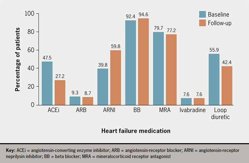
More patients were on ACEi/ARB as compared with ARNI upon the initiation of dapagliflozin (figure 1). However, 17 (18.5%) patients on ACEi/ARB at baseline were switched to ARNI during their three-month follow-up. Overall, most patients were able to remain on the same dose for all HF medications post-initiation of dapagliflozin (table 3).

Tee - Figure 1. Heart failure medications taken by patients in this cohort at baseline and follow-up
Figure 1. Heart failure medications taken by patients in this cohort at baseline and follow-up

Table 3. Changes to patients’ heart failure medications post-initiation of dapagliflozin

Heart failure medication Dose decreased Dose increased Switched to ARNI Newly added Discontinued
ACEi/ARB 5 (5.4) 4 (4.3) 17 (18.5) 1 (1.1) 3 (3.3)
ARNI 5 (5.4) 8 (8.7) 0 (0.0) 1 (1.1)
BB 8 (8.7) 9 (9.8) 1 (1.1) 0 (0.0)
MRA 3 (3.3) 7 (7.6) 0 (0.0) 6 (6.5)
Ivabradine 1 (1.1) 0 (0.0) 0 (0.0) 0 (0.0)
All data expressed as number of patients (%), where N=92
Key: ACEi = angiotensin-converting enzyme inhibitor; ARB = angiotensin-receptor blocker; ARNI = angiotensin-receptor/neprilysin inhibitor; BB = beta blocker; MRA = mineralocorticoid receptor antagonist
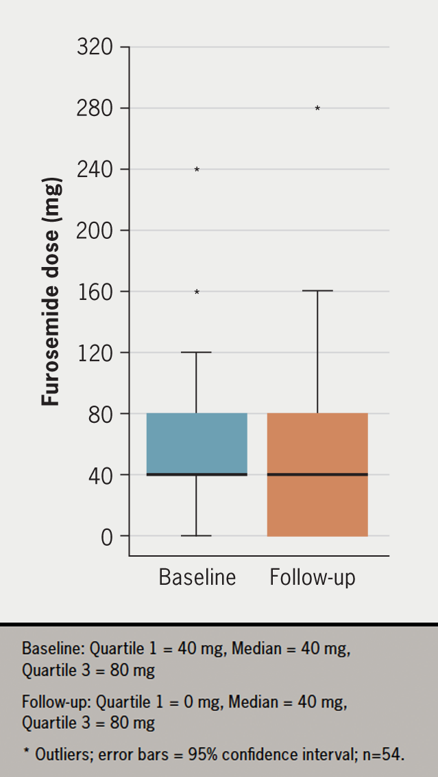
Tee - Figure 2. Furosemide dose taken by patients at baseline and follow-up
Figure 2. Furosemide dose taken by patients at baseline and follow-up

During the three-month follow-up, 42.6% (n=23) of patients either discontinued or had a dose reduction in loop diuretics, while 48.1% (n=26) of patients remained on the same dose (figure 2). For patients taking bumetanide, the furosemide-equivalent dose was calculated and used to simplify the reporting of the results. The estimated median reduction in furosemide dose post-initiation of dapagliflozin was 20 mg (95% confidence interval [CI] –20 to 0, p=0.001).

Safety and tolerability of dapagliflozin

Dapagliflozin was discontinued for reasons other than death in 12 (13.0%) patients. Of the 12 patients, dapagliflozin was temporarily discontinued in two (2.2%) patients. The main reasons dapagliflozin was discontinued were genital thrush infections (n=3, 25.0%), not tolerated for other reasons (n=2, 16.7%) and unacceptable renal insufficiency (n=2, 16.7%). Other reasons (n=5, 41.7%) included increased dyspnoea, hypotension, dehydration, recurrent urinary tract infection and polyuria.

Discussion

The present study highlights the potential benefits of the early initiation of dapagliflozin, particularly the changes to other HF pharmacological treatments, and the tolerability of dapagliflozin. The early initiation of dapagliflozin in this study led to the discontinuation or dose reduction in loop diuretics in 42.6% (n=23) of patients at three months. This is higher than the results from the DAPA-HF trial, which only initiated dapagliflozin in patients when treatment had already been optimised, where only 10.4% of patients had a dose reduction in diuretics at six months.16 Interestingly, both the DAPA-HF and Empagliflozin Outcome Trial in Patients with Chronic Heart Failure with Reduced Ejection Fraction (EMPEROR-Reduced) trial also found that fewer patients in the treatment group required intensification of diuretic treatment during their follow-up.10,16 These are significant benefits as loop diuretics are used only to relieve congestion symptoms and improve exercise capacity without reducing mortality.17 Additionally, patients who remain on diuretics long term, particularly on high doses, are associated with higher mortality, which may indicate the presence of more severe disease.18 Therefore, reduction in diuretic dose and relief from congestion without the need for long-term diuretics is prognostically advantageous for HF patients.

The discontinuation of loop diuretics may also provide opportunities for clinicians to further optimise other disease-modifying HF medicines. The present study found that 18.5% (n=17) who were on ACEi/ARB at baseline switched to ARNI three months post-initiation of dapagliflozin. The superiority of ARNI to ACEi in reducing mortality and HF hospitalisation has been documented in several studies.11,12,19,20 While this study found reductions in blood pressure and weight, these changes may result from the concomitant up-titration of other HF therapies and not just the effect of dapagliflozin alone.

The results of the present study indicate that dapagliflozin is safe and well-tolerated by HFrEF patients with early use in combination with other prognostic therapies. The discontinuation rate of dapagliflozin in this study was similar to the other SGLT2 inhibitor studies.9,20,21 There were several reasons dapagliflozin was discontinued permanently in these patients, with the main reason being recurrent genital thrush infections. Similar reasons have also been reported in previous trials, where a higher incidence of genital fungal infections was found in the treatment group, resulting in the discontinuation of dapagliflozin.22-24 Proper counselling on perineal hygiene, signs and symptoms of genital fungal infections, and sick day rules are required to avoid discontinuation of dapagliflozin.

Ultimately, the greatest benefits will be achieved at a population level if SGLT2 inhibitors can be safely initiated and managed collaboratively in primary and secondary care, given their benefits in treating and preventing multiple conditions, including HF, T2DM and chronic kidney disease with proteinuria.9,25-27 Many patients affected by these conditions may never require referral to secondary care. Hence, there is a need for simplification of a practical guideline to cover initiation and management for a variety of indications, with universal application in primary and secondary care, to avoid unnecessary variation in practice and confusion. HF patients, particularly those with heart failure with preserved ejection fraction (HFpEF), will also benefit from the approval of other SGLT2 inhibitors, such as empagliflozin, which was recently approved for the treatment of HFrEF and HFpEF, as this will extend the treatment options for patients across the HF spectrum.25,28

Limitations

This study has a number of limitations worthy of mention. First, the study was a single-centre, retrospective, observational study with a relatively small sample size, which could affect the generalisability of the results. Second, there were a few differences between the patient demographic at GSTT and the rest of the UK, as London has a significantly higher ethnic diversity. The mean age in this study is younger than the average age of HF diagnosis in the NHS (66 vs. 77 years).14 Seventy-two per cent of the patients (n=85) recruited in this study were male, 61.0% (n=72) of the cohort were white and 15.3% (n=18) were black.14 Therefore, the population in the study may not reflect other local HFrEF populations across the UK.

In this study, some patients did not have a follow-up as they were discharged back to their local communities outside of South East London, where we have restricted access to patient data. Besides that, the follow-up timeframes were inconsistent due to the nature of the appointment booking system within the Trust.

Finally, and most importantly, the effects described in the present study are not to be attributed to dapagliflozin alone, as concomitant up-titration of other HF medicines and treatment responses related to cardiac devices also occurred simultaneously. Therefore, the outcomes measured may represent changes resulting from the composite effects of therapy with these potential confounding factors, and not purely the effect of the initiation of dapagliflozin alone.

Conclusion

In conclusion, this study has demonstrated the benefits and safety of the early initiation of dapagliflozin in patients with HFrEF in line with the updated ESC HF guideline. In clinical practice, early initiation of dapagliflozin resulted in earlier discontinuation or dose reduction in loop diuretics, providing opportunities to further optimise other HF medicines. Dapagliflozin is safe and well-tolerated in a real-world population, outside the setting of a clinical trial. Counselling on perineal hygiene may avoid discontinuation of dapagliflozin due to minimising the risk of genital thrush infection. This study supports the safety of the updated ESC guideline position to initiate all four key HF medicines concurrently, without stepwise maximal up-titration and sequential addition. This new treatment algorithm will minimise delays in HF treatment optimisation, which should translate to reduced NHS healthcare costs through fewer HF hospitalisations and, as such, increase hospital inpatient capacity with greater bed availability.

Key messages

  • The early initiation of dapagliflozin resulted in the early discontinuation or dose reduction in loop diuretics, providing opportunities for clinicians to further optimise other heart failure (HF) medications
  • Dapagliflozin is safe and well-tolerated in the real-world setting
  • Dapagliflozin should be considered as a key HF pharmacological therapy rather than an add-on therapy, hence, it should be initiated earlier in the treatment pathway in order to minimise delays in treatment optimisation.

Conflicts of interest

None declared.

Funding

None.

Study approval

As this was a service evaluation, no ethical approval was needed. The evaluation was registered on the Trust’s audit database. Patient consent was not required.

Acknowledgements

Thank you to the heart failure team at Guy’s and St. Thomas’ Hospital for the care of these patients and support of this work. We would particularly like to acknowledge Professor Gerald Carr-White, Dr Jessica Webb, Dr Tevfik F Ismail, Dr Stamatis Kapetanakis, Dr Laura-Ann McGill and Dr Syed Gardezi.

References

1. British Heart Foundation. Heart & Circulatory Disease Statistics 2021. London: BHF, 2021. Available from: https://www.bhf.org.uk/what-we-do/our-research/heart-statistics [accessed 7 November 2021].

2. British Heart Foundation. An integrated approach to managing heart failure in the community. London: BHF, 2015. Available from: https://www.heartfailurehubscotland.co.uk/wp-content/uploads/2016/04/HF-an-integrated-approach-to-managing-heart-failure-in-the-community-sirhf1.pdf

3. Cowie MR, Wood DA, Coats AJS et al. Survival of patients with a new diagnosis of heart failure: a population-based study. Heart 2000;83:505–10. https://doi.org/10.1136/heart.83.5.505

4. Ho KKL, Anderson KM, Kannel WB et al. Survival after the onset of congestive heart failure in Framingham Heart Study subjects. Circulation 1993;88:107–15. https://doi.org/10.1161/01.CIR.88.1.107

5. Shah A, Gandhi D, Srivastava S et al. Heart failure: a class review of pharmacotherapy. P T 2017;42:464. Available from: https://www.ncbi.nlm.nih.gov/pmc/articles/PMC5481297/

6. Crespo-Leiro MG, Anker SD, Maggioni AP et al. European Society of Cardiology Heart Failure Long-Term Registry (ESC-HF-LT): 1-year follow-up outcomes and differences across regions. Eur J Heart Fail 2016;18:613–25. https://doi.org/10.1002/ejhf.566

7. National Institute for Cardiovascular Outcomes Research. Heart Failure. Available at: https://www.nicor.org.uk/category/heart-failure/ [accessed 17 December 2021].

8. Marti CN, Fonarow GC, Anker SD et al. Medication dosing for heart failure with reduced ejection fraction – opportunities and challenges. Eur J Heart Fail 2019;21:286–96. https://doi.org/10.1002/ejhf.1351

9. McMurray JJV, Solomon SD, Inzucchi SE et al. Dapagliflozin in patients with heart failure and reduced ejection fraction. New Engl J Med 2019;381:1995–2008. https://doi.org/101056/NEJMoa1911303

10. Tomasoni D, Fonarow GC, Adamo M et al. Sodium-glucose co-transporter 2 inhibitors as an early, first-line therapy in patients with heart failure and reduced ejection fraction. Eur J Heart Fail 2022;24:431–41. https://doi.org/10.1002/ejhf.2397

11. Tromp J, Ouwerkerk W, van Veldhuisen DJ et al. A systematic review and network meta-analysis of pharmacological treatment of heart failure with reduced ejection fraction. JACC Heart Fail 2022;10:73–84. https://doi.org/10.1016/j.jchf.2021.09.004

12. Vaduganathan M, Claggett BL, Jhund PS et al. Estimating lifetime benefits of comprehensive disease-modifying pharmacological therapies in patients with heart failure with reduced ejection fraction: a comparative analysis of three randomised controlled trials. Lancet 2020;396:121–8. https://doi.org/10.1016/S0140-6736(20)30748-0

13. Bauersachs J. Heart failure drug treatment: the fantastic four. Eur Heart J 2021;42:681–3. https://doi.org/10.1093/eurheartj/ehaa1012

14. National Institute for Health and Care Excellence. Dapagliflozin for treating chronic heart failure with reduced ejection fraction. TA679. London: NICE, 2021. Available from: https://www.nice.org.uk/guidance/ta679

15. McDonagh TA, Metra M, Adamo M et al. 2021 ESC guidelines for the diagnosis and treatment of acute and chronic heart failure: developed by the Task Force for the diagnosis and treatment of acute and chronic heart failure of the European Society of Cardiology (ESC) with the special contribution of the Heart Failure Association (HFA) of the ESC 2021. Eur Heart J 2021;42:3599–726. https://doi.org/10.1093/eurheartj/ehab368

16. Jackson AM, Dewan P, Anand IS et al. Dapagliflozin and diuretic use in patients with heart failure and reduced ejection fraction in DAPA-HF. Circulation 2020;142:1040–54. https://doi.org/10.1161/CIRCULATIONAHA.120.047077

17. Casu G, Merella P. Diuretic therapy in heart failure – current approaches. Eur Cardiol Rev 2015;10:42. https://doi.org/10.15420/ecr.2015.10.01.42

18. Hasselblad V, Stough WG, Shah MR et al. Relation between dose of loop diuretics and outcomes in a heart failure population: results of the ESCAPE trial. Eur J Heart Fail 2007;9:1064. https://doi.org/10.1016/j.ejheart.2007.07.011

19. Okumura N, Jhund PS, Gong J et al. Effects of sacubitril/valsartan in the PARADIGM-HF trial (Prospective Comparison of ARNI with ACEI to Determine Impact on Global Mortality and Morbidity in Heart Failure) according to background therapy. Circ Heart Fail 2016;9:e003212. https://doi.org/10.1161/CIRCHEARTFAILURE.116.003212

20. Packer M, Anker SD, Butler J et al. Cardiovascular and renal outcomes with empagliflozin in heart failure. N Engl J Med 2020;383:1413–24. https://doi.org/10.1056/NEJMoa2022190

21. Bhatt DL, Szarek M, Steg PG et al. Sotagliflozin in patients with diabetes and recent worsening heart failure. N Engl J Med 2021;384:117–28. https://doi.org/10.1056/NEJMoa2030183

22. Wiviott SD, Raz I, Bonaca MP et al. Dapagliflozin and cardiovascular outcomes in type 2 diabetes. N Engl J Med 2019;380:347–57. https://doi.org/10.1056/NEJMoa1812389

23. Shah SR, Ali A, Ikram S. Sotagliflozin and decompensated heart failure: results of the SOLOIST-WHF trial. Expert Rev Clin Pharmacol 2021;14:523–5. https://doi.org/10.1080/17512433.2021.1908123

24. Anker SD, Butler J, Filippatos G et al. Effect of empagliflozin on cardiovascular and renal outcomes in patients with heart failure by baseline diabetes status: results from the EMPEROR-Reduced trial. Circulation 2021;143:337–49. https://doi.org/10.1055/s-0041-1727472

25. Anker SD, Butler J, Filippatos G et al. Empagliflozin in heart failure with a preserved ejection fraction. N Engl J Med 2021;385:1451–61. https://doi.org/10.1056/NEJMoa2107038

26. Zinman B, Wanner C, Lachin JM et al. Empagliflozin, cardiovascular outcomes, and mortality in type 2 diabetes. N Engl J Med 2015;373:2117–28. https://doi.org/10.1056/NEJMoa1504720

27. Heerspink HJL, Stefánsson BV, Correa-Rotter R et al. Dapagliflozin in patients with chronic kidney disease. N Engl J Med 2020;383:1436–46. https://doi.org/10.1056/NEJMoa2024816

28. Heidenreich PA, Bozkurt B, Anguilar D et al. AHA/ACC/HFSA guideline for the management of heart failure: a report of the American College of Cardiology/American Heart Association Joint Committee on Clinical Practice Guidelines. J Am Coll Cardiol 2022;79:e263–e421. https://doi.org/10.1016/j.jacc.2021.12.012

Pectus excavatum with right ventricular compression-induced ventricular arrhythmias

Br J Cardiol 2023;30:39–40doi:10.5837/bjc.2023.004 Leave a comment
Click any image to enlarge
Authors:
First published online 18th January 2023

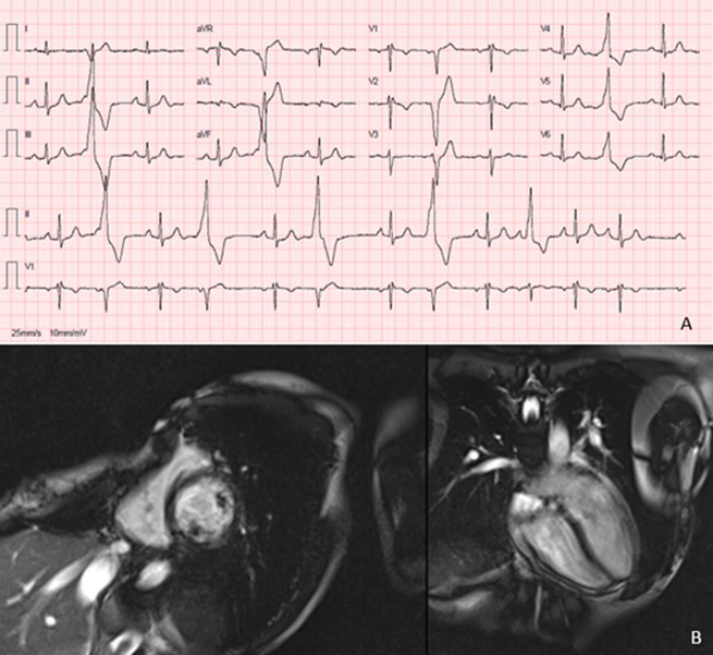
A 33-year-old woman, with palpitations since the age of 15, was referred to a cardiology consultation due to very frequent ventricular extrasystoles with morphology of left bundle branch block, inferior frontal axis, late precordial transition, rS in V1, R in V6 and rS in DI. She had pectus excavatum. The cardiac magnetic resonance showed severe pectus excavatum associated with exaggerated cardiac levoposition, compression and deformation of the right cardiac chambers. However, the patient became pregnant, and follow-up was delayed.

Introduction

Pectus excavatum is the most common congenital anomaly of the anterior chest wall, most often being benign, however, it can sometimes have structural and haemodynamic consequences secondary to mechanical compression.1 Most patients are asymptomatic, but may experience symptoms due to impaired cardiac function and, occasionally, cardiac arrhythmias.2

Case

A 33-year-old woman, without relevant personal and family history, with palpitations since the age of 15, was referred to a cardiology consultation due to very frequent ventricular extrasystoles (28,423/day, 26.6%). On objective examination, she had pectus excavatum, without other alterations. Blood tests showed no significant changes, namely in the electrolytes, blood count or thyroid function. Her resting 12-lead electrocardiogram (ECG) showed sinus rhythm and ventricular extrasystoles with morphology of left bundle branch block, inferior frontal axis, late precordial transition, rS in V1, R in V6 and rS in DI (figure 1A), suggesting an origin in the free wall of the right ventricle (anterior).3 Transthoracic echocardiogram showed no structural heart disease, there was no evidence of arrhythmogenic right ventricular cardiomyopathy. Coronary artery disease was excluded by coronary computed tomography angiography. She underwent cardiac magnetic resonance with perfusion study that showed severe pectus excavatum (Haller index 4.9), associated with exaggerated cardiac levoposition, compression and deformation of the right cardiac chambers (figure 1B), no delayed enhancement or ischaemia, and good biventricular function. However, the patient became pregnant, and the follow-up to the study and therapeutic guidance (namely catheter ablation and surgical correction for her pectus excavatum) were delayed.

Ferraz - Figure 1. A. 12-lead electrocardiogram showing ventricular extrasystole. B. Cardiac magnetic resonance study revealing severe pectus excavatum with compression and deformation of the right cardiac chambers
Figure 1. A. 12-lead electrocardiogram showing ventricular extrasystole. B. Cardiac magnetic resonance study revealing severe pectus excavatum with compression and deformation of the right cardiac chambers

Discussion

Pectus excavatum is considered to be a benign condition, rarely requiring treatment, commonly undertaken only for cosmetic and psychological reasons. However, it can be associated with arrhythmias, mainly supraventricular arrhythmias, and, more rarely, ventricular arrhythmias. We present a case of focal electrocardiomyopathy that resulted from sternal compression and gave rise to symptomatic ventricular extrasystole.

Less common causes of ventricular tachycardia, including compression cardiomyopathy, should be considered if there is unusual ECG-based morphology for right ventricular tachycardia, not consistent with right ventricular outflow tract origin.

Literature regarding cardiac arrhythmias, namely ventricular arrhythmias, caused by pectus excavatum is limited. Winkens et al. reported a case presenting with progressive heart palpitations, fatigue and postural dyspnoea, due to recurrent supraventricular tachycardia and a nodal tachycardia caused by pectus excavatum, in which the physical condition of the patient improved rapidly with surgical correction.3

Surgical correction should be considered as the initial treatment in cases of persistent right ventricular compression with structural and haemodynamic consequences. Catheter ablation may not be necessary.

Conclusion

This case highlights an uncommon cause of ventricular extrasystole, pectus excavatum, which despite being a common skeletal anomaly of the chest wall and usually having a benign course, may also be associated with symptomatic tachyarrhythmias.

Conflicts of interest

None declared.

Funding

None.

Patient consent

A written patient consent was obtained.

References

1. Ahn J, Choi J, Shim J, Lee S, Kim Y. Right ventricular compression mimicking Brugada-like electrocardiogram in a patient with recurrent pectus excavatum. Case Rep Cardiol 2017;2017:3047937. https://doi.org/10.1155/2017/3047937

2. Geiser G, Epstein S, Stampfer M, Goldstein R, Noland S, Levitsky S. Impairment of cardiac function in patients with pectus excavatum, with improvement after operative correction. N Engl J Med 1972;287:267–72. https://doi.org/10.1056/NEJM197208102870602

3. Winkens R, Guldemond F, Hoppener P, Kragten H, Leeuwen Y. Pectus excavatum, not always as harmless as it seems. BMJ Case Rep 2009;2009:bcr10.2009.2329. https://doi.org/10.1136/bcr.10.2009.2329

The prognostic impact of HDL-C level in patients presenting with ST-elevation myocardial infarction

Br J Cardiol 2023;30:31–4doi:10.5837/bjc.2023.005 Leave a comment
Click any image to enlarge
Authors:
First published online 18th January 2023

Low high-density lipoprotein-cholesterol (HDL-C) concentration is among the strongest independent risk factors for cardiovascular disease, however, studies to assess the cardioprotective effect of normal or high HDL-C level are lacking.

To determine the prognostic impact of initial serum HDL-C level on in-hospital major adverse cardiovascular and cerebrovascular events (MACCE) and the one-year all-cause mortality in patients presenting with ST-elevation myocardial infarction (STEMI) we performed a retrospective analysis of the data from 1,415 patients presenting with STEMI in a tertiary-care centre equipped with a 24-hour-ready catheterisation laboratory. The period from June 2014 to June 2017 was reviewed with a follow-up as regards one-year all-cause mortality. Patients were divided into two groups according to HDL-C level. HDL-C <40 mg/dL (2.22 mmol/L) was considered low, while HDL-C ≥40 mg/dL was considered normal.

There were 1,109 patients with low HDL-C, while 306 had normal HDL-C levels, which was statistically significant (p<0.001). Total MACCE and all-cause mortality were significantly lower in patients with normal HDL-C (p=0.03 and p=0.01, respectively).

In conclusion, this retrospective study to assess the prognostic effect of HDL-C in patients presenting with STEMI, found normal HDL-C level was associated with lower in-hospital MACCE and all-cause mortality at one-year follow-up.

Introduction

Multiple risk factors have been identified for myocardial infarction (MI). Hypercholesterolaemia is one of the major risk factors for cardiovascular diseases, and has an important pathophysiological role in the development of acute ST-elevation myocardial infarction (STEMI).1 Endothelial dysfunction, inflammation, increased thrombogenicity, and plaque vulnerability are the main underlying mechanisms explaining the complex interaction between hypercholesterolaemia and STEMI.2–4

Low-density lipoprotein-cholesterol (LDL-C) has been shown to be a major risk factor in patients with MI, and aggressive LDL-C lowering remains a main target for medical therapy. However, there are still residual risks in MI patients being treated with LDL-C lowering therapy, and this may draw the attention to the prognostic impact of the level of high-density lipoprotein-cholesterol (HDL-C) in acute MI.5–7

Lipid-lowering therapies are an important component of the early treatment for patients with acute coronary syndromes (ACS), including MI, because randomised-controlled trials have shown that rapid lowering of LDL-C levels with high-intensity statins improves clinical outcomes.8,9 Reduced levels of HDL-C are a component of the metabolic syndrome, which is associated with an increased risk of cardiovascular events, but the prognostic impact of HDL-C levels has only been studied in patients with stable coronary artery disease, or in those patients at high risk for cardiovascular diseases.6,10,11

Low HDL-C levels have been shown to be associated with an increased risk of cardiovascular events, but the prognostic impact of low HDL-C levels on clinical outcomes in patients with ACS is still controversial. Moreover, there are no guidelines or recommendations for the treatment or risk stratification of ACS patients with low HDL-C levels.12,13

Aim

To detect the prognostic impact of the initial level of serum HDL-C on in-hospital major adverse cardiovascular events (MACE), as well as the one-year all-cause mortality in patients presenting with ST-elevation myocardial infarction (STEMI).

Method

A retrospective analysis of the data from 1,415 patients presenting with STEMI in a tertiary-care centre equipped with a 24-hour-ready cardiac catheterisation laboratory in the period from June 2014 to June 2017. We excluded 126 patients who did not have measured HDL-C values recorded during the index hospitalisation, and 242 patients were excluded because they refused consent to collect their data. We also excluded 230 patients who were not taking statin therapy or were lost to follow-up. Follow-up for one-year, regarding all-cause mortality, was done for the included patients.

Fasting peripheral venous blood samples were drawn the next day, after admission to the coronary care unit. Total cholesterol, HDL-C, LDL-C cholesterol, and triglyceride concentrations were measured using the Cobas 8000 modular analyzer series (Roche Diagnostics, Vienna, Austria). Peak high-sensitivity cardiac troponin T (hs-cTnT), troponin I, troponin T, and peak high-sensitivity C-reactive protein (hs-CRP) concentrations were also determined.

Patients were divided into two groups according to HDL-C level. HDL-C level less than 40 mg/dL (2.22 mmol/L) was considered low, while HDL-C level of 40 mg/dL (2.22 mmol/L) or more was considered normal.

The diagnosis of STEMI was based on clinical signs or symptoms of typical angina or angina equivalent symptoms, specific changes on 12-lead electrocardiogram (ECG) (ST-segment elevation in continuous leads or new left bundle branch block), and laboratory changes including increases of serum cardiac biomarkers (creatine kinase-MB, troponin I, or troponin T).

The study was approved by the local ethics committee and all patients signed an informed consent. Retrospective data were retrieved from the electronic database of the hospital. In-hospital data and one-year follow-up data were obtained, including demographics, risk factors, clinical presentation, angiographic data, in-hospital MACE and one-year mortality incidence.

Statistical analysis

The Statistical Package for Social Science (SPSS, Chicago, IL, USA) for Windows, version 22.0 was used for statistical analysis. Continuous variables were expressed as mean ± standard deviation (SD) and compared with Student’s t-test or Mann–Whitney test. Categorical variables were expressed as percentage and compared with Chi-square test. All variables were considered statistically significant when the p value was less than 0.05.

Results

Table 1. Lipid profile (n=1,415)

Lipid
Mean ± SD, mmol/L
Low HDL-C group
n=1,109
p value Normal HDL-C group
n=306
TC 14.02 ± 2.23 0.69 13.89 ± 2.16
LDL-C 8.82 ± 1.33 0.23 7.87 ± 1.58
HDL-C 1.71 ± 0.29 <0.001* 2.64 ± 0.44
TG 10.44 ± 1.17 0.06 8.44 ± 1.04
*Statistically significant.
Key: HDL-C = high-density lipoprotein-cholesterol; LDL-C = low-density lipoprotein-cholesterol; SD = standard deviation; TC = total cholesterol; TG = triglycerides

From June 2014 to June 2017, 1,415 patients presenting with STEMI were included and were treated with primary percutaneous coronary intervention (pPCI). All the included patients were taking high-intensity statin therapy (atorvastatin 40–80 mg/day or rosuvastatin 20–40 mg/day). Included patients were divided into two groups according to HDL-C level; HDL-C <40 mg/dL (2.22 mmol/L) was considered low, while HDL-C ≥40 mg/dL was considered normal.

There were 1,109 patients with low HDL-C level, while 306 had a normal HDL-C level, which was statistically significant (p<0.001) (table 1). Patients with normal HDL-C were older, and more likely to be male and/or diabetic; but nothing statistically significant. Patients with low HDL-C had significantly more history of previous ACS (p<0.001). Regarding clinical and angiographic data, anterior STEMI was more prevalent (p<0.001) in patients with low HDL-C, with culprit left anterior descending artery (LAD) during pPCI (table 2).

In-hospital MACCE were significantly lower in patients with normal HDL-C (p=0.03). In-hospital mortality and all-cause mortality at one year were also significantly lower in patients with normal HDL-C (p=0.03 and p=0.01, respectively) (table 2).

Table 2. Baseline patient characteristics, in-hospital major adverse cardiovascular and cerebrovascular events (MACCE) and all-cause mortality at one year (n=1,415)

Characteristic Low HDL-C group
n=1,109
p value Normal HDL-C group
n=306
History
Mean age, years (SD) 52 (±11.1) 0.69 59 (±10.5)
Male, n (%) 665 (59.96%) 0.78 187 (61.11%)
Hypertension, n (%) 611 (55.09%) 0.09 124 (40.52%)
Smoking, n (%) 654 (58.97%) 0.06 113 (36.93%)
Diabetes, n (%) 211 (19.03%) 0.33 63 (20.59%)
Previous MI, n (%) 99 (8.93%) <0.001* 22 (7.19%)
Clinical data
Anterior STEMI, n (%) 855 (77.1%) <0.001* 111 (36.27%)
Killip class IV, n (%) 7 (0.63%) 0.12 3 (0.98%)
GRACE risk score, mean (SD) 140 (±25) 0.23 144 (±21)
IRA, n (%)
LM 9 (0.81%) 0.2 4 (1.31%)
LAD 611 (55.1%) 0.11 155 (50.65%)
RCA 319 (28.76%) 0.23 80 (26.14%)
LCx 170 (15.33%) 0.08 67 (21.9%)
In-hospital MACCE, n (%)
Death 27 (2.43%) 0.03 5 (1.63%)
MI 13 (1.17%) 0.09 3 (0.98%)
TVR 19 (1.71%) <0.001* 1 (0.33%)
CVS/TIA 7 (0.54%) 0.06 1 (0.33%)
Total 66 (5.95%) 0.03* 10 (3.27%)
All-cause mortality at 1 year 44 (3.97%) 0.01* 3 (0.98%)
*Statistically significant.
Key: CVS = cerebrovascular stroke; GRACE = Global Registry of Acute Coronary Events; IRA = infarct-related artery; LAD = left anterior descending; LCx = left circumflex; LM = left main; MACCE = major adverse cardiovascular and cerebrovascular events; MI = myocardial infarction; RCA = right coronary artery; SD = standard deviation; STEMI = ST-elevation myocardial infarction; TIA = transient ischaemic attack; TVR = target-vessel revascularisation

Discussion

The main finding in this study was that low HDL-C (<40 mg/dL; 2.22 mmol/L) was associated with higher in-hospital and one-year mortality in STEMI patients. Previous studies have shown controversial data about the prognostic impact of low HDL-C on clinical outcomes in non-ST-segment-elevation acute coronary syndrome (NSTE-ACS) patients, while there is a scarcity of data in STEMI patients.14–16

Some studies,17,18 have suggested that sex difference might affect the prognosis of patients with acute MI; however, our results showed that sex difference itself was not statistically different between low and normal HDL-C levels.

Our study provides an important new clinical view on the prognostic significance of cholesterol metabolism in patients with acute STEMI. Our results could highlight, particularly, the prognostic importance of HDL-C status in these patients. Indeed, the prognostic value of low HDL-C level for worse outcomes in STEMI, may emphasise the importance of HDL-C assessment on coronary care unit admission for routine daily clinical and laboratory workup. Above and beyond these prognostic implications, the present results may suggest HDL-C levels as a prophylactic or therapeutic target against the development of adverse cardiovascular events.

Some studies had shown that lower HDL-C levels are associated with a higher risk of cardiovascular events and more burden of atherosclerosis, even among cardiovascular patients with lower LDL-C levels, including those patients treated with statins.6,11 In our study, we have shown that lower HDL-C levels were associated with a higher risk of adverse outcomes, and appear to correlate with younger age upon presentation in patients with STEMI. In contrast, higher HDL-C levels were associated with older age at presentation in STEMI, which may indicate that higher HDL-C levels delay symptomatic coronary artery disease progression until older age. Nevertheless, lower HDL-C levels appear to be a unique, and potentially modifiable, risk factor for STEMI patients.

Limitations

This was a retrospective study, with the possibility of selection bias or missing data. We could not fully characterise the extent of the metabolic syndrome in our study because waist circumference measures were not routinely collected. There are no specific cut-off values for the cholesterol levels in the Egyptian population, but it was assumed that the Egyptian population level may be similar to those of the Mediterranean region. HDL-C levels before admission were not collected and recorded, HDL-C levels during the hospitalisation may differ from chronic HDL-C levels before presentation, so the impact of low HDL-C levels on the progression of coronary disease may not be accurately represented by in-hospital HDL-C levels.

Conclusion

We can conclude that low HDL-C level was associated with a significantly higher risk of in-hospital and one-year mortality in STEMI patients. Thus, early detection of low HDL-C is important in STEMI patients, and proper treatment with high-intensity statin therapy during the early stage should be considered in STEMI patients with low HDL-C, aiming both to reduce LDL-C levels and increase HDL-C levels.

Key messages

  • Low high-density lipoprotein-cholesterol (HDL-C) level is associated with higher risk of in-hospital and one-year mortality in ST-elevation myocardial infarction (STEMI) patients
  • Early detection of low HDL-C is important in STEMI patients, and proper treatment with high-intensity statin therapy should be considered in STEMI patients, aiming to reduce LDL-C levels and increase HDL-C levels

Conflicts of interest

None declared.

Funding

None.

Study approval

All procedure aspects were approved by the local ethics committee.

References

1. Catapano AL, Graham I, De Backer G et al. 2016 ESC/EAS guidelines for the management of dyslipidaemias: the Task Force for the management of dyslipidaemias of the European Society of Cardiology (ESC) and European Atherosclerosis Society (EAS) developed with the special contribution of the European Association for Cardiovascular Prevention & Rehabilitation (EACPR). Eur Heart J 2016;37:2999–3058. https://doi.org/10.1093/eurheartj/ehw272

2. Steinberg D, Witztum JL. Oxidized low-density lipoprotein and atherosclerosis. Arterioscler Thromb Vasc Biol 2010;30:2311–16. https://doi.org/10.1161/ATVBAHA.108.179697

3. Carnevale R, Bartimoccia S, Nocella C et al. LDL oxidation by platelets propagates platelet activation via an oxidative stress-mediated mechanism. Atherosclerosis 2014;237:108–16. https://doi.org/10.1016/j.atherosclerosis.2014.08.041

4. Stampfer MJ, Sacks FM, Salvini S et al. A prospective study of cholesterol, apolipoproteins, and the risk of myocardial infarction. N Engl J Med 1991;325:373–81. https://doi.org/10.1056/NEJM199108083250601

5. Hausenloy DJ, Yellon DM. Targeting residual cardiovascular risk: raising high-density lipoprotein cholesterol levels. Heart 2008;94:706–14. https://doi.org/10.1136/hrt.2007.125401

6. Barter P, Gotto AM, LaRosa JC et al.; Treating to New Targets Investigators. HDL cholesterol, very low levels of LDL cholesterol, and cardiovascular events. N Engl J Med 2007;357:1301–10. https://doi.org/10.1056/NEJMoa064278

7. Olsson AG, Schwartz GG, Szarek M et al. High-density lipoprotein, but not low-density lipoprotein cholesterol levels influence short-term prognosis after acute coronary syndrome: results from the MIRACL trial. Eur Heart J 2005;26:890–6. https://doi.org/10.1093/eurheartj/ehi186

8. De-Lemos JA, Blazing MA, Wiviott SD et al. Early intensive vs a delayed conservative simvastatin strategy in patients with acute coronary syndromes: phase Z of the A to Z trial. JAMA 2004;292:1307–16. https://doi.org/10.1001/jama.292.11.1307

9. Cannon CP, Braunwald E, McCabe CH et al.; Pravastatin or Atorvastatin Evaluation Infection Therapy – Thrombolysis in Myocardial Infarction 22 Investigators. Intensive versus moderate lipid lowering with statins after acute coronary syndromes. N Engl J Med 2004;350:1495–504. https://doi.org/10.1056/NEJMoa040583

10. Gami AS, Witt BJ, Howard DE et al. Metabolic syndrome and risk of incident cardiovascular events and death: a systematic review and meta-analysis of longitudinal studies. J Am Coll Cardiol 2007;49:403–14. https://doi.org/10.1016/j.jacc.2006.09.032

11. deGoma EM, Leeper NJ, Heidenreich PA. Clinical significance of high-density lipoprotein cholesterol in patients with low low-density lipoprotein cholesterol. J Am Coll Cardiol 2008;51:49–55. https://doi.org/10.1016/j.jacc.2007.07.086

12. Collet JP, Thiele H, Barbato E et al. 2020 ESC guidelines for the management of acute coronary syndromes in patients presenting without persistent ST-segment elevation: the Task Force for the management of acute coronary syndromes in patients presenting without persistent ST-segment elevation of the European Society of Cardiology (ESC). Eur Heart J 2021;42:1289–367. https://doi.org/10.1093/eurheartj/ehaa909

13. Ibanez B, James S, Agewall S et al. 2017 ESC guidelines for the management of acute myocardial infarction in patients presenting with ST-segment elevation: the Task Force for the management of acute myocardial infarction in patients presenting with ST-segment elevation of the European Society of Cardiology (ESC). Eur Heart J 2018;39:119–77.

14. Wolfram RM, Brewer HB, Xue Z et al. Impact of low high-density lipoproteins on in-hospital events and one-year clinical outcomes in patients with non-ST-elevation myocardial infarction acute coronary syndrome treated with drug-eluting stent implantation. Am J Cardiol 2006;98:711–17. https://doi.org/10.1016/j.amjcard.2006.04.006

15. Roe MT, Ou FS, Alexander KP et al. Patterns and prognostic implications of low high-density lipoprotein levels in patients with non-ST-segment elevation acute coronary syndromes. Eur Heart J 2008;29:2480–8. https://doi.org/10.1093/eurheartj/ehn364

16. Acharjee S, Roe MT, Amsterdam EA et al. Relation of admission high-density lipoprotein cholesterol level and in-hospital mortality in patients with acute non-ST segment elevation myocardial infarction (from the National Cardiovascular Data Registry). Am J Cardiol 2013;112:1057–62. https://doi.org/10.1016/j.amjcard.2013.05.050

17. Jneid H, Fonarow GC, Cannon CP et al. Sex differences in medical care and early death after acute myocardial infarction. Circulation 2008;118:2803–10. https://doi.org/10.1161/CIRCULATIONAHA.108.789800

18. Lawesson SS, Alfredsson J, Fredrikson M et al. A gender perspective on short- and long-term mortality in ST-elevation myocardial infarction – a report from the SWEDEHEART registry. Int J Cardiol 2013;168:1041–7. https://doi.org/10.1016/j.ijcard.2012.10.028