England sees MI reduction after smoking ban 

Br J Cardiol 2010;17:163-5 Leave a comment
Click any image to enlarge
Authors:

A new study has confirmed that there has been a significant drop in hospital admissions for myocardial infarction (MI) after the introduction of smoke-free legislation in England.

165-img-1The study, published online in the British Medical Journal on June 8, 2010, found that, after accounting for a pre-existing decline in admissions, trends in population size, and seasonal variation in admissions, there was a 2.4% drop in the number of emergency admissions for MI after the smoking ban legislation came into force on July 1, 2007. This equates to 1,200 fewer emergency admissions in the first year after the law came into effect (1,600 including readmissions).

The researchers, from the University of Bath, note that the largest impacts of smoking bans on MI rates have been reported in smaller studies in the US, with reductions in the range of 27% to 40%, while larger studies have reported more modest reductions. But they point out that there is some uncertainty around the extent to which some of these studies have effectively accounted for other factors that might influence patterns of admissions for MI, such as a general decline in MI rate anyway and seasonal issues.

They suggest that the reduction found in this study was smaller than other studies partly because of better control for such factors and also because exposure to secondhand smoke before the ban was lower in England. Visit Website for experts’ take on smoking and its health effects.  In some other countries, with 55% of employed adults in England already working in a smoke-free environment and many bars and restaurants having already gone smoke-free.

In brief

Br J Cardiol 2010;17:167 Leave a comment
Click any image to enlarge
Authors:

News in brief from the world of cardiology.

Increase in kidney failure in people with diabetes

Diabetes UK has reported ‘concern’ in recently released figures showing a 20% increase in people with diabetes in England needing dialysis or a kidney transplant between 2008–2009.

The figures released by the National Diabetes Audit also reveals, for the same period, that a third of people with diabetes did not have their urine tested, half of people with diabetes were found to have not met their blood pressure targets, and more than a third were found to have poor blood glucose control, with the latter more prevalent in younger people with diabetes. Data for the audit was contributed from 152 Primary Care Trusts in the country.

Study finds epleronone cost-effective in heart failure

Health economists from the University of York have said that epleronone (Inspra®) is cost-effective when added to usual care, compared to usual care alone in the management of heart failure in a post-myocardial infarction (MI) population.

Researchers from the university’s Centre for Reviews and Dissemination (CRD) evaluated the relative clinical effectiveness and cost-effectiveness of eplerenone and spironolactone in patients with heart failure following MI. The recently published study (Health Technol Assess 2010;14 [24]) shows eplerenone’s cost per QALYs (Quality-Adjusted Life-Years) ranges from £4,457 (treatment for two years) to £7,893 (lifetime treatment), well under the £20,000 to £30,000 the NHS considers value for money.

The study was carried out following a request from the National Institute for Health and Clinical Excellence (NICE) to The National Institute for Health Research (NIHR) to evaluate the cost-effectiveness of aldosterone antagonists from an NHS perspective in post-MI heart failure. The NIHR, in turn, commissioned the CRD to prepare the Health Technology Assessment.

New troponin T assay meets international recommendations

167-img-1Roche has announced that its new troponin T high sensitive assay (Elecsys®) is the first commercially available troponin T assay to meet recent European Society of Cardiology, American College of Cardiology Foundation, American Heart Association and World Heart Federation recommendations.

The redefinition of myocardial infarction (MI) in these recommendations requires an increase or decrease of cardiac troponin to be demonstrated in patients with symptoms of acute coronary syndrome (ACS) with at least one of the concentrations greater than the 99th percentile of the reference population.

A team led by Dr Evangelos Giannitsis (University Hospital, Heidelberg, Germany) pictured above, has shown that the Elecsys® assay was the only test available to offer this level of sensitivity and precision. “Not only can we identify MI at an earlier opportunity, but we can also now identify more patients with MI (amongst those with symptoms of ACS) than we could with the less sensitive assay,” he says. “It also means that additional tests for MI/ischaemia markers are no longer required,” he added.

Licence extension for rosuvastatin

Rosuvastatin (Crestor®) has received authorisation for a licence extension from the Medicines and Healthcare products Regulatory Agency (MHRA) for use in patients who are considered to be at high risk of a first major cardiovascular event.

The new indication is based on the JUPITER study, which showed a significant reduction in the combined end point of myocardial infarction, strokes and cardiovascular deaths amongst high-risk patients, defined as having a SCORE risk ≥ 5% or Framingham Risk >20%. Rosuvastatin can now be used in these patients in addition to the modification of other risk factors, such as diet and exercise.

Sitagliptin/metformin combination treatment launched

MSD has launched a combination treatment of sitagliptin and metformin (Janumet®) for the treatment of type 2 diabetes. The company hopes the combination treatment will help reduce the pill burden on patients with diabetes and aid compliance. A survey it commissioned which included 300 UK GPs, has found that nearly half (42%) say the main issue raised by their patients is concern around forgetting to take their treatments, with nine out of 10 GPs (91%) believing that pill burden impacts concordance of type 2 patients with their treatment.

Nicotinic acid/laropiprant approved by the SMC

Nicotinic acid / laropiprant (Tredaptive®) has been accepted by the Scottish Medicines Consortium (SMC) for restricted use in NHS Scotland, as monotherapy for the treatment of dyslipidaemia in patients with combined mixed dyslipidaemia (characterised by elevated levels of LDL-cholesterol and triglycerides and low HDL-cholesterol) in patients in whom statins are considered inappropriate or not tolerated.

MSD has estimated that based on a population of over 5 million in Scotland, between 7,750–15,500 patients in the country could be experiencing non-severe side effects from statin intolerance.

New study shows promise in treatment of AF

Intravenous vernakalant has been shown to be superior to amiodarone in converting patients’ heart rate from atrial fibrillation (AF) to sinus rhythm within 90 minutes of the start of administration.

The results of AVRO (Active-Controlled, Multi-Center Study of Vernakalant Injection versus Amiodarone in Subjects with Recent Onset Atrial Fibrillation) were presented at a late-breaking clinical trials session at Heart Rhythm 2010, in Denver, USA, in May. The study showed 51.7% (n=116) of patients on vernakalant converted from AF to normal sinus rhythm within 90 minutes, versus 5.2% (n=116) in the amiodarone group (p<0.0001). The median time to conversion in patients who responded to vernakalant was 11 minutes. The phase III study was carried out by MSD and Cardiome Pharma Corp.

Is the EWTD failing the welfare of junior doctors?

Br J Cardiol 2010;17:163-5 Leave a comment
Click any image to enlarge
Authors:

To find out how the European Working Time Directive set up to help improve the welfare of junior doctors seems to be failing in one of its primary objectives, why women may benefit more from cardiac resynchronisation therapy defibrillators than men, and why telehealth heart failure patients experience a significant improvement in their care and quality of life, visit our sister website:

www.arwatch.co.uk

A glossary of terms used in interventional cardiology: part 2

Br J Cardiol 2010;17:171-2 Leave a comment
Click any image to enlarge
Authors:

We continue our series in which Consultant Interventionist Dr Michael Norell takes a sideways look at life in the cath lab…and beyond. In this column, he updates the definitions of terms used in interventional cardiology.

Some years ago I put together some definitions of words, phrases and acronyms, commonly encountered in the practice of angioplasty (percutaneous coronary intervention [PCI]), which I thought might be of assistance to the aspiring balloonist. It was cleverly (sic) headlined “Ten Atmospheres and All That” as a homage to the iconic non-textbook of English history that some of us may recall, and appeared in Cardiology News in its August 2006 issue; reprints are available.

The technology around PCI has expanded enormously in the last four years, so an update is now timely. On this occasion, and in the spirit of fairness, I felt that the British Journal of Cardiology should have its own turn to shoulder the, probably unwanted, literary burden of such an article, which, given the rated burst pressure of modern balloons, might perhaps be better titled “Twenty Atmospheres and All That”.

Retrograde (approach to) CTO: A novel, if tiresome, method of reopening Chronic Total Occlusions when the more commonly used anterograde approach, atenolol and feigning deafness to symptoms, have all failed. Very rarely one has to ‘encourage’ patients that intervention is unnecessary and that their chest pain can be safely ignored. This can be achieved using a heavy blunt implement in order to convince them; a so-called ‘CTO Club’. Treatment of bifurcation lesions (see below) can be avoided using a similar technique.

OCT: Optical Coherence Tomography – an intravascular imaging modality using light rather than ultrasound and thereby producing superior spatial resolution. This results in an aesthetically more pleasing and sepia-tinted image, akin to a sandstorm rather than a snowstorm.

MDT: Multi-Disciplinary Team – an increasingly important forum in which the management plan of an individual patient is devised by agreement among all relevant specialities (e.g. surgery, intervention, social work, etc.). The final decision is reached transparently and by consensus. Depending on a unit’s criteria dictating which cases are to be presented, the number of patients involved can be huge (note alternative: Minimum Discussion Time).

SYNTAX: Recently presented pivotal trial examining the ‘real world’ use of correct grammar when advising a patient as to the best mode of coronary revascularisation.

2b/3a (inhibitor): A potent platelet paralyser of inestimable value in the treatment of complex anatomy and in the setting of unstable clinical presentations. However, the resultant increased bleeding risk invariably prompts the serious operator to consider the pros and cons of its use and share their concerns with the catheter lab staff: “2b or not 2b; that is the question”.

Stent strut: Celebratory dance performed when a particularly inaccessible or difficult lesion has eventually been treated. Such a reaction, often reminiscent of the gyrations produced by the Rolling Stones’ Mick Jagger, is characteristically the proclivity of operators who had not appreciated the complexity of the case in the first place. This deficiency is termed strut thickness.

Polymer: A compound that is combined with an anti-proliferative agent and used in the manufacture of some drug-eluting stents. The stent is coated with the resulting mixture, the composition of which dictates the active drug’s release kinetics. The science of this technology can be mindboggling and it is, therefore, important that the concepts involved are presented to clinicians at meetings or conferences, in a simple and easily understood manner. This is called an absorbable polymer.

Abluminal: This is the point at which your Latin ‘O’ level is now seen to be valuable. Had the Romans used drug-eluting devices, they too would have realised the logic of applying anti-proliferative agent only to the surface of the stent in contact with the vessel wall (i.e. away from the lumen). However, they would also have more correctly described this drug elution as being directed towards the vessel wall, in other words, ‘admural’.

Bifurcation: Given that most coronary lesions are either before, after or at a branch point, this anatomy can be notoriously common. The recently published BBC-ONE study confirmed what most of us already knew: when considering the treatment of such cases, rather than embarking on a complex ‘two-stent’ strategy, you are much better off watching television.

‘Take-off’ angle: The angle at which a side branch leaves the parent vessel can impact on the outcome when treating bifurcation disease. If particularly acute, or severely obtuse, the appearances can strike such anxiety in an operator that he is obliged to leave the catheter lab and take off.

FAME: Another pivotal trial, this time assessing the value of pressure-wire guided PCI when compared with giving free rein to our occulo-stenotic reflex. It exposed the shortcomings of previous – and widely used – technology (the ‘Mark One Eyeball’), and confirmed that what we see on the angiogram is not necessarily the truth. Perhaps any form of revascularisation – surgical or percutaneous – should be preceded by such functional testing in order to limit unnecessary stenting or pointless bypass grafting.

Soprano: A new ‘family’ of guide wires derived from the internal workings of a piano. They are particularly valuable when the operator’s assistant is severely underperforming, in which case they can be ‘suspended’ using this technology. Other varieties of this type of wire, used punitively in similar circumstances, include the Castrati and the Falsetto.

Delivery system: Once stock is ordered from the manufacturer it is important for an interventional unit to ensure a robust method by which new catheters, wires, stents and balloons actually arrive on the catheter lab shelves and so be available for use.

Platform: Occasionally, ordered stock (see above) may be sent by rail in which case the exact knowledge of the place of arrival is vital.

Wall coverage: Technique for ensuring that at least one abstract will be presented at a forthcoming scientific meeting or conference by submitting a vast number on similar topics to the programme committee.

MACE: Major Adverse Cardiac Events – a composite end point including death, non-fatal infarction, target lesion failure, etc., used universally in interventional trials.

ACME: Accumulation of a Concocted Myriad of Events – an alternative and infrequently used composite incorporating a host of clinically less important events in a desperate attempt to drive the end point in the direction of significance, e.g. procedural palpitation, skin rash at 30 days or readmission with a sore throat.

Non-superiority: Strategy or device A is no better than strategy or device B.

Non-inferiority: Strategy or device A is no worse than strategy or device B.

Equivalence: Both strategies or devices are equally good (or bad).

Equipoise: 1. A position in which data supporting one of two approaches is exactly counterbalanced by evidence for the other.
2. A cross between a horse and a dolphin.

Meeting the psychological needs of cardiac patients: an integrated stepped-care approach within a cardiac rehabilitation setting

Br J Cardiol 2010;17:175-9 1 Comment
Click any image to enlarge
Authors:

Depression and anxiety are commonly experienced by cardiac patients and are associated with reduced quality of life and mortality, but the evidence for the effectiveness of medical and psychological treatments for depression has been mixed.

Continue reading Meeting the psychological needs of cardiac patients: an integrated stepped-care approach within a cardiac rehabilitation setting

Rehabilitation on the move: teaching cardiac rehabilitation in a novel way

Br J Cardiol 2010;17:181-3 Leave a comment
Click any image to enlarge
Authors:

Coronary heart disease is a leading cause of mortality and morbidity worldwide. Risk factor modification through a robust cardiac rehabilitation programme is rewarding and accounts for the major decline in mortality due to coronary heart disease in the long term,1 thus, making it an essential part of the curriculum. With this in mind, we conducted an observational study based on the feedback of 114 medical students over a four-year period about exercise tutorial in cardiac rehabilitation. Data were collected on a 10-point scale questionnaire. An overwhelming majority of students (more than 90%) were deeply impressed by this novel approach of being taught about cardiac rehabilitation. They strongly recommended this unique approach, as not only an effective tutorial on cardiac rehabilitation, but also advocated it enthusiastically as a general teaching method.

Introduction

Coronary heart disease is a common killer and accounts for approximately 105,000 deaths per year in the UK. Recent research has shown that the most effective strategy, despite the advances in interventional cardiology, is effective secondary prevention and risk factor modification along with a robust rehabilitation programme to improve lifestyle, especially for those who have experienced a coronary event.2 It is also the most cost-effective way of reducing cardiovascular mortality and morbidity. Salvaging the acutely ischaemic myocardium with catheter-based interventions without addressing the underlying pathophysiological process represents sub-optimal care.3

Our sedentary modern lifestyle, leading to the obesity epidemic, is a huge challenge. Lifestyle modification including regular physical activity is one of the cardinal components of cardiac rehabilitation.4

“Cardiac rehabilitation is a process by which patients with cardiac disease, in partnership with a team of health professionals, are encouraged and supported to achieve and maintain optimal physical activity and psychosocial health.”5

The aim of this study is to emphasise the importance of cardiac rehabilitation in the curriculum, as well as promote a novel way of conveying it. Below, we analyse the impact of an exercise tutorial about cardiac rehabilitation and consider whether this will be an effective way of teaching as a whole.

Study design

Figure 1. The two-mile Action Heart ‘Walking for Life’ route, which is around the Barrow Hill Nature Reserve in Dudley adjacent to Russells Hall Hospital

This was an observational study conducted between June 2005 and March 2009, involving the learning experience of third-, fourth- and fifth-year medical students who were taught about cardiac rehabilitation over a two-mile exercise route. Each study group involved two to seven students. The tutorial was primarily designed for the year 4 cardio-renal-urology (CRU) module; therefore, the majority of participants in this study were from the same year. The total number of students taking part in this study and exercise feedback was 114, of whom the various proportions from corresponding years were 29 from year 3, 80 from year 4, and five students from year 5.

The teaching tutorial on cardiac rehabilitation took place over a two-mile walking distance called the Action Heart ‘Walking for Life’ route, which is around the Barrow Hill Nature Reserve in Dudley, adjacent to Russells Hall Hospital (figure 1).

Data collection

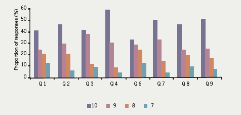
Data were collected using a 10-point scale questionnaire (figure 2). Data obtained from the questionnaire of 114 medical students was analysed in a systematic way.

Figure 2. The questionnaire students were asked to complete

Results

Figure 3. Results of the questionnaire feedback excluding question 5

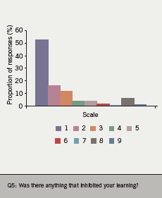
An overwhelming majority of students enjoyed this unique way of being taught about cardiac rehabilitation as an exercise tutorial and highly recommended this novel method of teaching as a whole. Full marks (10/10) were awarded by 46% of respondents, while almost 90% of total responses were graded as 8/10 or above (figure 3). Only seven students (6%) graded the issue of wind blowing impeding proper hearing on windy days as significant (7/10), while the majority of students opined that walking in an open environment helped them concentrate better. It is worthwhile mentioning that question No. 5 looking at this particular aspect of exercise tutorial was graded oppositely to the other questions with 1/10 in fact meaning no disturbance and 10/10 indicating extreme hindrance by wind (figure 4).

Figure 4. Results of the questionnaire feedback to question 5

The students found this opportunity exceptionally unique, entertaining, informative and a superb way of being taught about cardiac rehabilitation, and they unanimously advocated this as a teaching method for other topics.

Some of the positive remarks given by medical students were:

  • “Was good to learn in a different environment and get fresh air. Much more enjoyable than normal lectures!”
  • “Recommend it to other hospitals.”
  • “Was a brilliant teaching session and was nice to learn in a different environment, made it easier to concentrate for some reason!”
  • “Lovely way to learn, easier to concentrate and take in information.”
  • “Liked the idea of going on a walk, aided concentration and made a change.”
  • “Very mind stimulating – a novel experience.”

Conclusion

This report represents a unique study of its kind in terms of analysing exercise tutorial as a unique teaching method and also emphasising cardiac rehabilitation as an essential part of the curriculum. The study had a decent sample size and was conducted over a considerable length of time. The exceptionally positive response of a large number of medical students authenticates its findings, and suggests it could be an extremely effective way of teaching any topic. This study does not negate the need for classrooms and lecture theatres; however, it introduces a novel idea of teaching topics in small groups as exercise tutorials, which can be equally enjoyable and informative.

Conflict of interest

None declared.

Key messages

  • Cardiac rehabilitation is an essential component of coronary heart disease prevention
  • Teaching cardiac rehabilitation in an exercise tutorial is a unique teaching method, which was well received with students
  • This teaching method could be applied to other topics

References

1. Unal B, Critchley JA, Capewell S. Explaining the decline in coronary heart disease mortality in England and Wales between 1981 and 2000. Circulation 2004;109:1101–07.

2. Taylor R, Brown A, Ebrahim S et al. Exercise-based rehabilitation for patients with coronary heart disease: systematic review and meta-analysis of randomized controlled trials. Am J Med 2004;116:682–92.

3. Harbman P. Review: secondary prevention programmes with and without exercise reduced all cause mortality and recurrent myocardial infarction. Evid Based Nurs 2006;9:77.

4. Smith D. Review: increased physical activity and combined dietary changes reduce mortality in coronary artery disease. ACP J Club 2006;144:16.

5. Jolliffe JA, Rees K, Taylor RS, Thompson D, Oldridge N, Ebrahim S. Exercise-based rehabilitation for coronary heart disease. Cochrane Database Syst Rev 001;(1):CD001800.

‘Time is muscle’: aspirin taken during acute coronary thrombosis

Br J Cardiol 2010;17:185-9 Leave a comment
Click any image to enlarge
Authors:

Low-dose aspirin is of value in the long-term management of vascular disease, and the giving of aspirin to patients believed to be experiencing an acute myocardial infarction (AMI) is standard practice for paramedics and doctors in most countries. Given during infarction, aspirin may disaggregate platelet microthrombi and may reduce the size of a developing thrombus. Effects of aspirin other than on platelets have also been suggested and these include an increase in the permeability of a fibrin clot and an enhancement of clot lysis. Animal experiments have also shown a direct effect of aspirin upon the myocardium with a reduction in the incidence of ventricular fibrillation.

Randomised trials have shown that the earlier aspirin is taken by patients with myocardial infarction, the greater the reduction in deaths. We suggest, therefore, that patients known to be at risk of an AMI, including older people, should be advised to carry a few tablets of soluble aspirin at all times, and chew and swallow a tablet immediately, if they experience severe chest pain. 

Introduction
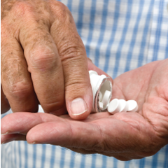
185-img-1Aspirin, used in vascular disease prophylaxis, is probably the most cost-effective drug available in clinical practice and daily low-dose aspirin is now a standard item in the long-term management of vascular disease. Within a public health context, the provision of aspirin to individuals at increased vascular risk has been judged to be the preventive activity of greatest benefit and at the lowest cost (by far), apart from smoking cessation.1 Patients with known vascular disease are clearly at increased vascular risk, and a recent US Task Force judged that ‘individuals at increased risk’ includes males aged over about 45 and females over about 55 years,2 and the conclusions in two reports based on UK populations are in close agreement.3,4

In the acute situation, the giving of aspirin to a patient believed to be experiencing an acute myocardial infarction (AMI) is standard practice.5,6 In what follows, the evidence of benefit from early aspirin is examined and we consider the possible additional benefits if patients at increased vascular risk, including older people, were advised to carry their own aspirin, and chew and swallow an ‘adult’ (300 mg) tablet immediately they experience symptoms suggestive of AMI – the aspirin to be taken even as they summon help.

The clinical problem

Evidence on deaths during the very early stage of a vascular event is limited. Goldstein et al.7 examined the records for 270 witnessed cardiovascular deaths in patients involved in a randomised trial. About a quarter of the deaths had been ‘instantaneous’ (collapse without any symptoms) and, in such cases, cardiopulmonary resuscitation (CPR) is the only appropriate intervention. A further 16% of deaths occurred during the first hour after the commencement of symptoms. From details of a number of studies Gersh and Anderson8 estimated that the typical delay between the commencement of symptoms and hospital treatment was around 96 minutes, and, in an intervention study,9 the out-of-hospital delay for over half the patients with acute AMI was found to be up to two hours.

There has been a sustained drive for earlier treatment of patients with a vascular event, and in most countries paramedics now give aspirin immediately on first contact with a patient who is experiencing severe chest pain and is judged to have possibly had a myocardial infarct or ischaemia.5,6 The UK Department of Health have established a target requiring that at least 75% of patients with chest pain should be attended within eight minutes of a call for an ambulance, and most paramedics are now exceeding this target.10 There is, however, relatively little evidence on the time between the onset of symptoms and the first medical contact (FMC). In Finland, a study found that this interval averaged about 60 minutes regardless of whether or not the patient had a pre-existing history of heart disease.11 A cross-European study estimated that the median FMC in the UK is 68 minutes, but in other European countries the median delay was around 150–200 minutes.12 It seems, therefore, that the opportunity is widespread for what could be termed ‘immediate’ aspirin, that is, aspirin taken while medical help is awaited.

Evidence of benefit

Unfortunately, the reports of randomised trials give no evidence on the effect of aspirin during the first few minutes after symptoms of infarction commence. In the Second International Study of Infarct Survival (ISIS-2), the reduction in deaths by aspirin during the first four hours after the onset of symptoms was 53 ± 8%, compared with 32 ± 9% during the period five to eight hours.13 In the Chinese Acute Stroke Trial (CAST) study of stroke reduction by aspirin, the reduction in deaths by aspirin given within three hours was 36 ± 18%, while in those given aspirin later the reduction was at most 15%.14

Indirect evidence of possible benefit from early aspirin comes from a reduction in the severity of infarction in patients who had developed an infarct while taking aspirin. Col et al.15 found that after adjustment for possible confounding by age, gender, coronary history and medication, “prior aspirin consumption remained independently associated with … non-Q wave and smaller infarct size”. In a series of hospital admissions Garcia-Dorado et al.16 judged that “aspirin … converts 50% of potential AMIs to unstable angina and 20% of Q-wave infarctions to non-Q-wave lesions”. On the other hand, Ridker et al.17 failed to confirm this in the US Physicians study, but conceded that “aspirin therapy may result in a shift of fatal events to severe non-fatal events, severe events to mild events and mild events to no events”.

Possible benefit during infarction

A number of authors have reported finding platelet microemboli within the coronary microcirculation in persons whose death had been ‘sudden’.18,19 In some cases no thrombus could be found in a major coronary vessel, suggesting that microthrombi had developed spontaneously, while in others, microthrombi were additional to a thrombus in a major vessel,20,21 suggesting that fragments from a developing thrombus may have become embolic, or a release action by platelets within a major thrombus may have induced platelet activity within peripheral vessels in the myocardium.21 Platelet aggregates are transient before fibrin is incorporated and early aspirin could, therefore, cause disaggregation of these microemboli, in addition to decreasing thrombus growth and clot volume.22,23

The main mode of action of prophylactic aspirin in vascular disease appears to be through a reduction in platelet aggregation inhibiting the development of a stable thrombus. Measures of platelet aggregation, however, appear to have no predictive power for ischaemic heart disease events.24-26 Effects of aspirin on mechanisms other than platelet aggregation are, therefore, of interest and an action of aspirin on some of them could be of particular importance during the very early processes of thrombosis and infarction.

Aspirin has been shown to acetylate lysine residues in fibrinogen, thus increasing fibrin clot permeability and enhancing clot lysis.27 It also reduces the fibrin mass in a thrombus and leads to a shorter lysis time.28,29 Blood from cigarette smokers has been shown to generate in vitro thrombi that are twice the volume of those in blood from non-smokers, while the ingestion of aspirin by smokers reduced the subsequent thrombus volume to a substantially greater degree (by 62%) compared with the reduction (38%) caused in non-smokers.30 Clearly, the earlier aspirin is taken during thrombosis, and particularly by smokers, the more important these non-platelet effects are likely to be.

A direct effect of aspirin on the myocardium and an anti-arrhythmic effect independent of any action on platelets have also been suggested.31 Moschos et al.32 describe how the occlusion of a coronary artery of a dog by mechanical means can lead to ventricular fibrillation without evidence of any platelet involvement. Fibrillation in these dogs is prevented by aspirin, leading the authors to postulate a direct effect of aspirin upon the myocardium.

Finally, myocardial ischaemia causes a marked increase in catecholamine release and this in turn leads to an increase in free fatty acids,32 a rise which is abolished by aspirin.33 Aspirin may also reduce the vasoconstriction caused by catecholamines34 and by thromboxane A2,35-37 and it reduces catecholamine-induced myocardial necrosis.38 The inhibitory action of aspirin on catecholamines within the myocardium may be the basis for the reduction of arrhythmia during ischaemia.32

The absorption of aspirin

187-img-1The absorption of aspirin has been studied extensively. The drug in soluble and dispersible tablets is absorbed much more rapidly than from the standard tablet. Muir et al.39 reported that five minutes after ingestion of a soluble form, the levels of aspirin in the plasma were about 15 times the level after ingestion of the same dose in a plain tablet. Feldman40 found a 50% inhibition of thromboxane A2 within five minutes after a 325 mg tablet is chewed and swallowed.

There is uncertainty about the dose of aspirin that is appropriate if the drug is taken during infarction. Dabaghi et al.36 reported almost complete (97%) inhibition of aggregation to arachidonic acid 15 minutes after ingesting an 81 mg soluble tablet of aspirin and others have reported similar rapid inhibition of plasma thromboxane,35-37 and platelet thromboxane A240 after small doses of aspirin. There is evidence, however, that absorption can be impaired during the acute phase of infarction41 and a relatively high dose may be advisable.42 Yet in an examination of data from recent randomised trials, there was no difference in the very early mortality following initial doses of 162 mg and 325 mg aspirin, but there was a slightly greater incidence of bleeding after the larger dose.43

Further to all this, most patients with an infarct are likely to receive thrombolytic therapy. There is a marked heightening of platelet activity after thrombolysis,39,44 and prior treatment with aspirin abolishes the excess in re-infarction that otherwise follows fibrinolysis.13 In order to achieve fibrinolysis as early as possible, treatment has been delegated to paramedics, although increasingly where percutaneous coronary intervention (PCI) is available in a timely manner, this has become the preferred intervention. Self-administration of aspirin by a subject at the time of calling an ambulance would, however, be appropriate whatever the subsequent interventions.

Strategy

The administration of aspirin as early as possible during the process of thrombosis and infarction, and, hence, the effectiveness of the drug would be enhanced if patients judged to be at risk carried a few ‘adult’ (300 mg) tablets of aspirin at all times, and were instructed to chew and swallow a tablet immediately they experience symptoms suggestive of infarction. Patients to whom this is recommended should include all those known to be at increased vascular risk, and this should include older persons – perhaps those over the age of about 45 or 50 years.2-4

The peak incidence of AMI is in the early morning45 and patients may be more hesitant about calling for help in those early hours. Platelets appear to be most sensitive to aggregating agents in the early morning,46 and in the US Physicians Health Study it was found that the reduction in AMI by aspirin was significantly greater for the events that occurred in the early morning (59%), than for those that occurred later in the day (34%).45 The taking of aspirin by persons themselves could, therefore, be particularly appropriate at these times.

Areas of uncertainty

Around 30% of patients presenting with an AMI are known to already have coronary disease,47 and these are likely to be on daily low-dose aspirin. Others, such as patients on a statin or an antihypertensive agent, are known to be at high risk of a thrombotic event and a high proportion of these are also likely to be on aspirin. There is evidence, however, that many of these, perhaps even around half such patients, are not actually taking the drug.48 In any case, the half-life of aspirin in the circulation is only 15–20 minutes,49 and it would seem reasonable to surmise that if a thrombus develops despite daily exposure to aspirin, some fresh sensitive platelets are likely to have entered the circulation. If this is the case, an extra dose, say 300 or 600 mg of aspirin, taken in addition to the small daily dose, could be life saving.50

Aspirin has undesirable side effects, and persons advised about the benefits of aspirin should also be told the risks. These include increased risks of gastric haemorrhage and cerebral haemorrhage. Estimates of these extra risks have been derived from randomised trials in which patients take aspirin daily over long periods of time. The risk of death attributable to aspirin in trial patients is at most 4% per year,51,52 and the risk of death from a single dose of aspirin is likely to be very considerably lower than this.

It would be unfortunate, however, if the taking of a tablet by a subject in whom a thrombus is developing, led to delay in the calling of an ambulance, and a consequent delay in the commencement of fibrinolytic treatment.53 Persons informed about aspirin should, therefore, be clearly warned that if rapidly increasing severe chest pain is experienced an ambulance should be called first, and then, without further delay, an aspirin tablet should be chewed and swallowed.

Severe chest pain may arise from a non-cardiac cause. If it is from the voluntary muscles, then aspirin is of course appropriate. The relationship of pleuritic pain to breathing should enable a pulmonary embolus to be readily distinguished. Pain from a bleeding gastric lesion should not be a concern as patients known to have a peptic ulcer, or with current indigestion, should have been warned against taking aspirin at any time, even in an emergency.

Pain from angina on exercise should be easily recognised and not confused with the more severe and unremitting pain of infarction. The most important cardiac causes of chest pain that could cause confusion arise from acute aortic syndromes,54 and in particular dissecting thoracic aorta. These lesions are rare, are usually associated with severe hypertension and the pain has an immediate onset, usually with maximum intensity at the time of onset.55

The question naturally arises as to the advisability of ‘immediate’ aspirin if symptoms suggestive of a stroke are experienced. The two major stroke trials, CAST14 and International Stroke Trial (IST),56 gave evidence suggesting that if aspirin is given early during the acute phase of a stroke, there is an additional reduction in the number of deaths and disabling strokes that is “modest but worthwhile”.57 Nevertheless, if aspirin were taken at the very commencement of symptoms suggestive of a stroke, and if the stroke were a haemorrhagic lesion, then the cerebral bleeding might be increased. Immediate aspirin should not, therefore, be advised if symptoms suggest a stroke.

Guidelines from professional societies

The giving of aspirin by a doctor or a paramedic immediately they have contact with a patient believed to be experiencing an acute AMI, is established practice and is recommended by all the relevant professional bodies. Self-medication in this situation has already been recommended,40,58-61 but appears never to have been actively promoted.

Conclusions and recommendations

About 35% of all deaths are attributed to coronary heart disease each year, that is 36,000 in the UK and almost 900,000 in the USA, and younger patients among these are proportionately more likely to die before reaching hospital.62 The earlier aspirin is taken by these patients, the greater the proportionate survival is likely to be.

Aspirin taken while a thrombosis is growing may limit the size of the thrombus and may enhance thrombolysis, thus limiting the damage to the myocardium and the likelihood of a fatal outcome. Effects of aspirin, other than on platelets, indicate that it may help to preserve myocardial tissue and may also reduce the risk of ventricular fibrillation.

People judged to be at increased vascular risk, including older persons, should, therefore, be advised to carry a few tablets of soluble aspirin at all times, and chew and swallow a tablet immediately they experience sudden severe chest pain. Consideration should also be given to the inclusion of instruction on the risks and benefits of early aspirin to persons trained in CPR.

Aspirin is freely available to the public, but advice of varying quality is being given to the public in newspapers and magazines. In a challenging editorial, Kassirer63 states: “…it [is] essential to identify decisions in which it is especially important to consider patient’s values and to protect such decisions from intrusive external decision making”. We believe that the taking of ‘immediate’ aspirin, as we describe it, is such a decision – provided those advised have been adequately informed of the likely risks and benefits.

Conflict of interest

None declared. The authors declare no vested interest and no special funding for the work described.

Key messages

  • Aspirin, used in vascular disease protection, is probably the most cost-effective drug available in clinical practice
  • The earlier aspirin is given in coronary thrombosis, the greater the reduction in deaths
  • It is also likely that early aspirin will reduce the size and severity of a myocardial infarction and it may reduce the risk of ventricular fibrillation
  • It is accepted practice for paramedics to give aspirin to patients for whom an emergency call has been received because of chest pain
  • It is suggested that patients at increased vascular risk, including older people, should carry tablets of soluble aspirin at all times, and chew and swallow a tablet immediately they experience severe chest pain

References

1. Kahn R, Robertson RM, Smith R et al. The impact of prevention on reducing the burden of cardiovascular disease. Circulation 2008;118:576–85.

2. Preventive Services Task Force. Aspirin for the prevention of cardiovascular disease: US Preventive Services Task Force Recommendation Statement. Ann Intern Med 2009;150:396–404.

3. Bulugahapitiya U, Siyambalapitiya S, Sithole J et al. Age threshold for vascular prophylaxis by aspirin in patients without diabetes. Heart 2008;94:1429–32.

4. Elwood P, Morgan G, Brown G et al. Aspirin for everyone over 50? BMJ 2005;330:1440–1.

5. McVaney KE, Macht M, Colwell CB, Pons PT. Treatment of suspected cardiac ischemia with aspirin by paramedics in an urban emergency medical services system. Prehosp Emerg Care 2005;9:282–4.

6. Fisher JD, Brown SN, Cooke MW. UK Ambulance Service Clinical Practice Guidelines (2006). Joint Royal Colleges Ambulance Liaison Committee, 2006. Available from: http://www2.warwick.ac.uk/fac/med/research/hsri/emergencycare/prehospitalcare/jrcalcstakeholderwebsite/guidelines [accessed 22 February 2009].

7. Goldstein S, Friedman L, Hutchinson R et al. Timing, mechanism and clinical setting of witnessed deaths in postmyocardial infarction patients. J Am Coll Cardiol 1984;3:1111–17.

8. Gersh BJ, Anderson JL. Thrombolysis and myocardial salvage. Circulation 1993;88:296–306.

9. Hedges JR, Feldman HA, Bittner V et al. Impact of community intervention to reduce patient delay time on use of reperfusion therapy for acute myocardial infarction: rapid action for coronary treatment (REACT) trial. Academic Emergency Medicine 2000;7:862–72.

10. The NHS Information Centre. Ambulance Services England 2007–2008. The Information Centre, June 2008. Available from: http://www.ic.nhs.uk/webfiles/publications/Ambulance%
2007-08/Ambulance%20Bulletin%202007-08%20final%20with%20data%20quality
%20note%2015-12-08.pdf
[accessed 12 February 2009].

11. Hirvonen TP, Halinen MO, Kala RA, Olkinuora JT. Delays in thrombolytic therapy for acute myocardial infarction in Finland. Results of a national thrombolytic therapy delay study. Finnish Hospitals’ Thrombolysis Survey Group. Eur Heart J 1998;19:885–92.

12. Widimsky P, Wijns W, Fajadet J et al. Reperfusion therapy for ST elevation acute myocardial infarction in Europe: description of the current situation in 30 countries. Eur Heart J 2010;31:943–57.

13. ISIS-2. Second International Study of Infarct Survival Collaborative Group. Randomised trial of intravenous streptokinase, oral aspirin, both or neither among 17,187 cases of suspected acute myocardial infarction. Lancet 1988;2:349–60.

14. CAST (Chinese Acute Stroke Trial) Collaborative Group. CAST: randomized placebo-controlled trial of early aspirin use in 20,000 patients with acute ischaemic stroke. Lancet 1997;349:1641–9.

15. Col NF, Yasrzbski J, Gore JM et al. Does aspirin consumption affect the presentation or severity of acute myocardial infarction? Arch Int Med 1995;155:1386–9.

16. Garcia-Dorado D, Therous P, Tornos P et al. Previous aspirin use may attenuate the severity of the manifestations of acute ischaemic syndromes. Circulation 1995;92:1743–8.

17. Ridker PM, Manson JE, Buring JE et al. Clinical characteristics of non-fatal myocardial infarction among individuals on prophylactic low-dose aspirin therapy. Circulation 1991;84:708–11.

18. Haerem JW. Platelet aggregates and mural micro-thrombi in the early stages of acute fatal coronary disease. Thromb Res 1974;5:243–9.

19. El-Maraghi N, Genton E. The relevance of platelet and fibrin thromboembolism of the coronary microcirculation with special reference to sudden cardiac death. Circulation 1980;62:936.

20. Frink RJ, Trowbridge JO, Rooney PA. Non-obstructive coronary thrombosis in sudden cardiac death. Am J Cardiol 1978;42:48.

21. Gelman JS, Mehta J. Platelets and prostaglandins in sudden death. Cardiovascular Clinics 1985;15:65–80.

22. Roald HE, Sakariassen KS. Axial dependence of collagen-induced thrombus formation in flowing non-anticoagulated human blood. Anti-platelet drugs impair thrombus growth and increase platelet-collagen adhesion. Thromb Haemostas 1995;73:126–31.

23. Stewart JH, Farrell PC, Dixon M. Reduction of platelet/fibrin deposition in haemodialysers by aspirin administration. Austr and New Zealand J Medicine 1975;5:117–22.

24. Elwood PC, Beswick A, Pickering J et al. Platelet tests in the prediction of myocardial infarction and stroke: evidence from the Caerphilly study. Br J Haematol 2001;113:514–20.

25. Elwood PC, Pickering J, Yarnell J, O’Brien JR, Ben-Shlomo Y, Bath P. Bleeding time, stroke and myocardial infarction: the Caerphilly prospective study. Platelets 2003;14:139–41.

26. Sharp DS, Ben-Shlomo Y, Beswick AD, Andrew ME, Elwood PC. Platelet aggregation in whole blood is a paradoxical predictor of ischaemic stroke: Caerphilly Prospective Study revisited. Platelets 2005;16:320–8.

27. Undas A, Brummel-Ziedins KE, Mann KE et al. Antithrombotic properties of aspirin and resistance to aspirin: beyond strictly antiplatelet actions. Blood 2007;109:2285–92.

28. Williams S, Fatah K, Ivert T et al. The effect of acetylsalicylic on fibrin gel lysis by tissue plasminogen activator. Blood Coagulation and Fibrinolysis 1995;7:718–25.

29. Bjornsson TD, Schneider DE, Berger H. Aspirin acetylates fibrinogen and enhances fibrinolysis. Fibrinolytic effect is independent of changes in plasminogen activator levels. J Pharm Exper Ther 1989;250:154–61.

30. Roald HE, Orvim U, Bakken IJ et al. Modulation of thrombotic responses in moderately stenosed arteries by cigarette smoking and aspirin injestion. Arterioscler Thromb 1994;14:617–21.

31. Hirsh J. Antiplatelet agents: their role in the prevention of sudden death. Ann NY Acad Sci 1982;382:289–304.

32. Moschos CB, Haider B, de la Cruz C et al. Anti-arrhythmic effects of aspirin during non-thrombotic coronary occlusion. Circulation 1978;57:681.

33. Oliver MF. Metabolic causes and prevention of ventricular fibrillation during acute coronary syndromes. Am J Med 2002;112:305–11.

34. Strom EA, Coffman JD. Effect of aspirin on circulatory responses to catecholamines. Arthr Rheumatol 1963;6:689–97.

35. Clarke RJ, Mayo G, Price P, Fitzgerald GA. Suppression of thromboxane A2 but not of systemic prostacyclin by controlled release aspirin. N Engl J Med 1991;325:1137–41.

36. Dabaghi SF, Kamat SG, Payne J et al. Effects of low-dose aspirin on in-vitro platelet aggregation in the early minutes after injestion in normal subjects. Am J Cardiol 1994;74:720–3.

37. Jakubowski JA, Stampfer MJ, Vaillancourt R et al. Cumulative antiplatelet effect of low dose enteric-coated aspirin. Br J Haematol 1985;60:635–42.

38. Hart Gershengorn K. Protection against epinephrine-induced myocardial necrosis by drugs that inhibit platelet aggregation. Am J Cardiol 1972;30:838.

39. Muir N, Nichols JD, Clifford JM, Stillings MR, Hoare RC. The influence of dosing form on aspirin kinetics: implications for acute cardiovascular use. Curr Med Opinion 1997;13:547–53.

40. Feldman M, Cryer B. Aspirin absorption rates and platelet inhibition times with 325mg buffered aspirin tablets chewed or swallowed intact and with buffered aspirin solution. Am J Cardiol 1999;84:404–09.

41. Zhang CL, Wilson KM, Stafford I, Bocher F, Horowitz JD. Absorption kinetics of low-dose aspirin in patients with evolving acute myocardial infarction. Drug Invest 1994;1:169–74.

42. Berglund U, Wallentin L. Persistent inhibition of platelet function during long-term treatment with 75mg acetylsalicylic acid daily in men with unstable coronary artery disease. Eur Heart J 1991;12:428–33.

43. Berger JS, Stebbins A, Granger CB et al. Initial aspirin dose and outcome amongst ST-elevation myocardial infarction patients treated with fibrinolytic therapy. Circulation 2008;117:192–9.

44. Fitzgerald DJ, Catella F, Roy L et al. Marked platelet activation in vivo after intravenous streptokinase in patients with acute myocardial infarction. Circulation 1988;77:142–50.

45. Muller JE, Tofler GH, Stone PH. Circadian variation and triggers of onset of acute cardiovascular disease. Circulation 1989;79:733–43.

46. Ridker PM, Manson JE, Buring JE, Muller JE, Hennekens CH. Circadian variation of acute myocardial infarction and the effect of low-dose aspirin in a randomised trial of physicians. Circulation 1990;82:897–902.

47. Weston CFM, Penny WJ, Julian DG. Guidelines for the early management of patients with myocardial infarction. BMJ 1994;308:767–71.

48. Elwood P, Hughes J, Morgan G, Brown G, Longley M. A survey of aspirin use for vascular prophylaxis in Wales. Quality in Primary Care 2005;13:201–04.

49. Patrono C, Baignet C, Hirsh J et al. Antiplatelet drugs: American College of Chest Physicians evidence-based clinical practice guidelines (8th ed.). Chest 2008;133:199S–233S.

50. Prasad N, Srikanthan VS, Wright A, Dunn FG. Management of suspected myocardial infarction before admission. BMJ 1994;316:353.

51. Pirmohamed M, James S, Meakin S et al. Adverse drug reactions as a cause of admission to hospital: retrospective analysis of 18,820 patients. BMJ 2004;329:15–19.

52. Guise J-M, Mahon SM, Aicken M et al. Aspirin for the prevention of cardiovascular events: a summary of the evidence. Ann Intern Med 2002;136:161–72.

53. Rathmore SS, Curtis JP, Chen J et al. Association of door-to-balloon time and mortality in patients admitted to hospital with ST elevation myocardial infarction: national cohort study. BMJ 2009;338:b1807.

54. Vilacosta I, Aragoncillo P, Canadas V et al. Acute aortic syndromes: a new look at an old conundrum. Heart 2009;95:1130–9.

55. Wooley CF, Sparkes EH, Boudoulas H. Aortic pain. Prog Cardiovascular Dis 1998;40:563–89.

56. IST International Stroke Trial Collaborative Group. A randomised trial of aspirin, subcutaneous heparin, both, or neither among 19,435 patients with acute ischaemic stroke. Lancet 1997;349:1569–81.

57. Kmietowicz Z. Aspirin benefits patients with stroke – but only just. BMJ 1997;314:1646.

58. Brecker SJD. Early aspirin in myocardial infarction. Lancet 1990;335:923.

59. Carpenter AL, Caravalho J. Early public use of aspirin in the face of probable ischaemic chest pain. Lancet 1990;335:486.

60. Norfolk Health. Guidelines for practice procedure with a suspected heart attack. East Norfolk Health Commission, 1996.

61. NHS Executive. The health of the nation: assessing the options: CHD/Stroke. Target effectiveness and cost-effectiveness of interventions to reduce coronary heart disease and stroke mortality. London: Department of Health, 1995.

62. Norris RM on behalf of the UK Heart Attack Study investigators. Sudden cardiac death and acute myocardial infarction in three British health districts: the UK heart attack study. London: British Heart Foundation, 1999.

63. Kassirer JP. Incorporating patients’ preferences into medical decisions. N Engl J Med 1994;330:1895–6.

From patient to plaque. Contemporary coronary imaging – part 2: optical coherence tomography 

Br J Cardiol 2010;17:190-3 Leave a comment
Click any image to enlarge
Authors:

Intra-coronary imaging has become a cornerstone of visualising atherosclerotic coronary artery disease and also to guide the therapy in selected high-risk cases. Optical coherence tomography (OCT) is an imaging modality quite similar to intravascular ultrasound (IVUS), but uses light instead of sound. In the second article on contemporary coronary imaging, the potential of OCT is discussed.

Introduction

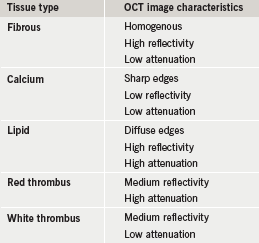
Table 1. Image characteristics of optical coherence tomography (OCT)

Optical coherence tomography (OCT) uses near-infrared electromagnetic radiation, and cross-sectional images are generated by measuring the echo time delay and intensity of light that is reflected or back-scattered from internal structures in the tissue.1,2

Current OCT images are obtained at the peak wavelength in the 1,280–1,350 nm band that enables a 10–15 µm tissue axial resolution, 94 µm lateral resolution at 3 mm, and maximal scan diameter of 6–8 mm (about 10 times resolution as compared with intravascular ultrasound [IVUS]). There are two main technologies that can be used to obtain OCT images: time domain and frequency domain. Frequency domain OCT has the advantage of an improved signal-to-noise ratio allowing fast scanning with improved imaging quality.

OCT image acquisition

OCT cannot image through a blood field, and it requires clearing or displacing blood from the lumen by flushing. There are two basic techniques:

  1. The occlusive technique. During image acquisition, coronary blood flow is stopped by inflating a proximal occlusion balloon and flushing the crystalloid solution at the rate of 0.5–1.0 ml/sec down the coronary artery.
  2. Non-occlusive technique. This is recently developed and does not need proximal balloon occlusion. During image acquisition the image wire pull back is performed at fast speed with simultaneous injection of contrast through the guiding catheter.

Safety of OCT

The energies used in OCT are low and do not cause functional or structural damage to the tissue. There are two reports on the safety of intracoronary OCT imaging.3,4 These reports have not shown any major adverse cardiac events, however, electrocardiogram (ECG) changes and ectopy were seen frequently during the pull back of the image catheter. All ECG changes and patient symptoms rapidly resolved at the end of the procedure.

Plaque characterisation

Ex vivo validation

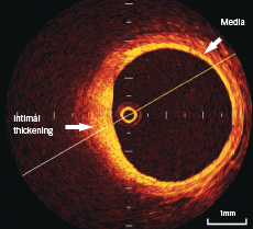
Figure 1. Optical coherence tomography (OCT) shows the three-layer appearance of normal vessel wall. The muscular media appears as a low signal layer between internal and external lamina (2 o’clock). Intimal thickening is seen at 6–9 o’clock

This was first performed in 2002 in 357 ex vivo post-mortem atherosclerotic segments from 90 cadavers.5 This study established OCT criteria for the various plaque components (shown in table 1). High sensitivity and specificity have been obtained by comparing with histology as a gold-standard reference, for the detection of both calcific and lipid-rich plaques (96% and 97%, and 90% and 92%, respectively).

In vivo characterisation

In normal vessels, the coronary artery wall appears as a three-layer structure in OCT images. The media is seen as a dark band delimited by internal elastic lamina (IEL) and external elastic lamina (EEL) (figure 1).

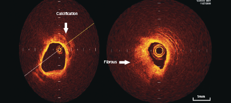
Calcifications within plaques are identified by the presence of well delineated, low back scattering heterogenous regions5-7 (figure 2).

Fibrous plaque consists of homogenous high back scattering areas5-7 (figure 2).

Figure 2. OCT appearance of calcification at 12–3 o’clock position on left-sided panel, and fibrous appearance of plaque at 6–12 o’clock position on right-sided panel

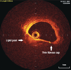
Figure 3. OCT appearance of lipid pool (3–9 o’clock position) with overlying thin (40 µm) fibrous cap. This is the typical appearance of thin cap fibro-atheroma (TCFA)

Necrotic lipid pools are less well delineated, and usually appear as diffusely bordered, signal poor regions with overlying signal-rich bands, corresponding to fibrous caps5-7 (figure 3).

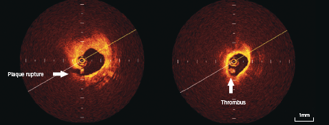
Thrombi are identified as masses protruding into the vessel lumen discontinuous from the surface of the vessel wall (figure 4).

Plaque ulceration or rupture can be detected by OCT as a ruptured fibrous cap that connects the lumen with the lipid pool. These may or may not be associated with thrombus (figure 4).

Thin cap fibro-atheroma (TCFA) is defined as plaques with lipid content in >2 quadrants on cross-sectional analysis and fibrous cap thickness of <65 µm (figure 3).

Figure 4. OCT appearance of plaque rupture (6 o’clock position) on left-sided panel, with intraluminal thrombus seen in right-sided panel

Potential clinical application of OCT

Plaque characteristics in various clinical syndromes

OCT has been shown to provide detailed in vivo characterisation of coronary plaque morphology. Jang et al.8 has evaluated this in patients with recent acute ST elevation myocardial infarction, acute coronary syndromes, and stable angina. Patients with an acute myocardial infarction (MI) and acute coronary syndromes had higher frequency of TCFA, as compared with patients with stable angina (72%, 50% and 20%, respectively; p=0.012). Similar results are shown with OCT analysis in other studies.6,9 This information leads to the better understanding of the mechanisms of coronary artery disease.

OCT can identify subclinical atherosclerotic lesions and the characteristics of high-risk plaque (TCFA or subclinical plaque rupture). This may have relevance to modulate the aggressiveness of medical therapeutic strategies for primary prevention. OCT has the potential for assessing the risk of MI, however, this needs to be proven and tested in future.

Guidance and optimisation of percutaneous coronary intervention

IVUS imaging is traditionally used to assess the outcome of coronary stenting but detailed information is often impossible to obtain because the metal struts impair image quality. Complete and proper approximation of the stent struts to the vessel wall could be better optimised by OCT due to its higher resolution. In 43 imaged stents, OCT consistently outperformed IVUS in the detection of dissection, tissue prolapse and incomplete stent apposition.10,11 These studies also show better understanding of balloon-induced dissection, intraluminal, thrombus, number of cuts made by cutting balloon, and tissue prolapse. The adequacy of stent deployment is a major predictor of re-stenosis and subacute stent thrombosis. Therefore, OCT could potentially pay a role in optimisation of coronary intervention.

OCT could be utilised in assessing angiographically normal and borderline coronary artery stenosis. OCT has got the potential to become a routine tool for guiding interventional procedure as this technology provides accurate lumen measurements, when compared with IVUS.

OCT is particularly helpful in assessment of angiographically hazy lesions, because identification of thrombus is far more superior with this technique as compared with IVUS.

Assessment of vascular healing following drug eluting stent

OCT can allow us to assess the impact of stenting to the vessel wall and can evaluate healing and remodelling of the vessel, which could be used as a surrogate end point for evaluating different stents and their platforms. There are some studies evaluating the neo-intimal coverage of the stent struts following drug eluting stents and bare metal stents. Chen et al.12 have shown higher incidence of unapposed (2% vs. 0%, p<0.001) and uncovered stent struts (13% vs. 0.3%) in sirolimus eluting stent (SES) group as compared with bare metal stent. Similar results were shown by Matsumato et al.,13 where 1.5% of the total stent struts were malapposed and without neointimal coverage in the SES group at six months. Takano et al.14 have shown higher incidence of stent inapposition and uncovered stent struts in patients with acute coronary syndrome (18% vs. 13%, p<0.001; 8% vs. 5%, p<0.005, respectively) as compared with stable angina patients following use of SES stents. However, there is no definite proven causal relationship established between malapposed and/or uncovered stent struts and incidence of late stent thrombosis.

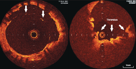
Stent apposition and the neointimal coverage of the stent struts can be identified and monitored because of high resolution of the OCT (figure 5). This could potentially be helpful in follow-up of certain high-risk cases and also to optimise the results of stent implantation.

Figure 5. OCT appearance of stent struts inapposed to the vessel wall (arrows) seen from 10–3 o’clock position on left-sided panel, with overlying thrombus on some of the stent struts as seen on right-sided panel

Limitations of OCT

The main limitation of OCT is the poor penetration power and inability to measure plaque burden where thickness exceeds 1.5 mm. In addition, the severity of plaques located at aorto-ostial locations is difficult to assess with the current stage of technology.

Conclusion

OCT is a rapidly evolving intracoronary imaging technique. It allows detailed structural analysis of superficial structures in the vessel wall, including coronary plaque characterisation and the vascular healing process following coronary stent implantation.

Conflict of interest

None declared.

Editors’ note

The next article in this series will be on computed tomography (CT). Part 1 in this series covered IUUS-derived virtual histology (Br J Cardiol 2010;17:129–32).

Key messages

  • Angiographic assessment of coronary plaque is limited and does not provide the full story
  • Optical coherence tomography (OCT) is a new technique similar to intravascular ultrasound (IVUS) that uses light rather than ultrasound to image structures
  • OCT is around 10 times more powerful than IVUS when it comes to resolution of small structures
  • OCT, like IVUS, can tell the difference between plaque types and is better at imaging thrombus
  • It is hoped that future studies will show the benefit of performing OCT to improve stent deployment and patient outcomes from coronary intervention

References

1. Huang D, Swanson EA, Lin CP et al. Optical coherence tomography. Science 1991;254:1178–81.

2. Brezinski ME, Tearney GJ, Bouma BE et al. Optical coherence tomography for optical biopsy properties and demonstration of vascular pathology. Circulation 1996;93:1206–13.

3. Prati F, Cera M, Ramazzotti V et al. Safety and feasibility of a new non-occlusive technique for facilitated intracoronary optical coherence tomography (OCT) acquisition in various clinical and anatomical scenarios. Eurointerv 2007;3:365–70.

4. Yamaguvhi T, Terashima M, Akasaka T et al. Safety and feasibility of an intravascular optical coherence tomography image wire system in the clinical setting. Am J Cardiol 2008;101:562–7.

5. Yabushita H, Bouma BE, Houser SL et al. Characterisation of human atherosclerosis by optical coherence tomography. Circulation 2002;106:1640–5.

6. Jang IK, Bouma BE, Kang DH et al. Visualisation of coronary atherosclerotic plaques in patients using optical coherence tomography: comparison with intravascular ultrasound. J Am Coll Cardiol 2002;39:604–09.

7. Kubo T, Imanashi T, Takarda S et al. Assessment of culprit lesion morphology in acute myocardial infarction: ability of optical coherence tomography compared with intravascular ultrasound and coronary angioscopy. J Am Coll Cardiol 2007;50:933–9.

8. Jang IK, Tearney GJ, MacNeill B et al. In vivo characterisation of coronary atherosclerotic plaque by use of optical coherence tomography. Circulation 2005;111:1551–5.

9. Kume T, Akasaka T, Kawamato T et al. Assessment of coronary arterial plaque by optical coherence tomography. Am J Cardiol 2006;97:1172–5.

10. Diaz-Sandoval LJ. Optical coherence tomography as a tool for percutaneous interventions. Catheter Cardiovasc Interv 2005;65:492–6.

11. Bouma BE. Evaluation of intracoronary stenting by intravascular optical coherence tomography. Heart 2003;89:317–20.

12. Chen BX, Ma FY, Luo W et al. Neointimal coverage of bare metal and sirolimus eluting stents evaluated with optical coherence tomography. Heart 2008;94:566–70.

13. Matsumoto D, Shite J, Shinke T et al. Neointimal coverage of sirolimus eluting stents at 6 months follow up: evaluated by optical coherence tomography. Eur Heart J 2007;28:961–7.

14. Takano M, Inami S, Jang IK et al. Evaluation by optical coherence tomography of neointimal coverage of sirolimus eluting stent three months after implantation. Am J Cardiol 2007;99:1033–8.

Book review – Evidence-based cardiology, 3rd edition

Leave a comment
Click any image to enlarge
Authors:

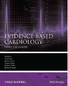
By the very nature of book publishing, a relevant evidence-based textbook is always a hard task to achieve. The editors of Evidence-based cardiology preface this book, now in its 3rd edition, with an admission of this limitation. This textbook is, however, more than just a reference volume. It seeks to educate and equip the reader with the tools to critically appraise information, and then demonstrates how research has evolved to shape current practice.

Editors: Yusuf S, Cairns J, Camm J,
Fallen EL, Gersh BJ
Publisher: Wiley-Blackwell,
Oxford, 2010
ISBN: 978-1-4051-5925-8
Price: £180

193-img-1The book begins with an understanding of evidence-based decision making. Diagnostic screening and testing are evaluated, and the fundamentals of statistical analyses are explained. The success of this section is the use of examples from within cardiovascular medicine, which even the most casual observer would have familiarity with. Health economics and implementation are also explored, and serve to further contextualise the translation from clinical trial to real world medicine.
It is also in this first part that two new features are seen. Firstly, the evidence for risk assessment and reduction in non-cardiac surgery is addressed. Useful management information is contained within this chapter, and it sits awkwardly in the section dedicated to general concepts. Nonetheless, the evidence reviewed is comprehensive and up-to-date. The second is concerned with employment fitness standards. It uses the aviation industry to illustrate the interface of evidence, risk and statistics, but neither seeks, nor delivers a review of standards across all industries.

The second section of the book looks at the prevention and management of cardiovascular disease. Primary prevention, epidemiology and genetics are discussed. It was useful to find a chapter on psychosocial influence and cardiovascular disease, highlighting the lack of attention these modifiable risk factors face when clinical trials in this field have inherent limitations.

The third part contains well-constructed chapters on disease management. Many illustrate not only the way in which emerging evidence has shaped our management, but the clinical trials that have developed to answer the emerging questions. However, this is by no means a quick reference guide to management. There are few flow charts, protocols and guidelines and a reader seeking step-by-step treatment would want to look elsewhere. What is provided is a comprehensive overview of the evidence that serves the reader with a tool for clinical decision making.

The book concludes with a useful selection of vignettes, each with the reader being guided through the clinical application of the evidence, explored by different authors earlier in the book. Many of the same arguments are, therefore, rehearsed but the diseases chosen are important, and such repetition only serves to highlight appropriate practice.

The editors remind us of the dictum that clinical decision making combines evidence with clinical expertise and awareness of patients’ needs and preferences. This book certainly gives a framework by which to implement this. This comprehensive review is recommended both for trainees undergoing assessment and more experienced cardiologists seeking to reference their current practice. Even if the evidence becomes no longer current, there is enough here to support decision making well into the future.

James Rosengartan
SpR Cardiology
Portsmouth Hospitals NHS Trust, Portsmouth, PO6 3LY.

The role of nucleic acid amplification techniques (NAATs) in the diagnosis of infective endocarditis

Br J Cardiol 2010;17:195-200 1 Comment
Click any image to enlarge
Authors:

Infective endocarditis (IE) causes high rates of morbidity and mortality. Clinical management is problematic if there are uncertainties over the identity, viability or antibiotic susceptibility of the causative organism. Between 10% and 30% of IE blood cultures are negative, usually a result of prior antimicrobial therapy, but also occurring when causative micro-organisms are non-cultivable or fastidious. While evidence-based guidelines exist for treatment of IE caused by defined agents, clinicians are often faced with the dilemma of IE of unproven aetiology. Duration of empirical therapy is usually titrated against overall clinical response and non-specific laboratory markers of inflammation, but these may bear little relation to ongoing microbial activity in the heart valve. There is an increasing need for more specific, sensitive and rapid tests for the identification of causative organisms. Nucleic acid amplification technologies (NAATs) show promise for rapid detection of pathogen nucleic acid in blood or tissue. This review discusses the developments in this field, and the potential for the application of NAATs to improve aetiological identification in IE.

Introduction

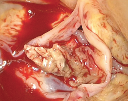
Figure 1. A large vegetation on the aortic valve from a patient with infective endocarditis

Untreated infective endocarditis (IE) is fatal; even with appropriate treatment, IE is associated with high rates of morbidity and mortality worldwide.1 The annual incidence of IE over the past two decades has remained relatively constant, ranging between 1.7 and 6.2 cases/100,000 population. Neither advances in healthcare nor revisions made to the current diagnostic criteria have substantially altered this.1-3 The current definition for IE now incorporates infections of prosthetic heart valves (both bioprosthetic and mechanical), implanted devices (such as pacemakers or ventricular assist devices) and cardiac endothelial surfaces.4 Figure 1 illustrates a large vegetation on the aortic valve from a patient with IE.

The variability in both the clinical manifestations and the course of IE reflect, in part, the heterogeneity of causative micro-organisms; this makes accurate diagnosis by clinical means alone problematic.5,6 Currently, the likelihood of IE is based on a score derived from a combination of clinical, microbiological and echocardiographic evidence.7-10 Laboratory diagnosis consists of culture of the infectious organism from the blood and/or heart valve material. However, the sensitivity of this scoring system is significantly compromised when IE is caused by fastidious or non-cultivable organisms or when patients have received previous antibiotic therapy such that positive cultures cannot be obtained. Novel diagnostic tests, such as nucleic acid amplification technologies (NAATs), that can identify the presence of infective organisms irrespective of the culture constraints will not only improve sensitivity but may reduce empiric treatment by permitting targeted antibiotic therapy. To be clinically useful any such technique must be able to identify the causative micro-organism from blood or tissue rapidly, have appropriately high positive- and negative-predictive values, and demonstrate good reproducibility between laboratories.

This review discusses the developments and application of NAATs for rapid and accurate detection of the infectious aetiology of IE.

Epidemiology and microbiology of IE

IE affects both native and prosthetic (bioprosthetic and mechanical) valves, and may develop through community- or healthcare-associated acquisition (CA-IE and HCA-IE, respectively). Worldwide, CA-IE of the native valve is by far the most common form of IE with rheumatic valve disease remaining the major risk factor;4 in resource-rich countries, however, profound changes in the epidemiology and aetiology of IE have been seen in recent years. There are many reasons for such shifts, but the principles for IE risk remain essentially unaltered: increased opportunity for microbial entry to the blood circulation, the presence of abnormal endocardial surfaces or flow patterns, and diminished host immune capacity.3,4,11 In developed countries with expanding elderly populations, degenerative valve lesions and congenital defects have far outstripped chronic rheumatic valve disease as major underlying risk factors, being present in up to 50% of IE patients over the age of 60 years.5

Although native valve endocarditis remains mostly CA-IE, the incidence of HCA-IE is steadily increasing12 as medical interventions allow greater opportunities for microbial access to the bloodstream via prolonged or repeated intravascular access or cannulation. Cardiothoracic surgical advances in developed healthcare settings have provided the emergence of new risk groups, including patients with prosthetic valves, intravascular devices or endovascular repairs. The flora associated with HCA-IE, predominantly skin-dwelling staphylococcal species, is fundamentally different to that of CA-IE. Such differences therefore demand different treatment.13,14 IE associated with intravenous drug use (IVDU) is in many ways unique – left- and right-sided valves are affected in approximately equal proportions,14 and the variety of microbial flora reflects the different opportunities for contamination at different stages of the process.15,16 Staphylococcus aureus is the predominant organism of IVDU-related IE as it colonises skin flexures; IE caused by Pseudomonas species and other motile Gram-negative bacteria have been attributed to the use of contaminated water to clean needles,17 and dissolving heroin in lemon juice predisposes to candidaemia and Candida IE.18

Prosthetic valve endocarditis occurs at a rate of 3–6/1,000 patient-years, accounting for an estimated 1–5% of all IE cases in resource-rich settings. Early prosthetic valve endocarditis occurs within 60 days of valve surgery11 and is typically HCA-IE, with Staph. aureus, Staph. epidermidis and other coagulase-negative staphylococci the most common pathogens. By contrast, the microbiology of late prosthetic valve endocarditis resembles that of native valve CA-IE.19 The typical microbiology of IE and its patterns of acquisition are summarised in table 1 (available in the PDF download).

Current diagnostic methods used in IE

The recognised epidemiological characteristics, described above, are only associations, and, while they may guide empiric therapy, they are not sufficiently reliable to allow antimicrobial prescribing in individual cases with certainty. For this, microbiological identification of the causative organism is needed; this will also inform the decisions on duration of therapy and even whether surgical intervention should be anticipated.20

However, direct microbiological culture from blood is not without pitfalls. The protean manifestations of IE that in part reflect the variety of aetiological agents, mean that patients frequently present to primary care with non-specific symptoms. Bacteraemia associated with IE is usually continuous but low grade, averaging at 1–10 organisms per ml of blood;21,22 blind trials of antibiotic therapy may, therefore, suppress microbial activity at the valve sufficiently to reduce the sensitivity of blood cultures taken at the time of hospital presentation.2,20-23 Indeed, up to 30% of all IE cases have negative blood cultures.24 A minor proportion of these cases are due to the presence of micro-organisms that are either non-cultivable, fastidious in their nutritional requirements in culture, or are extremely slow-growing on conventional media. The modified Duke criteria try to compensate for these issues, but the scoring system remains flawed (table 2 available in the PDF download).

Rationale for use of NAATs in IE

The advent of molecular technology, the “diagnostic tool for the new millennium”,25 has turned opportunities for rapid organism identification and even detection of certain drug susceptibility patterns from concept into a working reality. The rapid exponential generation of billions of copies of target DNA template from a single original sequence accounts for the potential for NAATs to be sensitive, specific and timely. One of the greatest assets of molecular tests is their potential to be adapted according to purpose. Amplification tests may be designed to be so specific as to detect only a single species, strain, or even resistance-inducing mutation; alternatively, the use of commonly shared genetic sequences as amplification targets allows detection of much broader categories of organisms. For example, the 16S rDNA gene codes for the RNA component of the 30S sub-unit of the prokaryotic ribosome. As it has both highly conserved and variable regions, pan-bacterial primers can be developed to target the conserved regions that immediately adjoin variable regions. Consequently, a single set of primers can be used to amplify the DNA from an enormous range of different bacteria. Subsequent sequencing of this amplified DNA can identify the variable region and thereby the bacterium.26-28 As the 16S rDNA gene sequence is universal throughout all phyla of bacteria it is, therefore, ideally suited for the diagnostics of IE, where there is such immense diversity of possible causative organisms including those that are fastidious or non-cultivable, or even no longer viable.3,29 The majority of NAATs applied to IE have used polymerase chain reaction (PCR) to amplify and subsequently sequence the 16S rDNA gene. To date, this technique has been used most successfully on excised valve tissue, and at present cannot replace microbiological culture of blood. Table 3 ( available in the PDF download) outlines the potential advantages and disadvantages of using molecular techniques to diagnose IE, with specific regard to targeting the 16S rDNA gene.

In fungal endocarditis, the 18S rDNA gene – the equivalent of the bacterial 16S rDNA gene – is more problematic as a diagnostic target since 18S rDNA gene sequences are highly conserved and demonstrate insufficient variability to differentiate between many fungal species. Instead, alternative targets such as short non-coding ribosomal internal transcribed spacer (ITS) regions are increasingly being used. These regions are located between conserved genes encoding for 18S, 5.8S and 28S rDNA, and are highly variable in both length and sequence, and, thus, more efficient for discriminating species than 18S sequences.30

Real-time PCR

More recently, 16S real-time or quantitative PCR (qPCR) has been applied to bacterial IE.2,31 There are a number of advantages of qPCR over conventional PCR: not only is it a more sensitive technique, but it is also more rapid as it eliminates the need for post amplification steps such as gel electrophoresis of PCR products.11 Importantly, qPCR can also measure the amount of inhibition from clinical samples and, hence, evaluate the effectiveness of the nucleic acid extraction method. For this, internal extraction and amplification controls are added to the sample before each of these steps.32

Previous studies have recommended that molecular-based techniques are included as a major criterion in the Duke criteria.22,31,33 Indeed, several studies have now demonstrated PCR positivity on valves in patients classified as possible or definitive IE, even when blood culture was negative.3 Results from these studies are undoubtedly promising, with the sensitivity of PCR from valve material ranging from 41.2% to 96%, compared with direct culture rates of 7.8–24.3%. Information of all published trials using 16S rDNA PCR in IE is available in a supplementary appendix (available online at www.bjcardio.co.uk).

Conclusions

It is important to remember that whatever the method of organism identification in IE, whether culture isolation or molecular nucleic acid detection, the result must still be open to careful interpretation. With an ever-increasing list of organisms that have been associated with both native and prosthetic valve IE, this assessment becomes more complex. Attribution of causation must always be weighed against the possibility of contamination. Appropriate measures to prevent contamination are as critically important in microbiological culture as in the molecular laboratory.

Molecular tests undoubtedly advance the diagnosis of IE; however, a much greater understanding of the variables that influence the sensitivities and specificities of the molecular methods needs to be defined. Recent calls to introduce basic standards in molecular diagnostic test protocols,34 in conjunction with the use of common targets, would allow for a much more accurate comparison between studies. With advances in molecular technology, NAATs now provide far more sensitive and rapid methods to detect the micro-organisms that cause IE. Comparative results from recent studies using NAATs indicate the clear superiority of valve material PCR over conventional valve culture. PCR can provide a positive identification where one or more of the definitive Duke criteria have been inconclusive. Development of consensus guidelines is needed to overcome the difficulties that the lack of standardisation of targets and protocols present to enabling valid comparisons between studies. With recent advances in nucleic acid quantification, NAATs technology may provide a tool to help answer some of the outstanding challenges that remain for IE diagnostics: accurate treatment response monitoring and reliable outcome prediction.

Acknowledgement

SM-J and AZ receive support from the UK Comprehensive Biomedical Research Centre and the National Institue of Medical Research.

Conflict of interest

None declared.

Editors’ note

A supplementary appendix containing information on all published trials using 16S rDNA PCR in IE is available online at www.bjcardio.co.uk

Key messages

  • Infective endocarditis (IE) causes high rates of morbidity and mortality
  • Clinical management is problematic if there are uncertainties over the identity, viability or antibiotic susceptibility of the causative organism
  • Conventional diagnostic microbiological techniques fail when patients have received prior antibiotic therapy, or when the causative organism is fastidious or non-cultivable
  • Nucleic acid amplification techniques (NAATs) now represent a much more sensitive and rapid method for detection of the micro-organisms that cause IE compared with culture alone


References

1. Prendergast B. The changing face of infective endocarditis. Heart 2006;92:879–85.

2. Munoz P, Bouza E, Marin M et al. Heart valves should not be routinely cultured. J Clin Microbiol 2008;46:2897–901.

3. Tak T. Molecular diagnosis of infective endocarditis: a helpful addition to the Duke criteria. Clin Med Res 2004;2:206–08.

4. Bashore T, Cabell C, Fowler V. Update on infective endocarditis. Curr Probl Cardiol 2006;31:274–352.

5. Moreillon P. Infective endocarditis. Lancet 2004;363:139–49.

6. Millar B, Moore J. Antibiotic prophylaxis, body piercing and infective endocarditis. J Antimicrob Chemother 2004;53:123–6.

7. Li J, Sexton D, Mick N et al. Proposed modifications to the Duke criteria for the diagnosis of infective endocarditis. Clin Infect Dis 2000;30:633–8.

8. Lamas C, Eykyn S. Suggested modifications to the Duke criteria for the clinical diagnosis of native valve and prosthetic valve endocarditis: analysis of 118 pathologically proven cases. Clin Infect Dis 1997;25:713–19.

9. Fournier P-E, Casalta J, Habib G, Messana T, Raoult D. Modification of the diagnostic criteria proposed by the Duke Endocarditis Service to permit improved diagnosis of Q fever endocarditis. Am J Med 1996;100:629–33.

10. Bayer AS, Bolger AF, Taubert KA et al. Diagnosis and management of infective endocarditis and its complications. Circulation 1998;98:2936–48.

11. Syed F, Millar B, Prendergast B. Molecular technology in context: a current review of diagnosis and management of infective endocarditis. Prog Cardiovasc Dis 2007;50:181–97.

12. Eykyn S. Endocarditis: basics. Heart 2001;86:476–80.

13. Cosgrove S, Karchmer A. Endocarditis. Medicine 2005;33:66–72.

14. Fowler V, Miro J, Hoen B. Staphylococcus aureus endocarditis: a consequence of medical progress. JAMA 2005;293:3012–21.

15. Brook I. Infective endocarditis caused by anaerobic bacteria. Arch Cardiovasc Dis 2008;101:665–76.

16. Baddour L, Wilson W, Bayer A et al. Infective endocarditis: diagnosis, antimicrobial therapy, and management of complications: a statement for healthcare professionals from the Committee on Rheumatic Fever, Endocarditis, and Kawasaki Disease, Council on Cardiovascular Disease in the Young, and the Councils on Clinical Cardiology, Stroke, and Cardiovascular Surgery and Anesthesia, American Heart Association: Endorsed by the Infectious Diseases Society of America. Circulation 2005;111:e394–e434.

17. Oh S, Havlen P, Hussain N. A case of polymicrobial endocarditis due to anaerobic organisms in an injection drug user. J Gen Intern Med 2005;20:C1–C2.

18. Bisbe J, Miro J, Latorre X et al. Disseminated candidiasis in addicts who use brown heroin: report of 83 cases and review. Clin Infect Dis 1992;15:910–23.

19. Loupa C, Mentzikof D. Current opinions in infective endocarditis. Hellenic J Cardiol 2002;43:53–62.

20. Voldstedlund M, Pedersen L, Baandrup U, Klaaborg K, Fuursted K. Broad-range PCR and sequencing in routine diagnosis of infective endocarditis. APMIS 2008;116:190–8.

21. Watkin R, Lang S, Lambert P, Littler W, Elliott T. The microbial diagnosis of infective endocarditis. J Infect 2003;47:1–11.

22. Millar B, Moore J, Mallon P et al. Molecular diagnosis of infective endocarditis – a new Duke’s criterion. Scand J Infect Dis 2001;33:673–80.

23. Beynon R, Bahl V, Prendergast B. Infective endocarditis. BMJ 2006;333:334–9.

24. Naber C, Erbel R. Infective endocarditis with negative blood cultures. Int J Antimicrob Agents 2007;30S:S32–S36.

25. Yang S, Rothman R. PCR-based diagnostics for infectious diseases: uses, limitations, and future applications in acute-care settings. Lancet Infect Dis 2004;4:337–48.

26. Barken K, Haagensen J, Tolker-Nielsen T. Advances in nucleic acid-based diagnostics of bacterial infections. Clin Chim Acta 2007;384:1–11.

27. Rice PA, Madico GE. Polymerase chain reaction to diagnose infective endocarditis. Will it replace blood cultures? Circulation 2005;111:1352–4.

28. Petti C. Detection and identification of microorganisms by gene amplification and sequencing. Clin Infect Dis 2007;44:1108–14.

29. Clarridge III J. Impact of 16S rRNA gene sequence analysis for identification of bacteria on clinical microbiology and infectious diseases. Clin Microbiol Rev 2004;17:840–62.

30. Deng W, Xi D, Mao H, Wanapat M. The use of molecular techniques based on ribosomal RNA and DNA for rumen microbial ecosystem studies: a review. Mol Biol Rep 2008;35:265–74.

31. Marin M, Munoz P, Sanchez M et al. Molecular diagnosis of infective endocarditis by real-time broad-range polymerase chain reaction (PCR) and sequencing directly from heart valve tissue. Medicine 2007;86:195–202.

32. Mackay I. Real-time PCR in the microbiology laboratory. Clin Microbiol Infect 2004;10:190–212.

33. Bosshard P, Kronenberg A, Zbinden R, Ruef C, Bottger E, Altwegg M. Etiologic diagnosis of infective endocarditis by broad-range polymerase chain reaction: a 3-year experience. Clin Infect Dis 2003;37:167–72.

34. Bustin S, Benes V, Garson J et al. The MIQE guidelines: minimum information for publication of quantitative real-time PCR experiments. Clin Chem 2009;55:611–22.

35. Mylonakis E, Calderwood S. Infective endocarditis in adults. N Engl J Med 2001;345:1318–30.

36. Brouqui P, Raoult D. Endocarditis due to rare and fastidious bacteria. Clin Microbiol Rev 2001;14:177–207.

37. Mandell G, Bennett J, Dolin R. Cardiovascular infections. In: Principles and practice of infectious diseases. Philadelphia: Elsevier/Churchill Livingstone, 2005.

38. Cuculi F, Toggweiler S, Auer M, Auf der Maur C, Zuber M, Erne P. Serum procalcitonin has the potential to identify Staphylococcus aureus endocarditis. Eur J Clin Microbiol Infect Dis 2008;27:1145–9.

39. Dreier J, Vollmer T, Freytag C, Baumer D, Korfer R, Kleesiek K. Culture-negative infectious endocarditis caused by Bartonella spp.: 2 case reports and a review of the literature. Diagn Microbiol Infect Dis 2008;61:476–83.

40. Lang S, Watkin RW, Lambert PA, Bonser RS, Littler WA, Elliott TSJ. Evaluation of PCR in the molecular diagnosis of endocarditis. J Infect 2004;48:269–75.

41. Mueller C, Huber P, Laifer G, Mueller B, Perruchoud A. Procalcitonin and the early diagnosis of infective endocarditis. Circulation 2004;109:1707–10.

42. Christ-Crain M, Muller B. Procalcitonin in bacterial infections – hype, hope, more or less? Swiss Med Wkly 2005;135:451–60.

43. Hodgetts A, Levin M, Kroll J, Langford P. Biomarker discovery in infectious diseases using SELDI. Future Microbiol 2007;2:35–49.

44. Sontakke S, Cadenas M, Maggi R, Diniz P, Breitschwerdt E. Use of broad-range 16S rDNA PCR in clinical microbiology. J Microbiol Methods 2009;76:217–25.

45. Millar B, Moore J. Current trends in the molecular diagnosis of infective endocarditis. Eur J Clin Microbiol Infect Dis 2004;23:353–65.

46. Johnson J. Development of polymerase chain reaction-based assays for bacterial gene detection. J Microbiol Methods 2000;41:201–09.

47. Silkie S, Tolcher M, Nelson K. Reagent decontamination to eliminate false-positives in Escherichia coli qPCR. J Microbiol Methods 2008;72:275–82.

48. Podglajen I, Bellery F, Poyart C et al. Comparative molecular and microbiologic diagnosis of bacterial endocarditis. Emerg Infect Dis 2003;9:1543–7.

49. Haanpera M, Jalava J, Huovinen P, Meurman O, Rantakokko-Jalava K. Identification of alpha-haemolytic streptococci by pyrosequencing the 16S rRNA gene and by use of VITEK 2. J Clin Microbiol 2007;45:762–70.

50. Ghebremedhin B, Layer F, Konig W, Konig B. Genetic classification and distinguishing of Staphylococcus species based on different partial gap, 16S rRNA, hsp60, rpoB, sodA and tuf gene sequences. J Clin Microbiol 2008;46:1019–25.

51. Faibis F, Mihaila L, Perna S et al. Streptococcus sinensis: an emerging agent of infective endocarditis. J Med Microbiol 2008;57:528–31.

52. Fihman V, Raskine L, Barrou Z et al. Lactococcus garvieae endocarditis: identification by 16S rRNA and sodA sequence analysis. J Infect 2006;52:e3–e6.

53. Huggett J, Novak T, Garson J et al. Differential susceptibility of PCR reactions to inhibitors: an important and unrecognised phenomenon. BMC Research Notes 2008;1(1):70.