Using the left trans-radial artery access route for coronary and bypass angiography has been frowned upon by the majority of operators due to several catheter changes during the procedure, patient discomfort because of spasm, positioning of the patients left arm and longer radiation exposure times, to name a few reasons. This short scientific article demonstrates one of a series of 22 cases where a single catheter was successfully used via this access route.
Introduction
The traditional approach to coronary catheterisation in patients with a history of coronary artery bypass grafting (CABG) with left internal mammary artery (LIMA) graft is via the femoral approach. However, trans-radial catheterisation still remains a distinct option for such patients. Operators tend to be cautious about using this approach due to the nature of having to exchange diagnostic catheters several times during the procedure, risk of spasm, alternative set-up for the procedure, arm positioning, etc. A retrospective audit of 22 single-operator coronary graft angiography cases over a six-month period demonstrates the feasibility and option to use the left trans-radial approach (LTRA) to perform a coronary and graft study with the use of a single diagnostic catheter – the TIGER (TIG) catheter (Terumo Medical Corporation).
Most operators, who opt to use the LTRA, routinely practice with the standard femoral diagnostic catheters to access the native coronary arteries and grafts.1 A disadvantage of using LTRA for graft angiography is because several catheter exchanges are necessary to engage both native coronary arteries and the bypass grafts.
My standard approach for performing coronary and graft angiography in patients is via the left radial artery (LRA) in those with a LIMA graft and via the right radial artery in those without. My personal preference is to use the TIG catheter as the exclusive standard catheter of choice for all coronary angiography via the radial artery. The TIG is designed to enable coronary angiography of the left coronary artery (LCA) and right coronary artery (RCA) via the right radial artery without the need for any catheter exchange.2 Over a six-month period, I performed a series of 22 cases using the TIG catheter via the left radial to successfully perform both coronary and graft angiography. This case description illustrates the last in the short series of successful procedures.
Case description
A 68-year-old man with a history of a three-vessel bypass in 2007. The bypass grafts were: saphenous vein graft (SVG) to obtuse marginal (OM), SVG to RCA and LIMA to the left anterior descending artery (LAD).
The patient was referred for coronary angiography because he was experiencing a central crushing chest pain radiating to his left arm, relieved by glyceryl trinitrate (GTN) spray. Exercise tolerance had reduced in recent weeks with increasing shortness of breath. Other significant medical history to note included hypertension, diabetes mellitus and peripheral vascular disease, and is an ex-smoker.
I performed a left radial Allen’s test before the procedure. Ulnar arch patency using pulse oximetry was confirmed with a Barbeau score of A.3 A 6f Terumo Glidesheath (Terumo Medical Corporation) was inserted using the standard Seldinger technique. My standard radial angiography protocol is to administer 200 µg of nitrate, and 5000 IU heparin intra-arterially through the radial sheath before the catheter is inserted. The TIG catheter was fed up through the left arm with a 0.35”/150 cm Emerald (Cordis, Johnson & Johnson) guidewire and into the left subclavian artery.
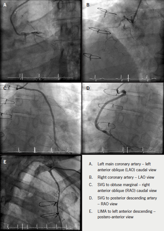
The catheter was used to first intubate the LCA. Figure 1A shows severe distal left main stem disease, a blocked proximal LAD and a severe lesion in the circumflex artery just proximal to a blocked OM artery. After disengagement, the TIG was rotated to the RCA (figure 1B) and showed it was blocked proximally. The TIG was disengaged and pulled back to engage the SVG to the obtuse marginal (OM) (figure 1C) showing that there was a mild ostial narrowing in an otherwise patent graft. The catheter was then disengaged and manipulated to intubate the SVG to posterior descending artery (PDA) (figure 1D). The SVG to PDA was patent with a good run off. The catheter was disengaged for a final time before being withdrawn slowly into the subclavian artery where the LIMA graft was successfully engaged to show a widely patent graft with a severe lesion just beyond the anastomosis site (figure 1E). The guidewire was re-inserted to remove the TIG catheter over the wire. Haemostasis was achieved using a TR band (Terumo Medical Corporation).

Discussion

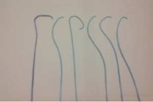
Operators who use the left trans-radial approach for coronary and graft angiography achieve comparable outcomes to those operators using the trans-femoral approach.4 However, this has been through the use of several different diagnostic catheters – Judkins, Amplatz, internal mammary or multi-purpose catheters (figure 2).
Several years ago, using the left trans-radial technique, initially meant another learning curve in what has now proven to be an effective and more patient friendly access route. Learning to handle and manipulate a single catheter through this particular access site will mean another exciting step in trans-radial education.
I found there was a learning curve of 15 cases before I had successfully adapted my practice. The structural association between the coronary arteries and aortic arch in the left radial approach varies from that of the right radial approach and even the femoral approach.5 This implies that re-education of certain manipulation techniques of the TIG catheter have to be developed to ensure a positive outcome, as well as patient satisfaction.
The TIG was originally designed to be used exclusively as a right radial catheter (Terumo Medical Corporation, 2008). It is my standard work-horse diagnostic catheter for right trans-radial procedures, and it is now my first choice for left trans-radial approaches as well.

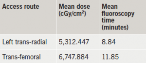
We found that there was no spasm present in any of the cases – this is attributed to the lack of catheter exchanges. Radiation and exposure times can be prolonged due to the catheter exchanges or spasm that may occur. We compared the mean doses and screening times for these cases against the same number (n=22) of retrospectively performed trans-femoral graft angiography cases. The results indicated that by using this single-catheter approach via the left trans-radial approach, the procedure dose and screening times were reduced by 25.4% and 21.3%, respectively (table 1).
Although challenging at first, due to its manipulation, the TIG was found to intubate the LIMA when it was the last artery to review. We have deduced that this may be due to the catheter having ‘softened’ during the procedure and, therefore, being more malleable to co-axially align itself to the angulation of the vessel.
Conclusion
When considering using the left radial artery for coronary and graft angiography, the TIG catheter can be safely used as a single catheter of choice to successfully engage all grafts, as well as the native coronary arteries, thereby, enhancing the patient experience, promoting less spasm, reducing radiation doses and exposure times.
Conflict of interest
None declared.
References
1. Suh WM, Kern M. Coronary and bypass graft angiography via the right radial approach using a single catheter. J Invasive Cardiol 2012;24:295–7. Available from: http://www.invasivecardiology.com/articles/coronary-and-bypass-graft-angiography-right-radial-approach-using-single-catheter
2. Terumo Medical Corporation (2008). Website: http://www.terumomedical.com
3. Barbeau GR, Arsenault F, Dugas L, Simard S, Lariviere MM. Evaluation of the ulno-palmar arterial arches with pulse oximetry and plethysmography: comparison with the Allen’s test in 1010 patients. Am Heart J 2004;147:489–93. http://dx.doi.org/10.1016/j.ahj.2003.10.038
4. Sanmartin M, Cuevas D, Moxica J et al. Trans-radial cardiac catheterization in patients with coronary bypass grafts: feasibility analysis and comparison with trans-femoral approach. Catheter Cardiovasc Interv 2006;67:580–4. http://dx.doi.org/10.1002/ccd.20633
5. Saito S. Right or left side? Catheter Cardiovasc Interv 2003;58:305. http://dx.doi.org/10.1002/ccd.10462
