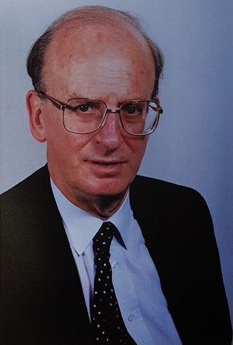
A personal tribute
I was deeply saddened by the news that Dr Derek Gibson had passed away. I have lost a teacher, a friend, an adviser and a father figure, for whom I had the highest respect.

Dr Gibson read Natural Sciences at Trinity College, Cambridge, followed by three years at Westminster Medical School, London, where he achieved his Bachelor of Surgery and Bachelor of Medicine degrees in 1962. His training continued at The National Heart Hospital and St Bartholomew’s Hospital, London, and he was appointed as a Consultant Cardiologist at the Royal Brompton Hospital, London, in 1971. He was awarded Fellowship to the Royal College of Physicians in 1975.
Dr Gibson made a hugely significant contribution to echocardiography. He was a unique master in applying echocardiography to physiology, pathophysiology, and the anatomy of the cardiovascular system, displaying his high level of expertise and clinical experience in lectures he gave nationally and internationally, as well as in the weekly echo meetings he gave at the Brompton. In many of these weekly echo meetings, we were unable to follow his observations or clinical reasoning from M-mode echocardiography, electrocardiography, phonocardiography, the apex cardiogram, sometimes the carotid or JVP pulse, or Doppler and two-dimensional images. We would defend our weakness by saying that he was using ‘the Gibson ruler’ and, in return, he would give a broad and gentle smile with a tinge of his pride and satisfaction. He was simply a unique and intelligent teacher!
He bravely pushed digital technology forward by combining it with echocardiography and, at a time when computing was only for a few, he, along with others, developed and applied digitised echocardiography to assess ventricular wall motion1 and to evaluate myocardial property by echo intensity.2,3 He also used digital techniques to reconstruct right and left ventricular pressure traces noninvasively.4,5
When there was insufficient appreciation or even opposition to M-mode echocardiography, Dr Gibson and his team persevered in its practice and research, particularly in the field of long-axis function.6,7 This persistence has led to M-mode echocardiography remaining part of the current guidelines rather than becoming a lost art, like phonocardiography.8,9
He was a true pioneer in studying cardiac physiology with echocardiography in the treatment of heart failure. At a time when the cardiology community was focussed on ejection fraction and contractility, he laid the foundations for resynchronisation to treat patients with heart failure by non-medical means. As early as 1971, he concluded biventricular pacing was superior to single ventricular pacing in increasing left ventricular force by synchronous contraction.10 Later on, his work on a controlled but synchronous atrioventricular contraction achieved by DDD pacing also proved to offer therapeutic potential in treating patients with congestive heart failure.11 It is no exaggeration to say that, as a result of his work, resynchronisation with biventricular pacing has now become a standard treatment for congestive heart failure in current practice.12
He has influenced many generations of cardiologists whom he trained in the UK and his contribution to the development, research and training in his field has spread all over the world. He sponsored and trained cardiology fellows from many nations – in my seven years at the Brompton, Dr Gibson’s fellows came from Australia, Canada, China, The Congo, Egypt, Italy, Japan, Malaysia, and the United States. His legacy runs deep and wide.
Dr Gibson published well over 400 original papers, with just a few of them referenced here in the areas I mention in this short tribute.
Outside cardiology, Dr Gibson was known to be a gifted musician, an accomplished collector of antiques and a mature enthusiast of architecture.
For me, this reserved man was far more than a teacher and a supervisor. He directly taught and encouraged much of my knowledge of English and cardiology. He guided me, often working with me, through all my research carried out at the Brompton and beyond, including my PhD thesis. He was always friendly, gentle and kind, teaching me on in a practical way on multiple occasions, the important principle of ‘family before career’.
Acknowledgement
I would like to express my profound gratitude to Dr Alison Duncan, Royal Brompton Hospital, London, for her invaluable help in preparing this tribute.
Han B Xiao
Consultant Cardiologist
Homerton University Hospital, Homerton Row, London, E9 6SR
([email protected])
References
1. Gibson DG, Brown D. Measurement of instantaneous left ventricular dimension and filling rate in man, using echocardiography. Br Heart J 1973;35:1141–9. http://doi.org/10.1136/hrt.35.11.1141
2. Logan-Sinclair R, Wong CM, Gibson DG. Clinical application of amplitude processing of echocardiographic images. Br Heart J 1981;45:621–7. http://doi.org/10.1136/hrt.45.6.621
3. Shaw TR, Logan-Sinclair RB, Surin C, McAnulty RJ, Heard B, Laurent GJ, Gibson DG. Relation between regional echo intensity and myocardial connective tissue in chronic left ventricular disease. Br Heart J 1984;51:46–53. http://doi.org/10.1136/hrt.51.1.46
4. Brecker SJ, Gibbs JS, Fox KM, Yacoub MH, Gibson DG. Comparison of Doppler derived haemodynamic variables and simultaneous high fidelity pressure measurements in severe pulmonary hypertension. Br Heart J 1994;72:384–9. http://doi.org/10.1136/hrt.72.4.384
5. Xiao HB, Jin XY, Gibson DG. Doppler reconstruction of left ventricular pressure from functional mitral regurgitation: potential importance of varying orifice geometry. Br Heart J 1995;73:53–60. http://doi.org/10.1136/hrt.73.1.53
6. Henein MY, Priestley K, Davarashvili T, Buller N, Gibson DG. Early changes in left ventricular subendocardial function after successful coronary angioplasty. Br Heart J 1993;69:501–06. http://doi.org/10.1136/hrt.69.6.501
7. Webb-Peploe KM, Henein MY, Coats AJS, Gibson DG. Echo derived variables predicting exercise tolerance in patients with dilated and poorly functioning left ventricle. Heart 1998;80:565–9. http://doi.org/10.1136/hrt.80.6.565
8. Mitchell C, Rahko PS, Blauwet LA, et al. Guidelines for performing a comprehensive transthoracic echocardiographic examination in adults: recommendations from the American Society of Echocardiography. J Am Soc Echocardiogr 2019;32:1-64. http://doi.org/10.1016/j.echo.2018.06.004
9. Popescu BA, Stefanidis A, Fox KF, et al. Training, competence, and quality improvement in echocardiography: the European Association of Cardiovascular Imaging Recommendations: update 2020. Eur Heart J Cardiovasc Imaging 2020;21:1305–19. http://doi.org/10.1093/ehjci/jeaa266
10. Gibson DG, Chamberlain DA, Coltart DJ, Mercer J. Effect of changes in ventricular activation on cardiac haemodynamics in man. Comparison of right ventricular, left ventricular, and simultaneous pacing of both ventricles. Br Heart J 1971;33:397–400. http://doi.org/10.1136/hrt.33.3.397
11. Brecker SJD, Xiao HB, Sparrow J, Gibson DG. Effects of dual-chamber pacing with short atrioventricular delay in dilated cardiomyopathy. Lancet 1992;340:1308–12. http://doi.org/10.1016/0140-6736(92)92492-x
12. McDonagh TA, Metra M, Adamo M, et al. 2021 ESC Guidelines for the diagnosis and treatment of acute and chronic heart failure: Developed by the Task Force for the diagnosis and treatment of acute and chronic heart failure of the European Society of Cardiology (ESC) With the special contribution of the Heart Failure Association (HFA) of the ESC. Eur Heart J 2021;2:3599–726. http://doi.org/10.1093/eurheartj/ehab368
