Viruses, vaccines and cardiovascular effects

Br J Cardiol 2022;29:43–5doi:10.5837/bjc.2022.016 Leave a comment
Click any image to enlarge
Authors:
First published online 31st May 2022

On the 31st March 2021, the German Health Ministry – on the advice of the Standing Committee on Vaccination (STIKO) – declared that the Astra Zeneca/Oxford Vaxzevria vaccine against SARS-CoV-2 (COVID-19), based on a chimpanzee adenovirus genetic scaffold, henceChAdOx1, would no longer be administered to those under the age of 60 years. In its hands were details of 31 cerebral venous sinus thrombosis (CVST) cases provided by the Paul Ehrlich Institute. These cases, of whom 19 had platelet deficiency, were seen after 2.7 million first and 767 second vaccine doses.

Professor Anthony Rees

The German decision somewhat pre-empted the European Medicines Agency analysis published a week later on 7th April 2021 in which 62 cases of CVST and 24 cases of splanchnic vein thrombosis, 18 of which were fatal, had been reported via the EudraVigilance database. After considering the cases, the EMA responded:

“The benefits of the vaccine continue to outweigh the risks for people who receive it. The vaccine is effective at preventing COVID-19 and reducing hospitalisations and deaths”.1

By the date of the EMA report, 25 million people in the European Economic Area (EEA) and the UK had received the vaccine. Speculation at the time was that the condition resembled atypical heparin-induced thrombocytopenia (HIT) and may have been induced by an auto-immune response triggered by the COVID-19 surface (spike) protein encoded in the vaccine.

Historical cases

Cardiovascular effects by viruses are not new. Numerous cases of myocarditis were reported during the 1918 Spanish flu (influenza A, H1N1) pandemic, while cases of myo/pericarditis in influenza B patients were seen during the late 1950s. In 1980, cases of viral myocarditis following influenza infection were reported from a military hospital in Finland. Of the serologically confirmed influenza A patients, 9% had developed acute myocarditis based on serial ECG ST-segment and/or T-wave changes, unresponsive to beta blockade. Multidirectional echocardiography revealed regional myocardial dysfunction with elevated MB-CK levels in three patients.2 In 1997, a case of fatal myocarditis in a six-year-old female infected with influenza B was reported in New Orleans, USA. Post-mortem examination led to elimination of direct viral invasion of the myocardium or coronary vascular damage as potential causes, the authors concluding that an autoimmune mechanism was the most likely.3 Other cases have been seen with enterovirus B where IgM autoantibodies were elevated in patients with chronic myocarditis, triggering T cells into an autoreactive state, possibly by a mechanism of molecular mimicry.4 Examples of excessive coagulation were reported in a Dutch study in 2000 where several viruses, influenza A and B, respiratory syncytial virus, adenovirus and cytomegalovirus, on infection of human umbilical cord endothelial cells, induced procoagulant activity in a one-stage clotting assay causing a 55% reduction in the clotting time. This induction was associated with a four-to-five-fold increase in the expression of tissue factor as measured by the generation of factor Xa. The authors’ conclusion was:

“…the procoagulant activity of endothelial cells in response to infection with respiratory viruses is caused by upregulation of the extrinsic pathway”.5

COVID-19 era

While cardiovascular effects from infection with different respiratory viruses have been widely described over many decades, the incidence of such events during the largest pandemic since the Spanish ‘flu, and the rapidity with which clinicians, academics, and the public have become aware of such adverse effects through the media, has brought the behaviour of COVID-19 into sharp focus. Of particular concern has been the incidence of thrombotic events that seem to have been triggered not only by the virus itself but, worryingly, by certain COVID-19 vaccines, particularly those based on adenovirus vectors.

Hospitalised COVID-19 patients have been shown to develop both cardiac and kidney dysfunction, potentially mediated by the receptor for COVID-19 (the ACE-2 receptor), a protein that is widely expressed in many tissues, including the heart and kidney. Myocardial damage can occur as a result of:

  • a decrease in oxygen levels
  • acute respiratory distress syndrome
  • the formation of microthrombi (as has been observed in some COVID-19 patients)
  • direct injury of cardiomyocytes by viral infection coupled with resulting inflammatory (cytokine) responses.

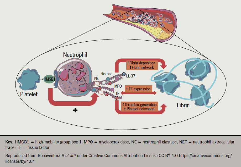
In vitro cardiomyocyte studies by Siddiq et al. suggest that direct injury from the infection is likely to be one of the causes of myocardial injury in COVID-19 patients.6 Raman et al.7 have proposed similar but subtly different aetiologies such as chronic inflammatory responses evoked by persistent viral reservoirs in the heart following the acute infection, or a mechanism where autoimmune responses to cardiac antigens are triggered through molecular mimicry, potentially explaining delayed damage. A further mechanistic candidate gathering interest has been proposed by Nappi et al. involving excessive activation of neutrophil extracellular traps (NETs). It is well known that neutrophils are players in the first line of defence against bacterial and fungal infections but their involvement in response to viruses has only been described in recent years. In a process known as NETosis (resembling apoptosis) triggered by pathogen entry, neutrophils self-disintegrate releasing protein granules and decondensed chromatin which form net-like structures that can engulf (trap) pathogens, including viruses. Evidence from autopsy studies on patients who died from serious COVID-19 disease, and studies on patients who underwent primary coronary interventions for ST-elevation myocardial infarction (STEMI), has implicated neutrophils and the resulting excessive production of aggregated NETs causing vascular-obstruction8 (figure 19).

Rees - Figure. 1 The interplay between NETs and platelets. NETs promote thrombosis by favouring fibrin deposition. Recently, a notion has been added by showing that NETs can express tissue factor further triggering thrombin generation and platelet activation and finally increasing the thrombogenic potential of NETs. This was reported especially at the site of plaque rupture during acute myocardial infarction when platelets and neutrophils interact with each other. In this view, activated platelets present HMGB1 to neutrophils and stimulate them to form NETs
Figure. 1 The interplay between NETs and platelets. NETs promote thrombosis by favouring fibrin deposition. Recently, a notion has been added by showing that NETs can express tissue factor further triggering thrombin generation and platelet activation and finally increasing the thrombogenic potential of NETs. This was reported especially at the site of plaque rupture during acute myocardial infarction when platelets and neutrophils interact with each other. In this view, activated platelets present HMGB1 to neutrophils and stimulate them to form NETs

The effects of the S-protein

While the exact mechanisms of cardiovascular damage from COVID-19 infection remain under study, with several candidate explanations, the story does not stop with the virus itself. Explanations of how COVID-19 vaccines can induce similar pathologies is of equal importance. The most extensively used vaccines are the mRNA vaccines of Pfizer (BNT162b2) and Moderna (mRNA-1273) and the adenovirus-based (AdV) vaccines of AstraZeneca (ChAdOx1) and Johnson & Johnson (Ad26.COV2.S), all of which share the COVID-19 spike protein gene (RNA or DNA) but which differ markedly in the case of the AdV vaccines by the additional presence of AdV genes.

A critical observation has been the presence of the S-protein in the circulation post-vaccination, seen with both types of vaccine. Aside from its affinity for ACE-2 receptors present on many different organs, the S-protein, generated either by COVID-19 infection10 or from vaccination with the mRNA vaccine,11 has been shown to damage the endothelium, disrupt the blood-brain barrier, and/or cause inflammation and damage to human cardiac pericytes i.e. multi-functional mural cells of the microcirculation that wrap around the endothelial cells lining the capillaries throughout the body. A novel route of entry for the S-protein via the CD147 receptor has recently been implicated in the pericyte damage10 (CD147 is an extracellular matrix metalloproteinase inducer and is present on cardiac pericytes; it is also known as an entry receptor for measles virus).

A more extensive study of myocarditis based on reports to the US Vaccine Adverse Event Reporting System (VAERS) exploring a possible causal relationship to the two mRNA vaccines, was carried out by the US Centres for Disease Control (CDC), the US Food and Drug Administration (FDA) and US medical centres, between December 2020 and August 2021. Out of more than 190 million people (median age of 21 years) with 354 million vaccinations, a total of 1,626 myocarditis cases were confirmed; an incidence rate of 8.6 per one million people. Of those affected and who reported their sex, 82% were males with the rates of myocarditis highest after the second vaccination. There was no difference in incidence for the two different mRNA vaccines. Of those aged under 30 years with detailed clinical information available, 87% had resolution of presenting symptoms by hospital discharge.12 Similar results were seen in a smaller 26-centre US study published in February this year.13 In a more recent analysis of Israeli army recruits who had received a third dose of the BNT162b2 vaccine, the incidence of myocarditis was lower than after the second dose, a result unexplained and requiring “future research”.14

The incidence of CVST

If science was easy everyone would be doing it. In a recently published meta-analysis in the Journal of Neurology, the authors stated that vector-based (e.g. ChAdOx1 or Ad26.COV2.S) COVID-19 vaccination was associated with an approximately 50-fold increase in the odds of thrombotic thrombocytopenia syndrome (TTS)-related CVST compared to non-TTS-related CVST. They also note that antigenic complexes of vaccine components have been shown to bind to platelet factor 4 (PF4) on platelet surfaces, inducing proinflammatory reactions, anti-PF4 antibody formation and prothrombotic cascades.15 The possibility that anti-PF4 antibodies form as a result of epitope mimicry with the COVID-19 S-protein has been previously proposed. So far, however, other studies on anti-PF4 antibodies found in patients have shown no binding of these antibodies to the viral S-protein.16 What is clear though is that in the rare cases of vaccine-induced immune thrombotic thrombocytopenia (VITT), associated with both the AD26.COV2.S and ChAdOx1 vaccines, anti-PF4 antibodies are not only present but in a recent study have been shown to persist for several months after acute presentation. Key to this observation by Kanack et al. was development of a diagnostic assay able to distinguish VITT from spontaneous HIT, another rare anti-PF4-mediated thrombotic thrombocytopenic disorder.17

In a related UK study, carried out between December 2020 and June 2021 on a combined cohort of approximately 11.6 million people with a 6.8 million person years follow-up, the authors found a ‘small elevated risk’ of CVST events following vaccination with ChAdOx1 but not with BNT162b2.18 To confuse matters further, a large US study took the step of comparing the incidence of CVST in COVID-19 vaccinated cohorts and those who had received other non-COVID vaccines during the four-year period 2017–2021. The conclusion of this study, which used databases from the US Mayo Clinic Health System, was:

“This real-world evidence-based study finds that CVST is rare and is not significantly associated with COVID-19 vaccination…”

“…the risk of CVST within 30 days of COVID-19 vaccination was similar to the risk of CVST within 30 days of all analysed non-COVID vaccinations…”19

Conclusion

The frustration that must be present among clinicians and virologists alike is palpable. There is no question that serious (thrombotic) and frequently recoverable (myocarditis) cardiovascular events occur both after COVID-19 infection and after vaccination, but they are rare. Further, when the incidence of such events is compared with other vaccinations, the picture of an elevated COVID-19 cardiovascular pathology causally related to vaccination is not well defined. Until then, the clinical communities eagerly await the mechanistic breakthroughs that will define effective therapies for these important pathologies.

Conflicts of interest

None declared.

Funding

None.

Editors’ note

Anthony R Rees is the author of recent book A new history of vaccines for infectious diseases available from Elsevier/Academic Press (ISBN 978-0-12-812754-4).

References

1. European Medicines Agency. News release 7th April 2021. AstraZeneca’s COVID-19 vaccine. https://www.ema.europa.eu/en/news/astrazenecas-covid-19-vaccine-ema-finds-possible-link-very-rare-cases-unusual-blood-clots-low-blood (last accessed 4th May 2022)

2. Karjalainen J, Nieminen MS, Heikkilä J. Influenza A myocarditis in conscripts. Acta Med Scand 1980;207:27–30. https://doi.org/10.1111/j.0954-6820.1980.tb09670.x

3. Craver R, Sorrells K, Gohd R. Myocarditis with influenza B infection. Pediatr Infect Dis J 1997;16:629–30. https://doi.org/10.1097/00006454-199706000-00018

4. Hussein HM, Rahal EA. The role of viral infections in the development of autoimmune diseases. Crit Rev Microbiol 2019;45:394–412. https://doi.org/10.1080/1040841X.2019.1614904

5. Visseren FL, Bouwman JJ, Bouter KP, Diepersloot RJ, de Groot PH, Erkelens DW. Procoagulant activity of endothelial cells after infection with respiratory viruses. Thromb Haemost 2000;84:319–24

6. Siddiq MM, Chan AT, Miorin L et al. Functional effects of cardiomyocyte Injury in COVID-19. J Virol 2022;96(2):e01063-21. https://doi.org/10.1128/JVI.01063-21

7. Raman B, Bluemke DA, Lüscher TF, Neubauer S. Long COVID: post-acute sequelae of COVID-19 with a cardiovascular focus. Eur Heart J 2022;43:1157–72. https://doi.org/10.1093/eurheartj/ehac031

8. Nappi F, Giacinto O, Eliouse O et al. Association between COVID-19 diagnosis and coronary artery thrombosis: a narrative review. biomedicines 2022;10:702. https://doi.org/10.3390/biomedicines10030702

9. Bonaventura A, Vecchié A, Abbate A, Montecucco F. Neutrophil extracellular traps and cardiovascular diseases: an update. Cells 2020;9:231. https://doi.org/10.3390/cells9010231

10. Avolio E, Carrabba M, Milligan R et al. The SARS-CoV-2 spike protein disrupts human cardiac pericytes function through CD147 receptor-mediated signalling: a potential non-infective mechanism of COVID-19 microvascular disease. Clin Sci (Lond) 2021;135:2667–89. https://doi.org/10.1042/CS20210735

11. Singh A, Nguyen L, Everest S, Afzal S, Shim A. Acute pericarditis post mRNA-1273 COVID vaccine booster. Cureus 2021;14(2):e22148. https://doi.org/10.7759/cureus.22148

12. Oster ME, Shay DK, Su JR et al. Myocarditis cases reported after mRNA-based COVID-19 vaccination in the US from December 2020 to August 2021. JAMA 2022;327:331–40. https://doi.org/10.1001/jama.2021.24110

13. Truong DT, Dionne A, Muniz JC et al. Clinically suspected myocarditis temporally related to COVID-19 vaccination in adolescents and young adults: suspected myocarditis after COVID-19 vaccination. Circulation 2022;145:345–56. https://doi.org/10.1161/CIRCULATIONAHA.121.056583

14. Friedensohn L, Levin D, Fadlon-Derai M et al. Myocarditis following a third BNT162b2 vaccination dose in military recruits in Israel. JAMA 2022;327:1611–12. https://doi.org/10.1001/jama.2022.4425

15. Palaiodimou L, Stefanou M-I, Aguiar de Sousa D, et al. Cerebral venous sinus thrombosis in the setting of COVID‑19 vaccination: a systematic review and meta‑analysis. J Neurol 2022:April 8:1–7. https://doi.org/10.1007/s00415-022-11101-2

16. Rees AR. A new history of vaccines for infectious diseases. London: Elsevier/Academic Press, 2022. Chapter 14, Immmunological challenges of the ‘new’ infections: corona viruses; p. 395–450.

17. Kanack AJ, Bandana S, George G, et al. Persistence of Ad26.COV2.S-associated vaccine-induced immune thrombotic thrombocytopenia (VITT) and specific detection of VITT antibodies. Am J Hematol 2022;97:519–26 https://doi.org/10.1002/ajh.26488

18. Kerr S, Joy M, Torabbi F et al. First dose ChAdOx1 and BNT162b2 COVID-19 vaccinations and cerebral venous sinus thrombosis: A pooled self-controlled case series study of 11.6 million individuals in England, Scotland, and Wales PLoS Med 2022;19(2):e1003927. https://doi.org/10.1371/journal.pmed.1003927

19. Pawlowski C, Rincón-Hekking J, Awasthi S, et al. Cerebral venous sinus thrombosis is not significantly linked to COVID-19 vaccines or non-COVID vaccines in a large multi-state health system. J Stroke Cerebrovasc Dis 2021;30(10):105923. https://doi.org/10.1016/j.jstrokecerebrovasdis.2021.105923

Total ischaemic time in STEMI: factors influencing systemic delay

Br J Cardiol 2022;29:60–3doi:10.5837/bjc.2022.017 Leave a comment
Click any image to enlarge
Authors:
First published online 31st May 2022

Total ischaemic time in ST-elevation myocardial infarction (STEMI) has been shown to be a predictor of mortality. The aim of this study was to assess the total ischaemic time of STEMIs in an Irish primary percutaneous coronary intervention (pPCI) centre. A single-centre prospective observational study was conducted of all STEMIs referred for pPCI from October 2017 until January 2019.

There were 213 patients with a mean age 63.9 years (range 29–96 years). The mean ischaemic time was 387 ± 451.7 mins. The mean time before call for help (patient delay) was 207.02 ± 396.8 mins, comprising the majority of total ischaemic time. Following diagnostic electrocardiogram (ECG), 46.5% of patients had ECG-to-wire cross under 90 mins as per guidelines; 73.9% were within 120 mins and 93.4% were within 180 mins. Increasing age correlated with longer patient delay (Pearson’s r=0.2181, p=0.0066). Women exhibited longer ischaemic time compared with men (508.96 vs. 363.33 mins, respectively, p=0.03515), driven by a longer time from first medical contact (FMC) to ECG (104 vs. 34 mins, p=0.0021).

The majority of total ischaemic time is due to patient delay, and this increases as age increases. Women had longer ischaemic time compared with men and longer wait from FMC until diagnostic ECG. This study suggests that improved awareness for patients and healthcare staff will be paramount in reducing ischaemic time.

Introduction

Despite primary percutaneous coronary intervention (pPCI) programmes,1-3 ST-elevation myocardial infarction (STEMI) is associated with significant morbidity and mortality.2,3 Total ischaemic time predicts mortality in STEMI,4,5 and was adopted by the European Society of Cardiology (ESC) in the most recent STEMI guidelines.3 This time-period starts at the onset of chest pain and ends at wire cross, including onset-to-door and door-to-balloon time, and outcomes worsen beyond 120 minutes.6 The ESC guideline advises optimal time cut-offs for each step. This document re-highlights ‘time is muscle’, first described by Braunwald 50 years ago: the extent of myocardial injury from coronary occlusion is significantly reversible up to three hours after coronary occlusion.7

Aim

To assess the total ischaemic time of patients presenting with STEMI in an Irish tertiary referral centre and determine factors influencing delays in presentation, treatment, and cardiovascular outcomes.

Method

Ethical approval was granted, and between October 2017 and January 2019 STEMI patients were prospectively enrolled into the study. Patients were included for analysis if they exhibited a culprit lesion that was successfully revascularised. Standard Bayesian statistics were employed to conduct the analysis, which was conducted in SPSS, and a confidence interval (CI) level of 95% was considered significant. Primary outcome was total ischaemic time. Follow-up was conducted during a six-week PCI clinic and 30-day mortality was assessed.

Results

During the study period, 279 patients presented with STEMI. Of these, 262 were deemed suitable candidates for pPCI (i.e. absence of contraindication, such as recent stroke or a late presentation). This included 213 patients who had an occluded coronary artery that was intervened upon, and were recruited to this study. The mean age was 63.9 years (range 29–96 years), with a male to female ratio of 3:1 (men n=163, women n=50).

The average individual total ischaemic time across all patients was 387.13 ± 451.70 minutes. The average time from chest pain to ‘call for help’ (i.e. patient delay) was 207.02 ± 396.8 minutes. Relative to systemic delay, patient delay (the time from onset of symptoms to first medical contact [FMC]) represented 54.76% of the total ischaemic time (figure 1A).

Where the patient sought medical attention influenced patient delay. Attendees of their general practitioner (GP) exhibited a higher patient delay compared with other forms of FMC (average 397 minutes vs. 172 minutes, respectively, p=0.0151). Conversely, patients who called for an ambulance called for help earlier compared with other forms of FMC (156.9 minutes vs. 304.5 minutes, respectively, p=0.00398) (figure 1B). The average time from ‘call for help’ to FMC (i.e. response time) was 18.49 ± 30.01 minutes.

O'Connor - Figure 1A. Average total ischaemic time for each phase of the ST-elevation myocardial infarction (STEMI) pathway
Figure 1A. Average total ischaemic time for each phase of the ST-elevation myocardial infarction (STEMI) pathway
O'Connor - Figure 1B. Flow diagram showing the different methods of presentation
Figure 1B. Flow diagram showing the different methods of presentation

The average time from FMC to ECG was 44.9 ± 151.16 minutes, and depended upon type of FMC. The time to ECG was significantly higher in patients who attended their GP/family physician (127 minutes) versus those who were attended by the ambulance service (25 minutes) (p=0.030932). After FMC, 48.7% of patients had an ECG performed in under 10 minutes as per the ESC guidelines. Following diagnostic ECG, 46.5% of patients had ECG to ‘wire-cross’ time within recommended guidelines; 73.9% were within 120 minutes and 93.4% were within 180 minutes.

Women exhibited a significantly longer total ischaemic time than men (508.96 vs. 363.33 minutes, p=0.03515), and this appears to be driven by a significantly longer time from FMC to ECG (104 minutes vs. 34 minutes, p=0.0021). There was no significant difference in the rate of GP attendance in men versus women (p=0.99). Once diagnosis was established there was no difference in ECG-to-wire time between men and women (106.3 minutes vs. 113.47 minutes, respectively, p=0.61708), or patient delay between men and women (254.12 minutes vs. 198.03 minutes, respectively, p=0.44726).

Inpatient death occurred in 5.5% of patients (12/219), of which nine (4.1%) were cardiovascular in origin. There were no additional deaths at 30-day follow-up. Haemodynamic support with intra-aortic balloon pump was required in four cases. Extracorporeal membranous oxygenation support or aortic micro-axial mechanical support was not employed. Death from either cardiovascular or non-cardiovascular causes was not observed to be associated with differences in patient delay, ECG-to-wire time or total ischaemic time in this cohort. There was no difference in mortality among men and women (p=0.4639).

The majority of patients (72.6%) had TIMI (Thrombolysis in Myocardial Infarction) 0 flow on presentation, with lower numbers of other flow grades (TIMI I 4.4%, TIMI II 5.7%, and TIMI III 16.6%). Reduced coronary flow in the culprit vessel on presentation (TIMI 0, I, and II) was associated with higher peak troponin (high-sensitivity troponin I [hs-TnI]) levels compared with patients with normal (TIMI III) flow (6,361 ng/L vs. 5,830 ng/L, respectively, p=0.0001). There was no difference in total ischaemic time between TIMI flow grades. Following intervention, 95.5% of patients exhibited TIMI III flow (1.9% TIMI 0, 1.2% TIMI I and 0.6% TIMI II flow).

Patients with previous myocardial infarction (MI)/coronary artery bypass graft (CABG) or PCI (n=12) exhibited no difference in total ischaemic time when compared with patients with no history (n=145) (403.9 minutes vs. 473 minutes, respectively, p=0.9681).

Those presenting to their GP as FMC were significantly less likely to have an ECG in under 10 minutes as per guidelines (relative risk [RR] 2.09, 95%CI 1.49 to 2.92; and number needed to harm [NNH] 2.59, 95%CI 1.68 to 5.69), however, there was no observed difference in ECG-to-wire time once ECG was performed (p=0.86). There was observed correlation between increasing age and total ischaemic time (Pearson’s r=0.1819, p=0.0244), which appears to be driven by patient delay, which exhibited stronger correlation (Pearson’s r=0.2181, p=0.0066). There was no correlation between age and ECG-to-wire time. There were six cases of failure to identify the initial ECG diagnosis (two in a non-PCI hospital emergency department [ED], two as an inpatient in a PCI hospital, one in a PCI hospital ED, and one as an inpatient in a non-PCI hospital).

Discussion

Total ischaemic time is employed by the ESC to assess performance in STEMI. Analysis of the time points that comprise total ischaemic time allow optimisation of the delivery of STEMI services. In this cohort, 54.76% of the total ischaemic time occurred before patients called for help, suggesting a role for awareness programmes. The type of FMC significantly impacted the total ischaemic times seen in patients; those seeing a GP exhibited longer total ischaemic times. The patient delay in this cohort (average 207 minutes) was seen to be higher than in previously reported studies (122 minutes8 and 108 minutes9), but, nevertheless, still comprised a significant proportion of total ischaemic time.

Campaigns have been variably successful in reducing patient delay; showing both success,10-13 and no benefit.14,15 These studies, in conjunction with this study, suggest blanket advice to all patient groups does not guarantee reduced delay times; instead, specific messages may be more effective, i.e. elderly patients are at a higher risk, and attending GP delays diagnosis. The ESC guidelines suggest that patients should call the emergency medical services (EMS) instead of attend their GP. In this cohort, 12% of this patient group attended their GPs, which significantly delayed their diagnosis and treatment. There may be a role for an awareness programme involving GP administrative staff when booking in patients for evaluation of chest pain.

Longer ischaemic time in women was an important finding, and is consistent with data from elsewhere.16,17 This study, however, suggests that a factor for this is a longer time until ECG following FMC. Campaigns for healthcare providers are warranted, highlighting that women may experience atypical symptoms when presenting with STEMI, and ECG should not be delayed if MI is suspected.

The majority of total ischaemic time was composed of patient delay and, thus, further study into the reasons that patients delayed seeking medical assistance would be useful; including factors influencing the decision to call for help (including the psychological factors associated with increased patient delay). These data would be useful in providing messages that overcome barriers to seeking medical care for suspected MI.

Limitations

The data presented are non-randomised and should be interpreted within the limitations of an observational study; such as underestimation of follow-up events/end points. Additionally, patients with no culprit lesion that was amenable to revascularisation (MINOCA) were not included in this study. Also, patients with a small branch culprit or medically managed STEMI that was not intervened upon were not included in the study.

This study was initiated within three months of the introduction of the total ischaemic time metric and so ambulance/GP services may not have been aware that the requirement was to have an ECG performed within 10 minutes.

Conclusion

Increasing age was associated with longer total ischaemic time and patient delay, indicating a need for directed awareness in this demographic. Women waited a significantly longer time for ECG following FMC, which resulted in a significantly higher total ischaemic time; highlighting the need for awareness among healthcare professionals of atypical clinical features associated with STEMI in women.

Patients who attended their GP as FMC experienced a longer wait for ECG and waited longer for an ECG and, once performed, were less likely to be revascularised within 90 minutes.

Key messages

  • Total ischaemic time is the recommended metric to measure effectiveness of primary percutaneous coronary intervention (PCI) programmes as per guidelines
  • Longer ischaemic times result in worse outcomes. The average total ischaemic time in this cohort was 387 minutes. Patient delay accounted for the majority of this (54.8%)
  • Women had a longer total ischaemic time, due to a longer time for electrocardiogram (ECG) following first medical contact (FMC)
  • Increasing age was also associated with both longer patient delay and longer total ischaemic time
  • Patients who attended their GP as FMC experienced a longer wait for ECG, and once performed, were less likely to be revascularised within 90 minutes
  • Future public health messages should clearly state that patients with chest pain should call for an ambulance; not to delay, and not wait to see their GP
  • For healthcare workers, it should be highlighted that women are more likely to present with atypical symptoms of acute coronary syndrome and not to delay performing ECG

Conflicts of interest

None declared.

Funding

None.

Study approval

Ethical approval was granted for this study by the University Hospital Limerick medical ethics board.

References

1. Granger CB, Henry TD, Bates WE et al. Development of systems of care for ST-elevation myocardial infarction patients: the primary percutaneous coronary intervention (ST-elevation myocardial infarction-receiving) hospital perspective. Circulation 2007;116:e55–e59. https://doi.org/10.1161/CIRCULATIONAHA.107.184049

2. O’Gara PT, Kushner FG, Ascheim DD et al. 2013 ACCF/AHA guideline for the management of ST-elevation myocardial infarction: a report of the American College of Cardiology Foundation/American Heart Association Task Force on Practice Guidelines. Circulation 2013;127:e362–e425. https://doi.org/10.1161/CIR.0b013e3182742cf6

3. Ibanez B, James S, Agewall S et al. 2017 ESC guidelines for the management of acute myocardial infarction in patients presenting with ST-segment elevation: the Task Force for the management of acute myocardial infarction in patients presenting with ST-segment elevation of the European Society of Cardiology (ESC). Eur Heart J 2018;39:119–77. https://doi.org/10.1093/eurheartj/ehx393

4. De Luca G, Suryapranata H, Ottervanger JP et al. Time delay to treatment and mortality in primary angioplasty for acute myocardial infarction: every minute of delay counts. Circulation 2004;109:1223–5. https://doi.org/10.1161/01.CIR.0000121424.76486.20

5. De Luca G, Suryapranata H, Zijlstra F et al. Symptom-onset-to-balloon time and mortality in patients with acute myocardial infarction treated by primary angioplasty. J Am Coll Cardiol 2003;42:991–7. https://doi.org/10.1016/S0735-1097(03)00919-7

6. Khowaja S, Ahmed S, Ullah Khan N et al. Acute and stable ischemic heart disease time to think beyond door to balloon time: significance of total ischemic time in patients with ST elevation myocardial infarction. J Am Coll Cardiol 2019;73(suppl 1):227. https://doi.org/10.1016/S0735-1097(19)30835-6

7. Maroko PR, Kjekshus JK, Sobel BE et al. Factors influencing infarct size following experimental coronary artery occlusions. Circulation 1971;43:67–82. https://doi.org/10.1161/01.CIR.43.1.67

8. Song JX, Zhu L, Lee CY et al. Total ischemic time and outcomes for patients with ST-elevation myocardial infarction: does time of admission make a difference? J Geriatr Cardiol 2016;13:658–64. https://doi.org/10.11909/j.issn.1671-5411.2016.08.003

9. Pereira H, Calé R, Pinto FJ et al. Factors influencing patient delay before primary percutaneous coronary intervention in ST-segment elevation myocardial infarction: the Stent for life initiative in Portugal. Rev Port Cardiol (Engl Ed) 2018;37:409–21. https://doi.org/10.1016/j.repc.2017.07.014

10. Herlitz J, Blohm M, Hartford M et al. Follow-up of a 1-year media campaign on delay times and ambulance use in suspected acute myocardial infarction. Eur Heart J 1991;13:171–7. https://doi.org/10.1093/oxfordjournals.eurheartj.a060142

11. Gaspoz JM, Unger PF, Urban P et al. Impact of a public campaign on pre-hospital delay in patients reporting chest pain. Heart 1996;76:150–5. https://doi.org/10.1136/hrt.76.2.150

12. Naegeli B, Radovanovic D, Rickli H et al. Impact of a nationwide public campaign on delays and outcome in Swiss patients with acute coronary syndrome. Eur J Cardiovasc Prev Rehabil 2011;18:297–304. https://doi.org/10.1177/1741826710389386

13. Bray JE, Stub D, Ngu P et al. Mass media campaigns’ influence on prehospital behavior for acute coronary syndromes: an evaluation of the Australian Heart Foundation’s Warning Signs Campaign. J Am Heart Assoc 2015;4:e001927. https://doi.org/10.1161/JAHA.115.001927

14. Ho MT, Eisenberg MS, Litwin PE et al. Delay between onset of chest pain and seeking medical care: the effect of public education. Ann Emerg Med 1989;18:727–31. https://doi.org/10.1016/S0196-0644(89)80004-6

15. Tummala SR, Farshid A. Patients’ understanding of their heart attack and the impact of exposure to a media campaign on pre-hospital time. Heart Lung Circ 2015;24:4–10. https://doi.org/10.1016/j.hlc.2014.07.063

16. Stehli J, Martin C, Brennan A et al. Sex differences persist in time to presentation, revascularization, and mortality in myocardial infarction treated with percutaneous coronary intervention. J Am Heart Assoc 2019;8:e012161. https://doi.org/10.1161/JAHA.119.012161

17. Velders MA, Boden H, van Boven AJ et al. Influence of gender on ischemic times and outcomes after ST-elevation myocardial infarction. Am J Cardiol 2013;111:312–18. https://doi.org/10.1016/j.amjcard.2012.10.007

Evaluation of the prognostic value of the admission ECG in COVID-19 patients: a meta-analysis

Br J Cardiol 2022;29:64–6doi:10.5837/bjc.2022.018 Leave a comment
Click any image to enlarge
Authors:
First published online 31st May 2022

The assessment of the prognostic value of the admission electrocardiography (ECG) (specifically of the duration of the PR and QTc intervals, the QRS complex and the heart rate [HR]) in COVID-19 patients on the basis of nine observational studies (n=1,424) indicates that relatively long duration of the QTc interval and QRS complex, as well as higher HR, are linked to a severe course of COVID-19, which may be of use in risk stratification. Since there are important differences in suggested indicators of adverse prognosis between observational studies, further research is necessary to clarify high-risk criteria.

Introduction

The COVID-19 pandemic has posed a serious threat to global health worldwide. In an acute disease process, which is the case in COVID-19, electrocardiography (ECG) abnormalities are common, present in up to 93% of hospitalised critically ill patients.1 Any specific ECG alteration criteria could help emergency clinicians establish the prognosis and assess the risk of adverse events resulting from coronavirus infection.

The purpose of this meta-analysis was to assess the prognostic value of the admission ECG (specifically: the duration of the PR interval, the QTc interval and the QRS complex and the heart rate [HR]) in COVID-19 patients.

Materials and method

A literature selection was performed in the PubMed database for articles up to 9 April 2021, using the keywords “(EKG or electrocardiography or ECG) and (COVID or COVID-19 or coronavirus)”. The eligibility criteria included prospective or retrospective observational cohort studies in which patients were diagnosed with COVID-19 confirmed with RT-PCR (reverse transcriptase polymerase chain reaction) test. The patients should have also undergone ECG on admission and their clinical outcomes should have been evaluated. The ECG results presented in the study should have contained the mean or median duration of the PR interval, of the QTc interval and of the QRS complex, as well as the mean HR, with their standard deviations or interquartile ranges.

We have divided the included patients into two groups: Group I – with favourable clinical outcome – no major adverse events during hospitalisation; Group II – with adverse clinical outcome – major adverse events, transfer to intensive care unit (ICU), death. Then the mean values of studied parameters with their standard deviations were calculated for both groups and compared using Student’s t-test. QTc intervals in almost all included studies were calculated based on Bazett formula. Those specific ECG parameters were selected based on data availability. Online Mean Variance Estimator – HKBU MATH2,3 was used to estimate the sample mean and standard deviation if the included study contained medians with interquartile ranges. Data were analysed in MS Excel 2016 and the significance of the tested differences was assumed on the p<0.05 level. Ethical approval was not required for our study.

Results

Our approach led to identification of a total of 502 potentially relevant studies out of which nine observational studies were eventually included.1,4-11 This brought the total number of examined patients to 1,424. The studies were published between June 2020 and February 2021, and 63 to 431 patients were included in each study, however, in four cases, only a part of the study group was suitable for this meta-analysis. Three studies were conducted in Italy, one in Germany, two in the United States, one in Iraq, one in China and one in Turkey.

Among the enrolled patients, 665 were assigned to Group I and Group II consisted of 759 patients. After calculation of means with standard deviations for Groups I and II, we found differences in every assessed parameter (table 1), but only QRS complex (p<0.001), QTc interval (p<0.000001) and HR (p<0.0001) were proven to be significantly different. Mean values for each parameter in included studies and in both analysed groups are displayed in figures 1A and 1B.

Table 1. Comparison of electrocardiogram (ECG) parameters between Groups I and II: descriptive statistics

ECG parameter Group I
(favourable outcome)
Group II
(adverse outcome)
Total
n mean ± SD n mean ± SD n mean ± SD
PR interval, ms 452 157.87 ± 28.57 667 159.98 ± 31.69 1,119 159.13 ± 30.47
QRS complex, ms 665 94.88 ± 16.20 751 98.43 ± 22.44 1,416 96.76 ± 19.83
QTc interval, ms 665 423.52 ± 32.25 759 446.76 ± 45.99 1,424 435.91 ± 41.79
Heart rate, bpm 566 82.11 ± 16.41 700 88.29 ± 23.56 1,266 85.52 ± 20.89
Key: bpm = beats per minute; ECG = electrocardiogram; SD = standard deviation
Wawrzenczyk - Figure 1A. The mean durations of the PR interval and QTc interval with confidence interval (CI) in selected studies and in both analysed groups. Red dashed lines represent proposed cut-off values of adverse prognosis
Figure 1A. The mean durations of the PR interval and QTc interval with confidence interval (CI) in selected studies and in both analysed groups. Red dashed lines represent proposed cut-off values of adverse prognosis
Wawrzenczyk - Figure 1B. The mean duration of the QRS complex and mean heart rate (HR) with confidence interval (CI) in selected studies and in both analysed groups. Red dashed lines represent proposed cut-off values of adverse prognosis
Figure 1B. The mean duration of the QRS complex and mean heart rate (HR) with confidence interval (CI) in selected studies and in both analysed groups. Red dashed lines represent proposed cut-off values of adverse prognosis

Discussion

In our study, the patients with worse clinical outcomes proved to have longer mean values of all studied admission ECG parameters than those whose clinical outcome was defined as favourable (table 1). However, statistical analysis revealed significant differences only in the duration of QRS complex, QTc interval and in the HR.

Our findings suggest that assessment of the duration of QRS complex, QTc interval and the HR performed on admission could be potentially beneficial in clinical management of patients with confirmed COVID-19. Clearly identified ECG predictors of adverse prognosis can be of help in pre-empting the appearance of more serious manifestations of the disease.

From this meta-analysis, we hypothesise that a potential high-risk profile for major adverse events, transfer to ICU or death may consist of:

  1. QRS complex ≥97 ms
  2. QTc interval >435 ms
  3. HR >85 bpm.

All those criteria are presented as red dashed lines in the charts (figure 1). Undoubtedly, this estimation should be verified in further studies, since all the included original research were observational studies.

In fact, similar ECG changes have been already proven to foreshadow increased risk of adverse events in myocarditis, where prolonged QRS duration was an independent predictor for cardiac death or heart transplantation, and QTc prolongation was associated with poor outcome as well.12 The possibility of such an application in COVID-19 patients is at least worth investigation.

We have to acknowledge the limitations of our study. First of all, the analysis is vulnerable to any deviation from a normal distribution in included study groups. Moreover, in our approach we did not take into account the course of COVID-19, such as the onset time of the patients’ COVID-19 symptoms. Furthermore, some patients in the selected studies were treated with hydroxychloroquine and/or azithromycin or lopinavir/ritonavir. The influence of those drug therapies could be relevant.

In summary, we found that ECG is of potential use in the triage of COVID-19 patients as patients with poor clinical outcome present significantly longer QRS complex, QTc interval and greater HR than patients with favourable prognosis. Since there are important differences in suggested indicators of adverse prognosis between observational studies, further research is necessary to clarify high-risk criteria.

Key messages

  • There is some evidence for the existence of statistically significant differences in the duration of QRS complex, QTc interval and in the heart rate (HR) between COVID-19 patients with favourable and unfavourable clinical outcome
  • Assessment of the duration of QRS complex, QTc interval and the HR performed on admission could be of help in pre-empting the appearance of more serious manifestations of COVID-19
  • A potential high-risk profile for major adverse events, transfer to intensive care unit (ICU) or death in COVID-19 may consist of: QRS complex ≥97 ms; QTc interval >435 ms; HR >85 bpm

Conflicts of interest

None declared.

Funding

None.

References

1. Bertini M, Ferrari R, Guardigli G et al. Electrocardiographic features of 431 consecutive, critically ill COVID-19 patients: an insight into the mechanisms of cardiac involvement. Europace 2020;22:1848–54. https://doi.org/10.1093/europace/euaa258

2. Luo D, Wan X, Liu J, Tong T. Optimally estimating the sample mean from the sample size, median, mid-range, and/or mid-quartile range. Stat Methods Med Res 2018;27:1785–805. https://doi.org/10.1177/0962280216669183

3. Wan X, Wang W, Liu J, Tong T. Estimating the sample mean and standard deviation from the sample size, median, range and/or interquartile range. BMC Med Res Methodol 2014;14:135. https://doi.org/10.1186/1471-2288-14-135

4. Moey MYY, Sengodan PM, Shah N et al. Electrocardiographic changes and arrhythmias in hospitalized patients with COVID-19. Circ Arrhythm Electrophysiol 2020;13:e009023. https://doi.org/10.1161/CIRCEP.120.009023

5. Lanza GA, De Vita A, Ravenna SE et al. Electrocardiographic findings at presentation and clinical outcome in patients with SARS-CoV-2 infection. Europace 2021;23:123–9. https://doi.org/10.1093/europace/euaa245

6. Bergamaschi L, D’Angelo EC, Paolisso P et al. The value of ECG changes in risk stratification of COVID-19 patients. Ann Noninvasive Electrocardiol 2021;26:e12815. https://doi.org/10.1111/anec.12815

7. Singh A, Akbar MS, McElroy D et al. The electrocardiographic manifestations and derangements of 2019 novel coronavirus disease (COVID-19). Indian Pacing Electrophysiol J 2021;21:156–61. https://doi.org/10.1016/j.ipej.2021.02.005

8. Rath D, Petersen-Uribe A, Avdiu A et al. Impaired cardiac function is associated with mortality in patients with acute COVID-19 infection. Clin Res Cardiol 2020;109:1491–9. https://doi.org/10.1007/s00392-020-01683-0

9. Barman HA, Atici A, Alici G et al. The effect of the severity COVID-19 infection on electrocardiography. Am J Emerg Med 2020;46:317–22. https://doi.org/10.1016/j.ajem.2020.10.005

10. Li Y, Liu T, Tse G et al. Electrocardiographic characteristics in patients with coronavirus infection: a single-center observational study. Ann Noninvasive Electrocardiol 2020;25:e12805. https://doi.org/10.1111/anec.12805

11. Alareedh M, Nafakhi H, Shaghee F, Nafakhi A. Electrocardiographic markers of increased risk of sudden cardiac death in patients with COVID-19 pneumonia. Ann Noninvasive Electrocardiol 2021;26:e12824. https://doi.org/10.1111/anec.12824

12. Ukena C, Mahfoud F, Kindermann I, Kandolf R, Kindermann M, Bohm M. Prognostic electrocardiographic parameters in patients with suspected myocarditis. Eur J Heart Fail 2011;13:398–405. https://doi.org/10.1093/eurjhf/hfq229

The use of PYP scan for evaluation of ATTR cardiac amyloidosis at a tertiary medical centre

Br J Cardiol 2022;29:73–6doi:10.5837/bjc.2022.019 Leave a comment
Click any image to enlarge
Authors:
First published online 31st May 2022

Cardiac transthyretin amyloidosis (ATTR) is an often underdiagnosed disease that can lead to significant morbidity and mortality for patients. In recent years, technetium-99m pyrophosphate scintigraphy (PYP) imaging has become a standard of care diagnostic tool to help clinicians identify this disease. With newly emerging therapies for ATTR cardiomyopathy, it is critical to identify patients who are eligible for therapy as early as possible. At our institution, we sought to describe the frequency of PYP scanning and how it has impacted the management of a patient suspected to have amyloid cardiomyopathy.

Between 1 January 2017 and 31 December 2020, we identified 273 patients who completed PYP scanning for evaluation of cardiac amyloidosis at Tufts Medical Center, a tertiary care centre. We reviewed pertinent clinical data for all study subjects. A PYP scan was considered positive when the heart to contralateral lung ratio was greater than or equal to 1.5, with a visual grade of 2 or 3, and confirmation with single-photon emission computerised tomography (SPECT) imaging.

In total there were 55 positive, 202 negative, and 16 equivocal PYP scans. Endomyocardial biopsies were rarely performed following PYP results. Of the seven patients with a positive PYP scan who underwent biopsy, five were positive for ATTR amyloid; of the patients with a negative scan who were biopsied, none were positive for ATTR amyloidosis and two were positive for amyloid light-chain (AL) amyloidosis. The biomarkers troponin I, B-type naturietic peptide (BNP), and N-terminal pro-BNP (NT-proBNP), as well as the interventricular septal end-diastolic thickness (IVSd) seen on echocardiogram, were all found to be statistically higher in the PYP positive cohort than in the PYP negative cohort using Mann-Whitney U statistical analysis. In total, 27 out of the 55 patients with a positive PYP scan underwent therapy specific for cardiac amyloid.

In conclusion, this study reinforces the clinical significance of the PYP scan in the diagnosis and management of cardiac amyloidosis. A positive scan allowed physicians to implement early amyloid-directed treatment while a negative scan encouraged physicians to pursue an alternative diagnosis.

Introduction

Transthyretin (ATTR) cardiac amyloidosis is a disorder characterised by the deposition of amyloid fibrils into the heart’s extracellular space.1,2 Over time, this condition leads to restrictive cardiomyopathy and heart failure. Most cases are caused by abnormal plasma cell proliferation leading to misfolded light chains (AL amyloidosis) or misaggregation of the transthyretin protein (ATTR).3,4

Cardiac amyloidosis is widely considered to be underdiagnosed as patients often present in the late stages of disease.2,5 Endomyocardial biopsy can be utilised to diagnose ATTR cardiomyopathy by demonstrating amyloid proteins on Congo red staining. While considered a gold standard for diagnosis, this procedure poses potential risks including pericardial tamponade, arrhythmia, haematoma and other morbidity. The frequency of such events, as reported by institution, ranges anywhere from <1% to 6%.6 Thus, non-invasive studies are crucial to evaluate patients for the presence of cardiac amyloidosis. Findings, such as increased ventricular wall thickness on echocardiography, delayed enhancement pattern on cardiac magnetic resonance imaging (MRI), and elevations in cardiac biomarkers, have been associated with cardiac amyloidosis, but on their own are not diagnostic.

In recent years, technetium-99m pyrophosphate scintigraphy (PYP) imaging has emerged as a sensitive and specific imaging modality to diagnose ATTR cardiac amyloidosis.7-9 This non-invasive nuclear imaging study poses little to no risk to the patient while also providing important information.

In this retrospective study, we reviewed all patients who underwent PYP imaging from 1 January 2017 to 31 December 2020 at Tufts Medical Center, to better understand how this imaging study is being used at a major tertiary academic centre. The purpose of this analysis is to examine the trends of PYP use, identify predictive factors associated with a positive result, and observe how this imaging study has changed clinical practice in recent years.

Method

Study population

The population of interest for this study included all patients who underwent a PYP scan for suspected cardiac amyloidosis at Tufts Medical Center between 1 January 2017 and 31 December 2020. Of the 278 PYP scans performed, three were excluded as they were performed for indications other than suspicion of cardiac amyloidosis and two were excluded as the tests were not completed due to patient discomfort. Therefore, a total of 273 patients were included in the study.

PYP scan

All PYP scans were performed by injection of 15mCi technetium-99-pyrophosphate. Cardiac images in the supine position were captured one hour following injection of the radioisotope. Single-photon emission computed tomography (SPECT) imaging was obtained immediately following static imaging. Repeat imaging was obtained at three hours following injection if there was persistent blood pool activity at one hour. Qualitative grade of myocardial uptake was obtained ranging from 0 to 3, with 0 representing no cardiac uptake, 1 representing cardiac uptake less than rib uptake, and 2 and 3 representing cardiac uptake equal to or greater than rib uptake.10 Heart to contralateral lung ratios (H:L) were obtained by measuring the circular target region of interest over the heart relative to the contralateral chest. Additionally, qualitative visual assessment of SPECT imaging was performed by an expert nuclear cardiologist or nuclear medicine radiologist to assess for radionuclide uptake in the myocardium versus the blood pool. A scan was considered positive with a H:L ratio of >1.5 and grade of myocardial uptake 2 or 3, with SPECT confirmation of uptake in the myocardium. A scan was considered negative with a H:L ratio of <1.5 with a grade of 0 or 1. A scan was considered equivocal with a H:L ratio of 1.3–1.5, a grade of 2, and an inconclusive qualitative assessment of myocardial uptake as determined by the interpreting physician.

Data collection

Patient demographics, clinical characteristics, laboratory studies, imaging results, biopsy results, and therapy-related information were identified via retrospective chart review of the electronic medical record. Troponin I, B-type natriuretic peptide (BNP), N-terminal pro-BNP (NT-proBNP), and echocardiography measurements were collected from the medical record of each enrolled patient when available; these values were from the time closest to the completion of the PYP scan. All data were collected by trained study staff. The study was approved by the Institutional Review Board at Tufts Medical Center. The Institutional Review Board at Tufts Medical Center granted a waiver of consent and a HIPAA (Health Insurance Portability and Accountability Act) waiver of research authorisation for this study in accordance with 45 CFR (Code of Federal Regulations) 46.116(d) and HIPAA.

Statistical analysis

Troponin I, BNP, and NT-proBNP, as well interventricular septal end-diastolic thickness (IVSd) from echocardiography were obtained when available in the medical chart. Given the non-normal distribution of values within each cohort, medians for these variables were measured. Statistical analysis was performed using the Mann-Whitney U two-tailed approach to compare the medians of each variable in the positive PYP scan group to the same variable of the negative PYP scan group. Gaussian approximation p values are reported. Patients with an equivocal scan were not included in this analysis as the number in the cohort was too small to assess for statistical significance.

Results

Patient characteristics

Background data of the study population are presented in table 1. The median age of participants ranged from 71 in the negative group to 79 in the equivocal group. The majority of patients who underwent a PYP scan were men and were white.

Table 1. Patient characteristics

Baseline characteristic Positive PYP scan Equivocal PYP scan Negative PYP scan
Number of patients 55 16 202
Median age, years 76 79 71
Gender, n (%) Female 8 (14.5%) 11 (68.8%) 88 (43.6%)
Male 47 (85.5%) 5 (31.2%) 114 (56.4%)
Race, n (%) White 46 (83.6%) 13 (81.3%) 145 (71.8%)
Black 1 (1.8%) 1 (6.25%) 21 (10.4%)
Hispanic 1 (1.8%) 0 4 (2%)
Asian 1 (1.8%) 0 14 (6.9%)
Other 6 (10.9%) 2 (12.5%) 18 (8.9%)
Key: PYP scan = technetium pyrophosphate scintigraphy

Scan positivity rate

Of the 273 patients evaluated in the study, 55 patients (20%) had a positive scan. Of the 202 negative scans, four were considered negative in the setting of a H:L ratio of greater than 1.5 but a visual grade of 1 and negative SPECT imaging, indicating uptake in the blood pool rather than myocardial uptake. Only 16 were considered equivocal.

Tissue confirmation

Table 2 outlines available biopsy results for the study population. A total of 27 patients in the study group underwent endomyocardial biopsy, which was pursued if the patient harboured a monoclonal protein to differentiate between ATTR and AL amyloidosis. Within the PYP positive group, five biopsies were positive for ATTR and two were negative for amyloid. Those that were positive had H:L ratios ranging from 1.7 to 2.8; four demonstrated grade 3 uptake and one demonstrated grade 2 uptake. The two patients with a negative endomyocardial biopsy had H:L ratios of 1.7 and 1.75, and both had grade 2 uptake.

Table 2. Biopsy results

Result Positive PYP scan Equivocal PYP scan Negative PYP scan
Endomyocardial biopsy
Number performed 7 2 18
Positive for ATTR amyloid (%) 5 (71.4%) 1 (50%) 0
Positive for AL amyloid (%) 0 0 2 (11.1%)
Fat-pad biopsy
Number performed 12 2 61
Positive for ATTR amyloid (%) 4 (33.3%) 0 1 (1.6%)
Positive for AL amyloid (%) 0 0 0
Key: AL = amyloid light-chain; ATTR = transthyretin; PYP scan = technetium pyrophosphate scintigraphy

Within the PYP negative group, two of the biopsies were positive for AL amyloid. These patients had H:L ratios of 1.2 and 1.43, and both had grade 1 uptake. Of the two equivocal patients who underwent endomyocardial biopsy, one was positive for ATTR. Scan results revealed an H:L ratio of 1.13 with grade 2 uptake.

Predictive markers

Table 3 outlines analysis of available predictive markers taken at the time of the scan result. Patients with a positive scan were found to have a statistically significantly higher measurement in troponin, BNP and pro-BNP, as well as IVSd measurement on echocardiography compared with those with a negative result.

Table 3. Predictive markers, Mann-Whitney analysis

Marker Positive PYP scan Negative PYP scan Mann-Whitney U score p value*
Median IVSd, cm 1.5 1.2 1,166.500 <0.001
Median BNP, pg/ml 330 114 2,154.500 <0.001
Median NT-proBNP, pg/ml 1,144 191.5 714.500 <0.001
Median troponin I, ng/ml 0.06 0.01 1,062.500 <0.001
*Gaussian approximation p values are reported.
Key: BNP = B-type naturietic peptide; IVSd = interventricular septal end-diastolic thickness; NT-proBNP = N-terminal pro-B-type naturietic peptide; PYP scan = technetium pyrophosphate scintigraphy
Treatment choices

Of the 55 patients with positive scans, 23 were treated with tafamidis and three were treated with an investigational agent as part of a clinical trial. One patient was treated with doxycycline for off-label management of cardiac amyloidosis. Seven were treated with diuretics alone for management of heart failure symptoms due to poor functional status. The remaining patients were treated expectantly or declined therapy.

Of the patients with equivocal scans, only one received tafamidis after endomyocardial biopsy was positive for ATTR amyloidosis. The remaining patients did not receive amyloid-directed therapy. Both patients with negative PYP scans but positive endomyocardial biopsies were treated for infiltrative AL cardiomyopathy with anti-plasma cell chemotherapy.

Discussion

Here we report on 273 patients who underwent PYP scanning to evaluate for cardiac amyloidosis at a tertiary care centre. This imaging modality has evolved as a critical diagnostic tool in the evaluation of cardiac amyloidosis, and a positive scan often leads the medical team to pursue therapeutic options for this disease entity.

Based on these data, we have been able to identify a number of factors that may help predict a positive scan. In our analysis, we found that patients who ultimately were found to have a positive PYP scan had statistically higher biomarkers (troponin, BNP and NT-proBNP) at the time of their PYP scan, as well as statistically thicker IVSd on echocardiogram.

Previous studies have revealed the PYP scan to be highly accurate in detection of cardiac amyloidosis, with a reported sensitivity of 99% and specificity of 86% in the absence of evidence of monoclonal gammopathy.11 This has led to adoption of the PYP scan as a non-invasive method for evaluation of cardiac ATTR. These data from our institution demonstrate that following these guidelines when evaluating patients limited the number of endomyocardial biopsies performed to a low number of cases. Of the 273 patients evaluated here, only 27 underwent biopsy, all due to the concomitant presence of a monoclonal gammopathy, which necessitated the need for biopsy confirmation. It has been well documented that AL amyloid can lead to a false-positive PYP scan, requiring biopsy to differentiate between AL and ATTR amyloidosis.11,12

In the PYP positive group, seven patients underwent biopsies of which five were positive for ATTR amyloid. Of the two that were negative, one patient was felt to have a very high clinical suspicion for ATTR and was managed accordingly, with the negative biopsy felt to be a false negative, possibly due to sampling error in the setting of patchy infiltration. The second patient did not have clinical or biochemical evidence of ATTR amyloid; the patient’s BNP and troponin were negative and she did not have signs or symptoms of heart failure on examination, but did have a monoclonal gammopathy on serum protein electrophoresis (SPEP). This patient was diagnosed with IgA lambda monoclonal gammopathy of undetermined significance (MGUS) and is being clinically monitored. Thus, patients with a positive PYP scan but negative clinical or biochemical signs of cardiac amyloid may need a confirmatory biopsy to help elucidate the diagnosis. This further supports the concept that a patient’s clinical exam and laboratory evaluations are important factors to consider in the context of PYP scan results.

In the negative group, 18 patients underwent biopsy, none of which were positive for ATTR amyloid while two were positive for AL amyloidosis. These results support the high accuracy of the imaging study when evaluating for this condition.

Tafamidis, a medication that stabilises the transthyretin tetramer, has been shown to reduce all-cause mortality and rate of cardiovascular hospitalisations among patients with ATTR cardiomyopathy.13,14 The encouraging results of treatment with this agent have elevated the importance of recognising and diagnosing cardiac amyloidosis earlier in the disease course. Within our study population, 23 of the patients in the PYP positive group were ultimately treated with tafamidis.

There are some limitations to this study due to its retrospective nature. Not all patients evaluated with a PYP scan had cardiac biomarkers and echocardiographic parameters available in the clinical record, making trends less conclusive. Similarly, a large cohort of patients were referred to Tufts Medical Center for amyloid consultation, underwent imaging and were ultimately lost to follow-up upon return to outside medical settings.

Furthermore, this study was unable to capture the patients who were considered for, but ultimately did not undergo, cardiac PYP scanning. At this time, the decision to pursue a cardiac PYP scan remains a clinical one, integrating the patient’s findings on diagnostic and clinical evaluation. Medical teams must consider a patient’s overall clinical status including symptoms, physical exam findings, laboratory and imaging results when deciding whether cardiac amyloidosis needs to be considered and whether a PYP scan would be indicated. Understanding the process behind forgoing the scan would be helpful in understanding how this disease remains underdiagnosed.

Conclusion

The PYP scan has emerged as a low-risk imaging study that helps medical teams diagnose ATTR cardiomyopathy. As demonstrated here, higher biomarker values and increased IVSd on echocardiogram can correlate with positive scans, and, thus, can help the medical team in deciding to pursue this study. PYP scan results have greatly impacted clinical decision making when diagnosing and treating cardiac amyloid. These data demonstrate that the high sensitivity and specificity of the test have resulted in limited need for endomyocardial biopsy, reinforcing this imaging modality to be a lower risk alternative for diagnosis. A positive result would justify the early introduction of disease-modifying agents for management of ATTR, and considering this test early, when there is an index of suspicion, could lead to improved overall outcomes for patients.

Key messages

  • Pyrophosphate scintigraphy (PYP scanning) is a clinically significant and effective imaging modality for use in the diagnosis and management of transthyretin (ATTR) cardiac amyloidosis
  • Positive PYP scan results were correlated with elevated cardiac biomarkers and larger interventricular septal diameter on echocardiography
  • Positive PYP scanning led to early amyloid-directed treatment in a real-world environment

Conflicts of interest

None declared.

Funding

None.

Study approval

All data for the study were collected by trained study staff. All data were de-identified to maintain the confidentiality of the subjects involved. The study was approved by the Institutional Review Board at Tufts Medical Center. The Institutional Review Board at Tufts Medical Center granted a waiver of consent and a HIPAA waiver of research authorisation for this study in accordance with 45 CFR 46.116(d) and HIPAA.

References

1. Fontana M, Ćorović A, Scully P, Moon JC. Myocardial amyloidosis: the exemplar interstitial disease. JACC Cardiovasc Imaging 2019;12(11 Pt 2):2345–56. https://doi.org/10.1016/j.jcmg.2019.06.023

2. Martinez-Naharro A, Hawkins PN, Fontana M. Cardiac amyloidosis. Clin Med (Lond) 2018;18(suppl 2):s30–s35. https://doi.org/10.7861/clinmedicine.18-2-s30

3. Yamamoto H, Yokochi T. Transthyretin cardiac amyloidosis: an update on diagnosis and treatment. ESC Heart Fail 2019;6:1128–39. https://doi.org/10.1002/ehf2.12518

4. Ruberg FL, Berk JL. Transthyretin (TTR) cardiac amyloidosis. Circulation 2012;126:1286–1300. https://doi.org/10.1161/CIRCULATIONAHA.111.078915

5. Gertz MA, Benson MD, Dyck PJ et al. Diagnosis, prognosis, and therapy of transthyretin amyloidosis. J Am Coll Cardiol 2015;66:2451–66. https://doi.org/10.1016/j.jacc.2015.09.075

6. Yilmaz A, Kindermann I, Kindermann M et al. Comparative evaluation of left and right ventricular endomyocardial biopsy: differences in complication rate and diagnostic performance. Circulation 2010;122:900–09. https://doi.org/10.1161/CIRCULATIONAHA.109.924167

7. Papantoniou V, Valsamaki P, Kastritis S et al. Imaging of cardiac amyloidosis by (99m)Tc-PYP scintigraphy. Hell J Nucl Med 2015;18(suppl 1):42–50.

8. Bokhari S, Castaño A, Pozniakoff T et al. (99m)Tc-pyrophosphate scintigraphy for differentiating light-chain cardiac amyloidosis from the transthyretin-related familial and senile cardiac amyloidoses. Circ Cardiovasc Imaging 2013;6:195–201. https://doi.org/10.1161/CIRCIMAGING.112.000132

9. Lo Presti S, Horvath SA, Mihos CG et al. Transthyretin cardiac amyloidosis as diagnosed by 99mTc-PYP scanning in patients with acute heart failure and preserved ejection fraction. Crit Pathw Cardiol 2019;18:195–9. https://doi.org/10.1097/HPC.0000000000000183

10. American Society of Nuclear Cardiology. Practice points. 99mTechnecium-pyrophosphate imaging for transthyretin cardiac amyloidosis. Fairfax VA, USA: ASNC, February 2019;1–5. Available from: https://www.asnc.org/practicepoints

11. Gillmore JD, Maurer MS, Falk RH et al. Nonbiopsy diagnosis of cardiac transthyretin amyloidosis. Circulation 2016;133:2404–12. https://doi.org/10.1161/CIRCULATIONAHA.116.021612

12. Varga C, Dorbala S, Lousada I et al. The diagnostic challenges of cardiac amyloidosis: a practical approach to the two main types. Blood Rev 2021;45:1007–20. https://doi.org/10.1016/j.blre.2020.100720

13. Maurer MS, Schwartz JH, Gundapaneni B et al.; ATTR-ACT Study Investigators. Tafamidis treatment for patients with transthyretin amyloid cardiomyopathy. N Engl J Med 2018;379:1007–16. https://doi.org/10.1056/NEJMoa1805689

14. Lamb YN, Deeks ED. Tafamidis: a review in transthyretin amyloidosis with polyneuropathy. Drugs 2019;79:863–74. https://doi.org/10.1007/s40265-019-01129-6

15. González-López E, Gagliardi C, Dominguez F et al. Clinical characteristics of wild-type transthyretin cardiac amyloidosis: disproving myths. Eur Heart J 2017;38:1895–904. https://doi.org/10.1093/eurheartj/ehx043

Angina pain associated with isolated R-IIP modified Lipton classification coronary artery anomaly

Br J Cardiol 2022;29:79–80doi:10.5837/bjc.2022.020 Leave a comment
Click any image to enlarge
Authors:

We report a case of a patient that presented with typical angina pain and associated risk factors for coronary artery disease (CAD). Subsequent cardiac catheterisation led to the discovery of an isolated R-IIP modified Lipton classification coronary artery anomaly with follow-up coronary computed tomography angiography (CCTA) confirmation. This case report includes images of the CCTA and left heart catheterisation results, along with a discussion of the potential for increased risk of atherosclerosis in our patient, and a proposed explanation of his presentation with prototypical angina pain, despite lack of apparent atherosclerosis.

Introduction

The human coronary system is normally comprised of a right and left coronary artery that feed respective regions of the heart. The right and left coronary arteries usually arise from the area superior to their respective coronary cusp, known as the sinus of Valsalva. However, it has been found that approximately 1.33% of humans have coronary artery anomalies. R-II Lipton classification coronary artery anomalies are found in only 0.015% of the population and 1.1% of coronary anomalies.1 We present the case of a 55-year-old man with a R-IIP modified Lipton classification coronary artery anomaly.

Case presentation

Coffey - Figure 1. Cardiac catheterisation with right coronary artery marked by green outlined star and left coronary artery marked by asterisk symbol
Figure 1. Cardiac catheterisation with right coronary artery marked by green outlined star and left coronary artery marked by asterisk symbol
Coffey - Figure 2. Coronary computed tomography angiography (CCTA) with right coronary artery marked by green outlined star and left coronary artery marked by asterisk symbol
Figure 2. Coronary computed tomography angiography (CCTA) with right coronary artery marked by green outlined star and left coronary artery marked by asterisk symbol

A 55-year-old man with a history of obesity, hypertension, uncontrolled diabetes, dyslipidaemia, and cerebral vascular accident presented with chest tightness, shortness of breath, and increased palpitations. An echocardiogram previously revealed a left ventricular ejection fraction of >55%. Cardiac catheterisation was performed, given the patient’s high risk for coronary artery disease (CAD) and typical anginal complaints. The catheterisation revealed no apparent obstructive CAD, but it did reveal that the left main coronary artery anomalously originated from the right coronary cusp, as can be seen in figure 1. Coronary computed tomography angiography (CCTA) was subsequently ordered to further assess the anatomy and pinpoint the origin of the coronary arteries. This study revealed a single coronary artery with common origin of the right and left coronary arteries arising from the right sinus of Valsalva with the left coronary artery coursing posteriorly between the aortic root and left atrium, as can be seen in figure 2. The patient was provided with supportive treatment and continued on prior medical therapy.

Discussion

Single coronary arteries are associated with other cardiac abnormalities approximately 40% of the time, which was not apparent in our patient.2 R-IIP single coronary abnormalities have been proposed to be a class II clinical classification, which means that these abnormalities are ‘related per se to myocardial ischaemia’. This rating was mostly attributed to the potential risk of myocardial ischaemia.3

R-IIP single coronary abnormalities could specifically have increased incidence of CAD. A case-control study found that anomalous left circumferential arteries that arose from the right side of the heart, and ran posteriorly to the great vessels, had a statistically significant increase in mean stenosis by 12.5% due to atherosclerotic disease compared with non-anomalous left circumferential arteries. These individuals were matched for age, gender, predominant symptoms, and degree of stenosis in non-anomalous coronary arteries. The act of running posterior to the great vessels most likely contributed to atherosclerosis due to the course the coronary arteries followed resulting in more tortuosity and angulation of the artery.4 This is most likely due to micro-environmental sheer forces and flow turbulence acting on different segments of the arteries due to their tortuosity and angulation, which leads to independent rates of atherosclerosis development in different regions of a coronary artery and between coronary arteries.3

However, the patient in this case report was found to have no apparent atherosclerotic disease. Thus, his angina pain could be the result of a combination of proposed processes. One proposed process is that a small coronary ostium size, relative to large distal coronary branches, may lead to angina symptoms. Similarly, a diseased or relatively small proximal vessel of a single coronary artery may make normally insignificant distal lesions more haemodynamically significant due to the idea of resistance in series.5 No uniform guidelines are established for the treatment of coronary artery abnormalities, with Rigatelli and Rigatelli recommending no pharmacological treatment, close follow-up, and immediate treatment only for coronary abnormalities associated with sudden cardiac death, such as anomalous coronary arteries running between the great vessels, or those that have superimposed CAD.3

Conclusion

Coronary artery anomalies are a rarity. Isolated R-IIP single coronary artery disease is an even rarer subtype seen in a small fraction of the population. This subtype can lead to myocardial ischaemia through increased risk of atherosclerosis in the anomalous coronary artery. Even without clear-cut atherosclerotic disease, mechanisms for blood flow restriction may lead to typical anginal presentation.

Conflicts of interest

None declared.

Funding

None.

Patient consent

The patient consented to anonymised publication of their case. All patient identifying information has been removed from this report.

References

1. Yamanaka O, Hobbs RE. Coronary artery anomalies in 126,595 patients undergoing coronary arteriography. Cathet Cardiovasc Diagn 1990;21:28–40. https://doi.org/10.1002/ccd.1810210110

2. Sharbaugh AH, White RS. Single coronary artery: analysis of the anatomic variation, clinical importance, and report of five cases. JAMA 1974;230:243–6. https://doi.org/10.1001/jama.1974.03240020033019

3. Rigatelli G, Rigatelli G. Coronary artery anomalies: what we know and what we have to learn. A proposal for a new clinical classification. Ital Heart J 2003;4:305–10. Available from: https://www.federcardio.it/pdf/2003/05/20030113.pdf

4. Click RL, Holmes DR, Vlietstra RE, Kosinski AS, Kronmal RA. Anomalous coronary arteries: location, degree of atherosclerosis and effect on survival – a report from the Coronary Artery Surgery Study. J Am Coll Cardiol 1989;13:531–7. https://doi.org/10.1016/0735-1097(89)90588-3

5. Lipton MJ, Barry WH, Obrez I, Silverman JF, Wexler L. Isolated single coronary artery: diagnosis, angiographic classification, and clinical significance. Radiology 1979;130:39–47. https://doi.org/10.1148/130.1.39

Book review

Br J Cardiol 2022;29:59 Leave a comment
Click any image to enlarge
Authors:
First published online 31st May 2022

Age proof
Author: Rose Anne Kenny

Publisher: Lagom, London 2022
ISBN: 978 1 7887 0504 2
Price: £20

Rose Anne Kenny - Age proof

There is only one certainty in life – that we will all grow older. The question that this book unpicks is whether ageing is within our control to change. Surely we are all after the elixir of youth? The pages of this book share the secrets of how we can stay younger as we grow older, through changes in lifestyle, and in doing so live longer, healthier and happier lives.

Professor Rose Anne Kenny is a Geriatrician and Professor of Medical Gerontology at Trinity College Dublin. She is respected internationally for her research and is founding Principal Investigator for the Irish Longitudinal Study on Ageing (TILDA). She draws on this research to shine a light on why we age and shares with us that 80% of our ageing biology is within our control.

Age proof is a wonderful mix of vignettes of storytelling, backed up and grounded by science. These link together seamlessly and stay with you long after you have put the book down, such as the retired dentist who took up bodybuilding aged 87 years of age and was still going in his mid 90s. Each chapter covers a key area: food, purpose, sex, laughter, exercise, sleep and downtime. I was delighted to hear there is science behind ‘you are as young as you feel’, as often I can convince myself I am still 30!

She shares a wonderful exposition of the seven ‘blue zones’ round the world, where people live the longest and are the healthiest. There is no magic blue zone potion or pill to take but rather a constellation of changes including movement, purpose and diet.

This book will inspire you to make some changes in your life, or may even confirm your life choices and will give you examples and scientific evidence to share with your patients about the choices they can make too.

Lara Mitchell
Consultant Geriatrician
National Clinical Lead Older Adult, Health Improvement Scotland

BSH position statement on heart failure with preserved ejection fraction

Br J Cardiol 2022;29(2) Leave a comment
Click any image to enlarge

Heart failure with preserved ejection fraction (HFpEF) is an increasingly recognised form of heart failure that has been described as an emerging epidemic. It presents many challenges to patients, healthcare services, and population health. The British Society for Heart Failure (BSH) HFpEF working group has produced this statement with the aims of increasing awareness of this syndrome, highlighting areas of uncertainty, and to promote discussion regarding measures that can support effective HFpEF diagnosis and care.

The British Society for Heart Failure

Prevalence

Among patients with a diagnosis of heart failure (HF), it is reported that up to 40-50% may have HFpEF.1 HFpEF also accounts for an increasing proportion of HF-related hospitalisations.2 There is a strong association between HFpEF, older age, and cardiovascular and non-cardiovascular comorbidities. As life expectancy and comorbidity rates rise, the proportion of HF patients with HFpEF and resulting impact of HFpEF on healthcare services is projected to increase.

Clinical presentation

Patients with HFpEF experience similar symptoms and signs to patients with HF with reduced ejection fraction (HFrEF), including breathlessness, fatigue, ankle swelling, and reduced quality of life. Some patients with HFpEF may not have symptoms at rest but develop moderate or severely limiting symptoms during exercise. It can be difficult to detect HFpEF in individuals who are obese or have co-existing cardiac and non-cardiac comorbidities with overlapping symptoms, including atrial fibrillation, chronic obstructive pulmonary disease (COPD), and renal failure. Since patients with HFpEF may present to different healthcare settings, all healthcare providers play a critical role in recognising patients with or at risk of HFpEF.

Diagnosis

Diagnosis of HFpEF currently requires consideration of multiple criteria, including symptoms and signs of heart failure, a left ventricular ejection fraction ≥50%, raised natriuretic peptides, and objective evidence of cardiac structural and functional alterations consistent with HF.3 It is necessary to exclude other conditions that mimic HFpEF, such as cardiac amyloidosis or hypertrophic cardiomyopathy, and additional specialist tests, such as exercise echocardiography or cardiac catheterisation, may be needed to confirm HFpEF in equivocal cases. Normal levels of natriuretic peptides do not necessarily exclude a diagnosis of HFpEF, for example in patients with obesity and symptoms and signs of HF; however, further objective measures of cardiac dysfunction should be sought in these patients to improve diagnostic specificity. Equally, it is recognised that natriuretic peptides may be elevated due to conditions other than HF. In order to achieve a timely and accurate diagnosis, the BSH working group proposes that all patients with suspected HFpEF (based on the presence of symptoms or signs of HF, elevated natriuretic peptide levels, and objective evidence of abnormal cardiac structure or function), should be referred to an appropriate specialist for evaluation.

Cardiac amyloidosis

A distinct cardiomyopathy that presents with the clinical syndrome of HFpEF is caused by cardiac amyloidosis. Transthyretin cardiac amyloid (ATTR) is increasingly detected due to better access to advanced imaging and should be suspected, particularly in patients who have had TAVI treatment for aortic stenosis and HF patients with African or Afro-Caribbean heritage. It is important that patients with HFpEF from at risk groups have access to the appropriate investigations when required, such as strain echocardiography imaging, cardiovascular magnetic resonance (CMR) or nuclear medicine DPD scanning (in line with the BSH’s Inclusion, Equality and Respect Charter). There are disease specific treatments for ATTR cardiomyopathy, and further treatments are at advanced stages of development. The BSH is working with patients and the HF charities to work towards access to these treatments in the UK.

Treatment

For patients with confirmed HFpEF, the main goals of treatment are to reduce HF symptoms, increase functional status, and reduce the risk of hospitalisation. At present, there is no clear evidence that pharmacological therapy, diet, or other treatments reduce the risk of mortality in patients with HFpEF. However, as for other types of HF, early recognition and treatment of fluid overload improves symptoms and may prevent a requirement for hospitalisation. Conditions commonly associated with HFpEF include hypertension, atrial fibrillation, coronary artery disease, diabetes, COPD, chronic kidney disease, anaemia, and sleep-disordered breathing. Screening for and treatment of these comorbidities is particularly relevant in HFpEF where the comorbidity burden is often high and drives additional healthcare needs and non-HF related hospitalisations.

Organisation of care

Specialist care can make an important difference in the lives of many patients with HFpEF. Organisation of HFpEF care requires consideration of the local population, resources, and existing care pathways. As a minimum, all patients with confirmed HFpEF should have access to a multidisciplinary HF team and appropriate non-HF specialists to define goals for comorbidity management. Community heart failure specialist nurses (HFSN) can provide invaluable support and education to patients after a HF diagnosis. To date, HFSN input has been shown to be effective for optimising treatment regimens and reducing recurrent hospitalisation in patients with HFrEF.4, 5 This is less well studied in HFpEF and has resulted in variable access to and funding for HFpEF care by community HFSN teams across the country. Importantly, however, some patients with HFpEF have repeated hospital admissions and both NICE and the national HF audit recommend that all patients admitted to hospital with a primary diagnosis of HF should have specialist follow-up within two weeks.2,6 The GIRFT report recommended a minimum of three to four whole-time equivalent community HFSN/advanced healthcare practitioners per 100,000 population,7 which is supported by the BSH.8 Therefore, to reduce existing inequities and achieve quality standards for HF care, including HFpEF, significant investment is needed to enable workforce expansion, training, and greater resource provision, including widespread access to natriuretic peptide testing and echocardiography in primary care.

Looking forward

As our understanding of HFpEF continues to evolve, so will our approach to diagnosis and treatment. For example, the sodium-glucose cotransporter-2 (SGLT2) inhibitors  have recently emerged as a promising therapy to reduce HF-related hospitalisations in HFpEF.9 Enrolment of patients in clinical research studies and registries will be important for continued progress. Studies are also needed to clarify which patients with HFpEF will benefit from community HFSN-led care as well as pragmatic models of implementing HFpEF care. Looking forward, the BSH HFpEF working group supports ongoing development of integrated and remote strategies for managing HFpEF that may help to optimise healthcare efficiency, broaden access, and contribute to an improved overall experience for patients living with HFpEF.

Working group members

Name Role
Rosita Zakeri Senior Clinical Lecturer and Honorary Consultant Cardiologist, London
Poppy Brooks Lead Advanced Care Practitioner Heart Failure, North Devon
Chris Miller Professor of Cardiovascular Medicine & Honorary Consultant Cardiologist, Wythenshawe, Manchester
Simon Williams Consultant Cardiologist, Past Chair BSH Board, Wythenshawe, Manchester
Patricia Campbell Consultant Cardiologist, Councillor BSH Board, Southern Trust, Northern Ireland
Zaheer Yousef Professor of Cardiovascular Medicine and Councillor BSH Board, Cardiff
Alan Japp Clinical Lead Heart Failure Hub, Scotland and Consultant Cardiologist, Edinburgh
Jayne Masters Consultant Heart Failure Nurse, Southampton
Rushabh Shah GPwSi Heart Failure, Radford Medical Centre, Nottingham
Lisa Anderson Chair Elect BSH and Consultant Cardiologist, St Georges, London
Mike Wardle BSH Patient Advisory Panel, Hull
Laurence Humphreys-Davies Chair BSH Patient Advisory Panel, London
Lynn Mackay Thomas Chief Executive, BSH
Janine Hogan Director of External Affairs, BSH

References

1. Savarese G, Lund LH. Global public health burden of heart failure. Card Fail Rev 2017;3:7–11. https://doi.org/10.15420/cfr.2016:25:2

2. NICOR. UK National Heart Failure Audit 2021 Summary Report. 2021. https://www.nicor.org.uk/wp-content/uploads/2021/10/NHFA-Domain-Report_2021_FINAL.pdf (last accessed 29 April 2022).

3. McDonagh TA, Metra M, Adamo M, et al. 2021 ESC Guidelines for the diagnosis and treatment of acute and chronic heart failure. Eur Heart J 2021;42:3599–726. https://doi.org/10.1093/eurheartj/ehab368

4. Stewart S, Blue L, Walker A, Morrison C, McMurray JJ. An economic analysis of specialist heart failure nurse management in the UK; can we afford not to implement it? Eur Heart J 2002;23:1369–78. https://doi.org/10.1053/euhj.2001.3114

5. Oyanguren J, Garcia-Garrido L, Nebot-Margalef M, et al.  Steering Committee on behalf of the ETIFIC research team group. Noninferiority of heart failure nurse titration versus heart failure cardiologist titration. ETIFIC multicenter randomized trial. Rev Esp Cardiol (Engl Ed) 2021;74:533–43. https://doi.org/10.1016/j.rec.2020.04.016

6. National Institute for Health and Care Excellence. Acute heart failure quality standards, 2015. https://www.nice.org.uk/guidance/qs103/chapter/List-of-quality-statements (last accessed 29 April 2022).

7. Clarke S, Ray S. NHS GIRFT Programme National Specialty Report: Cardiology. February 2021 www.GettingItRightFirstTime.co.uk (last accessed 29 April 2022)

8. Masters J, Barton C, Blue L, Welstand J. Increasing the heart failure nursing workforce: recommendations by the British Society for Heart Failure Nurse Forum. Br J Cardiac Nurs 2019;14:1–12. https://doi.org/10.12968/bjca.2019.0109

9. Anker SD, Butler J, Filippatos G, et al.  EMPEROR-Preserved Trial Investigators. Empagliflozin in heart failure with a preserved Ejection Fraction. N Engl J Med 2021;385:1451–61. https://doi.org/10.1056/NEJMoa2107038

Marijuana: cardiovascular effects and legal considerations. A clinical case-based review

Br J Cardiol 2022;29:55–9doi:10.5837/bjc.2022.011 Leave a comment
Click any image to enlarge
Authors:
First published online April 20th 2022

Though coronary artery disease primarily occurs in those over the age of 40 years, younger individuals who use recreational drugs may be afflicted with coronary events. Cannabis is one such perilous agent that can cause myocardial infarction (MI) and is one of the most common psychoactive drugs used worldwide. Cannabis (also known as marijuana, weed, pot, dope or grass) is the most widely used illegal drug in the UK. The desired euphoric effects are immediate, as are life-threatening hazardous ones.

In this article, we briefly describe a case series of two unique but similar cases of cannabis-induced ST-elevation MI witnessed at our hospital in quick succession. We will analyse the composite pathophysiology in acute coronary syndromes provoked by cannabis and discuss the evolving legality around the use of the drug.

Marijuana: cardiovascular and legal considerations

Background and history

In recent times, medical marijuana has been a popular topic that has necessitated legal regulation. Annual prevalence of marijuana consumption in 2017 was 147 million or roughly 2.5% worldwide,1 making it the most widely grown, distributed and consumed recreational drug.

The cannabis plant as botanical product has 480 natural components, 66 of which are classified as cannabinoids. The most commonly studied component, delta-9-tetrahydrocannabinol (THC) interacts with internal cannabinoid (CB) receptors of the human body. This activates an intricate physiological cascade, i.e. the endocannabinoid system described by Raphael Mechoulam, a regulatory system promoting balance and well-being in all mammals.2 Limited research into the benefits of cannabis has given impetus to its application as a medicinal agent.

“Weed day” is celebrated unofficially on April 20th every year. A chemical foot-print, dating back to 400 AD, marijuana’s healing properties were first described in British Medicine by Dr W O’Shaughnessy in 1842. He was an army surgeon who used it for a variety of ailments including muscle spasm, convulsions of tetanus and rheumatism. At the time, this played a partial role in overriding some of the bad press given to cannabis, such as Samuel Carey’s intoxicant depiction of cannabis in the British Pharmacopeia (1833).

Marijuana, often described as royal drug, was rumoured to be used by Queen Victoria for period pains. It was advocated for dysmenorrhoea by her personal physician Sir Robert Russell.3 Categorised as Schedule IV by the Drug Abuse convention in 1912, and banned in 1928, its medicinal use in Great Britain was outlawed in 1971. It is now regulated under Schedule I. Hemp oil, which is extracted from cannabis (THC) was allowed to be prescribed in the NHS in July 2019. It is currently awaiting the green signal from the European Medicines Agency (EMA).

Pathophysiology

The risk of developing MI has been demonstrated to be increased about fivefold in the initial first hour following cannabis smoking.4 Coronary artery spasm is the main proposed phenomenon, however, the exact hypothesised mechanism for cannabis-induced MI is likely to be much more complex. This encompasses an intricate interplay between three processes, which are increased oxygen demand, reduced oxygen supply and coronary vasospasm.

An array of mechanisms with overlapping pathophysiology have been described for marijuana-associated heart disease. Coronary vascular tone is regulated by several vasodilators and paracrine chemicals including nitric oxide (NO), endothelin-1 and prostacyclin. Nitric oxide is perhaps the most integral of these and is produced by coronary endothelial cells. Susceptibility to coronary artery spasm arises when vasodilation is impaired by endothelial damage, particularly in response to a vasoconstrictor stimulant. Smoking is well known to cause endothelial dysfunction and, when used concomitantly with cannabis, the foundation is laid for a vasospastic event. Marijuana triggers CB1 receptors inside the endothelium leading to a ROS-MAPK (reactive oxygen species – mitogen-activated protein kinase) activation cascade. This then promotes release of several reactive oxygen species (ROS) resulting in coronary smooth muscle constriction and promoting endothelial damage.5 This is just one facet, perhaps a direct consequence of cannabis and a cog in the wheel of toxicity. The pathophysiology will be expanded upon further in the discussion below.

Cases

A brief synopsis of the two cases we treated in our region are as follows. There was brisk transfer to a tertiary centre offering percutaneous coronary intervention (PCI) for both men. This was on the basis of ST-elevation on electrocardiogram (ECG) and unremitting chest pain, i.e. an indication for primary PCI.

Case 1

A 46-year-old man with a history of chronic smoking and recreational drug abuse, presented with severe chest pain following cannabis smoking hours earlier. He had no significant past medical history and was not on any prescription medications. His description of pain sounded cardiac in origin and ECG showed ST elevation in the inferior leads (II, III, aVF) as shown in figure 1. He was transferred to the tertiary cardiac hospital for consideration of primary PCI. He underwent coronary angiogram, which showed normal appearances of the coronary arteries with no flow-limiting disease (figures 2 and 3). Echocardiogram did not show any significant abnormality either and revealed a preserved biventricular systolic function. The patient most likely had coronary spasm of the right coronary artery (RCA) as a result of smoking cannabis. He was then advised to quit smoking and counselled on abstaining from cannabis and other recreational drugs.

Ahmad - Figure 1. ST elevation in inferior leads (with reciprocal anterior changes)
Figure 1. ST elevation in inferior leads (with reciprocal anterior changes)
Ahmad - Figure 2. Normal right coronary artery
Figure 2. Normal right coronary artery
Ahmad - Figure 3. Left coronary system on coronary angiogram
Figure 3. Left coronary system on coronary angiogram

As there was no significant rise in troponin, the deduction made from his presentation and subsequent investigations was that he had not had a significant MI. Nonetheless, the initial ECG was alarming enough to warrant a diagnosis of an acute coronary syndrome related to cannabis. The presumption was that this was the result of coronary artery spasm. Given the lack of risk factors and clear trigger for the event, it was elected from him not to be put on usual secondary prevention. The focus on prevention was indeed education around the hazards of cannabis. He was, however, started on a calcium-channel blocker and planned for follow-up by his local cardiologist.

Case 2

A 39-year-old man presented to our acute district general hospital with intermittent cardiac chest pain, which started after smoking a large dose of cannabis. He provided a history of very high alcohol intake, tobacco smoking and cannabis use. He had no other significant past medical history. His ECG showed ST elevation in the anterior leads (V1–V3) as shown in figure 4. Troponin I was raised at more than 25,000 ng/L at three hours. He was subsequently transferred to a cardiac centre for an urgent coronary angiography, which showed ostial left anterior descending (LAD) artery disease (figure 5). There was a 40–50% proximal lesion with thrombus, as well as minor atheromatous changes, in the mid-segment of the vessel. There was associated significant spasm, which responded to intra-arterial nitrates. The other coronary arteries were unremarkable without any flow-limiting disease.

Ahmad - Figure 4. ST elevation in anterior leads
Figure 4. ST elevation in anterior leads
Ahmad - Figure 5. Ostial left anterior descending (LAD) thrombus
Figure 5. Ostial left anterior descending (LAD) thrombus

There was TIMI (Thrombolysis in Myocardial Infarction) III flow in all the vessels. Although his ostium was slightly hazy, the lesion was non-occlusive and the patient soon became pain free. Hence, he was advised to continue on medical therapy without the need for angioplasty. Left ventricular (LV) systolic function was significantly reduced on LV angiogram, later confirmed on transthoracic echocardiography. It was deemed that this man had had a plaque rupture event causing acute coronary syndrome. He was also advised to quit cannabis smoking and to reduce alcohol consumption. He was commenced on medications with dual antiplatelet therapy, angiotensin-converting enzyme (ACE) inhibitor, beta blocker, high-dose statin and a diuretic. It was arranged for him to have follow-up with cardiology and the heart failure service with a repeat echocardiogram in six weeks’ time. Cardiac magnetic resonance imaging (MRI) to further assess the aetiology of left ventricular impairment was also arranged and this confirmed ischaemic cardiomyopathy. It is quite likely that this man already had mild underlying coronary artery disease with endothelial dysfunction. He suffered a significant MI as demonstrated by grossly elevated cardiac enzymes and impaired ventricular systolic function. No doubt, this was provoked by cannabis use.

Discussion

Chest pain in association with illicit drug use is a scenario encountered by many clinicians over the course of their careers. Often the patients in question are young healthy men with no cardiovascular history. Traditionally, case reports of the clinical presentation described have depicted men with a mean age of 30 years. In fact the overwhelming majority, i.e. 92%, were male according to one literature review.5 Acute coronary syndrome and drug-induced myopericarditis are two cardiac differentials to consider. The former, as witnessed in our cases, is more serious and has the potential to be fatal. There is growing evidence to support the detrimental effects of cannabis and other illegal drugs on coronary vasculature. Some of this is perhaps anecdotal with the postulated mechanism being cannabinoid-induced coronary vasospasm and hypoperfusion. It is believed that cannabis, specifically delta-8- and delta-9-THC cause vasoconstrictor activity.6 This is just one theory and the overall mechanism is likely to be intricate and multi-factorial.

Cannabis is also known as marijuana in many parts of the Western hemisphere. Adverse, although desired, neurological effects like euphoria and drowsiness are typically seen with marijuana. It is a psychoactive drug with the main stimulant ingredient being THC. The plant from which the drug is derived contains more than 400 different chemicals. Of these, at least 66 are cannabinoids. The sheer number of constituent chemicals makes the plant difficult to study for medicinal purposes.7

Drastic myocardial hypoxaemia secondary to marijuana leads to infarction, arrhythmias or even sudden cardiac death. There is thought to be a dose-dependent pathophysiology causing a supply and demand mismatch. The effect is on the autonomic nervous system and, when consumed acutely, sympathetic-mediated tachycardia results. Parasympathetic activation is probably seen when cannabis is taken over a longer period of time. An acute-on-chronic effect manifests with hypotension and bradycardia, and, ultimately, cardiac sequelae leading to myocardial ischaemia. Nonetheless, huge amounts of cannabis taken for the first time can trigger a cascade culminating in ischaemia or death. Sustained abuse of these drugs will pose a continued danger.

Other studies suggest that marijuana causes elevated carboxyhaemoglobin levels,8 hence, another reason for hypoxaemia. Vasospasm of coronary arteries and marijuana as a trigger for MI have been elucidated to have a direct association.9 The full effects of cannabinoids on coronary circulation are not entirely understood. It is plausible that those with underlying atherosclerosis who habitually use marijuana are at highest risk of coronary events.10 The culprit lesion in a drug-induced MI is often a site with pre-existing atherosclerotic plaque.11 Endothelial dysfunction and uncontrollable vasospasm may occur in response to haemodynamic stress. CB receptors and the ROS-MAPK activation cascade are triggered as outlined above. Furthermore, cannabis could have an effect on platelet function. Another proposed mechanism is enhanced platelet aggregation at the point of focal spasmodic stenosis. THC may be the chemical component to inhibit normal platelet function, but the exact mechanism is yet to be proven.12 Theoretically, the coagulation cascade may also be implicated, as is the case with coronary events in a traditional risk-factor ridden MI. Factor VII activity may be marginally accentuated by marijuana according to preliminary reports.

Considering anecdotal case reports of cardiac events in the young who consumed cannabis, the legality of the drug, thereby, becomes a serious issue. Described as a Class B drug, cannabis is illegal for recreational use in the UK. Only very recently, as of November 2018, has the medicinal use of cannabis been legalised. This too has been based on limited, non-rigorous clinical research given logistic and ethical concerns. The efficacy and safety is yet to be determined.13 In recent years, there has been a trend to legalise even recreational marijuana in a number of US states. There is a dearth of conclusive data to support favourable medicinal use. There is perhaps a misperception among young adults (those <30 years of age) that it is harmless. In some parts of the world, cannabis is being used more than tobacco in this age group. It should, therefore, be argued that this is a public health concern. If widespread legalisation prevails, a marijuana epidemic could ensue.

Critics would say the potential recreational risks outweigh theoretical benefits. Colorado was the first state to take this step and make recreational and medicinal cannabis legal. A study in 2016, conducted by the Colorado Health Department, suggested that there was an alarming increase in marijuana-related hospital admissions since it was legalised in the state.14

It is worth reviewing, briefly, the use of medicinal marijuana. One setting in which it may be beneficial is as an anti-emetic in chemotherapy-related nausea and vomiting. It may also have an application in treating muscle spasms and chronic pain.5,15 As for the use of cannabis in treating specific diseases, Tourette syndrome and severe epilepsy are conditions where preliminary research has shown possible unproven efficacy. It seems to have an anticonvulsant effect in managing refractory seizures.16 Again, these observations are based on scant data and definitive conclusions cannot be drawn. The jury is still out on whether any potential benefits outweigh harm and the risk of misuse.

MI presenting with ST elevation (STEMI), as described in our cases, is a grave vasospastic consequence of cannabis use. ST-segment elevation seen on an ECG signifies complete occlusion of a coronary artery. In the second case, the LAD artery was implicated. Severe spasm occurred such that there must have been a 100% focal obstruction to blood flow, albeit transiently. The result of this was a significant MI with troponinaemia and ensuing heart failure, i.e. a large area of infarction. The first case was lucky in that ischaemia and focal complete coronary occlusion did not result in a discernible infarct. The RCA was the culprit vessel here but was perhaps a non-dominant artery, whereby temporary cessation of blood flow because of severe spasm did not result in myocardial death. In any case, the precarious situation he exposed himself to was clear to see and on another day he could well have been less fortunate.

Given the detrimental effects in younger individuals without underlying cardiovascular disease, those with established cardiovascular disease are surely at greater risk. Marijuana smoking compounds pre-existing endothelial dysfunction and atherosclerosis. There is a consequential increase in cardiac work and supply/demand mismatch. The mechanisms involve vasospasm, increased catecholamine levels and carboxyhaemoglobin.17

Though other modulating pathophysiology is likely, an entirely normal coronary angiogram makes spasm all but certain. Provocation testing could have a role to play in this context, but equally could be catastrophic in an already spasm-prone artery. Much is yet to be learnt about the true cardiovascular effects of cannabis. Regardless, given that it is taken to achieve a fleeting ‘high’ and consumed and shared recreationally among impressionable youths, the risks are certainly not worth taking.

Key messages

  • Cannabis has been presumed to have a number of detrimental cardiac effects, particularly causative in acute coronary syndrome
  • The exact mechanisms by which toxicity is seen are multi-factorial; there is thought to be a supply/demand deficit resulting from haemodynamic instability, reduced oxygen-carrying capacity, vasospasm, alteration of the clotting cascade and heightened platelet aggregation
  • Vasospasm is elucidated as a key pathophysiological occurrence, leading to coronary ischaemia
  • Medicinal use has gained approval in recent years with cannabis being legalised in many parts of the world for a limited number of conditions; however, it remains unproven as a treatment
  • Hazards of the drug likely supersede any potential benefits
  • There are public health concerns associated with the evolving legality of marijuana, such that a misperception and unawareness of danger emerges

Conflicts of interest

None declared.

Funding

None.

Patient consent

The authors had full patient consent for the write-up of the clinical cases described.

References

1. Bridgeman MB, Abazia DT. Medicinal cannabis: history, pharmacology, and implications for the acute care setting. P T 2017;42:180–8. Available from: https://www.ncbi.nlm.nih.gov/pmc/articles/PMC5312634/

2. California Cannabis Industry Association. Promoting Safe and Responsible Cannabis Use. Available from: https://www.safecannabisuse.com/ [accessed 18 November 2020].

3. Parliament UK. Select Committee on Science and Technology Ninth Report. Chapter 2 History of the use of cannabis. Available from: https://publications.parliament.uk/pa/ld199798/ldselect/ldsctech/151/15103.htm [accessed 18 November 2020].

4. Gunawardena MD, Rajapakse S, Herath J, Amarasena N. Myocardial infarction following cannabis induced coronary vasospasm. BMJ Case Rep 2014;2014:bcr2014207020. https://doi.org/10.1136/bcr-2014-207020

5. Patel KH, Kariyanna PT, Jayarangaiah A, Khondakar N, Abduraimova M, McFarlane SI. Myocardial infarction secondary to marijuana-induced coronary vasospasm. Am J Med Case Rep 2020;8:76–8. https://doi.org/10.12691/ajmcr-8-3-4

6. Yurtdas M, Aydin MK. Acute myocardial infarction in a young man; fatal blow of the marijuana: a case report. Korean Circ J 2012;42:641–5. https://doi.org/10.4070/kcj.2012.42.9.641

7. Cottencin O, Karila L, Lambert M et al. Cannabis arteritis: review of the literature. J Addict Med 2010;4:191–6. https://doi.org/10.1097/ADM.0b013e3181beb022

8. Schubart CD, Sommer IE, Fusar-Poli P, de Witte L, Kahn RS, Boks MP. Cannabidiol as a potential treatment for psychosis. Eur Neuropsychopharmacol 2014;24:51–64. https://doi.org/10.1016/j.euroneuro.2013.11.002

9. Aronow WS, Cassidy J. Effect of marihuana and placebo-marihuana smoking on angina pectoris. N Engl J Med 1974;291:65–7. https://doi.org/10.1056/NEJM197407112910203

10. Mittleman MA, Lewis RA, Maclure M, Sherwood JB, Muller JE. Triggering myocardial infarction by marijuana. Circulation 2001;103:2805–09. https://doi.org/10.1161/01.CIR.103.23.2805

11. Slavich M, Patel RS. Coronary artery spasm: current knowledge and residual uncertainties. Int J Cardiol Heart Vasc 2016;10:47–53. https://doi.org/10.1016/j.ijcha.2016.01.003

12. Jouanjus E, Lapeyre-Mestre M, Micallef J. Cannabis use: signal of increasing risk of serious cardiovascular disorders. J Am Heart Assoc 2014;3:e000638. https://doi.org/10.1161/JAHA.113.000638

13. Mach F, Montecucco F, Steffens S. Cannabinoid receptors in acute and chronic complications of atherosclerosis. Br J Pharmacol 2008;153:290–8. https://doi.org/10.1038/sj.bjp.0707517

14. Release the strains. Nat Med 2015;21:963. https://doi.org/10.1038/nm.3946

15. Whiting PF, Wolff RF, Deshpande S et al. Cannabinoids for medical use: a systematic review and meta-analysis. JAMA 2015;313:2456–73. https://doi.org/10.1001/jama.2015.6358

16. Jensen B, Chen J, Furnish T, Wallace M. Medical marijuana and chronic pain: a review of basic science and clinical evidence. Curr Pain Headache Rep 2015;19:50. https://doi.org/10.1007/s11916-015-0524-x

17. Jones RT. Cardiovascular system effects of marijuana. J Clin Pharmacol 2002;42(S1):58S–63S. https://doi.org/10.1002/j.1552-4604.2002.tb06004.x

Drive-by collection and self-fitting of ambulatory electrocardiogram monitoring

Br J Cardiol 2022;29:52–4doi:10.5837/bjc.2022.012 Leave a comment
Click any image to enlarge
Authors:
First published online April 20th 2022

Ambulatory electrocardiogram (AECG) monitoring is a common cardiovascular investigation. Traditionally, this requires a face-to-face appointment. In order to reduce contact during the COVID-19 pandemic, we investigated whether drive-by collection and self-fitting of the device by the patient represents an acceptable alternative.

A prospective, observational study of consecutive patients requiring AECG monitoring over a period of one month at three hospitals was performed. Half underwent standard (face-to-face) fitting, and half attended a drive-by service to collect their monitor, fitting their device at home. Outcome measures were quality of the recordings (determined as good, acceptable or poor), and patient satisfaction.

A total of 375 patients were included (192 face-to-face, 183 drive-by). Mean patient age was similar between the two groups. The quality of the AECG recordings was similar in both groups (52.6% good in face-to-face vs. 53.0% in drive-by; 34.9% acceptable in face-to-face vs. 32.2% in drive-by; 12.5% poor in face-to-face vs. 14.8% in drive-by; Chi-square statistic 0.55, p=0.76). Patient satisfaction rates were high, with all patients in both groups satisfied with the care they received.

In conclusion, drive-by collection and self-fitting of AECG monitoring yields similar AECG quality to conventional face-to-face fitting, with high levels of patient satisfaction.

Background

Ambulatory electrocardiogram (AECG) monitoring is a common investigation performed as part of the assessment of patients with known or suspected cardiac arrhythmias. This normally requires the patient to attend a face-to-face hospital appointment. The role of patient-led collection and self-fitting of AECG within the National Health Service (NHS) has not previously been investigated. In order to reduce patient contact during the COVID-19 pandemic, and in order to maintain AECG services while other non-urgent diagnostics were suspended, we sought to assess the feasibility, reliability, and patient acceptability of a drive-by collection and self-fitting AECG service.

Method

This was a prospective, observational study of patients scheduled for AECG within our trust (Doncaster and Bassetlaw Teaching Hospitals NHS Foundation Trust), comprised of three separate hospitals (Doncaster Royal Infirmary [DRI], Bassetlaw District General Hospital [BDGH], and Montagu Hospital [MH]). All patients attending between 1 and 30 November 2020 were included. There were no exclusion criteria. All AECG appointments at DRI were defaulted to drive-by, unless the patient requested a face-to-face appointment or was an inpatient at the time of fitting; all services at BDGH and MH were maintained as face-to-face, with this cohort acting as a control group. The project was registered internally as a quality improvement project within the trust; we sought ethical approval from our ethics committee, who deemed that this was not required.

Face-to-face fittings were performed as per usual clinical practice. Patients attending for drive-by appointments parked in a designated area outside the hospital entrance close to the cardiology department and a cardio-respiratory assistant delivered a monitor pack to the patient (including laminated instructions, alcohol wipe, skin preparation pad, electrodes, monitor, and patient diary). Patients were asked to fit the monitor as soon as possible and return to a drop-off box by reception after the designated monitoring period.

The primary outcomes were the quality of the AECG recording, and patient satisfaction rates. The AECG recordings were analysed by associate practitioners and cardiac physiologists, and categorised as good, acceptable, or poor. Recordings were classed as good if the electrocardiogram (ECG) recording was easily analysed without having to make any significant adaptations in order to provide sufficient data/information; acceptable, if the ECG could be analysed, but required significant adaptations and/or took markedly more time than expected, although the referral question could still be effectively answered; poor, if the referral indication/question could not be answered due to insufficient analysable ECG data.

Satisfaction rates were assessed through a custom-designed questionnaire, consisting of the following five questions, with yes or no answers.

  1. If you attended the drive-by service, was it easy to park?
  2. Are you happy with the care you received?
  3. Given the COVID pandemic, did you feel safe attending?
  4. Were the instructions for the equipment clear?
  5. Would you recommend the department to friends and family?

Data are presented as mean ± standard deviation (SD), or number (percentage) as appropriate. For the comparison of ECG quality between the two groups, a two by three Chi-squared test of independence was performed. A p<0.05 was deemed statistically significant. All analyses were performed using IBM SPSS version 26.

Results

In total, 375 patients were included: 183 in the drive-by and self-fitting group, and 192 in the face-to-face fitting group. Baseline demographics are summarised in table 1. In the drive-by group 51.3% of patients were female, and 51.6% in the face-to-face group. The mean patient age was similar in both groups (61.6 ± 19.2 years in the drive-by group, 63.9 ± 21.0 years in the face-to-face group, p=0.29). The indications for referral for AECG are summarised in table 1, and were similar in the two groups. The most common indications for referral were: assessment of ventricular rate in the context of known atrial arrhythmia; symptoms of palpitations, dyspnoea, or chest pain; symptoms of syncope or pre-syncope; recent stroke or transient ischaemic attack.

Table 1. Patient demographics and indication for ambulatory electrocardiogram (AECG)

Drive-by and self-fitting group Face-to-face fitting group
Total number, n 183 192
Mean age, years ± SD 61.6 ± 19.2 63.9 ± 21.0
Female, n (%) 94 (51.3%) 99 (51.6%)
Indication for AECG, n (%)
Assessment of ventricular rate in context of known atrial arrhythmia 44 (24.0%) 54 (28.1%)
Symptoms of palpitations, dyspnoea, or chest pain 44 (24.0%) 35 (18.2%)
Symptoms of pre-syncope or syncope 33 (18.0%) 37 (19.3%)
Recent stroke or transient ischaemic attack 26 (14.2%) 30 (15.6%)
Suspected bradyarrhythmia 10 (5.5%) 12 (6.3%)
Monitoring in the context of structural heart disease 10 (5.5%) 6 (3.1%)
Suspected ventricular arrhythmia or bundle branch block 5 (2.7%) 9 (4.7%)
Other 3 (1.6%) 3 (1.6%)
Not known 8 (4.4%) 6 (3.1%)
Key: SD = standard deviation

No significant differences were observed in the quality of the AECG recordings between the two groups: 52.6% were deemed of good quality in the face-to-face group, compared with 53.0% in the drive-by group; 34.9% acceptable in the face-to-face group compared with 32.2% in the drive-by group; 12.5% poor in the face-to-face compared with 14.8% in the drive-by group (Chi-squared statistic 0.55, p=0.76). The reasons for poor quality recordings were: artefactual baseline (drive-by n=6; face-to-face n=11), loss of channels (drive-by n=3; face-to-face n=6), and no ECG data recorded (drive-by n=8; face-to-face n=0). Twelve patients within the drive-by group were re-booked for a face-to-face fitting and repeat recording due to poor quality.

All of the patients in both groups indicated that they were ‘happy’ with the care that they received (table 2). In the drive-by group, 99.2% (128/129) felt safe attending their appointment, and 99.2% (128/129) felt the instructions provided for the equipment were clear (table 2).

Table 2. Responses to patient satisfaction questionnaire

Drive-by and self-fitting group Face-to-face fitting group
Yes
n (%)
No
n (%)
Yes
n (%)
No
n (%)
If drive-by, was it easy to park? 123 (95.3%) 4 (3.1%)
NR: 2 (1.6%)
N/A N/A
Are you happy with the care you received? 129 (100%) 0 (0%) 75 (100%) 0 (0%)
Given the COVID pandemic, did you feel safe attending? 128 (99.2%) 0 (0%)
NR: 1 (0.8%)
74 (98.7%) 1 (1.3%)
Were the instructions for the equipment clear? 128 (99.2%) 0 (0%)
NR: 1 (0.8%)
74 (98.7%) 0 (0%)
NR: 1 (1.3%)
Would you recommend the department to friends and family? 129 (100%) 0 (0%) 75 (100%) 0 (0%)
Key: N/A = not applicable; NR = no response

Discussion

This is the first study to compare drive-by collection and self-fitting of AECG monitoring with traditional face-to-face fitting. In a cohort of nearly 400 unselected cardiology outpatients, we have demonstrated that the quality of recording using this novel method is similar to the traditional method, while maintaining patient satisfaction levels.

We observed a number of advantages to a drive-by and self-fitting AECG service, compared with a traditional face-to-face fitting service. First, the drive-by method was, in our experience, time-efficient. In our trust, face-to-face appointments are typically allocated 20 minutes for fitting and configuration of the monitor, with additional time between slots during the COVID-19 pandemic in order to prevent over-crowding. The drive-by issuing of all AECG monitors in our study hospital was possible in half a day; in contrast, an entire day was usually required to issue the same number of monitors via a face-to-face service prior to the pandemic. This enabled us to reconfigure our departmental staffing in order to transition to a seven-day service, with all available monitors issued on Saturday and Sunday mornings, keeping waiting times from referral-to-investigation to a minimum. A second benefit of a drive-by AECG service is its high satisfaction rate among patients, during a time when patient apprehension towards face-to-face consultations was high. Indeed, as many services transitioned to virtual appointments, our drive-by AECG service enabled patients, who may not have otherwise opted to attend the hospital, to undergo investigation within a timely manner. Third, we believe that a drive-by AECG service has the potential for integration within wider community services. Indeed, the British Cardiovascular Society’s recent report, The Future of Cardiology, recommends a move towards more diagnostic hubs in the community.1 Further development of a drive-by AECG service like ours, which does not necessitate co-location in a hospital setting, could facilitate such hubs.

We recognise the limitations of our study, and of a drive-by AECG service more generally. First, the study was undertaken in a single trust, and was not randomised or blinded. Second, we did not formally assess staff feedback and satisfaction regarding this new way of working. Finally, not all patients will be suitable for a drive-by AECG service. Specifically, in patients who cannot drive, or in those who are older, frailer, or who have a physical or cognitive disability, a face-to-face fitting may remain preferable.

Conclusion

Drive-by collection and self-fitting of 24-hour ambulatory ECG monitoring represents a feasible and reliable alternative to face-to-face 24-hour ambulatory ECG fitting. In the context of the COVID-19 pandemic, this method can be adopted to minimise face-to-face patient contact.

Key messages

  • Ambulatory electrocardiography (AECG) is a common investigation, usually requiring a face-to-face appointment for fitting. The COVID-19 pandemic has impacted the delivery of regular clinical services
  • We examined whether drive-by collection and self-fitting of AECG monitoring represents a suitable alternative to face-to-face fitting in terms of ECG quality and patient satisfaction
  • We found that the quality of ECG data was similar in the drive-by fitting group compared with the face-to-face fitting group, with only 15% of patients having poor quality traces in the drive-by group, and 12.5% in the face-to-face group. Patient satisfaction levels were high among both groups

Conflicts of interest

None declared.

Funding

None.

Study approval

The project was registered internally as a quality improvement project within the trust; we sought ethical approval from our ethics committee, who deemed that this was not required.

Acknowledgement

The authors would like to thank and acknowledge the contributions of all colleagues from the Cardio-Respiratory Department at DBTH involved in this project.

Reference

1. British Cardiovascular Society. The future of cardiology. A paper produced by the British Cardiovascular Society Working Group on The Future of Cardiology. London: BCS, August 2020. Available from: www.britishcardiovascularsociety.org/__data/assets/pdf_file/0010/21142/BCS-Future-of-Cardiology-17-Aug-2020.pdf [accessed 15 May 2021].