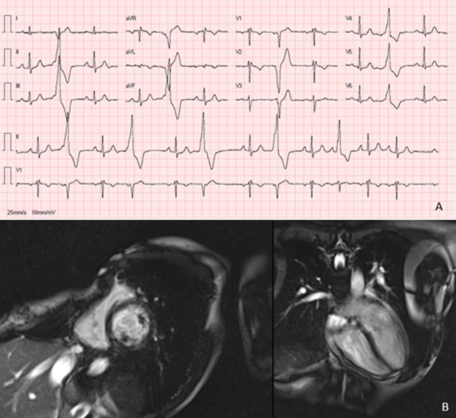
A 33-year-old woman, with palpitations since the age of 15, was referred to a cardiology consultation due to very frequent ventricular extrasystoles with morphology of left bundle branch block, inferior frontal axis, late precordial transition, rS in V1, R in V6 and rS in DI. She had pectus excavatum. The cardiac magnetic resonance showed severe pectus excavatum associated with exaggerated cardiac levoposition, compression and deformation of the right cardiac chambers. However, the patient became pregnant, and follow-up was delayed.
Introduction
Pectus excavatum is the most common congenital anomaly of the anterior chest wall, most often being benign, however, it can sometimes have structural and haemodynamic consequences secondary to mechanical compression.1 Most patients are asymptomatic, but may experience symptoms due to impaired cardiac function and, occasionally, cardiac arrhythmias.2
Case
A 33-year-old woman, without relevant personal and family history, with palpitations since the age of 15, was referred to a cardiology consultation due to very frequent ventricular extrasystoles (28,423/day, 26.6%). On objective examination, she had pectus excavatum, without other alterations. Blood tests showed no significant changes, namely in the electrolytes, blood count or thyroid function. Her resting 12-lead electrocardiogram (ECG) showed sinus rhythm and ventricular extrasystoles with morphology of left bundle branch block, inferior frontal axis, late precordial transition, rS in V1, R in V6 and rS in DI (figure 1A), suggesting an origin in the free wall of the right ventricle (anterior).3 Transthoracic echocardiogram showed no structural heart disease, there was no evidence of arrhythmogenic right ventricular cardiomyopathy. Coronary artery disease was excluded by coronary computed tomography angiography. She underwent cardiac magnetic resonance with perfusion study that showed severe pectus excavatum (Haller index 4.9), associated with exaggerated cardiac levoposition, compression and deformation of the right cardiac chambers (figure 1B), no delayed enhancement or ischaemia, and good biventricular function. However, the patient became pregnant, and the follow-up to the study and therapeutic guidance (namely catheter ablation and surgical correction for her pectus excavatum) were delayed.

Discussion
Pectus excavatum is considered to be a benign condition, rarely requiring treatment, commonly undertaken only for cosmetic and psychological reasons. However, it can be associated with arrhythmias, mainly supraventricular arrhythmias, and, more rarely, ventricular arrhythmias. We present a case of focal electrocardiomyopathy that resulted from sternal compression and gave rise to symptomatic ventricular extrasystole.
Less common causes of ventricular tachycardia, including compression cardiomyopathy, should be considered if there is unusual ECG-based morphology for right ventricular tachycardia, not consistent with right ventricular outflow tract origin.
Literature regarding cardiac arrhythmias, namely ventricular arrhythmias, caused by pectus excavatum is limited. Winkens et al. reported a case presenting with progressive heart palpitations, fatigue and postural dyspnoea, due to recurrent supraventricular tachycardia and a nodal tachycardia caused by pectus excavatum, in which the physical condition of the patient improved rapidly with surgical correction.3
Surgical correction should be considered as the initial treatment in cases of persistent right ventricular compression with structural and haemodynamic consequences. Catheter ablation may not be necessary.
Conclusion
This case highlights an uncommon cause of ventricular extrasystole, pectus excavatum, which despite being a common skeletal anomaly of the chest wall and usually having a benign course, may also be associated with symptomatic tachyarrhythmias.
Conflicts of interest
None declared.
Funding
None.
Patient consent
A written patient consent was obtained.
References
1. Ahn J, Choi J, Shim J, Lee S, Kim Y. Right ventricular compression mimicking Brugada-like electrocardiogram in a patient with recurrent pectus excavatum. Case Rep Cardiol 2017;2017:3047937. https://doi.org/10.1155/2017/3047937
2. Geiser G, Epstein S, Stampfer M, Goldstein R, Noland S, Levitsky S. Impairment of cardiac function in patients with pectus excavatum, with improvement after operative correction. N Engl J Med 1972;287:267–72. https://doi.org/10.1056/NEJM197208102870602
3. Winkens R, Guldemond F, Hoppener P, Kragten H, Leeuwen Y. Pectus excavatum, not always as harmless as it seems. BMJ Case Rep 2009;2009:bcr10.2009.2329. https://doi.org/10.1136/bcr.10.2009.2329
