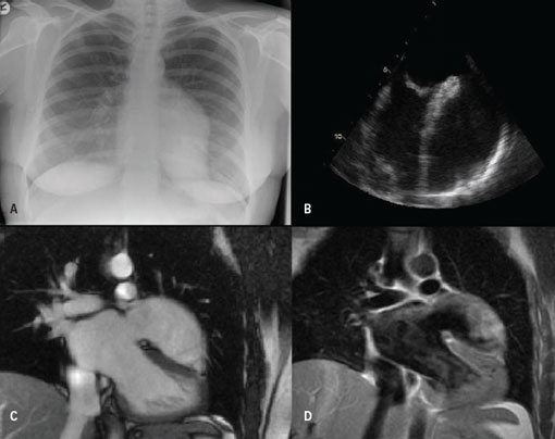
A previously fit and well 39-year-old Caucasian female patient was referred from the local district general hospital for further assessment and management of recurrent atrial tachyarrhythmias. A plain chest radiograph exhibited an abnormal left heart border (figure 1A), and initial transthoracic echocardiographic evaluation demonstrated a large intracardiac mass near the left ventricle.
To further characterise this mass, she then underwent imaging with transoesophageal echocardiography (figure 1B) and magnetic resonance imaging (MRI) of the thorax (figures 1C and 1D). These confirmed a large aneurysm arising from the left atrial appendage. An uncomplicated surgical resection of this aneurysm was performed. Intra-operative findings confirmed both a mass with a cystic structure and the dimensions, established with earlier imaging, of 60 x 50 x 10 mm. Subsequent histological studies confirmed the origin to be of cardiac muscle demonstrating nuclear enlargement consistent with hypertrophy.

At subsequent clinical follow-up the patient remained well with evidence of sinus rhythm on Holter monitoring. A transthoracic echocardiogram performed five months after surgery did not demonstrate any residual aneurysm.
Conflict of interest
None declared.
