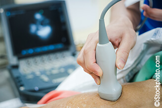
This study aimed to understand the national echocardiography assessment pathway in heart donation. We carried out a prospective national specialist nurses in organ donation (SNOD) audit of UK donor offers between 20 August and 31 November 2022, and a prospective national recipient transplant centre audit of all donor offers between 22 September and 19 December 2022.
The SNOD audit identified median time delay between requesting and performing an echocardiogram of 17.9 hours (interquartile range [IQR] 13.9–33.2). The staff group performing the echo were a cardiac physiologist in 57% (17/30) of cases and a medical doctor in 43% (13/30) of cases. Only 30% (9/30) of providers held comprehensive accreditation, 13% (4/30) were focused accredited, 33% (10/30) had no accreditation, and 23% (7/30) were unknown. Only 50% (15/30) of images were transferred for review to the transplant centre. Images were transferred via email (10/15, 67%), WhatsApp (4/15, 27%) and a standard picture archiving system (PACS) (1/15, 3%).
The transplant centre audit revealed that in 21% of donors, the transplant team felt that the echo performed at the referring centre contained inadequate information, and in 11% of donors, no echo was performed at all. Only 52% of potential donors had echo images available for direct review by the transplant centre. In 17% of cases, the transplant team felt that if good quality echo data and imaging had been available, the decision regarding mobilising the retrieval team may have been altered.
In conclusion, to improve donor heart utilisation rates we believe there is a need to recognise the contribution of focused echo and improve guidance for echo image acquisition. There is also a need for a robust system for image transfer to transplant centres.
Introduction

In the financial year 2022–23 there were 185 heart transplants performed in the National Health Service (NHS) of the UK.1 These were performed across six adult centres: Queen Elizabeth Birmingham, Golden Jubilee Glasgow, Harefield London, Wythenshawe Manchester, Royal Papworth Cambridge, Freeman Newcastle upon Tyne; and two paediatric centres: Great Ormond Street London and Freeman Newcastle. Each hospital has an allocation zone, which encompasses 336 referring hospitals across the UK.2
The heart donations are classified as either donation after brainstem death (DBD) or donation after circulatory death (DCD). The donation process in the UK starts with clinicians identifying patients who are either brainstem dead or progressing towards circulatory death. Based on the patient’s beliefs and wishes, specialist nurses in organ donation (SNODs) are contacted to assess if organ donation may be suitable and to obtain consent from families, in collaboration with the medical team. Once consent is obtained, crucial patient information, such as clinical history, clinical status and investigations including blood tests, electrocardiograms and echocardiograms, are relayed via NHS Blood and Transplant (NHSBT) to the transplant centres.
Based on this information, retrieval teams are mobilised to assess and retrieve hearts if they are considered suitable for transplantation. Upon retrieval, the heart can be placed in a hypothermic preservation system or a warm perfusion beating heart organ care system. Ensuring minimal cold ischaemic time, typically less than four hours, the heart is rapidly transported to a transplant centre for transplantation.
Worldwide, the countries with the highest heart transplantation rates, Slovenia and US, performed over 12 transplants per million population in 2022.3 The UK ranked 28th, performing three heart transplants per million population. This is despite 311 patients on the transplant waiting list as of April 2023, and the recent introduction of the DCD heart programme, which led to an additional 52 heart transplants in the 2022–2023 financial year.1
The authors propose a number of potential reasons for this low ranking:
- High bed occupancy rates within the NHS, leading to a lower prioritisation of donor optimisation in referring hospitals due to competing interests for intensive care beds. Notably, almost all solid organ donation in the UK occurs from ventilated patients in the intensive care unit.
- Low public and staff awareness regarding potential organ donation, particularly concerning the increasing utilisation of heart DCD.
- Ethical constraints on conducting additional investigations, such as coronary arterial assessment through invasive or non-invasive angiography and transoesophageal echocardiography.
- Inadequate provision of timely transthoracic echo in potential donors.
As a broad, multi-specialty and multi-centre working group, we were tasked by NHSBT to explore the potential role of echo to improve donor utilisation. This work builds on our prior research,4 which examined the current landscape of echo provision in intensive care units across the UK.
Method
We started by establishing a widely representative group of key stakeholders in this field of medicine from across the UK. This included representatives from cardiology, intensive care, paediatrics, cardiothoracic surgery and specialists in focused echo, all operating under the umbrella of NHSBT.
Through a modified Delphi approach, we developed two national surveys to better understand the national echo assessment pathway in donation, the quality of imaging, and the availability of image transfer from both the donation and transplant centre perspective.
We carried out a prospective national SNOD audit of all UK donor offers between 20 August and 31 November 2022. We collected data on Microsoft Forms© distributed through the NHSBT regional SNOD team network (appendix 1).
Appendix 1. UK SNOD donor echo audit
Survey link: https://forms.office.com/r/Bq0FKnewNg
|
1. What is the donor number? Required to answer. Single-line text. 2. What was the date of consent for donation? Required to answer. Date. 3. What was the time of consent for donation? Required to answer. Single-line text. 4. Was the donor suitable for heart transplant assessment? (<65 with no cardiac history). Required to answer. Single choice. Yes No 5. What was the time of echo request? Required to answer. Single-line text. 6. What was the date of echo request? Required to answer. Date. 7. Was an echo performed? Required to answer. Single choice. Yes No 8. What time was the echo performed? Required to answer. Single-line text. 9. What date was the echo performed? Required to answer. Date. 10. Who performed the echo? Required to answer. Single choice. Sonographer Cardiologist Intensivist Other 11. Do you know if they were echo accredited? Required to answer. Single choice. Level 2 – Full BSE or equivalent Level 1 – FUSIC/FICE/Level 1 BSE No Other 12. Were images sent for review by the transplant team or specialist? Required to answer. Single choice. Yes No Other 13. What method was used to transfer pictures? Required to answer. Single-line text. 14. Was the heart retrieved? Required to answer. Single choice. Yes No Retrieval team assessed but heart not removed Other |
Additionally, we carried out a prospective national recipient transplant centre audit of all donor offers between 22 September and 19 December 2022. We collected data on Microsoft Forms© distributed through the NHSBT recipient coordinator network (appendix 2). Statistical analysis was completed using Microsoft Excel©.
Appendix 2. UK surgical donor echo audit
Survey link: https://forms.office.com/r/hKKcaFAwEJ
|
1. What is the donor number? Required to answer. Single-line text. 2. Which Transplant Centre are you reporting from? Required to answer. Single choice. Freeman Hospital, Newcastle Golden Jubilee National Hospital, Glasgow Harefield Hospital, London Papworth Hospital, Cambridge Queen Elizabeth Hospital, Birmingham Wythenshawe Hospital, Manchester 3. Was the donor heart accepted? Required to answer. Single choice. Yes No 4. Were there any echo images available for review? Required to answer. Single choice. Yes No 5. What was the quality of echo or echo report available for decision making? Required to answer. Single choice. Good Inadequate No echo available 6. In what way was the echo inadequate? Required to answer. Multi-line text. 7. If a good quality echo had been present would this have potentially changed the decision of accepting the heart? Required to answer. Single choice. Yes No 8. If the patient underwent a repeat echo or a retrieval echo were the findings different and did it change outcome? Required to answer. Single-line text. |
Results
Table 1. UK specialist nurses in organ donation (SNOD) audit for echocardiography availability
| Responses | N=30 |
| In-hours echo, n (%) | 24 (80) |
| Median time delay (IQR), hours | 17.9 (13.9–33.2) |
| Accreditation, n (%) | |
| Full | 9 (30) |
| Focused | 4 (13) |
| Unknown | 7 (23) |
| None | 10 (30) |
| Image transfer, n (%) | |
| 10 (33) | |
| 4 (13) | |
| PACS | 1 (3) |
| Not done | 15 (50) |
| Key: IQR = interquartile range; PACS = picture archiving communication system | |
From the national SNOD audit, a total of 30 potential donors were assessed during the study period (table 1). Analysis revealed that the majority of consents for donation (19/30, 60%), echo requests (19/30, 63%), and echo studies (24/30, 80%) were obtained during daytime hours (0800–1800). The median time between the request and the performance of an echo was 17.9 hours (interquartile range [IQR] 13.9–33.2).
Cardiac physiologists performed the scan in 57% (17/30) of cases, while medical doctors conducted the remaining 43% (13/30) of scans. Comprehensive echo accreditation (e.g. British Society of Echocardiography Level 2) was held by 30% (9/30) of providers, 13% (4/30) had focused echo accreditation (e.g. FUSIC Heart©), 33% (10/30) had no accreditation, and the accreditation status of 23% (7/30) was unknown.
Half (50%, 15/30) of images were transferred for review by the transplant centre, with 13% (4/30) remaining unknown or determined as unnecessary. Of the cases where images were transferred, images were transferred via email (10/15, 67%), WhatsApp (4/15, 27%) and a standard picture archiving system (PACS) (1/15, 3%).
From the national recipient transplant centre audit, a total of 96 donor offers were assessed: 7% from Golden Jubilee National Hospital, 29% from Harefield Hospital, 10% from Papworth Hospital, 47% from Queen Elizabeth Hospital and 6% from Wythenshawe Hospital. In total, 29% of the hearts were accepted for transplantation.
In 21% of potential donors, the transplant team deemed that the echo performed at the referring centre did not contain adequate information to make a transplant decision, while in a further 11% of potential donors, no echo was performed at all. Of potential donors, 52% had echo images available for direct review by the transplant centre.
In echo studies where incomplete echo data were available to the transplant team, reasons for this included lack of images, poor quality images available for review (e.g. only static images), lack of measurements and lack of reporting information, such as left ventricular wall thickness. In 17% of cases, the transplant team felt that, if good quality echo data and imaging had been available, the decision regarding mobilising the retrieval team may have been altered.
Discussion
This prospective data collection has brought to light three key issues in donor echo that require attention in the UK. First, there is significant reliance on staff who lack comprehensive echocardiography accreditation. As identified previously in our analysis,4 medical personnel with focused echo accreditation provide the primary cover for out-of-hours services in the UK. The limited coverage and availability of this workforce contribute to prolonged delays in acquiring imaging, which can impact on the donation process, prolonging the ordeal for families and reducing hospital capacity.
Second, the data have highlighted the inadequacy of echo imaging results provided to the transplant centres. In 17% of cases, transplant teams reported that this deficiency may have influenced the subsequent decision making. In a review of all national organ retrieval team dispatches for hearts in the UK between April 2019 and October 2021, of 1,158 offers, 398 were accepted for retrieval by the National Organ Retrieval Service (NORS) team. Upon local assessment by the NORS team, 47% of these hearts were accepted for transplantation, 29% were declined on repeat echo assessment and 24% were declined for other reasons (unpublished data). The heart may have declined in function since initial echocardiography, explained by neurohormonal changes,5 however, it could also stem from incorrect initial assessments by inexperienced scanners. Inadequate echo data could lead to missed opportunities for heart donation or inappropriate and costly dispatch of retrieval teams. ‘Good quality’ echocardiography images was a subjective assessment, however, the working group consensus for a minimum dataset was 2D and colour imaging in parasternal, apical and subcostal views with measurements of left ventricular wall thickness and end diastolic diameter.
Third, both audits revealed only half of potential donors had imaging transferred for review, and in most cases, this was not done through the standard interhospital imaging exchange portal and PACS system, but through mobile messaging apps and emails. This limits the information available for review by transplant centres. Given that a significant proportion of scans are performed by non-comprehensively accredited staff, this further raises the risk of incomplete or inaccurate echo information being available for assessment.
These surveys had multiple limitations, including incomplete data capture over the period, as evidenced by low rates of form completion. Additionally, data collection was also vulnerable to recollection bias and poor documentation, such as that concerning staff accreditation levels. These limitations need to be acknowledged when assessing the results and implications for practice change.
Historically, attempts have been made to address these issues, including a ‘scout programme’.6 This involved sending practitioners to perform detailed echo assessment ahead of the retrieval team. However, ongoing costs have limited its implementation beyond the pilot project.
We have demonstrated that a significant proportion of echos are performed by staff with focused echo accreditation. To improve donor heart utilisation rates, we believe there is a pressing need to improve guidance for echocardiography image acquisition in donor hearts. Furthermore, there needs to be a robust system in place for image transfer and review by transplant centres. This involves, not only providing high-quality imaging software for reviewing images, but also ensuring compliance with information governance standards when sharing patient-sensitive data.
Key messages
- Echocardiography delays: there is a median delay of 17.9 hours for echocardiograms, with many (13%) performed by staff holding focused accreditation
- Poor image transfer: only 50% of images were sent to transplant centres for review, often using non-standard methods like email or WhatsApp
- Inadequate data: 21% of echocardiograms lacked sufficient information, impacting decision-making in potential heart donations
- Positive collaboration: despite challenges, the study demonstrates strong collaboration between multiple UK hospitals and transplant centres to improve heart donation outcomes
Conflicts of interest
None declared.
Funding
None.
Study approval
This was an observational audit of current clinical practice within the NHS, so ethical approval was not needed, as judged by the NHSBT working group.
Acknowledgement
We would like to thank all the medical, nursing, and allied health team who helped collect these data and contributed to the development of the project at the NHS and NHSBT.
References
1. NHS Blood and Transplant. Annual report on heart transplantation 2022/23. Bristol: NHSBT, 2023. Available from: https://nhsbtdbe.blob.core.windows.net/umbraco-assets-corp/30881/nhsbt-heart-transplantation-report-2223.pdf [accessed 9 January 2024].
2. NHS Blood and Transplant. DAT2982/5 Heart Allocation Zones by Hospital. Bristol: NHSBT, 2024. Available from: https://www.odt.nhs.uk/transplantation/tools-policies-and-guidance/allocation-zones/
3. Global Observatory on Donation and Transplantation. GODT database. Available at: https://www.transplant-observatory.org/export-database/ [accessed 9 January 2024].
4. Akhtar W, Marshal L, Buglass H et al. A survey of United Kingdom intensive care echocardiography provision. J Intensive Care Soc 2024;online first. https://doi.org/10.1177/17511437241264978
5. Apostolakis E, Parissis H, Dougenis D. Brain death and donor heart dysfunction: implications in cardiac transplantation. J Card Surg 2010;25:98–106. https://doi.org/10.1111/j.1540-8191.2008.00790.x
6. NHS Blood and Transplant. Taking organ utilisation to 2020. Bristol: NHSBT. Available from: https://bts.org.uk/wp-content/uploads/2018/01/odt-organ-utilisation-strategy.pdf [accessed 9 January 2024].
