A 55-year-old smoker with no significant past medical history was admitted following an episode of dyspnoea and intrascapular pain. Clinical examination was normal. His blood pressure (BP) was 80/40 mmHg and his electrocardiogram (ECG) showed a sinus tachycardia and right bundle branch block.

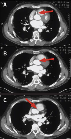
A computed tomography (CT) scan of the chest excluded a pulmonary embolism and aortic dissection, and, although not a dedicated cardiac CT, suggested an occlusion of the left main coronary artery (LMCA) (figure 1). Echocardiography showed impaired left ventricular function with an akinetic anterior, inferior and lateral wall. An intra-aortic balloon pump (IABP) was inserted and coronary angiography was performed, which confirmed an occlusion of the LMCA (figure 2, panel B). This was pre-dilated, and then stented with a bare-metal stent (3.5 x 16 mm Liberté) producing an excellent final angiographic result (figure 2, panel D). Despite continued IABP support, he required ventilation for refractory pulmonary oedema, and died five days later.
This case highlights the benefit, and shows the potential of CT imaging in helping establish a rapid diagnosis in patients presenting with non-specific chest pain and non-specific ECG changes where it is not possible to localise pathology to lungs, heart or aorta. In this case, the coronary pathology was picked up by chance, but dedicated multi-detector CT scanners are available that can perform a single examination in less than 10 minutes (‘the triple scan’), which will exclude lung, aortic and coronary pathology.1 These scanners will ultimately play a comprehensive role in the diagnosis and subsequent management of these challenging patients.

Conflict of interest
None declared.
Reference
- Gershlick AH, de Belder M, Chambers J et al. Role of non- invasive imaging in the management of coronary artery disease: an assessment of likely change over the next 10 years. A report from the British Cardiovascular Society Working Group. Heart 2007;93:423–31.
