In brief

Br J Cardiol 2013;20:136-7 Leave a comment
Click any image to enlarge
Authors:

News in brief from the world of cardiology

Caffeine intake may reduce risk of type 2 diabetes

Coffee and caffeine intake may significantly reduce the incidence of type 2 diabetes, according to a new meta-analysis published in the European Journal of Clinical Nutrition. 

Pertinent studies were identified by a search of PubMed and EMBASE. The fixed- or random-effect pooled measure was selected based on between-study heterogeneity. Dose–response relationship was assessed.

Commenting on the implications of this study (doi: 10.1007/s00394-013-0603-x), London general practitioner Dr Sarah Jarvis said: “There is growing evidence to suggest that moderate coffee consumption, that’s four to five cups per day, may have a protective effect against the development of type 2 diabetes. This new analysis adds to the weight of research in the area and is very relevant to us here in the UK given the high prevalence of type 2 diabetes.”

E-cigarettes comparable to nicotine patches in helping smokers quit

The first clinical trial to compare e-cigarettes with an established smoking cessation aid, nicotine patches, has found that both methods result in comparable quitting success. Roughly similar proportions of smokers who used either method remained abstinent from smoking for six months after a 13-week course of patches or e-cigarettes, the authors report.

The study, published in The Lancet (doi: 10.1016/S0140-6736(13)61842-5), divided 657 smokers who wanted to quit into three groups. 292 received 13 weeks’ supply of commercially available e-cigarettes, each of which contained around 16 mg nicotine. The same number received 13 weeks’ supply of nicotine patches, and 73 received placebo e-cigarettes, which contained no nicotine.

At the end of the six-month study period, around one in 20 study participants (overall, 5.7%) had managed to remain completely abstinent from smoking. While the proportion of participants who successfully quit was highest in the e-cigarettes group (7.3%, compared to 5.8% for those in the nicotine patches group, and 4.1% in the placebo e-cigarettes group), these differences were not statistically significant.  

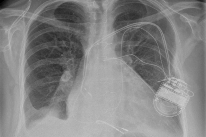
3D holographic imaging arrives in cardiology

Screen shot 2013-12-04 at 15.38.14Live 3D holographic visualisation and interaction technology can be used to guide minimally-invasive structural heart disease procedures, according to a recently completed clinical study.

The pilot study involved eight patients and was conducted in collaboration with the Schneider Children’s Medical Center in Petach Tikva, Israel. Visualisation technology (RealView Imaging Ltd.) was used to display interactive, real-time 3D holographic images, acquired by interventional X-ray and cardiac ultrasound systems (Philips).

In addition to viewing the patient’s heart on a 2D screen, doctors in the interventional team were able to view detailed 3D holographic images of the heart appearing in free space, without using special eyewear. The doctors were also able to manipulate the projected 3D heart structures by touching the holographic volumes in front of them.

The results of this pilot study were presented at the 25th annual Transcatheter Cardiovascular Therapeutics scientific symposium held recently in San Francisco, sponsored by the Cardiovascular Research Foundation. For more information, please visit http://www.tctconference.com/agenda/agenda.html 

Womens’ greater shortness of breath when exercising explained

Women have greater shortness of breath than men when exercising due to greater electrical activation of their respiratory muscles, shows a new study from Experimental Physiology (doi: 10.1113/expphysiol.2013.074880).

50 healthy, non-smoking men and women aged 20–40 years completed a maximum exercise test on a stationary bicycle. During exercise, researchers monitored their cardiovascular, metabolic and ventilatory responses.

At regular intervals during exercise, participants rated the intensity of their breathlessness using a 10-point scale. Using a multipair electrode catheter placed in the participants’ oesophagus, authors also recorded the electromyogram of the diaphragm throughout exercise. These measurements were then analysed and compared between men and women.

Lead author Dr Dennis Jensen (McGill University, Canada) said: “Our study uniquely showed that sex differences in activity-related breathlessness could be explained by the awareness of greater electrical activation of the respiratory muscles – specifically the diaphragm – needed to achieve any given ventilation during exercise in healthy young women compared to men.

No patient cholesterol checks after ezetimibe discontinued

An analysis of a primary care database has shown that in a sample group of over 7,000 patients who were discontinued from ezetimibe, approximately one third did not receive a cholesterol measurement in the six months prior to the treatment being stopped, and of patients who did have a measurement, 30% were removed in spite of not having achieved the minimum QOF target for total cholesterol.

The study published in Curr Med Res Opin (doi: 10.1185/03007995.2013.842164) also showed that in the most high-risk patient population only 26% achieved their NICE recommended target cholesterol measurement, i.e. total cholesterol measurement of 4 mmol/l and an low-density lipoprotein (LDL) cholesterol measurement of 2 mmol/l, compared to 39% of patients who were reaching target before ezetimibe was discontinued.

General practitioner Professor Michael Kirby (University of Bedfordshire) said of the results: “It is concerning to note that patients were being removed from a treatment in the absence of cholesterol levels being known. What is more concerning, however, is the safety implications of not retesting once a medication has been stopped or changed. Yes, cost constraints are ever present; however, leaving risk factors suboptimally controlled just for short-term financial wins is of concern.”

Psychological interventions halve cardiovascular deaths

Psychological interventions halve deaths and cardiovascular events in heart disease patients, according to research presented at the Acute Cardiac Care Congress 2013 held recently in Madrid, Spain.

Researchers from Athens, Greece, conducted a meta-analysis of nine randomised controlled trials. They evaluated whether psychological interventions could improve outcomes of patients with coronary heart disease when combined with a conventional rehabilitation programme.

The researchers found that the addition of psychological interventions reduced mortality and cardiovascular events by 55% after two years or more (relative risk [RR]=0.45, 95% confidence interval [CI]=0.37–0.54, p<0.001). The benefits were not significant during the first two years (RR=0.77, 95% CI=0.55–1.09, p=0.145).

New approach to phosphate control in CKD

Colestilan (BindRen®) is now available for the treatment of hyperphosphataemia in adult patients with chronic kidney disease (CKD) Stage V, receiving haemodialysis or peritoneal dialysis in the UK, according to Mitsubishi Pharma Europe Ltd.

Hyperphosphataemia is characteristic of end-stage renal disease (ESRD) and occurs when the kidneys do not properly filter phosphate. Control of serum phosphate presents a significant challenge in the management of CKD with more than half of ESRD patients not adhering to phosphate binding medication.

Dr Alastair Hutchison (Manchester Institute of Nephrology & Transplantation) said: “Adherence to phosphate binding medication remains a major issue. The unique granule formulation of BindRen® represents a new approach to phosphate management – since in my view the most effective treatment is to prescribe a phosphate binder that patients will actually take.”

Please also see our report on the recent Cardiorenal Forum meeting (pages 133–5).

Post-MI oxygen not supported by evidence

Oxygen therapy following myocardial infarction (MI) may do more harm than good, according to a Cochrane Review (doi: 10.1002/14651858.CD007160.pub3) conducted by academics from City University London, the University of Birmingham and the University of Surrey, together with colleagues in Spain. 

Authors analysed high-quality randomised controlled trials that compared oxygen and air, their findings highlighting a paucity of research behind this intervention:

Only four trials of oxygen were available, which had enrolled a total of 430 participants

There were 17 deaths in total, and more than twice as many people given oxygen died compared to those given air in these trials

This result is not statistically significant but shows a clear need for more research into the use of oxygen therapy for the treatment of acute MI.

Co-author Professor Amanda Burls (City University) said: “There is an urgent need for an adequately powered randomised controlled trial to establish the effectiveness of, or harm from, the administration of oxygen to people with an acute MI.”

Lower mortality with TAVI for aortic valve surgery patients with diabetes

Patients with diabetes at high risk for aortic valve surgery had a 35% lower all-cause mortality one year after treatment with transcatheter aortic valve implantation (TAVI), than those treated with surgical aortic valve replacement (AVR), according to a new post-hoc data analysis from the PARTNER (Placement of Aortic Transcatheter Valves) trial.

In the analysis of patients with diabetes in the as-treated high-risk cohort of the PARTNER trial, one-year all-cause mortality was 18% for patients treated with TAVI, and 27.4% for patients treated with surgical AVR. Additionally, the analysis showed that diabetic patients treated with TAVI experienced quicker quality of life improvement and lower one-year rates of renal failure than those treated with surgery.

The data were presented at the ESC Congress 2013 held recently in Amsterdam, by PARTNER investigator Dr Brian Lindman (Washington University School of Medicine, St. Louis, USA). “While this was not a pre-specified analysis of the trial and should be considered hypothesis generating, our study raises the possibility that TAVI may be the preferred approach for diabetic patients with severe symptomatic aortic stenosis who are at high surgical risk,” he said.

Correspondence: national survey of patients with AF in the acute medical unit: a day in the life survey

Br J Cardiol 2013;20:160 Leave a comment
Click any image to enlarge
Authors:

Correspondence from the world of cardiology

National survey of patients with AF in the acute medical unit: a day in the life survey

Dear Sirs,

The first national survey examining the management of atrial fibrillation (AF) within acute medical units up and down the country has just been published in the British Journal of Cardiology.1 Essentially it seems to show that secondary care is just as bad as primary care in initiating warfarin for AF patients. This group of patients is five times more likely to have a thromboembolic cerebrovascular accident than matched populations in sinus rhythm and yet doctors are ineffective at influencing change. This study took place over a 24-hour period and involved 31 hospitals and 149 patients.

The results showed that 80% of patients with a significant risk of stroke (CHA2DS2-VASc score of over 2) did not receive anticoagulation. Stroke risk scores were documented in less than a quarter of patients and the same fraction received digoxin as monotherapy. The rationale for this treatment decision was only documented in a quarter of cases and was not consistent with national standards. Although the study was small, it does strongly suggest that both general practitioners (GPs) and hospital doctors are struggling to anticoagulate AF patients and are also selecting the wrong drugs for AF rate management.

I have spent several years trying to understand why we as GPs are performing below par in anticoagulation2 and now it seems that hospital doctors are just the same. Of course it takes time to explain and organise warfarinisation and patients are naturally apprehensive. When we do have the discussion, it is easy to persuade patients to take aspirin – it reduces stroke risk by 20%. In contrast, it’s a different issue to persuade them to take warfarin, which reduces risk by nearly 70%. I believe we as GPs encourage the British compromise of aspirin because we are unduly and disproportionately influenced by the risk of bleeds.

It is a paradox that the BAFTA (Birmingham Atrial Fibrillation Treatment of the Aged) study showed that in the elderly, aspirin was actually more dangerous than warfarin in causing both intracerebral and extracerebral bleeds! Nevertheless we tend to feel if we prescribe warfarin then we take on enduring risk both of bleeding and prescribing/compliance errors, and so we push the discussion away from the real data. Patients and GPs are therefore complicit in not preventing 15,000 AF strokes a year – and now hospital doctors are embroiled as well!The risk of stroke needs to be balanced with the risk of major haemorrhage. Despite this, the study showed only 36% (n=54) of patients with a CHA2DS2-VASc score >2 had a HAS-BLED score of 3 (8.7 major bleeds per 100 patient-years). In addition, 24% (n=15) of patients at risk of major haemorrhage (HAS-BLED score of 3) were discharged on anticoagulation therapy. Four of this group were concomitantly discharged on antiplatelet therapy.

Decades have gone by with new tools and education to try to influence the prescription of more anticoagulants in AF, but performance has not improved significantly. Surely the time has now come for a more radical and aggressive way forward for stroke prevention in AF. The mantra needs to be that every patient with AF should be on warfarin unless a consultant cardiologist or stroke physician feels this is clinically inappropriate. If the patient declines – as is their informed right – then a letter needs to be in the electronic notes signed by the patient absolving the medical attendants of clinical responsibility and explaining the risks. 

This letter will confirm informed decision making and make both patients and their physicians think more about the real risks – and could have some significant impact on stroke prevention. This is a large clinical performance gap that needs to be closed, and patently it will not happen without a more assertive approach.

Conflict of interest

None declared.

Dr John Havard
General Practitioner

Saxmundham Health, Lambsale Meadow, Saxmundham, IP17 1AS
([email protected])

References

1. Soong J, Balasanthiran A, MacLeod DC, Bell D. National survey of patients with AF in the acute medical unit: a day in the life survey. Br J Cardiol 2013;20:106. http://dx.doi.org/10.5837/bjc.2013.021

2. Havard J. Why are we so bad in primary care at initiating warfarin in atrial fibrillation patients? Br J Cardiol 2009;16:237–40. 

The authors’ reply

We are grateful to Dr Havard for his detailed response to our article, highlighting in particular the recurring issue of anticoagulation in AF. 

It should be appreciated that this study focused on acute management strategies. Outwith the context of DC cardioversion during the index episode, the initiation of anticoagulation for the majority of AF patients is a deferred process involving the patient and their GP, supported by secondary care, commonly in relation to echocardiographic assessment.

However, Dr Havard appropriately draws attention to the low rate (<25%) of risk scoring in our study, and it might reasonably be proposed that all unanticoagulated AF patients discharged from acute medical units should have both CHA2DS2-VASc and HASBLED scores documented to guide decision making.

Barriers to good practice need to be studied. Interventions must be locally relevant and embedded within existing systems to be sustainable. Quality improvement methodology may provide the necessary framework for “real world” investigation and implementation of best practice.

Dr John Soong
RCP Clinical Quality Improvement Research Fellow

Chelsea & Westminster Hospital NHS Foundation Trust, 369 Fulham Road, London, SW10 9NH

Me doctor, you patient

Br J Cardiol 2013;20:138-9 Leave a comment
Click any image to enlarge
Authors:

We continue our series in which Consultant Interventionist Dr Michael Norell takes a sideways look at life in the cath lab…and beyond. In this column, the perils of being a patient, as a doctor, are discussed.

With regard to my title, I think that we sometimes struggle to get this right.

As the years advance, it is almost a certainty that we will acquire some form of health problem along the way – and definite that there will be a terminal issue eventually. That said, our ability to deal with whatever pathology emerges might be seriously hampered by the small matter of being in possession of a medical degree.

In a previous column (BJC passim), I alluded to the sad truth that female spouses systematically underestimate any disease process affecting their male partner. The example I used to illustrate this phenomenon was the notoriously unpleasant Influenza Hominis Gravis, or IHD – universally passed off as ‘man flu’ and roundly ignored, particularly by those who are meant to care for us the most.

If then, added to the already disabling symptoms of a condition, you have some medical knowledge, the traumatising impact of any illness is compounded. This was also demonstrated in another piece (BJC passim – yet again) when I had contracted ‘pine mouth’ – a persistent bitter taste resulting from eating a specific variety of nut. Thankfully, I chanced eventually upon the correct diagnosis courtesy of Google, but not before I had assumed (naturally enough) that I had acquired an illness that – although unspecified – was undoubtedly going to polish me off within the next three months.

Heal thyself

My contention is that when illness strikes and we are obliged to assume the role of patient, doctors are at a major disadvantage compared with their lay counterparts.

To start with, we are decidedly reluctant even to accept the designation of patient in the first place. Our knowledge and training allows – if not mandates – us to assess whatever symptom complex has befallen us and make what we think is a dispassionate judgement as to the pathology, its significance, the potential seriousness and treatment (if any). As long as the responses to this personalised diagnostic run do not appear to cross an imaginary line of acceptability that we ourselves have somehow constructed, we feel content to manage the situation ‘in house’ – as it were.

The much quoted aphorism “Physician, heal thyself”, presupposes that the individual in question has insight into the fact that he has succumbed to some condition or other, but refuses to deal with it. My contention is that he cannot have insight in terms of true objectivity and, rather than reluctance to address issues, his training results in him being unable to.

Subjective objectivity

In order to help our patients, we need to be empathetic, understanding and sensitive, but, in addition, we also need to be objective and make our clinical judgements without being clouded by emotion. When we ourselves become the recipients of a disease process, then, as per our training, we try in vain to rationalise our own predicament. The result can vary from mere inconvenience and distraction to downright misery if not, occasionally, disaster.

We tend to either hyper-interpret or over-analyse our own symptoms, ending up with a diagnosis as rare as hens’ teeth, or, alternatively, play down and even ignore them by explaining them away on the basis of relatively innocent pathology. Remember, a runny nose is most likely to represent a common cold rather than cerebrospinal fluid rhinorrhoea due to an expanding frontal lobe tumour, and oppressive, central chest discomfort on exertion does not lose its significance because you just happen to have MBBS after your name.

A hopeless case

Essentially, it is hopeless, and we cannot possibly win. Even if we eventually present ourselves to independent medical opinion, we still lose out. Why? Because the essence of the doctor-patient relationship is that ‘the patient gives their illness to the doctor’ to sort out, in the same way that we might involve a solicitor in a house purchase. The mental burden of disease is, thus, transferred to someone else qualified to deal with it, and the patient will, thereby, experience some sense of relief.

Not with us. Even though we may eventually submit ourselves to independent medical opinion and see a colleague (usually), I suspect that we still do not share a sensation of that weight of anguish being lifted from our shoulders. We continue to lie awake at night and worry about it, only now it’s worse; we have an even better idea of what it is. We read about that at Medical School, and it wasn’t very pleasant!

The analogy is illustrated by the business man whose wife asks him why he is still awake at 3 a.m. “I owe Mr Cohen (a next door neighbour) 10,000 pounds and I haven’t got it”, he answers. The exasperated wife goes to the open window and yells out, “Hey, Mr Cohen! That 10 grand my husband owes you? He hasn’t got it so you aren’t getting it!” The husband is astounded. “What on earth are you doing?” to which his wife calmly replies; “Now it’s his problem”.

Is it about trying to distance ourselves from those for whom we are caring? We see our profession in the context of ‘us and them’; in order to act as doctor, we must be seen as separated from the patient and somehow protected or shielded against the problems that have befallen them. How else can we possibly be in a position to assist?

This is, of course, a falsehood; once you yourself have experienced grief, pain or disease you become a far better physician. I speak from personal knowledge (as usual), having experienced symptoms related to ventricular ectopic beats some years ago and which disappeared in the same way they had begun a few months before – without any explanation. Nevertheless, the whole experience made me assess outpatients with palpitation in a very different light.

As well as with other physical conditions (cardiac or not), the same phenomenon occurs with emotional events, such as parenthood or the loss of a loved one. You become far more in tune with your patients and that is not such a bad thing.

Special treatment

There is another downside to being a doctor/ patient, which is well recognised and potentially far more serious. It can be summed up in the oft quoted words of Professor Harold Ellis, a renowned teacher of surgery at the Westminster Medical School (as it then was): “Mistakes happen when special patients get special treatment”.

Whether you like it or not, a doctor will not be managed along the same care pathway that directs the care of other, mere mortal, patients. To avoid any inconvenience (naturally), they will come in on the morning of the procedure rather than the night before; they will go home the same afternoon rather than the next day, because (of course) they will be far better able to look after themselves at home (“their wife is a nurse as well, so that will be fine”); they will either have investigations skipped because they are deemed unnecessary (“I doubt we should need to check that in your case”) or, conversely, be over investigated because they themselves have more knowledge or concerns about their illness (“we’ll do that just to be sure”).

They are never straightforward, because they either presented way too early in the course of their disease because of undue anxiety, or way too late because they rationalised it as something else. Whatever management plan, procedure, diagnostic test, operation or treatment is proposed, any quoted risk should be doubled.

Here’s my advice. If you become unwell and need hospital admission, turn up at an Accident and Emergency (A&E) department in Fife or Aberystwyth and book in under an assumed name; McKay or Jones are some helpful examples. Claim to be currently unemployed and deny, not only any medical background or knowledge, but also the existence of any living medical relatives. You will get excellent treatment and do absolutely fine.

Footnote: For any readers currently working in the A&E departments in either Fife or Aberystwyth, I look nothing like my photograph

Renal denervation for hypertension: where are we now?

Br J Cardiol 2013;20:142–7doi:10.5837/bjc.2013.33 Leave a comment
Click any image to enlarge
Authors:

Hypertension is a growing clinical burden associated with significant morbidity and mortality. Those patients who remain with uncontrolled blood pressure despite multiple appropriate tablets are labelled as resistant hypertension. This cohort faces the highest risk. A key driving factor in resistant hypertension is an abnormally elevated sympathetic nervous system (SNS). It is now possible to attenuate this non-pharmacologically by performing radiofrequency ablation to the renal sympathetic nerves using a transcatheter approach. Currently available trial data show impressive blood pressure reductions with this therapy and, more importantly, its relative safety. The National Health Service (NHS) experience with this procedure is at an early stage, but is likely to grow with guidance already published by the joint British Societies and National Institute for Health and Care Excellence (NICE).

Continue reading Renal denervation for hypertension: where are we now?

Renal sympathetic denervation: cautious optimism and careful next steps

Br J Cardiol 2013;20:128–29doi:10.5837/bjc.2013.32 Leave a comment
Click any image to enlarge
Authors:

Despite the high and growing prevalence of hypertension worldwide, and the increasing attention focused on the challenge of resistant hypertension (RHTN), it is somewhat extraordinary to note the lack of data attesting to the epidemiology and management of RHTN at the present time. Few studies have described the incidence and prevalence of this condition, yet, it is very clear that, once diagnosed with RHTN, patients are at strikingly elevated risk of cardiovascular events, and thus clearly defined treatment strategies are urgently required.1 Quite remarkably, when reviewing the evidence base in RHTN for the recent National Institute for Health and Care Excellence (NICE) hypertension guidelines, the authors could find just one head-to-head randomised-controlled trial in this patient cohort, and only six retrospective cohort studies, with the largest being a post hoc analysis of the Anglo-Scandinavian Cardiac Outcomes Trial (ASCOT) study, where the use of spironolactone as a fourth-line agent was associated with blood pressure (BP) reductions of ~20/10 mmHg.2 Currently, we are hopeful that trials, such as the British Hypertension Society (BHS)-led Pathway 2 study (UKCRN.org.uk ID 4500) and the Resistant Hypertension Optimal Treatment (ReHOT) study (Clinicaltrials.gov ID NCT01643434), will help improve our drug therapy of RHTN. Nonetheless, it should be recognised that, while pharmacotherapy of hypertension is proven (at least up until the point of RHTN), issues with physician inertia, poor concordance and drug intolerance continue to undermine our efforts to get patients to target BP. 

Screen shot 2013-12-04 at 16.16.22
Dr Melvin D Lobo (William Harvey Heart Centre, Barts and the London School of Medicine and Dentistry, Queen Mary University of London, Charterhouse Square, London)

The past three years has serendipitously seen the emergence of a novel class of device therapies for hypertension with renal sympathetic denervation (RSD) and baroreflex activation therapy currently leading the way. The intense interest in RSD is reflected in the fact that there are now more than 60 device manufacturers competing in this environment to produce technologies that cause renal nerve destruction through a variety of energy modalities based upon the fact that renal nerves are exquisitely sensitive to thermal and vibrational energy. Thus RSD is now feasible, not only with radiofrequency energy, but also low-intensity and high-intensity ultrasound energy, cryoablation techniques and microwave energy. Importantly, all of these therapies are delivered via an endoluminal approach and, to a variable extent, result in endothelial injury, which may have ramifications for renal artery health in the long term, although no data exist to support or challenge this notion at present. However, opportunities to achieve RSD through direct neurolysis with alcohol or guanethidine microinjection into the adventitia without intimal damage look promising, and, certainly, are worthy of further study.

Issues

A major issue with RSD at present is the fact that the treatment is predicated on the crucial role of renal sympathetic nerve signalling in hypertension perpetuation, and, while impressive data have accrued indicating an important role for these nerves in hypertension in both animal and human models, it is by no means clear that the renal sympathetic nervous system (SNS) is a critical component of all forms of RHTN.3 Furthermore, a major disadvantage of current RSD technologies is the inability to determine whether or not ablation has been delivered successfully to result in renal nerve destruction. While the limited clinical trials dataset to date has indicated encouraging responses to RSD, a number of criticisms have been voiced and there is a paucity of well-constructed, randomised-controlled studies in rigorously screened RHTN patients.4 In the published clinical trials, encompassing only very small numbers of patients, the non-responder rate of 10–15% seems overly optimistic and the lack of ambulatory blood pressure monitoring (ABPM) data is a concern. Even smaller datasets pertaining to Hypertension Centres of Excellence indicate, worryingly, that success rates of the therapy are much lower and, thus, the hypertension community eagerly awaits the 2014 publication of the Symplicity HTN-3 study, which is a prospective, single-blind, masked procedure trial with ABPM mandated at entry.5-7 

RSD has been taken up with unbridled enthusiasm globally and, in Germany alone, more than 100 centres are offering the procedure with several thousand patients denervated to date. The UK uptake, however, remains restricted, given that presently the treatment is not commissioned and, thus, centres wishing to treat patients with RSD have had to do this within the framework of sponsored clinical trials or using their own funds. In recognition of both the unmet need in RHTN for better therapies, and also the need for the UK to develop experience in the procedure and remain competitive in the areas of therapeutic innovation and research, the Joint UK Societies’ Consensus Statement on Renal Denervation has outlined a clear framework for the screening of patients and the place of RSD in the hypertension treatment algorithm as a final last step when conventional measures to treat RHTN have failed.8 Furthermore, NICE has now produced interim guidance to assist clinicians with patient selection for RSD.9 

Progress in the UK

Recently, National Health Service (NHS) England has announced the intention to progress adoption of renal denervation for hypertension as a specialised service within the ‘Commissioning through Evaluation’ programme.10 This will allow 10–15 centres throughout the UK the opportunity to undertake RSD in several hundred patients per annum over the next three years, permitting centres to develop and maintain proficiency by undertaking reasonable numbers of the procedure. It is expected that centres selected will be able to demonstrate expertise in the investigation and management of secondary and resistant hypertension and have relevant multi-disciplinary assessment to safely plan and carry out RSD. Reimbursement for the procedure will be contingent upon operators submitting full datasets for collection into the National Registry for renal denervation – currently in the final stages of development – to be hosted at the National Institute for Cardiovascular Outcomes Research (NICOR). While there may be disappointment at restriction of RSD to such small numbers of procedures/centres, this seems an eminently practical and sensible approach to introducing the therapy in the absence of data supporting its use as a standard of care within the NHS. 

There is a real danger that RSD is perceived by clinicians and patients alike as a panacea for RHTN, and possibly even milder forms of hypertension, yet, there is still much to learn about the technology and many questions remain to be answered. These include defining precisely the mechanisms of action and time to full effect of the therapy, durability, safety and who may benefit most. As such, it is encouraging to consider that, apart from potential value to the hypertensive population, RSD has already been of proven benefit in two vitally important areas. First by bringing together clinicians from disparate specialties (including interventional cardiologists/radiologists, nephrologists, endocrinologists, general physicians and hypertension specialists) to work in teams in a way that no other organ-based specialty has previously established. Second, it has refocused our attention on the role of renal sympathetic nerves in hypertension and illustrated how much there is still for us to learn about nervous system regulation of BP. Further progress in this arena must inevitably lead to the involvement of yet another specialty – the autonomic physiologist

Conflict of interest

Dr Lobo is a co-author of the Joint UK Societies’ Consensus statement on renal denervation and a Specialist Adviser to NICE on interventional therapy of hypertension. He is on the medical advisory board of St. Jude Medical.

Editors’ note

See also the article by Patel and Di Mario entitled ‘Renal denervation for hypertension: where are we now?’ on pages 142–7 of this issue.

References

1. Pierdomenico SD, Lapenna D, Bucci A et al. Cardiovascular outcome in treated hypertensive patients with responder, masked, false resistant, and true resistant hypertension. Am J Hypertens 2005;18:1422–8. http://dx.doi.org/10.1016/j.amjhyper.2005.05.014 

2. National Institute for Health and Care Excellence. Hypertension: full guideline. CG127. London: NICE, 2011. Available from: http://guidance.nice.org.uk/CG127/Guidance/pdf/English

3. DiBona GF, Kopp UC. Neural control of renal function. Physiol Rev 1997;77:75–197. Available from: http://physrev.physiology.org/content/77/1/75.abstract

4. Persu A, Renkin J, Thijs L, Staessen JA. Renal denervation: ultima ratio or standard in treatment-resistant hypertension. Hypertension 2012;60:596–606. http://dx.doi.org/10.1161/HYPERTENSIONAHA.112.195263 

5. Fadl Elmula FE, Hoffmann P, Fossum E et al. Renal sympathetic denervation in patients with treatment-resistant hypertension after witnessed intake of medication before qualifying ambulatory blood pressure. Hypertension 2013;62:526–32. http://dx.doi.org/10.1161/HYPERTENSIONAHA.113.01452

6. Kandzari DE, Bhatt DL, Sobotka PA et al. Catheter-based renal denervation for resistant hypertension: rationale and design of the SYMPLICITY HTN-3 trial. Clin Cardiol 2012;35:528–35. http://dx.doi.org/10.1002/clc.22008

7. Brinkmann J, Heusser K, Schmidt BM et al. Catheter-based renal nerve ablation and centrally generated sympathetic activity in difficult-to-control hypertensive patients: prospective case series. Hypertension 2012;60:1485–90. http://dx.doi.org/10.1161/HYPERTENSIONAHA.112.201186

8. Caulfield M, De Belder M, Cleveland T et al. Joint UK Societies’ Consensus Statement on Renal Denervation for Resistant Hypertension. 2012. Available from: http://www.bhsoc.org/docs/The-Joint-UK-Societies’-Consensus-on-Renal-Denervation-for-resistant-hypertension.pdf 

9. National Institute for Health and Care Excellence. Percutaneous transluminal radiofrequency sympathetic denervation of the renal artery for resistant hypertension. IPG418. London: NICE, 2012. Available from: http://www.nice.org.uk/ipg418

10. NHS England. NHS England invites specialised services providers to take part in its innovative new programme ‘Commissioning through Evaluation’. Redditch: NHS England, 2013. Available from: http://www.england.nhs.uk/2013/09/26/com-through-eval/

Giant aortic sinus fistula

Br J Cardiol 2013;20:156doi:10.5837/bjc.2013.35 Leave a comment
Click any image to enlarge
Authors:

A 34-year-old Filipino computer engineer with no previous medical history presented to the emergency department with sudden onset exertional breathlessness and intermittent palpations for 10 days. He had no associated dizziness, syncope or chest pain, and no significant family history or recent travel. Blood pressure was 126/69 mmHg and pulse 104 bpm, regular with normal volume and character. The jugular venous pressure was normal. A 4/6 continuous murmur with diastolic accentuation was heard loudest at the left lower sternal edge, associated with a diastolic thrill. Electrocardiogram (ECG) showed sinus tachycardia and no other abnormality.

Echocardiography showed a right coronary (anterior) aortic sinus aneurysm protruding into the right ventricle with a large (1.5 cm) fistula (figure 1). Biventricular size and function was normal; there were no other structural cardiac defects or evidence of infective endocarditis seen. Percutaneous device closure was not possible due to the large size of the aneurysm. He underwent cardiac surgery. There was a right coronary (anterior) aortic sinus aneurysm, which had ruptured into the right ventricle. Patch closure with bovine pericardium and aortic valve replacement with a 25 mm St. Jude Regent™ mechanical valve were

Figure 1
Figure 1. Echocardiography showing right coronary (anterior) aortic sinus aneurysm protruding into the right ventricle with a large (1.5 cm) fistula (arrows) seen in the transthoracic parasternal long axis (A); mid-oesophageal three-chamber unenhanced (B); and contrast-enhanced views (C). Doppler signal was present throughout systole and diastole (D) with a left-to-right (aorta-to-right ventricle) shunt on colour-flow Doppler (E)

Ruptured aortic sinus (sinus of Valsalva) aneurysms are rare, but are an important differential diagnosis of a continuous murmur that can be diagnosed with echocardiography. Aortic sinus aneurysms can be congenital or acquired (e.g. due to infective endocarditis or Kawasaki’s disease), occur more often in males and have been reported in 0.15–3.5% of patients undergoing cardiac surgery. Right aortic coronary sinus aneurysms are most common, and have typically ruptured into the right ventricle; early surgical intervention can be life-saving

Conflict of interest

None declared.

Recurrent syncope in head and neck cancer: a case report with literature review

Br J Cardiol 2013;20:157–9doi:10.5837/bjc.2013.36 Leave a comment
Click any image to enlarge
Authors:

 A 68-year-old male with a background fhistory of squamous cell carcinoma of the epiglottis presented with recurrent syncope. During a witnessed collapse in Accident and Emergency (A&E), his heart rate decreased to 38 bpm and blood pressure dropped to 74/50 mmHg. Electrocardiogram (ECG) confirmed sinus bradycardia. Magnetic resonance imaging (MRI) of his neck revealed disease recurrence with a mass encasing the left internal carotid artery. He was diagnosed with reflex syncope secondary to mechanical stimulation of the carotid sinus. He had a dual-chamber pacemaker inserted, and re-presented with one further episode of collapse shortly afterwards. This report discusses the different options in managing this rare but debilitating symptom in head and neck cancers invading the carotid sinus.

Introduction

Syncope is defined as a transient loss of consciousness due to cerebral hypoperfusion followed by rapid and complete recovery. It is a common complaint, accounting for 3–5% of Accident and Emergency (A&E) attendances, and up to 50% of these patients are admitted to hospital.1 One rare cause of reflex syncope is mechanical stimulation of the carotid sinus by cancers of the head and neck, and this case demonstrates the challenges in its management.

Case report

A 68-year-old male presented to the A&E department having suffered from nine episodes of collapse on the day of admission. With the aid of collateral history from his wife, the collapses were described as feeling ‘funny’ and turning pale, immediately followed by complete loss of consciousness for a few seconds. Having fallen to the ground, he would quickly regain consciousness with a return of skin colour. He felt tired but not confused after each episode. The collapses were not associated with shaking of the limbs, tongue-biting or incontinence. He reported four or five similar independent episodes over the preceding five weeks, but he did not seek medical attention as he had made a quick and full recovery each time. He had also noticed progressive dysphagia and symptoms of gagging over the same period. 

His past medical history included a squamous cell carcinoma of the epiglottis for which he had had radical surgery and radiotherapy 18 months previously. Since surgery, he had needed percutaneous endoscopic gastrostomy (PEG) feeding and suffered from recurrent oropharyngeal infections. He had no other medical problems. A recent follow-up computed tomography (CT) scan of his neck had shown a new mass, for which an appointment with the ear, nose and throat (ENT) surgeon had been arranged. His medication included lansoprazole, long-term co-amoxiclav, cyclizine, liquid morphine and tramadol. He lived with his wife and was independent with his activities of daily living. He was an ex-smoker and did not drink alcohol.

In A&E, he had a normal clinical examination. Initially, his blood pressure (BP) was 122/66 mmHg and pulse rate 67 bpm, regular. There were no cardiac murmurs on auscultation. He had a further witnessed collapse in the department, during which his BP fell to 74/50 mmHg and pulse to 38 bpm.

A 12-lead electrocardiogram (ECG) recorded during the collapse showed sinus bradycardia with a heart rate of 38 bpm (figure 1). Telemetry over the next 24 hours showed no further bradycardia or other arrhythmias. Blood results showed a mild normocytic anaemia (haemoglobin 116 g/L, mean cell volume [MCV] 91 fL), normal calcium (2.56 mmol/L) with raised alkaline phosphatase (123 U/L) and low albumin (30 g/L). White cell count was normal (6.9 × 109/L), but C-reactive protein was slightly raised (18 mg/L). Renal function was normal (creatinine 56 µmol/L, urea 5.3 mmol/L). Chest X-ray and CT of the brain were also normal.

Figure 1
Figure 1. A standard 12-lead electrocardiogram (ECG) taken during a syncopal episode in Accident and Emergency (A&E) demonstrating extreme sinus bradycardia

Thus, the diagnosis of this patient was syncope. The differential diagnoses included cardiac syncope (e.g. brady/tachyarrhythmia, structural heart disease including severe aortic stenosis and hypertrophic obstructive cardiomyopathy), reflex syncope (e.g. vasovagal, situational, carotid sinus) and orthostatic hypotension (e.g. primary and secondary autonomic dysfunction, drug-induced, volume depletion).

Figure 2
Figure 2. Magnetic resonance imaging (MRI) of neck showing a mass encasing the left internal carotid artery (arrow)

A magnetic resonance imaging (MRI) scan of his neck showed tumour extending from just beyond the left common carotid artery bifurcation, encasing the left internal carotid artery all the way to the skull base and extending to the jugular foramen (figure 2). His syncope was clearly secondary to malignant invasion of the carotid sinus and most certainly of reflex type.

He was treated with a dual-chamber (DDDR) pacemaker insertion and was discharged. However, he re-presented with a further collapse 24 hours after discharge. His presenting BP was 101/47 and fell to 80/52 mmHg, but his pulse was consistently 80–90 bpm. After that he remained asymptomatic with no further collapses. There were no documented arrhythmias on the follow-up 24-hour ECG (heart rate range 84–124 bpm) or further BP drops. He was discharged and died shortly afterwards due to disease progression.

Discussion

The carotid sinus comprises baroreceptors located in the internal carotid artery near the carotid bifurcation and is principally involved in immediate blood pressure modulation, particularly during changes in posture. It is innervated by the sinus nerve of Hering, a branch of the glossopharyngeal nerve to which it joins at the inferior hypoglossal ganglion. The glossopharyngeal nerve passes up through the jugular foramen and synapses in the nucleus tractus solitaris of the medulla, the site of sympathetic and parasympathetic nerve modulation. Stimulation of the afferent nerve fibres in the carotid sinus increases vagal tone to the sinoatrial node and inhibits sympathetic tone to the peripheral vasculature, leading to bradycardia and reduced blood pressure. 

Reflex syncope is a manifestation that these physiological reflexes involved in blood pressure control are abnormally triggered. Malignant invasion of the carotid sinus by head and neck tumours is a rare, but well-documented, cause of recurrent reflex syncope.2 

Management of reflex syncope in advanced head and neck malignancy can be difficult.3 Double-blinded randomised trials investigating the role of pacemakers in these patients have not shown significant benefit,4 but the impact on symptoms may be greater if patients are selected according to documented episodes of asystole or severe bradycardia.5 Thus, physiological pacing can still be considered as one therapeutic approach in selected cases. Some case reports have demonstrated success with this approach.6,7 However, in other instances this has been unsuccessful.8,9 This may partly be attributed to the use of single chamber (VVI) rather than DDDR pacemakers.6 Our patient had a DDDR pacemaker inserted to manage the reflex bradycardia. He re-presented with a further collapse despite a functioning pacemaker, suggesting that though it prevented bradycardia, DDDR pacing, in our case, was insufficient to maintain adequate blood pressure during carotid sinus reflex activity. This is not surprising as multi-centre trials have shown similar results in patients with reflex syncope but no malignancy invading the carotid sinus.5,10 

Finally, complete tumour regression and symptom resolution can be achieved in suitable cases by chemotherapy11 or surgical resection with glossopharyngeal and limited vagal neurectomy.12

In conclusion, this case shows that DDDR cardiac pacing can effectively abolish reflex bradycardia, but hypotension remains a challenge in managing carotid sinus syncope secondary to head and neck cancer

Conflict of interest

None declared.

Key messages

  • The carotid sinus baroreflex is an important physiological mechanism in the fine adjustment of blood pressure
  • Malignant invasion of the carotid sinus by head and neck tumours can cause refractory syncopal collapses, which are difficult to manage
  • Cardiac pacing in this situation has mixed results, and alternative approaches need to be considered

References

1. Nyman JA, Krahn AD, Bland PC, Griffiths S, Manda V. The costs of recurrent syncope of unknown origin in elderly patients. Pacing Clin Electrophysiol 1999;22:1386–94. http://dx.doi.org/10.1111/j.1540-8159.1999.tb00633.x

2. Macdonald DR, Strong E, Nielsen S, Posner JB. Syncope from head and neck cancer. J Neurooncol 1983;1:257–67. http://dx.doi.org/10.1007/BF00165610

3. Sharma J, Dougherty AH. Recurrent syncope in a cancer patient: a case report and review of the literature. Cardiol Res Pract 2011, Article ID 678237. http://dx.doi.org/10.4061/2011/678237

4. Connolly SJ, Sheldon R, Thorpe KE et al.; VPS II investigators. Pacemaker therapy for prevention of syncope in patients with recurrent severe vasovagal syncope. Second Vasovagal Pacemaker Study (VPS II): a randomized trial. JAMA 2003;289:2224–9. http://dx.doi.org/10.1001/jama.289.17.2224

5. Brignole M, Menozzi C, Moya A et al.; International Study on Syncope of Uncertain Etiology 3 (ISSUE-3) investigators. Pacemaker therapy in patients with neurally mediated syncope and documented asystole. Third International Study on Syncope of Uncertain Etiology (ISSUE-3): a randomized trial. Circulation 2012;125:2566–71. http://dx.doi.org/10.1161/CIRCULATIONAHA.111.082313

6. Yue AM, Thomas RD. Neurocardiogenic syncope due to recurrent tonsillar carcinoma: successful treatment by dual chamber cardiac pacing with rate hysteresis. Pacing Clin Electrophysiol 2002;25:121–2. http://dx.doi.org/10.1046/j.1460-9592.2002.00121.x

7. Campbell S, Walker D, Lanzon-Miller S, Gurr P. Nasal carcinoma a rare indication for a permanent pacemaker. Clin Med 2004;4:165–7. http://dx.doi.org/10.7861/clinmedicine.4-2-165

8. Ju JH, Kang MH, Kim HG et al. Successful treatment of syncope with chemotherapy irresponsive to cardiac pacemaker in head and neck cancer. Yonsei Med J 2009;50:725–8. http://dx.doi.org/10.3349/ymj.2009.50.5.725

9. Patel AK, Yap VU, Fields J, Thomsen JH. Carotid sinus syncope induced by malignant tumors in the neck: emergence of vasodepressor manifestations following pacemaker therapy. Arch Intern Med 1979;139:1281–4. http://dx.doi.org/10.1001/archinte.1979.03630480061019

10. Sutton R, Brignole M, Menozzi C et al.; Vasovagal Syncope International Study (VASIS) investigators. Dual-chamber pacing in the treatment of neurally mediated tilt-positive cardioinhibitory syncope pacemaker versus no therapy: a multicenter randomized study. Circulation 2000;102:294–9. http://dx.doi.org/10.1161/01.CIR.102.3.294

11. Choi YM, Mafee MF, Feldman LE. Successful treatment of syncope in head and neck cancer with induction chemotherapy. J Clin Oncol 2006;24:5332–3. http://dx.doi.org/10.1200/JCO.2006.08.0713

12. Lin HW, Rho MB, Amin-Hanjani S, Barker FG, Deschler DG. Glossopharyngeal and limited vagal neurectomy for cancer-related carotid sinus syncope. Skull Base 2009;19:369–73. http://dx.doi.org/10.1055/s-0029-1220204

The art of bluffing

Br J Cardiol 2013;20:140–1 2 Comments
Click any image to enlarge
Authors:
First published online September 18th 2013

In this new regular series ‘ECGs for the fainthearted’ Dr Heather Wetherell will be interpreting ECGs in a non-threatening and simple way. She hopes this will help keep the art alive in primary care. In this first article, she looks at ECG methodical analysis

Normal values

Firstly, the most important things to know, are normal values.

Providing the paper speed is standard at 25 mm/second, then each small square = 0.04 seconds. So the only other thing you need to know, in order to correctly identify ECG abnormalities, is your 4 times table! Simple.

Looking at figure 1, you can see the following:

  • 1 small square on an ECG trace (at 25 mm/s speed) = 0.04 s
  • The P wave 0.08–0.11 seconds (2–3 small squares)
  • PR interval 0.11–0.20 seconds (3–5 small squares)
  • QRS complex 0.06–0.11 seconds (1.5–2.5 small squares)
  • QT interval 0.36–0.44 (9–11 small squares).
Figure 1
Figure 1. What’s what in an ECG

The basics of ECG analysis

Call me sad, but when I see an ECG, I like to play that ‘Keep talking’ quiz game when you talk for three minutes without hesitation, interruption or repetition.

It’s a great exam technique for students – don’t let the examiner get a word in edgeways. Fill the time with FACTS, and don’t make guesses (for three minutes, at least) and you can’t go wrong!

For every ECG, all you need to do is describe what you see and you’re half way there.

Always consider the following aspects, learn them by rote, and talk away.

First ask yourself three questions, and then just talk away:

  1. Is there electrical activity seen?
  2. Is this a 12 lead, or single lead, analysis?
  3. Is the paper speed (25 mm/s) and gain (1mv=1cm) standard?

Then comment on:

Rhythm

  • Atrial rhythm – regular or irregular?
  • Ventricular rhythm – regular or irregular?
  • Overall regularity – regular or irregular?

Rate

  • Atrial rate (P waves)
  • Ventricular rate (QRS complexes)

P-wave

  • Presence
  • Appearance (morphology)
  • Size  (<3 mm tall)
  • Duration
  • Consistency (in appearance)
  • Relationship to QRS

P-R interval

  • Duration
  • Consistency (does it alter between complexes?)

QRS complex

  • Presence
  • Appearance – morphology (narrow, duration <3 small squares)
  • Consistency – same in all leads?
  • QT interval

T-wave

  • Presence
  • Morphology (rounded? tented? axis/orientation? – should point in the same direction as the associated R wave, but be of smaller amplitude)

ST segment

  • Isoelectric (normal) or Depression? / Elevation?

Axis  (if 12 lead)

All of this is just factual stuff. You can talk away for three minutes without even attempting to analyse, let alone have a stab at a diagnosis!

If you methodically consider the above whenever faced with an ECG, you’ll always end up with some sort of sensible diagnosis.  Even if you guess wrong, the intelligent banter that you’ve just displayed will as least make people think it was a very educated guess!

The main things to remember, and to know by heart, are:

  1. The lead appearances, and orientation, of a normal 12-lead ECG.
  2. The normal values for size, and duration, of the waves forms.

So, for starters, here’s a NORMAL 12-lead ECG (figure 2)

Figure 2
Figure 2. The normal ECG

Always bear in mind the direction of a normal electrical impulse through the myocardium, and the position of the electrodes on the external chest wall/limbs. That way, you should be able to remember, which lead complexes/waveforms should always be positive. For this reason the QRS wave aVR should always be negative.

Remember – ECG interpretation is an ‘art form’. Factual descriptions, based on the above, can never be truly ‘wrong’.

That’s reassuring for us dummies  :-).

Echocardiography is not indicated for an enlarged cardiothoracic ratio

Br J Cardiol 2013;20:149–150doi:10.5837/bjc.2013.30 1 Comment
Click any image to enlarge
Authors:
First published online November 12th 2013

An increased cardiothoracic ratio (CTR) on chest X-rays is a not uncommon reason for requesting echocardiography. To assess how often the echocardiogram was abnormal in patients with an increased CTR, the results of 62 open-access echocardiograms requested with this indication were analysed. 

Means, standard deviations and 95% confidence intervals were calculated for the left ventricular diameters of the patient group investigated. Two-tailed t-tests were used to compare those with and without reported breathlessness, and those with additional radiology consistent with heart failure. Positive predictive values (PPVs) were calculated.

Only four echocardiograms were abnormal, giving a PPV for CTR of 6%. This increased only slightly to 15% with the inclusion of another radiological abnormality, and to 19% with a symptom or sign. We, therefore, conclude that an increased CTR alone is not a valid reason for requesting echocardiography. 

Continue reading Echocardiography is not indicated for an enlarged cardiothoracic ratio

Pacing in patients with congenital heart disease: part 2

Br J Cardiol 2013;20:151–3doi:10.5837/bjc/2013.31 Leave a comment
Click any image to enlarge
Authors:
First published online November 12th 2013

We continue our series looking at pacing in patients with congenital heart disease. In the second article, we discuss the challenge of device implantation in patients with more complex congenital structural cardiac defects.

Introduction

In the previous article, we discussed those anomalies that are usually encountered by chance at, or just prior to, implantation: patent foramen ovale/atrial septal defect, Ebstein’s anomaly and ventricular septal defect, and the potential problems that they may provide to the device implanter. In this and the next article, we will discuss the challenge of device implantation in patients with more complex congenital structural cardiac defects, which the operator should be aware of prior to device implantation. In this paper we include congenitally corrected L-transposition of great arteries (L-TGA), tetralogy of Fallot and tricuspid atresia/univentricular heart, and in the final article D-transposition of the great arteries and those cases where trans-SVC pacing is practically impossible.

Congenitally corrected L-transposition of great arteries

Figure 1a
Figure 1a. Schematic representation of congenitally corrected transposition of the great arteries. (1) is the right-sided left ventricle while (2) is the left-sided right ventricle

In this rare condition, a morphologic right atrium (RA) drains into a right-sided morphologic left ventricle (LV), which gives rise to the pulmonary artery (double atrioventricular and ventriculo-arterial discordance). The pulmonary venous blood enters the left atrium (LA) and then a left-sided right ventricle (RV), which functions as a systemic pumping chamber (figure 1a). In only 5% of the cases, L-TGA can be isolated (not associated with other congenital abnormality) and, therefore, asymptomatic until late adulthood. In the remaining 95% majority, it is associated with ventricular septal defect (VSD) (75%), sub/pulmonary stenosis (75%), or Ebstein-like anomaly of the tricuspid valve (75%).

Congenital atrioventricular block will occur in 25% of patients and require permanent pacing.1 Implanting physicians should understand the anatomy of this condition as ventricles lie side-by-side to each other rather than the RV being anterior to the LV in a normal heart. The septum is thus antero-posteriorly (AP) positioned rather than left to right. The atrial lead should position normally but the ventricular lead may pass inferiorly through the anatomic LV and the tip point vertically downwards (figure 1b). However, the lead tip may also point anteriorly or posteriorly, and may even point to the right on AP fluoroscopy. An intracardiac electrocardiogram (ECG) will confirm good endocardial contact. Active fixation leads are preferred because of the smaller trabeculae in the right-sided LV.

Figure 1b
Figure 1b. Postero-anterior and lateral chest X-ray in a patient with congenitally corrected transposition of the great arteries and evidence of multiple previous pacing system implants including an epicardial procedure (1). Two endocardial right ventricle (RV) leads with the tips pointing vertically downwards (2) with one right atrium (RA) lead with excessive redundancy (3)

Tetralogy of Fallot

The four components of tetralogy of Fallot are: RV outflow obstruction, VSD, overriding of the aorta and RV hypertrophy. Patients with this anomaly who require permanent pacing or an implantable defibrillator (ICD) will usually have had a previous surgical procedure.2 This would usually have been a preliminary systemic-pulmonary artery anastomosis as an infant. Waterston (ascending aorta–left pulmonary artery), Cooley (ascending aorta–right pulmonary artery), Potts (descending aorta–left pulmonary artery) or Blalock-Taussig (subclavian artery–pulmonary artery) shunt are the usual temporary procedures performed prior to a more definitive intracardiac repair when the individual gets older. Surgery usually consists of patch closure of the VSD and infundibular widening using a synthetic or pericardial patch up to or across the pulmonary valve annulus. Pulmonary regurgitation and a dilated RV, prosthetic material and myocardial fibrosis make endocardial lead placement difficult and active fixation leads should be preferred (figure 2). A left superior vena cava (SVC) draining into a large coronary sinus may co-exist in 10% of patients.

Figure 2
Figure 2. Following failure of the RV apical lead in this patient with Fallot’s tetralogy, a second RV lead is actively fixed to the interventricular septum. The proximal end of the dysfunctional lead is capped and then fixed with a suture to the fascia over the pectoralis major

Tricuspid atresia/univentricular heart

Figure 3a
Figure 3a. Total cavo-pulmonary anastomosis (the most recent form of modified Fontan operation) achieved via an extracardiac conduit made of Dacron graft (D) bypassing the right atrium. It connects the inferior vena cava (IVC) to right pulmonary artery (RPA) with the superior vena cava (SVC) anastomosed directly to the RPA. Endocardial access to both the right ventricle and the right atrium is not possible in such cases

In its classic form, tricuspid atresia is best described as the absence of the right atrioventricular (AV) connection. Consequently, there must be an atrial septal defect (ASD). There is usually hypoplasia of the morphological RV, which communicates to the dominant ventricle via a VSD. Surgery to separate and redirect venous blood flow involves the Fontan repair or one of its modifications. In the classic Fontan procedure (early 1970s), the ASD is closed and a direct right atrial-pulmonary artery anastomosis is created.3,4 Frequently, the ultimately elevated atrial pressures result in severe atrial dilatation, increase in wall thickening, sinus node dysfunction, and atrial arrhythmias. Variations of the Fontan repair include a number of procedures aimed at connecting the venous circulation to the pulmonary artery by directly connecting the venae cavae to the pulmonary artery either in two steps or in one (total cavo-pulmonary anastomosis, 1990s). The same can be achieved by using an intra-atrial or, more recently, extra cardiac tunnel/conduit (figure 3a).

After the Fontan procedure (or variants) and patch closure of a VSD, atrial bradyarrhythmias and AV block are common and, therefore, pacing is indicated. Endocardial access to the RV is not possible due to the tricuspid atresia, while endocardial RA pacing is only possible if the SVC is not anastomosed to the pulmonary artery (figure 3b). Epicardial pacing is necessary in all other cases. A hybrid approach can be used in that the endocardial RA lead can be placed via the pectoral region and the epicardial ventricular lead (usually from the LV) tunnelled to the pectoral pocket for pacemaker connection (figure 3c). Alternatively, the atrial lead can be extended and tunnelled to the anterior abdominal wall and the pacemaker buried behind the rectus sheath.

Figure 3b
Figure 3b. Following a Fontan procedure, an active-fixation lead is placed in this dilated RA and connected to a pectorally placed generator to function as an atrial inhibited (AAI) system. In this case the systemic venous system is still connected to the right atrium
Figure 3b
Figure 3c. Following a Fontan procedure, the large and dilated RA can be accessed to allow transvenous implantation of an atrial lead. If atrioventricular block is present then epicardial dual-chamber pacing may be more appropriate, although a hybrid procedure of endocardial RA and epicardial RV pacing may be possible. In this situation, tunnelling of one or other leads to the pectoral or abdominal sites will be necessary. This patient has two endocardial RV leads, one placed through the left SVC and one across the bridging venous connection with the right SVC

Conflict of interest

None declared.

Editors’ note

Part 1 of this article was published in the last issue Br J Cardiol 2013;20:117–20. It is also available online at www.bjcardio.co.uk.

References

  1. Rutledge JM, Nihill MR, Fraser CD et al. Outcome of 121 patients with congenitally corrected transposition of the great arteries. Pediatr Cardiol 2002;23:137–45. http://dx.doi.org/10.1007/s00246-001-0037-8
  2. Hokanson JS, Moller JH. Adults with tetralogy of Fallot: long-term follow-up. Cardiol Rev 1999;7:149–55. http://dx.doi.org/10.1097/00045415-199905000-00012
  3. Gentles TL, Gauvreau K, Mayer JE Jr et al. Functional outcome after the Fontan operation: factors influencing late morbidity. J Thorac Cardiovasc Surg 1997;114:392–403. http://dx.doi.org/10.1016/S0022-5223(97)70184-3
  4. Gentles TL, Mayer JE Jr, Gauvreau K et al. Fontan operation in five hundred consecutive patients: factors influencing early and late outcome. J Thorac Cardiovasc Surg 1997;114:376–91. http://dx.doi.org/10.1016/S0022-5223(97)70183-1