A comparison between CTCA and functional testing for stable chest pain and moderate-to-high CAD risk

Br J Cardiol 2013;20:155doi:10.5837/bjc.2013.34 Leave a comment
Click any image to enlarge
Authors:
First published online November 12th 2013

The National Institute for Health and Care Excellence (NICE) guidelines on chest pain recommended the use of computed tomography coronary angiography (CTCA) in patients with low pre-test probability, functional tests in patients with moderate pre-test probability, and invasive coronary angiography (ICA) in patients with high pre-test probability, of having coronary artery disease (CAD). A previous audit demonstrated low incidence of CAD in patients with moderate and high pre-test probabilities. We investigated these patients non-invasively and assessed outcome.

We retrospectively reviewed 213 consecutive patients who were seen in the outpatient setting and had a moderate or high risk of CAD based on NICE CAD score. We compared the performance of the tests. 

CTCA was performed in 107, stress echo in 67 and myocardial perfusion scintigraphy (MPS) in 39 patients. The MPS group were older (p<0.01) and had a higher incidence of risk factors (p<0.01). Of the patients undergoing CTCA, 9.4% were found to have significant CAD requiring revascularisation. Functional testing led to revascularisations in 4.7%. The higher rate of revascularisation in the CTCA cohort was not statistically significant (p=0.28).

Our real-world data suggest that CTCA can be at least as effective as functional tests in detecting significant CAD and may lead to more revascularisations than functional tests. CTCA should be considered as an effective alternative to functional tests in patients with higher pre-test probability of CAD in hospitals with limited access to functional tests. 

Introduction

In 2010, the UK National Institute of Health and Care Excellence (NICE) published guidelines on the investigation of patients with chest pain and recommended the use of cardiac computed tomography (CT) in patients with low pre-test likelihood of having coronary artery disease (CAD).1 They also recommended that patients with moderate pre-test likelihood should have an imaging functional test such as myocardial perfusion scintigraphy (MPS) or dobutamine stress echo (DSE), and that patients with a high pre-test likelihood should be investigated with invasive coronary angiography (ICA) as the first-line investigation. NICE recommended the use of a risk score (CAD score) to assess pre-test probability. The CAD score was adapted from the Duke clinical score.2

Before implementing the NICE guidelines in our hospital, we audited the incidence of CAD in patients attending rapid access chest pain (RACP) clinics and found that the majority of patients with a high pre-test probability (CAD score >60%) who underwent ICA had normal coronary arteries. The incidence of confirmed CAD in patients with CAD score of 30–60% was 8.7% and in patients with CAD score >60% was 23.2%.3 This may either reflect a lower incidence of CAD in our population, or that the CAD score overestimates the prevalence of CAD in primary care populations, which is acknowledged in the NICE guidelines.1 Hence, we elected to investigate patients with both moderate and high pre-test likelihood with imaging functional tests.

NICE recommend the use of imaging functional tests in place of exercise tolerance tests (ETT) because of their superior sensitivity and specificity.1 The drawbacks of functional tests are their cost, and DSE can be labour-intensive. Furthermore, the American College of Cardiology (ACC)/American Heart Association (AHA) appropriateness criteria for CT coronary angiography (CTCA) recommend its use in patients with low-to-moderate likelihood of having CAD.4 This is based on numerous studies, including three recent clinical trials that demonstrated CTCA to have an excellent negative predictive value (NPV) and very good sensitivity for the detection of CAD.5-7 Hence, we elected to extend the use of CTCA to patients with CAD score 30–90%. The availability, speed and reliability of CTCA in our hospital encouraged clinicians to refer such patients for CTCA.

We conducted a retrospective study to look at the clinical effectiveness and six-month outcome of the use of CTCA in patients with chest pain, who presented to the RACP or general cardiology clinics. We calculated the CAD score for all patients and, for the purposes of this study, excluded patients with low (10–29%) CAD scores. We compared the performance of CTCA with that of imaging functional tests (DSE and MPS) in patients with moderate (30–60%) and high (60–90%) CAD scores.

Methods

Patients

We analysed data from August 2010 to April 2012 for patients who underwent CTCA for the investigation of chest pain after attending RACP or general cardiology clinics. There were 107 patients with moderate and high CAD scores. We also looked at 106 consecutive patients from April 2012 backwards who had undergone functional testing (DSE or MPS) and who attended these clinics. Inclusion criteria required patients to be in the moderate-to-high CAD score categories, as calculated by NICE guidelines 95. The choice of functional test in our hospital is down to the cardiologist’s preference. Baseline demographic data were obtained retrospectively from clinical notes and electronic radiology and biochemistry results. Follow-up data were obtained from clinic letters. Hospital records were checked at six months for re-admissions and deaths. Baseline patient characteristics are summarised in table 1.

Table 1
Table 1. Baseline patient characteristics

CTCA

Patients were beta-blocked by the referring clinician (atenolol 50 mg) and/or intravenously with metoprolol (5–30 mg) aiming to achieve a heart rate of <60 bpm. All patients received two 400 µg doses of sublingual glycerol trinitrate. All CTCA were performed with a 64-slice LightSpeed VCT XTe GE scanner (GE Healthcare) and prospective gating using the commercially available protocol (SnapShot Pulse, GE Healthcare) and the following scanning parameters: slice acquisition 64 × 0.625 mm, scan field of view (SFOV) cardiac, Z-axis detector coverage 40 mm, gantry rotation time of 350 ms. Patient’s size was visually judged for adapted tube voltage; 100 kV was used for small patients, 120 kV for average size patients. Similarly, effective tube-current ranged between 500 mA and 650 mA based on patient’s size judged visually. We only acquire the 75% phase or the RR-cycle. CTCA images were reconstructed with slice thickness of 0.625 mm, on an external workstation (ADW 4.5, GE Healthcare). Significant CAD on CTCA was defined as >50% diameter stenosis. The decision on further investigation was down to the cardiologist’s judgment. ICA +/- proceed was chosen for the more proximal and severe stenosis.

DSE

All patients were scanned using a Philips IE33 echocardiography machine. The images were acquired by a trained cardiac physiologist with a consultant cardiologist reporting the scans. Intravenous dobutamine was administered via a syringe pump starting with 10 µg/kg/min and increased up to 40 µg/kg/min. Boluses of atropine up to a maximum of 1 mg were added if the target heart rate of 85% of maximum predicted was not achieved. Images were captured at rest, low-dose, pre-peak and peak heart rates. Sonovue contrast was used at the discretion of the cardiologist if the image quality was suboptimal.

MPS

Studies were performed under standard departmental protocols with a conventional gamma camera with sodium iodide detector and single photo emission computed tomography (SPECT) with technetium radiotracer. Images were acquired at rest and at stress, which was induced with adenosine intravenously at a rate of 140 µg/kg/min for six minutes with injection of radiotracer four minutes after the start of the infusion. All images were reviewed by a radiologist and a cardiologist.

ICA

Patients underwent ICA at King’s College Hospital, and were all reviewed by a consultant interventional cardiologist. Severe CAD on ICA was defined as ≥70% diameter stenosis of at least one major epicardial artery. Functional analysis with fractional flow reserve (FFR) was used at the discretion of the interventional cardiologist before revascularisation.

Statistical analysis

A chi-squared test was used to compare the proportions of male/female patients, the presence of risk factors between the CTCA, DSE and MPS groups and the rate of revascularisations. For comparing the risk factors between the MPS, CTCA and DSE groups and the total sample an ANOVA test was used. A p value <0.05 was considered to indicate statistical significance.

Results

The average age of the subjects was higher in the MPS group (64.7 years), when compared with the CTCA group (53.5 years, p<0.001) and the DSE group (57.5 years, p=0.002). In total there were 132 males and 81 females, but there was no significant difference between the sexes in any of the groups (p=0.71). There was no significant difference in the incidence of diabetes and smoking, but there was a higher incidence of hypertension and hypercholesterolaemia in the MPS cohort (p<0.001).

The CTCA cohort

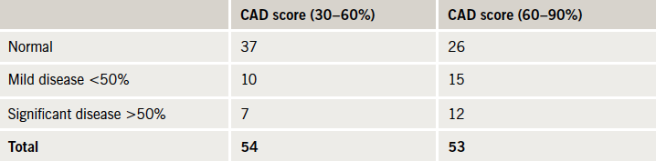
In the CTCA cohort, 107 had CTCA, of whom 54 had a CAD score of 30–60% and 53 had a CAD score of 60–90%. In total, 19 patients (17.9%) were found to have significant CAD (>50% stenosis) (table 2).

Table 2
Table 2. Incidence of coronary artery disease (CAD) in the computed tomography
coronary angiography (CTCA) group

In the patients with CAD score 30–60%, seven patients had significant CAD (13.0%). Six of them went on to have further investigation, one had a positive MPS study, but did not attend his follow-up, three patients were investigated further with a DSE, which were negative and they were discharged, two were referred for ICA, and both were revascularised. In the 60–90% CAD score group, 12 patients had significant CAD on CTCA (22.6%), two had negative DSEs and were discharged. The other 10 were referred for ICA, seven of them proceeded to have revascularisation with percutaneous coronary intervention (PCI) and one with coronary artery bypass grafting (CABG). A total of 10 (9.4%) patients were revascularised in the CTCA cohort.

The functional test cohort

In the functional test cohort, 106 patients had functional tests: 39 had MPS, of which 12 patients were in the 30–60% CAD score group and 27 in the 60–90% CAD score group. In total, five patients (all in the 60–90% group) were found to have a positive result (12.8%) and all proceeded to have ICA. Four (10.3%) were revascularised with PCI.

There were 67 patients who underwent DSE, of which 29 were in the 30–60% CAD score group and 38 patients were in the 60–90% CAD score group. Only one patient (from the 60–90% CAD score group) was found to have significant reversible ischaemia, and went on to have ICA and was revascularised with PCI (1.5%). In total, five (4.7%) patients were revascularised in the functional test cohort. The higher revascularisation rate in the CTCA cohort of 9.4% was not statistically significant (p=0.28).

Six-month outcome

In total, three patients presented to accident and emergency (A&E) and were admitted for investigation of chest pain (1.4%). None of these three patients were deemed to have had a cardiac cause for chest pain and they were all discharged. There was no mortality at six months.

Discussion

The use of CTCA for the evaluation of chest pain in patients with moderate prevalence of CAD is well validated in three clinical trials that demonstrated very good sensitivity and excellent NPV.5-7 More recently, clinical trials from the USA demonstrated CTCA to be clinically effective, safe and economical compared with the current standard of care and MPS, in the emergency room.8-10

Our real-world data in a London district general hospital (DGH) demonstrate that CTCA can be at least as effective as functional tests in detecting significant CAD, and may lead to more revascularisations than functional tests. CTCA should be considered as an effective alternative to functional tests in patients with higher pre-test probability of CAD, in hospitals with limited access to functional tests. A further advantage of CTCA is that it can detect milder degrees of atherosclerosis, and these patients would benefit from secondary prevention.

Although functional testing is a test for ischaemia, and CTCA is an anatomical test, they are both used to rule out significant CAD in patients with chest pain. The NICE guidelines1 recommend the use of functional imaging tests in patients with moderate likelihood. However, the incidence of CAD in our population of patients with moderate and high likelihood appears to be relatively low. The NICE guidelines state that the CAD score is likely to overestimate risk in primary care populations and recommend that further research is needed in this area.1 CTCA is the best test available to rule out CAD with a NPV of 99% in published data,6 and, hence, we believe its use can be extended to patients with moderate and even high CAD scores. The estimated cost of a CTCA by NICE is £173, which compares well with the costs of DSE and MPS in the same costing report (£236 and £293, respectively).11 More importantly for busy clinical services, the advantage of CTCA over functional tests is its speed. The total time taken to perform a CTCA is less than 20 minutes per patient, which compares very well with DSE, MPS and cardiac magnetic resonance, all of which take around an hour per patient.

Although there were fewer MPS studies performed than DSEs, there were more positive studies in the MPS cohort (five vs. one), perhaps indicating physician preference to use MPS when they have a higher index of suspicion for CAD, thus, introducing a real-world selection bias. Our demographic data do demonstrate that the MPS cohort appeared to be older and have more risk factors.

The limitations of our study are those common to retrospective studies. Outcome data were obtained only from our hospitals’ notes, and any adverse events recorded in other hospitals would not have been automatically available. At six months, only three patients were re-admitted, all found to have non-cardiac chest pain. Not all patients underwent ICA, the gold standard for evaluation of CAD. Four patients were lost to follow-up, which represents 1.9% of the total study population.

In conclusion, we found CTCA to be at least as effective as functional tests in detecting significant CAD and may lead to more revascularisations than functional tests. Furthermore, our data demonstrate the CAD score to overestimate the probability of CAD in a RACP clinic population (table 2). Hence, we believe the use of CTCA can be extended to patients with moderate and even high CAD scores. Although CTCA is a low-cost test, a trend to higher revascularisation rate was also seen in the trials of CTCA in the emergency room.8,10 This may lead to an increase in the overall cost of managing CAD. A clinical trial is needed to evaluate the clinical effectiveness, as well as the cost-effectiveness, of using CTCA in patients with higher CAD scores.

Conflict of interest

There was no funding for this project and the authors have decared no conflict of interest.

Key messages

  • The National Institute for Health and Care Excellence (NICE) coronary artery disease (CAD) score is likely to overestimate risk in primary care populations
  • Computed tomography coronary angiography (CTCA) is an excellent test for ruling out CAD in patients with low-to-moderate likelihood of CAD
  • A clinical trial is needed to evaluate the clinical and cost-effectiveness of CTCA in patients with higher pre-test likelihood of CAD

References

  1. National Institute for Health and Care Excellence. Chest pain of recent onset. Clinical guideline 95. London: NICE, 2010. Available from: www.nice.org.uk/CG95
  2. Pryor DB, Shaw L, McCants CB, et al. Value of the history and physical in identifying patients at increased risk for coronary artery disease. Ann Intern Med 1993;118:81–90.
  3. Rogers T, Dowd R, Yap HL, Claridge S, Alfakih K, Byrne J. Strict application of NICE clinical guideline 95 ‘chest pain of recent onset’ leads to over 90% increase in cost of investigation. Int J Cardiol 2013;166:740–2. http://dx.doi.org/10.1016/j.ijcard.2012.09.180
  4. Taylor AJ, Cerqueira M, Hodgson JM et al. ACCF/SCCT/ACR/AHA/ASE/ASNC/SCAI/SCMR 2010 appropriate use criteria for cardiac computed tomography. J Am Coll Cardiol 2010;56:1864–94. http://dx.doi.org/10.1016/j.jacc.2010.07.005
  5. Budoff MJ, Dowe D, Jollis JG et al. Diagnostic performance of 64-multidetector row coronary computed tomographic angiography for the evaluation of coronary artery stenoses in individuals without known coronary artery disease. Results from the prospective multicentre ACCURACY trial. J Am Coll Cardiol 2008;52:1724–32. http://dx.doi.org/10.1016/j.jacc.2008.07.031
  6. Meijboom WB, Meijs MF, Schuijf JD et al. Diagnostic accuracy of 64 slice computed tomography coronary angiography: a prospective multicentre multivendor study. J Am Coll Cardiol 2008;52:2135–44. http://dx.doi.org/10.1016/j.jacc.2008.08.058
  7. Miller JM, Rochitte CE, Dewey M et al. Diagnostic performance of coronary angiography by 64-row CT. N Engl J Med 2008;359:2324–36. http://dx.doi.org/10.1056/NEJMoa0806576
  8. Litt HI, Gatsonis C, Snyder B et al. CT angiography for safe discharge of patients with possible acute coronary syndromes. N Engl J Med 2012;366:1393–403. http://dx.doi.org/10.1056/NEJMoa1201163
  9. Goldstein JA, Chinnaiyan KM, Abidov A et al. The CT-STAT (Coronary Computed Tomographic Angiography for Systematic Triage of Acute Chest Pain Patients to Treatment) trial. J Am Coll Cardiol 2011;58:1414–22. http://dx.doi.org/10.1016/j.jacc.2011.03.068
  10. Hoffmann U, Truong QA, Schoenfeld DA et al. Coronary CT angiography versus standard of evaluation in acute chest pain. N Engl J Med 2012;367:299–308. http://dx.doi.org/10.1056/NEJMoa1201161
  11. National Institute for Health and Care Excellence. Chest pain of recent onset – costing report. London: NICE, 2011. Available from: www.nice.org.uk/nicemedia/live/12947/55738/55738.pdf

News from the ESC Congress 2013

Br J Cardiol 2013;20:130–2 Leave a comment
Click any image to enlarge
Authors:
First published online October 11, 2013

This year’s European Society of Cardiology (ESC) Congress took place in Amsterdam, The Netherlands, on 31st August – 4th September 2013. We report highlights from some of the hundreds of studies reporting including more on the cardiovascular effects of drugs for diabetes to the latest in intervention, the effects of anticoagulants in valve disease and new lipid lowering agents.

PRAMI: preventive PCI of other lesions beneficial in STEMI

Patients undergoing percutaneous coronary intervention (PCI) for ST-elevation myocardial infarction (STEMI) have better outcomes if non-culprit lesions are also treated, according to results from the PRAMI (Preventive Angioplasty in Myocardial Infarction Trial) study published recently in the New England Journal of Medicine (http://dx.doi.org/10.1056/NEJMoa1305520).

In the trial, patients who also had PCI of the non-culprit lesions had a 65% reduction in event rate, driven by reductions in subsequent myocardial infarction (MI) and refractory angina.

Presenting the results, Dr David Wald (Queen Mary University of London) said the benefit of preventive PCI of non-culprit lesions occurred early, within days, and the full benefit was maximised after a few months.

The study involved 465 STEMI patients who underwent PCI of the infarct-related artery. Of these, 234 patients were randomised to PCI of non-culprit lesions and the other 231 patients received no further PCI treatment. The trial was stopped early after a significant benefit was seen favouring preventive PCI.

After a mean follow-up of 23 months, the primary end point, defined as death from cardiac causes, nonfatal MI, or refractory angina, occurred in 21 patients treated with preventive PCI and 53 patients treated with PCI of the culprit lesion only.

In an accompanying editorial (http://dx.doi.org/10.1056/NEJMe1309383), Dr Laura Mauri (Harvard Clinical Research Institute, Boston, USA) said this practice of preventative PCI during STEMI is not currently recommended in the guidelines. However, she concluded that: “We can no longer assume that secondary lesions in acute myocardial infarction are innocent until proven guilty.”

RE-ALIGN: dabigatran shows hazard with mechanical valves

Dabigatran is not appropriate as an alternative to warfarin in the setting of mechanical heart valves, say results from the RE-ALIGN (Randomised, phase II study to Evaluate the Safety and Pharmacokinetics of Oral Dabigatran Etexilate in Patients After Heart Valve Replacement) trial.

The European Medicines Agency issued a statement in December last year that dabigatran was contraindicated in mechanical heart valves as a result of an increase in strokes, myocardial infarction (MI), and thrombosis forming on the valves seen in RE-ALIGN. The full data, presented at the congress, showed that ischaemic or unspecified stroke occurred in 5% of the dabigatran patients versus 0% of the warfarin group, and major bleeding occurred in 4% of dabigatran patients versus 2% of warfarin patients. All patients with major bleeding had pericardial bleeding. Asymptomatic valve thrombosis was detected in 3% of the dabigatran group versus 0% of those on warfarin.

The investigators concluded that the different mechanism of thrombosis in patients with mechanical heart valves, as compared to patients with atrial fibrillation (AF), may explain the failure of dabigatran in RE-ALIGN.

The study was published online in the New England Journal of Medicine (http://dx.doi.org/10.1056/NEJMoa1300615). In an accompanying editorial (http://dx.doi.org/10.1056/NEJMe1310399), Dr Elaine M Hylek (Boston University School of Medicine, USA) suggested several reasons for the failure of dabigatran in this setting. These include the fact that most patients received dabigatran early after surgery, when thrombogenicity is enhanced, the dose may not have been appropriate for this population, and the drug may not have been absorbed as well as expected. Because of these possibilities, she says further research in this field should be continued.

ACCOAST: No benefit of prasugrel pretreatment in ACS

There was no benefit of pretreatment with prasugrel in acute coronary syndrome (ACS) patients in the ACCOAST (A Comparison of Prasugrel at PCI or Time of Diagnosis of Non-ST Elevation Myocardial Infarction) trial. And giving the drug at the time of diagnosis, rather than at the time of PCI, increased major bleeding complications,

While clopidogrel is recommended for preloading, prasugrel has a more rapid onset of action and these results suggest early treatment is not necessary and could be harmful.

ACCOAST was designed to randomise 4,100 patients to an early or standard strategy. The trial was stopped after enrollment of 4,033 patients last November because of an increase in major and life-threatening bleeding and no reduction in cardiovascular events.

The primary efficacy end point (a composite of cardiovascular death, myocardial infarction (MI), stroke, urgent revascularisation, or glycoprotein IIb/IIIa bailout) was not significantly different between the two treatment groups at seven or 30 days. However, bleeding measures were significantly higher in the pretreatment group (see table 1).

Table 1. ACCOAST – main results

The study was published in New England Journal of Medicine (http://dx.doi.org/10.1056/NEJMoa1308075).

PARIS: limited impact of stopping dual antiplatelet therapy

The overall impact of stopping dual antiplatelet therapy is modest and may have been mitigated with the introduction of safer stent platforms. This was the conclusion of Dr Roxana Mehran (Mount Sinai Medical School, New York, USA) presenting results from the PARIS (Patterns of non-Adherence to anti-platelet Regimens In Stented patients) study. The results did suggest different effects of stopping dual antiplatelet therapy in different situations, she noted.

“When physicians recommend discontinuation, presumably because patients are stable, there is no risk of adverse events, but when patients simply don’t comply or are forced off antiplatelet therapy because they are bleeding, their risks are significantly elevated”. She added that these results highlight the need for potentially tailoring guidance of antiplatelet therapy cessation to the different categories.

The PARIS study enrolled more than 5,000 patients following stent implantation. “Discontinuation” was defined as physician-recommended cessation, “interruption” was a temporary stop (up to 14 days) due to surgical necessity, and “disruption” was unplanned cessation, due to bleeding or patient noncompliance.

The majority (74%) of cardiac events in the trial occurred when patients were actually taking dual antiplatelet therapy (DAPT). In patients who had disrupted therapy, the risk was highest in the first seven days (hazard ratio 7.04, compared with remaining on therapy, p<0.001) and continued out to 30 days.

Stent thrombosis most commonly occurred in patients still taking their medication (80.3% of the events) but was also seen in 14.1% of patients with disrupted therapy. Numbers were negligible in patients with recommended discontinuation or interruption.

Dr Mehran noted that the hazard was much greater if both drugs were stopped, so she advised that if there was a bleeding concern that only one of the drugs was stopped. She added that she would voluntarily discontinue both drugs in patients at higher risk for bleeding complications after the highest risk period was over – usually three to six months post-percutaneous coronary intervention with the newer generation of drug eluting stents.

LINC: mechanical and manual CPR gives similar results

Mechanical chest compression with an automated system device (Lucas™, Jolife AB) was not more effective than manual compressions for delivering cardiopulmonary resuscitation (CPR) to patients in cardiac arrest, according to results from the LINC (LUCAS in Cardiac Arrest) study.

Presenting the results, Dr Sten Rubertsson (Uppsala University Hospital, Sweden) said the fact that the mechanical device was equally effective and showed no safety concerns meant there was now scientific data to support its use.

He added that there was only one patient out of 111 treated with the mechanical device who was left with a bad neurological outcome (clinical performance category score of three or four) compared with six of 104 patients given manual compressions.

Dr Rubertsson suggested the device would be useful when prolonged CPR is needed, such as when a long journey to the hospital is necessary or if the patient is taken to the cath lab. Discussant of the trial, Dr Patrick Goldstein (University Hospital of Lille, France), noted the good neurological outcomes in both groups – around 7.5% to 8.5% at six months. “If we go back just five years these figures would not be over 3%. So we can see what improvements have been made in the field.”

The LINC study included 2,589 patients from six European sites who had suffered an out-of-hospital cardiac arrest. Manual chest compressions were started on all patients as soon as paramedics arrived on the scene. Patients were then randomised to be kept on manual chest compressions or be switched to mechanical compressions with defibrillation during ongoing chest compressions.

Results showed that four hours after the start of CPR, survival rates were similar in both groups. Later outcomes were also similar, including the rate of restoration of spontaneous circulation (ROSC), the number of patients who arrived at the emergency room with a palpable pulse, the number of patients who survived until discharge from intensive care, and neurological outcomes at one and six months.

Two diabetes drugs show neutral CV effect

Two oral antihyperglycaemic agents – saxagliptin and alogliptin – did not increase or decrease the risk of major cardiovascular events in patients with type 2 diabetes mellitus according to the SAVOR (Saxagliptin Assessment of Vascular Outcomes Recorded in Patients with Diabetes Mellitus) -TIMI 53 and EXAMINE (Cardiovascular Outcomes Study of Alogliptin in Subjects With Type 2 Diabetes and Acute Coronary Syndrome) studies respectively.

Saxagliptin, however, was associated with a significant increased risk of hospitalisations for heart failure, a component of the pre-specified secondary end point in SAVOR-TIMI 53. Secondary end points have not yet been analysed in EXAMINE.

While saxagliptin and alogliptin significantly reduced glycated haemoglobin levels, there was some debate about the role of the drugs, which are dipeptidyl peptidase-4 (DPP-4) inhibitors, in clinical practice.

At an ESC press conference on the trials, SAVOR-TIMI 53 investigator Dr Deepak Bhatt (Brigham and Women’s Hospital, Boston, USA) said: “What’s clear is that neither of these drugs, and I would be willing to say that none of the DPP-4 inhibitors, seem to impact favourably on cardiovascular outcomes in this intermediate-term follow-up.”

Noting that metformin is widely regarded as first-line therapy for patients with diabetes, EXAMINE investigator Dr William White (University of Connecticut School of Medicine, USA) suggested these latest results, which support lack of a safety signal in a very high-risk population, gave enhanced credibility to use a DPP-4 inhibitor as a second drug in addition to metformin. He pointed out that these agents lower the glycaemic index significantly, and therefore should reduce the risk of microvascular complications.

In SAVOR-TIMI 53, 16,492 patients with type 2 diabetes with a high risk of cardiovascular events were randomised to saxagliptin 5 mg/day or placebo. After a median of 2.1 years, the primary end point — a composite of cardiovascular death, myocardial infarction (MI), or ischaemic stroke — had occurred in 613 patients treated with saxagliptin and 609 patients treated with placebo. Glycated haemoglobin levels were reduced from 8.0% at baseline to 7.7% with saxagliptin, a significant reduction versus placebo.

In the EXAMINE trial, 5,380 patients with diabetes and acute MI/unstable angina were randomised to alogliptin or placebo. The primary end point of the trial – a composite of cardiovascular death, nonfatal MI, and nonfatal stroke – were similar in the two groups, and glycated haemoglobin levels were significantly reduced by -0.36%.

The studies were published in the New England Journal of Medicine (http://dx.doi.org/10.1056/NEJMoa1307684 and http://dx.doi.org/10.1056/NEJMoa1305889). An accompanying editorial (http://dx.doi.org/10.1056/NEJMp1309610) makes the point that the optimal approach for reducing cardiovascular risk in diabetic patients is the aggressive management of cardiovascular disease risk factors rather than aggressive glycaemic control.

ASSURE: new HDL drug disappoints

A new oral drug that boosts production of the high-density lipoprotein (HDL) precursor protein apoA1 did not produce significant regression of atherosclerosis in the ASSURE (ApoA-I Synthesis Stimulation and Intravascular Ultrasound for Coronary Atheroma Regression Evaluation) study.

The study included 323 patients with low HDL and coronary disease. All patients received statins and were also randomised to receive either RVX-208 100 mg (n=244) or placebo (n=80) twice daily for 26 weeks. Atherosclerosis was assessed by intravascular ultrasound (IVUS) at baseline and the end of the study.

Both groups showed increases in HDL and reductions in low-density lipoprotein (LDL), as well as trends toward a regression of atherosclerosis. Presenting the results with RVX-208, Dr Stephen Nicholls (Royal Adelaide Hospital, Australia) noted that a large placebo effect led to no significant difference in HDL changes between the active treatment and placebo groups, so this study had not addressed the HDL hypothesis.

The primary end point of the study – change from baseline in percent atheroma volume – just failed to meet significance in the RVX-208 group at six months, and it was suggested that longer treatment may be needed to show an effect.

AQUARIUS: no effect of aliskiren on atherosclerosis

The renin-inhibitor aliskiren had no effect on atherosclerosis progression in patients with coronary disease and prehypertension, according to results from AQUARIUS (Aliskerin QUantitative Atherosclerosis. Regression Intravascular Ultrasound Study).

The drug was tested in this intravascular ultrasound (IVUS) study after atheroprotective effects with renin inhibition were observed in preclinical studies. For the study, 458 patients were randomised to aliskiren or placebo and underwent IVUS screening at baseline and again after at least 72 weeks. Results showed that percent atheroma volume decreased slightly in both groups (-0.33% with aliskiren versus -0.24% with placebo), a non-significant difference.

BIC-8: rule out ACS with negative troponin and copeptin tests

Acute coronary syndrome (ACS) can be ruled out with negative results from two simple tests – troponin and co-peptin – allowing the majority of individuals to be discharged quickly and easing overcrowding in the emergency department, a new study suggests.

Presenting the results of the BIC-8 (Effect of the Biomarker Copeptin in Managing Patients With Suspected Acute Coronary Syndrome) trial, Dr Martin Möckel (Charité-Universitätsmedizin Berlin, Germany) noted that 10% of patients in the emergency department present with chest pain, but only 10% of those patients are having a real ACS event.

Co-peptin is a marker of severe haemodynamic stress and is detectable immediately in patients with ACS. In observational studies, a combination of cardiac troponin and co-peptin was shown to be a significant predictor of acute events, Dr Möckel explained.

In the BIC-8 study, 902 patients at low to intermediate risk of ACS and an initial negative troponin test, were randomised to co-peptin test or standard care, including serial cardiac troponin testing. If the co-peptin test was positive, defined as >10 pmol/L, they were treated with standard ACS care. If the co-peptin assay was negative, they were discharged and scheduled for an outpatient visit within 72 hours.

At 30 days, the rate of major adverse cardiovascular events was almost identical – 5.46% in those undergoing co-peptin testing and 5.5% in the standard-care arm. The discharge rate from the emergency department was 66% in the co-peptin group versus 12% in those given standard care.

Targeted approach best for sodium restriction

Only certain groups in most countries actually experience blood-pressure benefits from restricting their sodium intake, and the guidelines need to be changed to target sodium restrictions to vulnerable groups such as hypertensive subjects and the elderly, new results from the PURE (Prospective Urban Rural Epidemiological) study suggest.

Screen shot 2013-10-11 at 17.37.30
Table 1. Systolic blood pressure changes associated with 1 g increases in sodium intake

Presenting PURE results (table 1), Dr Andrew Mente (McMaster University, Hamilton, Canada) said that whole population recommendations for daily sodium limits are pointless in many parts of the world.

In the study of almost 100,000 subjects, none of the populations surveyed had a usual intake of sodium below the 2.3 g/day recommended in most guidelines. Intakes were lowest in Malay; ranged from 4.2–4.8 g/day in North America, Europe, South Asia, Africa, and South America; and were highest in China at more than 5.5 mg/day.

And while there was a linear relationship between sodium levels and blood pressure, meaningful systolic blood pressure changes in response to 1 g increases in sodium consumption were only seen in certain groups – hypertensive subjects, the elderly, and people consuming more than 5 g/day of sodium.

Dr Mente said: “For those in the 3–5 g/day range, there is little benefit of reducing levels apart from in those vulnerable groups, so in most countries, strategies to reduce blood pressure via sodium restriction are best targeted specifically at those groups. Whereas in China where intakes are higher, a population strategy appears more reasonable”.

Statins linked to reduced dementia?

A new observational study from Taiwan suggested that high-dose statins may be linked to a reduced risk of dementia in the elderly.

Screen shot 2013-10-11 at 17.42.12
Table 1. Hazard ratio (vs. control) of developing dementia with statins at different dosage tertiles

The researchers used Taiwan’s national health insurance data to identify 58,000 individuals over 65 years who had no history of dementia in 1997 and 1998. Of these, 15,200 were taking statins. Propensity scoring was used to match these patients with controls not taking statins. There were 5,516 new diagnoses of dementia during the 4.5 years of follow-up. Results showed a dose-related inverse relationship between statin use and new-onset nonvascular dementia (see table 1).

Statins and dementia is a controversial area, with previous studies having shown conflicting results. Two randomised trials – PROSPER (Pravastatin in elderly individuals at risk of vascular disease) with pravastatin in the elderly and the LEADe (Lipitor’s Effect in Alzheimer’s Dementia) study with atorvastatin in early Alzheimer’s patients – have not shown any benefit of statins on risk of dementia. Possible cognitive impairment has recently been added to the safety information for statins in the US, the study concluded.

CHAMPION meta-analysis suggests cangrelor benefit

Cangrelor is indeed effective for the reduction of periprocedural thrombotic complications during percutaneous coronary intervention (PCI) compared with placebo or clopidogrel, at the expense of an increase in minor bleeding, according to a pooled analysis of three trials with the new IV antiplatelet.

Table 1. CHAMPION main results

Presenting the data (see table 1), Dr Christian Hamm (Kerckhoff Heart and Thorax Centre, Germany) explained that the pre-specified patient-level analysis combines data from CHAMPION-PCI, CHAMPION-PLATFORM, and CHAMPION-PHOENIX studies in a total of 25,000 patients with ST-elevation myocardial infarction (STEMI), without STEMI, and with stable coronary disease, all undergoing PCI.

The primary outcome – a combination of death, MI, ischaemia-driven revascularisation, or stent thrombosis at 48 hours – was significantly lower in the cangrelor group. Stent thrombosis was also reduced in the cangrelor group. The primary safety outcome – non-coronary artery bypass graft (CABG)-related Global Use of Strategies to Open Occluded Arteries (GUSTO) moderate bleeding or transfusions – was not significantly different between the groups. GUSTO mild bleeding was higher with cangrelor.

The analysis was published recently in The Lancet (http://dx.doi.org/10.1016/S0140-6736(13)61615-3). In an accompanying editorial (http://dx.doi.org/10.1016/S0140-6736(13)61840-1), Dr Shamir Mehta (McMaster University, Hamilton, Canada) points out that two of the three trials did not show significant results on their own, but that benefit has been achieved in the meta-analysis.

Dr Mehta notes that cangrelor, which is given by intravenous injection, produces almost-immediate platelet inhibition, and has a very short biological half-life of three to six minutes, with normalisation of platelet function within one hour of discontinuation. He suggests the drug could have a major role in high-risk STEMI, patients with nausea/vomiting, intubated patients, and those in cardiogenic shock.

UK doctor payment system linked to good lipid results

More patients in the UK are achieving cholesterol goals than in Germany, a finding that might have been influenced by the different payments systems used in those countries, suggests a new study

Dr Anselm Kai Gitt  (Herzzentrum Ludwigshafen, Germany) presented a subanalysis of DYSIS (Dyslipidemia International Study), which examined the possible impact of reimbursement systems on the achievement of low-density lipoprotein (LDL)-cholesterol targets in 4,260 German patients and 540 UK patients at high cardiovascular risk. The aim was to compare the German system, which restricts chronic medical treatment by budget constraints, with the UK’s incentive system where GPs receive payment linked to achieving cholesterol targets.

All patients in the study were taking statins, but 42% of German patients achieved the target of LDL <100mg/dl (< 5.5 mmol/L) compared to 79.8% of UK patients. The potent statin, atorvastatin, was used in 3.9% of German patients (mean dose 24 mg) compared to 24.8% of UK patients (mean dose 34.2 mg), whereas the weaker statin, simvastatin, used in 83.9% of Germans (mean dose 27.2 mg) compared to 67.6% of UK patients (mean dose 36.6 mg). Daily dosages were significantly lower in Germany than in the UK, independent of the statin used.

Dr Gitt concluded: “Our study showed that healthcare reimbursement systems appear to impact on the achievement of cholesterol lowering targets. The German system was put in place to control costs but it remains to be seen whether it will achieve this in the long term. The UK system has higher short term costs, with more GP visits, use of potent statins and high doses, but it may ultimately be more cost effective because of fewer complications.”

Dual antiplatelet therapy and upper gastrointestinal bleeding risk: do PPIs make any difference?

Br J Cardiol 2013;20:148doi:10.5837/bjc.2013.029 Leave a comment
Click any image to enlarge
Authors:
First published online September 5th 2013

Dual antiplatelet therapy (DAT) with aspirin and clopidogrel is recommended for up to one year following acute coronary syndrome (ACS). Gastrointestinal bleeding is the main hazard of this treatment and proton pump inhibitors (PPIs) are often prescribed in selected patients to reduce this risk. The main purpose of this study was to analyse the effect of PPIs in reducing the subsequent risk of gastrointestinal bleeding. 

The medical records of 177 consecutive patients treated with DAT following ACS, were specifically reviewed for the study parameters over a 12-month period. 

The mean age was 66 years (range 24–96) with a median value of 68 years; 67% were males and 33% females, 74% Caucasians and 26% Asians. Patients were divided into two groups: the PPI group (patients on DAT and PPIs, n=91) and the control group (patients on DAT only, n=86). In the PPI group, 55% were on lansoprazole, 34% on pantoprazole and 11% on omeprazole. 

Out of the 177 patients, evidence of upper gastrointestinal bleeding was found in 10 patients, with the mean age of these patients being 77 years in the PPI group and 53 years in the control group. In the PPI group, endoscopy findings from six patients (6.6%) revealed gastritis in four, bleeding angiodysplasia in one, and bleeding oesophagitis in one; while the findings for the four patients in the control group (4.6%) showed gastritis in two, gastric ulcer in one and Mallory Weiss tear in one (odds ratio: 1.45, 95% confidence interval 0.39–5.32, p=0.58). None of these patients had a previous history of gastrointestinal bleeding.

In conclusion, empirical prophylactic prescription of PPIs for patients on DAT following ACS is of no significant benefit in reducing their predisposition to upper gastrointestinal bleeding. However, studies utilising larger populations are warranted to confirm this conclusion. 

Introduction

Dual antiplatelet therapy (DAT) with aspirin and clopidogrel is recommended for up to one year following acute coronary syndrome (ACS) in order to reduce the risk of further cardiac events.1,2 Gastrointestinal bleeding is the main hazard of this treatment; however, although the incidence of bleeding is low, it results in significantly increased morbidity and mortality in these patients,3-5 and proton pump inhibitors (PPIs) are often prescribed to selective patients to reduce this risk. PPIs act by reducing the secretion of gastric acid, neutralising gastric pH, increasing clot formation and decreasing the lysis of blood clots.

There are no formal guidelines concerning the initiation and continuation of PPIs for such patients. In addition, evidence regarding the treatment effect of this group of medications in lowering the risk of subsequent gastrointestinal bleeding is absent. Therefore, the main purpose of this study was to analyse the effect of PPIs in reducing the subsequent risk of gastrointestinal bleeding in patients on DAT with aspirin and clopidogrel following ACS.

Methods and materials

The medical records of 177 patients treated with DAT following ACS were specifically reviewed for the study parameters over a 12-month period of time. These patients were consecutive admissions following index myocardial infarction, admitted to the cardiology department of Bradford Teaching Hospital: 66 patients (37.3%) were admitted with ST-elevation myocardial infarction (STEMI) and 111 (62.7%) with non-ST elevation myocardial infarction (NSTEMI). All the patients in the study were given 300 mg aspirin and 300 mg clopidogrel after index myocardial infarction followed by 75 mg aspirin and 75 mg clopidogrel daily. Records of re-admissions with gastrointestinal problems, particularly gastrointestinal bleeding, were retrieved from the medical records department.

Patients were divided in two groups: the PPI group (patients on DAT and PPIs, n=91) and the control group (patients on DAT only, n=86). At the time of data collection, there were no hospital guidelines in terms of prescribing PPIs for these patients. The database was analysed for re-admissions with upper gastrointestinal problems over one year while patients were receiving DAT. The unpaired t-test was used to compare age between the two groups, while Fisher’s exact test was used for the remaining demographics, all of which were measured on a categorical scale. Subsequent analysis re-examined the difference adjusting for demographic factors found to significantly vary between groups. These analyses were performed using logistic regression. The end points were re-admissions with gastrointestinal bleeding or death.

Results

The mean age of patients was 66 years (range 24–96) with a median value of 68 years; 67% were males and 33% females, 74% Caucasians and 26% Asians. In the PPI group, 27 patients (29.7%) were admitted with STEMI and 64 patients (70.3%) with NSTEMI, while the corresponding values for the control group were 39 (45.3%) and 47 (54.7%), respectively. In the PPI group, 39 patients (42.9%) were treated with percutaneous coronary intervention (PCI), four (4.4%) with a coronary artery bypass graft (CABG) and 48 (52.7%) were managed conservatively, while the corresponding values for the control group were 11 (12.8%), five (5.8%) and 70 (81.4%), respectively. Baseline demographics are illustrated in table 1.

Table 1
Table 1. Baseline demographics of the patients

Out of the 177 patients, evidence of upper gastrointestinal bleeding was found in 10 patients, six in the PPI group and four in the control group (odds ratio 1.45, 95% confidence interval [CI] 0.39–5.32, p=0.58). In the PPI group (six patients, 6.6%), the endoscopy findings were as follows: gastritis in four, bleeding angiodysplasia in one and bleeding oesophagitis in one. The results for the control group (four patients, 4.6%) showed gastritis in two, gastric ulcer in one and Mallory Weiss tear in one. None of these patients had a previous history of gastrointestinal bleeding. Table 2 explains these results in more detail.

Table 2
Table 2. Results

Out of the six patients admitted with gastrointestinal bleeding, 4/50 (8%) were on lansoprazole, 1/31 (3.2%) on pantoprazole and 1/9 (11.1%) on omeprazole. Gastrointestinal bleeding was sufficiently severe to require a blood transfusion in two patients in the PPI group and one in the control group. In the PPI group, two patients required endoscopic intervention in the form of an adrenaline injection and diathermy, and only one patient was treated with heater probe coagulation and clips. In contrast, only one patient was treated with an adrenaline injection and haemostatic clips in the control group.

There was no statistically significant difference in gastrointestinal bleeding between the two groups, both unadjusted and adjusted for potentially confounding variables. Although there were no significant differences, the differences in outcome between groups were reduced after adjusting for differences in demographics between groups. Table 3 outlines the upper gastrointestinal bleeding episodes, both unadjusted and adjusted for variables.

Table 3
Table 3. Unadjusted and adjusted analyses

There was no gastrointestinal bleeding related mortality in any of the study patients. Overall, 12 deaths were observed, seven in the PPI group (four ACS, one pulmonary embolism, one pneumonia, one heart failure) and five in the control group (three ACS, one metastatic disease with unknown primary, one pneumonia).

Discussion

Gastrointestinal bleeding is the main hazard identified for DAT with aspirin and clopidogrel following ACS. However, our study suggests that the co-prescription of PPIs with aspirin and clopidogrel does not alter the risk of gastrointestinal bleeding in these patients (odds ratio 1.45, p=0.58).

The findings reported here are consistent with another retrospective study that was carried out to determine the efficacy of PPIs in lowering the risk of gastrointestinal bleeding for patients receiving DAT with aspirin and clopidogrel. In a retrospective study of 1,023 patients, no difference was found in the incidence of upper gastrointestinal bleeding between those patients receiving and those not receiving PPIs (0.7% vs. 0.6%, p=0.88).6 A cohort analysis of a pharmacy database of 385 patients,7 which assessed the role of PPIs in reducing the probability of gastrointestinal bleeding in patients who possessed additional risk factors for gastrointestinal bleeding, found that only those with an additional risk factor had a lower incidence of gastrointestinal bleeding with PPIs compared with those without PPIs (1.7% vs. 11.1%, p=0.05).7 However, a retrospective study of 666 patients found a reduced risk of gastrointestinal bleeding with PPIs for patients receiving DAT.8

The benefits of prescribing PPIs should, therefore, be weighed in individual cases, and only patients with an increased risk of gastrointestinal bleeding should be considered for PPIs. Risk factors for gastrointestinal bleeding are not well characterised, but the most important are a prior history of peptic ulcers or gastrointestinal bleeding and advanced age.4,9

The limitations of our study include its retrospective nature and the small sample size.

In summary, empirical prophylactic prescription of PPIs in patients on DAT with aspirin and clopidogrel following ACS is of no significant benefit in reducing a predisposition to upper gastrointestinal bleeding; however, studies with larger population numbers are warranted in order to confirm this conclusion.

Conflict of interest

None declared.

Key messages

  • Empirical prophylactic prescription of proton pump inhibitors in patients on dual antiplatelet therapy following acute coronary syndrome is of no significant benefit in reducing predisposition to upper gastrointestinal bleeding
  • Studies with larger population numbers are warranted in order to confirm this conclusion

References

  1. Yusuf S, Zhao F, Mehta SR et al. Effects of clopidogrel in addition to aspirin in patients with acute coronary syndromes without ST-segment elevation. N Engl J Med 2001;345:494–502. http://dx.doi.org/10.1056/NEJMoa010746
  2. CAPRIE Steering Committee. A randomized, blinded, trial of Clopidogrel versus Aspirin in Patients at Risk of Ischaemic Events (CAPRIE). Lancet 1996;348:1329–39. http://dx.doi.org/10.1016/S0140-6736(96)09457-3
  3. Popławski C, Jakubczyk P, Jakubczyk M. Analysis of the upper gastrointestinal tract bleeding prevalence in patients treated due ischaemic heart disease. Adv Med Sci 2007;52:288–93.
  4. Alli O, Smith C, Hoffman M et al. Incidence, predictors, and outcomes of gastrointestinal bleeding in patients on dual antiplatelet therapy with aspirin and clopidogrel. J Clin Gastroenterol 2011;45:410–14. http://dx.doi.org/10.1097/MCG.0b013e3181faec3c
  5. Ng FH, Chan P, Kwanching CP et al. Management and outcome of peptic ulcers or erosions in patients receiving a combination of aspirin plus clopidogrel. J Gastroenterol 2008;43:679–86. http://dx.doi.org/10.1007/s00535-008-2215-4
  6. Barada K, Karrowni W, Abdullah M et al. Upper gastrointestinal bleeding in patients with acute coronary syndromes: clinical predictors and prophylactic role of proton pump inhibitors. J Clin Gastroenterol 2008;42:368–72. http://dx.doi.org/10.1097/MCG.0b013e31802e63ff
  7. Luinstra M, Naunton M, Peterson GM et al. PPI use in patients commenced on clopidogrel: a retrospective cross-sectional evaluation. J Clin Pharm Ther 2010;35:213–17. http://dx.doi.org/10.1111/j.1365-2710.2009.01089.x
  8. Ng FH, Wong SY, Lam KF et al. Gastrointestinal bleeding in patients receiving a combination of aspirin, clopidogrel and enoxaparin in acute coronary syndrome. Am J Gastroenterol 2008;103:865–71. http://dx.doi.org/10.1111/j.1572-0241.2007.01715.x
  9. Nikolsky E, Stone GW, Kirtane AJ et al. Gastrointestinal bleeding in patients with acute coronary syndromes: incidence, predictors, and clinical implications: analysis from the ACUITY (Acute Catheterization and Urgent Intervention Triage Strategy) trial. J Am Coll Cardiol 2009;54:1293–302. http://dx.doi.org/10.1016/j.jacc.2009.07.019

Cardiovascular magnetic resonance training in the UK: an update from the BSCMR trainee observers

Br J Cardiol 2013;20:103-5doi:10.5837/bjc.2013.022 Leave a comment
Click any image to enlarge
Authors:
First published online July 17th 2013

Cardiovascular magnetic resonance (CMR) imaging is a rapidly developing subspecialty with a clear training structure and good career prospects. Demand for CMR demand is growing rapidly, with an 85% increase in cases scanned nationally in only two years, and this demand is predicted to continue with the British Cardiovascular Society working group predicting a further trebling of demand in the five years from 2010 to 2015. The most recent British Junior Cardiology Association survey identified cardiovascular imaging as an increasing preference for subspecialty training with 22% of trainees choosing imaging in 2012 (up from 10% in 2005) and CMR as the preferred imaging modality (selected by 45%). However, it was highlighted that there were still difficulties in accessing training by around one third of trainees. We describe the common indications for CMR, what CMR training involves (including the accreditation process), as well as how trainees can access current training opportunities.

Continue reading Cardiovascular magnetic resonance training in the UK: an update from the BSCMR trainee observers

In brief

Br J Cardiol 2013;20:94-96 Leave a comment
Click any image to enlarge
Authors:

News in brief from the world of cardiology

NICE update aims to reduce premature deaths after MI

The National Institute for Health and Care Excellence (NICE) is updating its guidance to improve the care of people who have survived a myocardial infarction (MI). The draft guideline, published recently for public consultation, contains a number of new recommendations and aims to improve the care received by patients in England and Wales.

The draft guideline, originally published in 2007, centres on stemming the progression of vascular disease as well as preventing further MI. It expands on previous recommendations for programmes to help people recover after an MI, including interventions which aim to ensure more people take up and complete cardiac rehabilitation programmes.

Major changes in the treatments given immediately after an MI, particularly the use of interventional procedures such as primary percutaneous coronary intervention rather than drugs, have also been considered in the updated guideline.

The draft guideline no longer recommends eating oily fish, or taking omega-3 fatty acid capsules or omega-3 fatty acid supplemented foods specifically for the prevention of further heart attacks. New evidence shows the risk of further cardiovascular events, such as MI or stroke, is very different today because of the new treatments now available, meaning that the preventative impact of an oily fish diet could be minimal. The draft guideline does, however, continue to recommend that people who have had a heart attack eat a Mediterranean-style diet.

Updated recommendations in the draft guideline also include those on the use of drugs after an MI. These reflect new findings on antithrombotic therapy, as well as the use of angiotensin-converting enzyme inhibitors and beta blockers.

New ESH/ESC guidelines for the management of hypertension

istockNew joint guidelines for hypertension were published recently by the European Society of Hypertension (ESH) and the European Society of Cardiology (ESC).

They recommend several significant changes to hypertension treatment including:

  • a single systolic blood pressure target of 140 mmHg for almost all patients
  • an increasing role for home blood pressure monitoring, alongside ambulatory blood pressure monitoring
  • greater emphasis on assessing the totality of risk factors for cardiovascular and other diseases, such as organ damage, diabetes, and other cardiovascular risk factors. These need be considered together before initiating treatment, and during follow-up
  • special emphasis on specific groups, such as patients with diabetes, the young, the elderly, and drug treatment of the over 80s. Women are considered separately. Consideration is given to new treatments such as renal denervation for resistant hypertension, which is described as “promising”
  • new guidance on how and when to take antihypertensive drugs. The report indicated no treatment for high-normal blood pressure, no specific preference for single-drug therapy, and an updated protocol for drugs taken in combination. The guidance takes a liberal attitude to choice of first step drugs, noting the evidence that the beneficial effect of hypertension depends largely on blood pressure lowering. Rather than presenting a hierarchy of drugs (a generic 1st, 2nd, 3rd choice and so on), the approach taken promotes individualised treatment, i.e. to help physicians to decide which drugs to give in which clinical/demographic condition.

The new guidelines, which replace the 2007 edition, were launched at the ESH congress in Milan, Italy, with simultaneous online publication in the Journal of Hypertension, European Heart Journal (doi: 10.1093/eurheartj/eht151) and Blood Pressure. The full guidance can be found at http://www.escardio.org/guidelines-surveys/esc-guidelines/Pages/arterial-hypertension.aspx

ACC/AHA update guideline for management of heart failure

An expanded clinical practice guideline for the management of patients with heart failure was released recently by the American College of Cardiology (ACC) and the American Heart Association (AHA). The guideline updates definitions and classifications for heart failure and puts increasing emphasis on patient-centric outcomes such as quality of life, shared decision-making, care coordination, transitions and palliative care.

The guideline is among the first ACC/AHA guidance documents to utilise a new designation for optimal treatment, termed guideline-directed medical therapy (GDMT), which enables clinicians to more easily determine the course of care deemed most important in the management of heart failure. The ACC/AHA hope this will be particularly helpful for primary care physicians.

The full guidance was published in the August 27th edition of Journal of American College of Cardiology (doi: 10.1161/ CIR.0b013e31829e8776) and online by the ACC (http://cardiosource.org) and AHA (http://my.americanheart.org).

British Undergraduate Cardiovascular Association launched

The British Undergraduate Cardiovascular Association (BUCA), a new student-led organisation for medical students with an interest in cardiology and for those who wish to pursue a career in cardiology, has been launched by the Cardiology Section at Royal Society of Medicine (RSM) and the British Cardiovascular Society (BCS).

Founder and President Mahvesh Rana Javaid, currently a 3rd year medical student at Barts & The London School of Medicine and Dentistry, has announced that BUCA will soon be running the first International Undergraduate Cardiovascular Conference in London, October 2013.

“We will be hosting a whole series of top-class presentations, workshops, competitions and much more. We will be having medical students and doctors from all over the UK and across the world attending this conference, the next generation of cardiologists. We will also be launching the first ever British Undergraduate Cardiovascular Journal, fulfilling our audience’s interest in cardiology,” she said.

More information is available at http://www.bucardiology.org.uk

Large-scale audit of UK nurse-led clinics

Drs Richard Hatchett, Jillian Riley and Theresa Wiseman, three nurse researchers with strong connections to cardiac nursing, are undertaking a large-scale audit of nurse-led clinics in the UK. They aim to identify factors that help and hinder the development of such clinics on a large scale, allowing those new to clinic development to progress with their service more smoothly.

They invite any nurses who run their own cardiac clinics to undertake a short survey, to help build this broad knowledge base and ultimately enhance service delivery. The data submitted will be anonymous and will form part of a publication to increase knowledge in this area. Survey available at: https://www.surveymonkey.com/s/LXV3FQS

Stroke Forum announces official links to BJC

stroke forum
UKSF Chair Professor Tom Robinson

The British Journal of Cardiology will be one of the official journals of this year’s UK Stroke Forum, which will be held from 3rd to 5th December, at the Harrogate International Centre.

UKSF Chair Professor Tom Robinson (pictured) said: “We are delighted to welcome the British Journal of Cardiology as one of the official journals of the UK Stroke Forum. We value its commitment to improving awareness of stroke and stroke prevention in both primary and secondary care and look forward to a successful partnership”.

“I firmly believe in the benefits of collaboration across disciplines and disease areas related to stroke research. The UK Stroke Forum is a coalition of over 30 stroke care organisations committed to improving stroke care in the UK. As Europe’s largest multidisciplinary stroke meeting, the UK Stroke Forum Conference attracts key decision makers, leading researchers and staff working on the frontline to debate key issues, be briefed
on major new trials results and, above all, to develop new collaborations to benefit stroke survivors,” he added.

The UKSF 2013 preliminary programme is now live. Full programme available at http://www.ukstrokeforum.org/sites/default/files/2013%20preliminary%20programme_03.07.1
3_for%20websitev2.pdf

Bovine valves show long-term durability in patients aged 60 and under

Bovine pericardial heart valves show unprecedented long-term durability, according to data presented recently at the 66th Congress of the French Society for Thoracic and Cardiovascular Surgery.

The study, which tracked patients aged 60 or younger at the time of operation between July 1984 and December 2008, achieved follow-up of 3,299 valve years and determed that expected valve durability for this population was greater than 17 years.

Dr Thierry Bourguignon (University Hospital of Tours, France), who presented the findings, also presented 25-year data on patients receiving mitral valve replacement with the valve (Perimount, Edwards Lifesciences) at the American Association for Thoracic Surgery (AATS) Mitral Conclave in May. This study followed 404 consecutive patients with a mean age of 68 at the time of their mitral valve surgery, which took place between August 1984 and March 2011. Data showed that the valve had an expected durability of more than 16 years in the mitral position, for the entire cohort and dependent on the patient’s age.

RELAX-AHF shows first positive findings in HFpEF patients

Serelaxin may be more effective for relieving dyspnoea in heart failure with preserved ejection fraction (HFpEF) than reduced (HFrEF) during the first 24 hours, according to results from the RELAX-AHF (Relaxin in Acute Heart Failure) trial presented recently at the Heart Failure Congress 2013, LIsbon, Portugal.

Some 1,161 acute heart failure (AHF) patients from 96 sites were randomised to 48-hour infusion of serelaxin or placebo within 16 hours of presentation. The primary efficacy end point was the effect on dyspnoea in the short term (6, 12 and 24 hours) and at five days. Secondary efficacy end points were cardiovascular death or rehospitalisation for heart or renal failure, and days alive and out of hospital through day 60. All-cause death and cardiovascular death through day 180 were also evaluated.

Serelaxin induced similar dyspnoea relief in HFpEF and HFrEF patients at day five but was more effective in the HFpEF group in the first 24 hours. There were no differences between HFpEF and HFrEF patients in the effect of serelaxin on the secondary end points. Serelaxin had similar benefits on mortality in patients with HFpEF and HFrEF.

Smoking leads to five-fold increase in CVD and stroke in under-50s

Smoking increases the risk of cardiovascular disease (CVD) and stroke five-fold in people under the age of 50 and doubles risk in the over-60s, according to the European Society of Cardiology (ESC), which is calling for stronger measures to discourage children and adolescents from taking up smoking.

The ESC is calling for a number of measures to prevent young people from taking up smoking, including the regulation of electronic cigarettes as a tobacco and medical product. Brands with flavours such as vanilla or chocolate attract children and put them at increased risk of experimenting with cigarettes or other nicotine containing products, says the statement.

Stopping young people from taking up smoking is a key goal of the ESC joint guidelines on prevention of CVD. The ESC also recommends that people avoid exposure to passive smoking, and that all smokers should be given advice and help to quit.

Passive smoking at home or in the workplace increases the risk of CVD by 30%, while smoking bans lead to rapid and sizeable reductions in hospitalisations for acute myocardial infarction, the ESC says. People who stop smoking also rapidly reduce their risk of CVD, the statement points out.

Professor Grethe Tell, ESC prevention spokesperson, said: “Passive smoking is much more dangerous than many people think. Increasing exposure to cigarette smoke, either active or passive, is significantly associated with atherosclerosis.’’

Regulation of e-cigarettes

science photo libraryStop smoking professionals have also called for regulation of e-cigarettes, according to a survey by The Advisor. They note that e-cigarettes are being increasingly used by clients. Three-quarters (198 out of 267) said they would recommend e-cigarettes if they were regulated.

The survey also showed that two-thirds of advisors (271 out of 331) felt tobacco harm reduction fitted within the remit of stop smoking activity, welcoming new guidance from the National Institute for Health and Care Excellence (NICE).

The NICE public health guidance on tobacco harm reduction (PH45) highlights other ways of reducing the harm from smoking, which involve continued use of nicotine. The guidance aims to help people, particularly those who are highly dependent on nicotine, who:

  • may not be able (or do not want) to stop smoking in one step
  • may want to stop smoking, without necessarily giving up nicotine
  • may not be ready to stop smoking, but want to reduce the amount they smoke.

The full guidance is available at http://guidance.nice.org.uk/PH45

New device enables blood conservation in operations

A new device – Hemosep – can help blood spilled during cardiac operations to be re-used and transfused back to the patient. This helps reduce the volume of donor blood required during operations and potential transfusion reactions.

The device works by using a haemoconcentration technology to concentrate blood salvaged at the end of an operation from the heart-lung machine. The processed blood is then re-transfused back to the patient. An article evaluating the device in the Annals of Thoracic Surgery (doi: 10.1016/j.athoracsur.2013.03.048) confirmed the device functioned as designed and without technical failures, saying it was “able to deliver a blood product that was in most respects very similar to the preoperative native blood in its essential composition”.

The device has been developed by Advancis Surgical in conjunction with researchers at Strathclyde University. Advancis Surgical is now working to make the device available for a wider range of surgical uses

The cycle of life

Br J Cardiol 2013;20:99-100 Leave a comment
Click any image to enlarge
Authors:

We continue our series in which Consultant Interventionist Dr Michael Norell takes a sideways look at life in the cath lab…and beyond. In this column, in case it is not immediately obvious, he considers cardiovascular fitness.

I didn’t have a bike as a kid. I suspect this was simply because I didn’t need one. Being brought up in North London, the roads were not exactly free of traffic, and school was only a two-mile walk away making it easily – and probably more safely – accessible on foot.

However, moving to Beverley in order to take up my first consultant post in Hull, I was seduced by the quiet, fairly straight and usually flat country lanes of East Yorkshire. This was the perfect opportunity to explore the possibilities of pedal power, and I later transported that enthusiasm to the Black Country when I moved to Wolverhampton and took up residence on the borders of South Staffordshire and Shropshire.

Legacy

One legacy that derived from the London Olympics in 2012 was the exponential growth in recreational cycling. This expansion may have received an extra shove with British success in the Tour de France, as well as the peculiar events preceding the resignation of the government’s chief whip, Andrew Mitchell, when he and his bicycle attempted to exit Downing Street last autumn.

The exact words used during that altercation with police officers remain a matter of mystery (if not sub judice), and so it has since been referred to as ‘cycle-gate gate’. This is akin to most high level, damage-limitation exercises and named after the infamous 1972 US Presidential cover-up of an attempted burglary at the Democratic headquarters in the Watergate offices, Washington DC. I prefer to dip into my scant knowledge of biochemistry and just call it the ‘Pleb’s cycle’.

Easy listening

As I used to pedal frantically in an attempt to whittle down the time for my 13-mile circuit, I listened to my radio via a pair of earplugs. This was not a sensible habit as seasoned peddlers among you will know – a view now shared by a small body of Shropshire motorists who crawled along behind me, unable to overtake.

I was, of course, unaware of their existence, such as might be hinted at by their car’s engine noise or ultimately their horn, as my lugholes instead were happily focused on Radio Five Live.

Currently, rather than just use one earpiece, I have adopted a more sensible approach and omitted this distraction altogether. However, with nothing of interest on which to concentrate now, my mind has been allowed to wander, and the result is that I have pondered upon some aspects of the physics and physiology involved with this form of transport.

Wind resistance

It is exceedingly clever. Jogging for instance, is high impact and hard work, and much effort is required to generate any meaningful speed; you get hotter but your pace is insufficient to allow wind to provide any temperature-lowering effect. In contrast, cycling – which is low impact, pleasant and far more efficient – ensures that any increase in effort to generate speed results in just the correct amount of cooling breeze.

This can be a disadvantage; cycling into wind is frankly unfair. There is only so much I personally can do to reduce the surface area that I present to an oncoming gust. Bending low over the handle bars is the usual response, but then forward vision can be compromised – as another group of Shropshire drivers have noticed.

This brings me onto the subject of slipstreaming. Apparently, tucking in behind a motor vehicle or another cyclist can increase your speed significantly as you pass through the ‘hole in the air’ made by your predecessor. I have no problems with the theory, but it does require you to match their speed in the first place, and that usually counts me out.

The right gear

Another subject that has occupied my mind – usually at around mile eight – is the whole question of gears. Is it better to pedal slower in a high gear (albeit with more effort) or faster (and thus easier) in a lower gear?

My own analysis is that you are seeking the fastest speed at the most acceptable (not necessarily, comfortable) cadence – a word I interpret as a combination of pedal revolutions and rhythm. Clear? I thought not, but it gives you an idea as to what life is like without being able to listen to my radio and Danny Baker’s iconic ‘Sausage Sandwich Game’. Let’s move on to something a bit closer to home … like personal safety.

I divide this section into two: acute and chronic. As for acute, I have always avoided wearing any form of head protection, my excuse being that I tend to avoid roads in which there is heavy traffic assuming that any injury is usually related to a third party interaction. I know; a big mistake of which I was reminded one morning last winter.

Bright, sunny and minus one. Although most of the snow that had fallen during the previous few days had thawed, there remained the odd frozen patch. I was on mile four, and riding flat and straight at 14 mph, give or take. Suddenly, I was on my bonce having presumably hit some ice; the lane’s slight camber was enough to mean that any lateral friction from the tyres was insufficient to counteract the moment of my centre of gravity (i.e. I fell over).

No major injury resulted, although I sustained a ‘beurmp’ on my head (as Detective Inspector Clouseau would say, courtesy of Peter Sellers in his 1964 classic, A Shot in the Dark). This injury was strikingly similar to one I had received years previously while sailing on the Solent on a 30-foot Beneteau, helmed by a long-standing friend, now a Professor of Interventional Cardiology, whom I won’t name – although he never reads this stuff anyway.

With a following wind and a corkscrewing sea, we gybed. The swinging boom delivered a clunk to my nut of sufficient sustenance to send me sprawling across the cockpit, lacerating my scalp to form a scab that would occupy my idle fingernails for weeks.

So, author’s message: always wear a helmet while cycling – and when sailing if he’s on the tiller.

Bringing up the rear

The other chronic, pathophysiological issue is more delicate, such that if you are under 18 years of age you should only read this after the nine o’clock watershed. Sustained pressure from the saddle on the more sensitive aspects of the male anatomy can result in a degree of neurological dysfunction. Believe me, this is not a mild case of ‘pins and needles’; we are talking total anaesthesia, equivalent to a lignocaine ring-block, if – God forbid – such a technique was ever allowed.

I have discussed this ailment with fellow cyclists, mostly male, and including my old sailing pal referred to above. The cause appears to be pressure on branches of the pudendal nerve and can be relieved (thank goodness) by occasionally taking weight off the seat, some cleverly placed padding (integral in most cycle apparel), a saddle specifically moulded to suit one’s individual anatomy and angling the seat very slightly downwards.

I have adopted some of these measures and am happy to let any interested readers know the outcome in a future column.

High-sensitivity troponin: six lessons and a reading

Br J Cardiol 2013;20:109–12doi:10.5837/bjc.2013.026 Leave a comment
Click any image to enlarge
Authors:

New high-sensitivity troponin assays will reduce the threshold for the diagnosis of myocardial infarction (MI), as specified in the 2012 third Universal Definition of MI. They will also allow earlier diagnosis of MI, but serial testing is required for adequate specificity. They convey prognostic information in both MI and in other acute conditions. Interpretation of troponin results must be in combination with a full assessment of the clinical context.

This review discusses these concepts and developments in this area.

Continue reading High-sensitivity troponin: six lessons and a reading

Audit of communication with GPs regarding renal monitoring in CHF patients: are we doing well?

Br J Cardiol 2013;20:113–16doi:10.5837/bjc.2013.027 Leave a comment
Click any image to enlarge
Authors:

Monitoring renal function is essential in chronic heart failure (CHF) patients on the combination of aldosterone antagonists (AA) and either angiotensin-converting enzyme inhibitors (ACEIs) or angiotensin receptor blockers (ARBs). The National Institute for Health and Care Excellence (NICE) recommends renal monitoring at weeks 1, 4, 8, 12 and then every three months. We audited the compliance of discharge notes to general practitioners (GPs) by hospital staff with NICE’s safety recommendation. We reviewed the notes of all consecutive CHF patients who were discharged in two periods (1st October to 20th November 2011 and 1st June to 30th June 2012) on the above combination therapy.

In the first audit, of 83 patients discharged on the combination (21 patients were commenced on it in the index admission), 43% met the audit standard. In the re-audit, 51 patients were discharged on the combination (12 had it commenced during the index admission), and 58% met the audit standard (p=not significant). In both audits, no advice at all was made to monitor renal function in 28% of the discharge notes.

Despite a trend of improvement in the rate of adherence to NICE’s safety recommendation between the two audits, almost a third of the patients were discharged without advice to the GP to monitor renal function.

Introduction

Therapeutic interventions in chronic heart failure (CHF) can lead to renal dysfunction. Combination of the aldosterone antagonist (AA) spironolactone with either angiotensin-converting enzyme inhibitors (ACEIs) or angiotensin receptor blockers (ARBs), reduced mortality and hospitalisation rates and improved the New York Heart Association (NYHA) functional class in patients recruited into the Randomised Aldactone Evaluation Study (RALES).1 That study showed no statistically significant difference in the incidence of hyperkalaemia between those on AA and those on placebo.1 However, when the results of the trial were implemented into clinical practice, there was an increased incidence of hyperkalaemia and renal failure among patients commenced on this combination.2 This was followed by published advice to practitioners on the safe monitoring of patients on this combination.3

The National Institute for Health and Care Excellence (NICE) guidance on the diagnosis and management of CHF in 2010 (CG108) stressed the importance of careful monitoring of renal function in these patients. It stated that potassium level should be checked at weeks 1, 4, 8 and 12, and then every
three months.4

We were concerned that some GPs were not alerted to the latter specific advice about monitoring renal function in the brief discharge notes (TTO) of patients discharged on this combination (AA with either ACEI or ARB). Thus, we conducted this audit.

Methods

We audited the adherence at Sheffield Teaching Hospitals NHS Foundation Trust (STH) to the safety recommendation by NICE with regards to advising GPs to monitor the renal function of patients with CHF discharged on the combination of AA and either ACEI or ARB. The aim was that 100% of patients should meet the standard (clear and specific advice to GP to monitor urea and electrolytes at the above-mentioned frequency). The Heart Failure Service database at STH was used to identify the heart failure patients discharged on the combination therapy of AA with ACEI/ARB. To ensure patients’ safety, the GPs were contacted post-discharge if the specific information regarding renal monitoring had not been transcribed on the TTO.

Two investigators (MZK and JB) reviewed the notes of consecutive CHF patients discharged on the combination therapy. The advice on the TTO was classified, on the basis of completeness of advice to the GP regarding the NICE safety recommendation, into three groups:

  • Group A: Specific instructions in the discharge notes were given to check the patient’s renal function on weeks 1, 4, 8 and 12, then every three months
  • Group B: The discharge note advised renal function monitoring without specifying the frequency
  • Group C: The discharge note did not mention renal function monitoring.

After the first audit, the results were publicised to the senior medical staff by email and then to all the medical staff in a grand-round. The audit was run again to look at whether there was any improvement of practice.

The results of the two audits were compared using Fisher’s exact test, in view of the small sample sizes, to look for statistically significant differences.

Results

The first audit was conducted between 1st October 2011 and 20th November 2011. During this period, 83 patients were discharged from hospital with CHF. Of these patients, 35 patients (42%) were on a combination of ACEI/ARB plus an AA. Of those patients, 60% had the combination therapy prescribed or its doses changed during the index admission. The latter group was the focus of the first audit.

In the first audit: nine of the 21 patients (43%) were in group A, 29% of the patients were in group B, and 28% of the patients were in group C (table 1).

Table 1
Table 1. The percentages of communication mode in the two audits

We repeated the audit in the month of June 2012. During this second period, 51 patients were discharged with CHF. Of these, 16 patients (31%) were discharged on the combination therapy in question. However, only 12 patients had the combination prescribed or its doses altered during the index admission, and were the focus of the second audit.

In the second audit, seven of the 12 patients (58%) were in group A, 17% were in group B and 25% were in group C (table 1).

The audit standard was achieved in the combined two audits in 48% of the patients (group A) and 24% were in group B (table 2).

Table 2
Table 2. The percentage of communication mode in both audits

Comparing the results of the two audits, no statistically significant difference was found despite the apparent rise in the percentage of patients in group A at the expense of group B (figure 1).

Figure 1
Figure 1. The percentage of patients in each of the three groups in both audits. Note that there is no statistically significant difference between the results of the two audits

Group C was comprised of 28% of the patients in both audits. We ensured patients’ safety was maintained by informing the GPs of the patients in groups B and C. This exercise proved to be time-consuming for the heart failure nurses. All patients have eventually received the same standard of care. However, the responsibility for safe practice ultimately lies in the hands of the prescribing physician writing up the discharge note (TTO) and the supervising consultant.

Discussion

The long-standing medical principle of doing no harm has withstood the test of time and remains relevant. CHF has a higher mortality rate than any other cardiovascular disease.

Patients with CHF caused by left ventricular systolic dysfunction (HF-LVSD), have been shown to achieve lower morbidity and mortality rates from being given the combination therapy of AA and ACEI or ARB. Derangement of kidney function as a result of this combination is not uncommon, and can be devastating especially if hyperkalaemia occurs. This has been demonstrated repeatedly since the results of the RALES study were implemented in clinical practice. The randomised-controlled trial did not show a statistically significant difference in the incidence of hyperkalaemia between those on the active therapy (AA) and those who were on placebo. However, the environment of the randomised-controlled study is slightly different from clinical practice. These differences include adherence to the strict protocol of frequent monitoring. The contrast between this and clinical practice led many authorities to alert practitioners to this danger. NICE proposed specific recommendations to ensure patients’ safety. We, in the heart failure services of Sheffield Teaching Hospitals, have come across patients discharged on this combination with no arrangements for monitoring the renal profile at regular intervals. We investigated the frequency of this problem and whether it reflects a systematic failure. We aim to correct any errors of practice.

We audited the degree of adherence to the safety recommendation by NICE on the need for and the frequency of monitoring the renal profile in CHF patients treated with the combination of ACEI/ARB and AA. Following the first audit, we publicised the results for educational purposes, and closed the audit loop by re-audit for a second period. Despite its small size, the audit allowed us to have an insight into the practices within the institution. The audit highlighted the concern that almost a third of the patients on this combination are discharged to their GPs without appropriate advice regarding monitoring of renal function.

There was a statistically insignificant trend of improvement in the rate of concordance with the safety recommendation from 43% to 58% between the two audits. The improvement was based on fewer patients discharged with vague requests to monitor renal function (group B: the percentage of these patients dropped from 29% to 17%). However, a fixed proportion of the patients (28%) were being discharged with no advice about renal monitoring, reflecting that a group of trainees are not recognising the importance of this safety recommendation being communicated to GPs.

Communication failure in healthcare is the source of many complications and complaints.5 We attempted to overcome the problem. We publicised the findings internally by email, departmental meetings and grand-round to announce the finding of the audit in the hope of educating the medical staff on the importance of the issue and the concerns raised by the findings of the first audit.

The issue of patients’ safety with the combination of ACEI/ARB and AA was raised almost 10 years ago when the Canadian study was published showing a real rise in the frequency of hyperkalaemia and renal failure following the implementation of the results of the RALES study.6 This raises two philosophical questions: whether the patients recruited into randomised clinical trials reflect real-life patients, and whether the medical practitioners are cherry picking when implementing evidence-based medicine!

On the first question, applying evidence into clinical practice creates new unencountered problems caused by comorbidities in some patients who would have been excluded from randomised clinical trials or by applying evidence to under-represented groups such as very old adults. In the latter group, we are increasingly realising how deceptive the level of serum creatinine could be when compared with the glomerular filtration rate (GFR), which more accurately reflects the renal function of the patient. The majority of the clinical trials used creatinine levels rather than GFR or estimated GFR (eGFR) when patients were recruited.7,8

On the second question, it is important that the outcome of a trial should be implemented along with any safety procedure applied within that trial. RALES and other trials followed stipulated frequent monitoring of the renal function of these patients.9,10 It is incumbent upon medical practitioners to be aware of and to adhere to the medical evidence in its entirety.

We are publishing notices on the wards to remind trainees about this safety recommendation, and we repeat it in the advice by the heart failure multi-disciplinary team ward rounds. We are also sending the recommendations of the heart failure multi-disciplinary team to the GPs, thus, raising their awareness of the plan of management as recommended by the specialist, including the safety advice. We recognise that changes in practice can only be achieved through education and the process is frequently iterative.

Acknowledgements

The authors wish to acknowledge the kind contribution of the heart failure specialist nurses at Sheffield Teaching Hospitals NHS Foundation Trust, who helped identify the patients included in the audit. They are Mr A Birchall, Ms D Barnett, Ms M Warriner and Ms N Buckley. We would also like to thank Dr V Watt, Consultant Cardiologist, for her supervision of the team and providing advice via the heart failure MDT.

Conflict of interest

None declared.

Editors’ note

See the comment on this article by John Pittard on pages 116 of this issue.

Key messages

  • Patients with heart failure due to left ventricular systolic dysfunction derive reduction of their morbidity and mortality rates from the combined suppression of the renin–angiotensin–aldosterone system by angiotensin-converting enzyme inhibitors (ACEIs)/angiotensin receptor blockers (ARBs) and aldosterone antagonists (AA)
  • Patients with heart failure on the combination of ACEI/ARB and AA should have their renal function checked at 1, 4, 8 and 12 weeks following commencement of therapy, and then every three months
  • Changes in practice can only be achieved through education and the process is frequently iterative

References

  1. Pitt B, Zannad F, Remme WJ et al.; for the Randomized Aldactone Evaluation Study Investigators. RALES: the effect of spironolactone on morbidity and mortality in patients with severe heart failure. N Engl J Med 1999;341:709–17. http://dx.doi.org/10.1056/NEJM199909023411001
  2. Juurlink DN, Mamdani MM, Lee DS et al. Rates of hyperkalemia after publication of the Randomized Aldactone Evaluation Study. N Engl J Med 2004;351:543–51. http://dx.doi.org/10.1056/NEJMoa040135
  3. McMurray J, Cohen-Solal A, Dietz R et al. Practical recommendations for the use of ACE inhibitors, beta-blockers and spironolactone in heart failure: putting guidelines into practice. Eur J Heart Fail 2001;3:495–502. http://dx.doi.org/10.1016/S1388-9842(01)00173-8
  4. National Institute for Health and Care Excellence. Clinical guidelines CG108. Chronic heart failure: NICE guideline. Appendix D – practical notes. London: NICE, August 2010. Available from: http://www.nice.org.uk/CG108
  5. Pincock S. Poor communication lies at heart of NHS complaints, says ombudsman. BMJ 2004;328:10.5. http://dx.doi.org/10.1136/bmj.328.7430.10-d
  6. Shah KB, Rao K, Sawyer R, Gottlieb SS. The adequacy of laboratory monitoring in patients treated with spironolactone for congestive heart failure. J Am Coll Cardiol 2005;46:845–9. http://dx.doi.org/10.1016/j.jacc.2005.06.010
  7. Stolberg HO, Norman G, Trop I. Randomized controlled trials. AJR Am J Roentgenol 2004;183:1539–44. http://dx.doi.org/10.2214/ajr.183.6.01831539
  8. Honore PM, Jacobs R, Joannes-Boyau O et al. Biomarkers for early diagnosis of AKI in the ICU: ready for prime time use at the bedside? Ann Intensive Care 2012;2:24.http://dx.doi.org/10.1186/2110-5820-2-24
  9. Pitt B, Remme W, Zannad F et al.; Eplerenone Post-Acute Myocardial Infarction Heart Failure Efficacy and Survival Study Investigators. EPHESUS trial: eplerenone, a selective aldosterone blocker, in patients with left ventricular dysfunction after myocardial infarction. N Engl J Med 2003;348:1309–21. http://dx.doi.org/10.1056/NEJMoa030207
  10. Zannad F, McMurray JJ, Krum H et al.; EMPHASIS-HF Study Group. Eplerenone in patients with systolic heart failure and mild symptoms. N Engl J Med 2011;364:11–21. http://dx.doi.org/10.1056/NEJMoa1009492

Pacing in patients with congenital heart disease: part 1

Br J Cardiol 2013;20:117–20doi:10.5837/bjc/2013.028 Leave a comment
Click any image to enlarge
Authors:

Only a small proportion of patients requiring pacemaker or defibrillator implantation have congenital cardiac abnormalities. Patients with such anomalies can be divided into two categories: those with undiscovered congenital abnormalities, which had not given rise to symptoms or other obvious physical signs, and those known to have congenital abnormalities having had surgical intervention or not.

Pacemaker implantation in these two groups of patients may give rise to practical challenges and the implanting physician should be familiar with them so that potential problems can readily be recognised. In this, and the subsequent articles, we will cover the most common congenital cardiac anomalies with relevance to cardiac device implantation.

Introduction

The vast majority of patients requiring pacemaker or defibrillator implantation have structurally normal hearts and patients with congenital cardiac abnormalities constitute only a small proportion. The latter can be divided into two groups. The first includes those with undiscovered congenital abnormalities, which do not give rise to symptoms or obvious physical signs, such as dextrocardia, persistent left-sided superior vena cava, atrial septal defect and patent foramen ovale. The second group includes those who are known to have structural cardiac abnormalities, such as Ebstein’s anomaly, ventricular septal defect, transposition of great arteries, tetralogy of Fallot and tricuspid atresia, and some patients will have already undergone corrective cardiac surgery. These patients require special consideration before proceeding to the pacing theatre. In particular, the operator will need to know whether a transvenous approach is feasible, what problems might be encountered during lead implantation and how to seek the best and most stable of lead positions.

Indications for device implantation

Many congenital cardiac defects are accompanied by abnormalities of the conduction system, either directly related to developmental abnormalities or as a consequence of the unique myocardial substrate created by large septal patches, extensive suture lines, long-standing cyanosis or abnormal pressure/volume status.1 Abnormalities vary from sick sinus syndrome, sino-atrial block, sinus arrest to atrio-ventricular (AV) disturbances, such as second- or third-degree AV block. Symptoms may include dizziness, syncope, heart failure or, simply, effort intolerance, lassitude or fatigue. Complete AV block carries a prognostic disadvantage and such patients require pacing whether they are symptomatic or not. Moreover, ventricular tachyarrhythmias may occur in patients with severe and complex congenital heart disease, particularly in those with complex cyanotic heart disease, Eisenmenger’s syndrome, after corrective surgical procedures, and in those with impaired left ventricular function. Such patients may require an implantable cardiac defibrillator (ICD), which may present a challenge to the implanting cardiologist.

Investigations prior to device implantation

All patients being put forward for elective device implantation require pre-operative investigations. These should include a 12-lead electrocardiogram (ECG) and a chest X-ray. These simple investigations may be diagnostic in some cases, such as dextrocardia, or may help to raise suspicion of congenital heart disease. Further investigations may include transthoracic echocardiography, transoesophageal echocardiography, computed tomography (CT), magnetic resonance imaging (MRI) and cardiac catheterisation. Where appropriate, the main aim of these tests is to clarify the cardiac anatomy in order to safely embark on transvenous lead placements. For those patients with known congenital cardiac defects, reviewing previous surgical records (following cardiac surgery in earlier life), or previous cardiac catheterisation information, may be useful in identifying anatomical abnormalities. In recent times, with the advent of biventricular pacing, it is relevant also to know the position of the coronary sinus, for example in patients with Ebstein anomaly who have already undergone tricuspid valve replacement.

It goes without saying that many young patients who receive permanent pacemakers as children following surgery for their congenital heart disease, will require several further procedures in the years to come. These range from generator change (at end of battery life), lead problems requiring replacement of a lead, to system revisions for infection and intervention for venous thrombosis/occlusion.

In this article, we will discuss those anomalies that are usually encountered by chance, at or just prior to, implantation: patent foramen ovale/atrial septal defect, Ebstein’s anomaly and ventricular septal defect. In the subsequent two papers, the challenge of device implantation in patients with more complex and usually recognised congenital structural cardiac defects will be discussed.

Dextrocardia

Figure 1
Figure 1. Active fixation leads are placed in the right ventricular outflow tract (white arrow) and in the low right atrium (black arrow) via the left-sided superior vena cava in this patient with dextrocardia. A redundant right atrial lead remains attached to the right atrial myocardium

The prevalence of situs inversus varies among different populations, but is less than one in 10,000 people. This positional abnormality, in which the cardiac apex is located on the right side of the chest, should not pose a problem to pacemaker implantation once the cardiologist is oriented, as long as no other cardiac structural defects are associated with the dextrocardia. Provided that the pre-paced rhythm is not complete heart block with a wide QRS complex, the 12-lead ECG should give the diagnosis before the patient enters the pacing theatre! If not, fluoroscopy will suggest the abnormality (figure 1). Some current fluoroscopy software allow live left–right inversion of the image, which can be helpful for the implanting physician as it provides a projection that looks more familiar.

Persistent left-sided superior vena cava

Figure 2a
Figure 2a. This patient is shown to have bilateral superior vena cavas (SVCs) having had pacing electrodes introduced via the left-sided SVC and coronary sinus (CS). An active fixation lead is placed into the right atrial (RA) appendage and a tined passive fixation lead into the right ventricular (RV) apex

This is the most common congenital venous anomaly in the chest. Although it is prevalent only in 0.3–0.5% of the general population, persistent left-sided superior vena cava (SVC) may be present in 4.3–11% of patients with congenital heart disease.2 The anomaly is due to failure of regression of the left anterior and common cardinal veins and left sinus horn. The persistent left SVC starts at the junction of the left subclavian vein and left internal jugular vein, passes lateral to the aortic arch and receives the left superior intercostal vein. It then passes anterior to the left hilum, is joined by the hemiazygous system, crosses the posterior wall of the left atrium and receives the great cardiac vein to become the coronary sinus.

In 92% of cases, the left-sided SVC enters the coronary sinus (CS) (figure 2a). In this configuration, there is little if any functional impact as blood from the head and arms still reaches the right atrium (RA). In the remaining 8% of cases, drainage is into the left atrium (LA), in which case the patient usually has a small right to left shunt.

Figure 2b
Figure 2b. This patient has a clear communication between right- and leftsided SVCs via the left brachiocephalic vein. Previously placed RA and RV leads have been placed via the right-sided SVC (A), and a new RV lead (because of loss of capture of previous RV lead) is shown to enter a left-sided SVC (B) and coronary sinus (CS) en route to the RV apex

In 65–75% of cases, the left brachiocephalic vein is absent or small and, therefore, there is no bridging connection to the right SVC (figure 2a). The majority (90%) of patients will have bilateral SVC, which is also termed SVC duplication (figures 2a and 2b).

To be able to achieve left-sided pacemaker implantation, the physician usually tries to manoeuvre the atrial and ventricular leads into ideal positions in the RA and right ventricle (RV), respectively, as they course through the coronary sinus. Although this can be challenging, persistence and use of active fixation leads usually proves successful (figure 2c). Thrombosis of a large coronary sinus following pacemaker lead implantation has been reported. Left ventricular pacing via a cardiac venous tributary is usually not difficult to achieve, but persistence in placing a lead into the RV is often worthwhile in pacemaker-dependent patients. Another way to achieve left-sided implantation is by passing the lead through the bridging venous connection (present in 25–35%) to the right SVC (figure 2b). If left-sided implantation is not feasible, then a right-sided venogram should be performed to ascertain the presence of a right SVC. Finally, if all previous attempts are exhausted, trans-IVC (inferior vena cava) or epicardial lead implant can be considered as a last resort.

Atrial septal defect/persistent foramen ovale

Figure 2c
Figure 2c. It is preferable to use an active fixation lead when placing electrodes via a left-sided SVC, as in this case where a new RV lead actually enters a left-sided SVC and enters RA/ RV via the coronary sinus. Clearly, this patient also has a right-sided SVC down which were placed the original RA and RV leads

Generally, isolated foramen ovale and atrial septal defect do not hinder permanent pacemaker implantation. However, it is possible for the lead to enter the LA and then left ventricle (LV), and if positioned there may inadvertently result in systemic embolisation – including stroke. They should be removed or the patient anticoagulated. The lateral chest X-ray will suggest a posterior position of the lead indicating left ventricular placement. The lead will arch over the atrial septum and the ECG will confirm LV pacing.

When an atrial septal defect has been repaired by a patch or a closure device, atrial septal pacing may not be possible because of fibrosis. Moreover, if the right-sided chambers are dilated as a result of a long-standing defect, active fixation leads should be preferred in order to prevent lead displacement.

Ebstein’s anomaly

Figure 3
Figure 3. The diagnosis of Ebstein’s anomaly is usually made by echocardiography. The left two-dimensional image shows the apically displaced tricuspid leaflet (S), grossly dilated right atrium (RA), atrialised portion of the right ventricle (*). The left ventricle (LV) is compressed due to the grossly enlarged right heart. The right colour-Doppler frame confirms the presence of severe tricuspid regurgitation secondary to failure of the tricuspid valve leaflets to co-apt

Ebstein’s anomaly is characterised by failure of delamination of the tricuspid valve leaflets with resultant apical displacement of the septal leaflet (± posterior leaflet) (figure 3). The displaced tricuspid valve divides the RV into two parts – an atrialised portion lying between the tricuspid annulus and the displaced tricuspid orifice, and the remainder of the true RV, which lies beyond the tricuspid valve.

The relevance to pacemaker implanters is that approximately 20–25% of such patients develop arrhythmias and conduction abnormalities and 3–4% require pacing.3 The most common indications for pacing include persistent atrial standstill and AV block (de novo, post-AV node ablation or post-surgery). The atrialised portion of the RV varies in size, muscularity and thickness, but it has the electrophysiological characteristics of the RV. Hence, the RV lead can be placed above the valve rather than through it. Active fixation leads should be used in the atrium, the atrialised portion of the RV and in the RV apex or RV outflow tract to avoid displacement in such patients, who often have significant tricuspid regurgitation. It is worth remembering that other congenital cardiac defects may co-exist with Ebstein’s anomaly, e.g. atrial septal defect, ventricular septal defect. When it is impossible to insert electrodes in the right atrium and ventricle, one may use the cardiac veins via the coronary sinus to achieve left ventricular pacing. However, in pacemaker-dependent patients, epicardial pacing may be more appropriate.

During the surgical correction for the tricuspid valve (reconstruction/replacement), a pacing lead can be inserted intra-operatively. After annuloplasty, the lead can be placed across the valve in the usual fashion. If valve replacement is required, a lead can be buried behind the sewing ring and the lead tunnelled to the anterior abdominal wall or pectoral region and connected to a generator, or capped for future use. Generally, however, if a mechanical prosthesis is implanted, epicardial pacing should be the method of choice, although, even here, endocardial pacing is not impossible if the coronary sinus is positioned on the atrial side of the prosthesis. Permanent pacing leads can be placed across a bio-prosthetic tricuspid valve without too much difficulty.

Ventricular septal defect

Patients with small ventricular septal defects that do not warrant closure may require pacemaker implantation. Care should be taken to be sure that the lead does not cross into the LV where systemic embolisation and stroke may be an associated risk.

Patients who have had surgical or device closure of a ventricular septal defect may be difficult to pace endocardially due to scarring/fibrosis or synthetic material at the RV apex or within the interventricular septum. A regurgitant tricuspid valve and enlarged/scarred right atrium/ventricle can result in difficulty placing leads into stable, effective positions, and, generally, active fixation leads should be used from the outset.

Conflict of interest

None declared.

References

  1. Franco D, Icardo JM. Molecular characterization of the ventricular conduction system in the developing mouse heart: topographical correlation in normal and congenitally malformed hearts. Cardiovasc Res 2001;49:417–29. http://dx.doi.org/10.1016/S0008-6363(00)00252-2
  2. Sarodia BD, Stoller JK. Persistent left superior vena cava: case report and literature review. Respir Care 2000;45:411–16.
  3. Celermajer DS, Bull C, Till JA et al. Ebstein’s anomaly: presentation and outcome from fetus to adult. J Am Coll Cardiol 1994;23:170–6. http://dx.doi.org/10.1016/0735-1097(94)90516-9 

Audit of communication with GPs regarding renal monitoring in CHF patients: a comment from primary care

Br J Cardiol 2013;20:116 Leave a comment
Click any image to enlarge
Authors:

Dr John B Pittard, a general practitioner in Staines, comments on whether implementing these research findings is achievable in primary care

The Sheffield audit of heart failure discharge advice given to GPs by Kanaan, Bashforth and  Al-Mohammed (see pages 113–16) illustrates perfectly the imperfections of implementing research findings and guidelines into every day clinical practice. The paper rightly points out the selective nature of the entry criteria of patients to RALES (Randomised Aldactone Evaulation Study).1 Most research trial patients are more scrupulously managed and monitored than in real world circumstances. The traditional way of organising discharge summaries usually defaults to the least experienced junior staff. The perception is often that a career in accountancy would be more exciting than this task.

The majority of hospitalised chronic heart failure (CHF) patients will be elderly, with challenging personal fitness and domestic circumstances. Most will be New York Heart Association (NYHA) grade III/IV with imperfect renal function aggravated by use of angiotensin-converting enzyme (ACE) inhibitors/antiotensin receptor blockers, diuretics and perhaps spironolactone. With assiduous medication supervision, many will enjoy some digitalis and beta blockers as well. Advice from the National Institute of Health and Care Excellence (NICE) to monitor bloods at weeks one, four, eight and 12 is easy to generate in a comfortable conference room before lunch by well meaning folks who will rarely call in at home of a grade IV CHF patient. It is certainly reasonable to add this advice in a one-line summary on the discharge letter. Perhaps a better way would be to have a template default letter generated by computer to be routinely added to the CHF patient discharge to the GP, rather than around a quarter of all letters failing to mention this.

In the community these high maintenance CHF cases are best shared by GPs and specialist heart failure nurses. The majority of contacts will be where these patients reside. The question of home visiting and access, then successful venesection and delivering samples in the first half of a ‘9 to 5’ day at a local laboratory takes on the logistical challenge of a NASA moon programme. The withdrawal for many patients of a home support service for such testing has occurred with the fragmentation of the provider services; many of whom win contracts by reducing services and employing fewer staff. There are many examples of good practice such as community matrons, virtual wards and some practices managing to fund home blood testing. For others a modified NICE guideline such as monitoring at one, six and 12 weeks might be better than currently is offered.

Pittard
John B Pittard

Ideally the discharge advice can have NICE monitoring guidance as a computer generated addendum. Clinical commissioning groups might explore local solutions to feasible testing provision for these CHF patients. They certainly generate excessive hospital costs if readmitted with electrolyte and drug concordance issues.The improvement in day-to-day well being with well-managed CHF patients remaining at home is every bit as important as the postulated survival benefit. Personally, I cannot recall a death certificate in 30 years that mentioned hyperkalaemia.

John B Pittard
General Practitioner
Chertsey Lane Surgery, 5 Chertsey Lane, Staines, TW18 3JH

Reference

1. Pitt B, Zannad F, Remme WJ, Cody R et al. for the Randomized Aldactone Evaluation Study Investigators. The effect of spironolactone on morbidity and mortality in patients with severe heart failure. N Engl J Med 1999;341:709−17. http://dx.doi.org/10.1056/NEJM199909023411001