Oral anticoagulation in hospitalised patients with newly diagnosed AF: a story of too little, too late

Br J Cardiol 2015;22:105–9doi:10.5837/bjc.2015.030 Leave a comment
Click any image to enlarge
Authors:

Current guidelines make no recommendations regarding the strategy for initiation of oral anticoagulant (OAC) therapy in patients who are hospitalised with newly diagnosed atrial fibrillation (AF). This was a single-centre, retrospective, observational study that included patients admitted in 2013 with newly diagnosed AF (ICD-10 I48). There were 234 patients hospitalised with newly documented AF. The mean CHA2DS2-VASc score was 3.8: 201 (86%) patients had a CHA2DS2-VASc score ≥2. Out of the 179 patients considered for anticoagulation, only 115 patients were intended to receive OAC therapy: 56 (49%) as an inpatient and 59 (51%) as an outpatient, either by anticoagulation clinic or primary care. In the outpatient group, only 41 patients (69%) were actually initiated on OAC, with a mean time delay of 10 and 93 days in anticoagulation clinic and primary care group, respectively. During mean follow-up of 194 days, there were two strokes in the outpatient group in patients intended to start anticoagulation but did not (2/59), while no episodes occurred in the inpatient group.

In summary, only 82% of patients with newly diagnosed AF and CHA2DS2-VASc score ≥2 were referred for initiation of OAC, and still fewer actually received such therapy. Outpatient anticoagulation is associated with poor uptake and significant delays.

Introduction

Atrial fibrillation (AF) is the most common cardiac arrhythmia with a reported worldwide prevalence of 0.6% in men and 0.4% in women.1 AF is independently associated with a five-fold increased rate of stroke, which is comparable with the risk seen in patients with three or more other stroke risk factors.2,3 Furthermore, strokes related to AF are associated with higher rates of disability and mortality than other strokes.4-6 The cost of AF-related strokes in the UK is estimated to be around £750 million per year.7

AF-related stroke is thought to be secondary to thromboembolism from the left atrium to the cerebral circulation. Oral anticoagulation (OAC) with warfarin and novel oral anticoagulants (NOACs) reduces the risk of AF-related stroke by about two-thirds.8,9 Clinical guidelines recommend that patients with AF should undergo formal risk stratification for stroke and those without contraindications should be treated with OAC.10

AF is commonly first diagnosed in hospitalised patients. In this patient group, OAC can be initiated either during the inpatient stay or following hospital discharge. There are no published data comparing these two strategies in relation to time to initiation of OAC, length of hospital stay, patient outcomes, or cost. Nor do current clinical guidelines specify the preferred strategy for initiation of OAC in hospitalised patients with newly diagnosed AF.

This study described the strategies used to initiate OAC in hospitalised patients with newly diagnosed AF, and the rate of uptake and time delay in initiating OAC in this group of patients. In addition, we aimed to determine whether there were factors that influenced the strategy used.

Method

This was a single-centre, observational, cohort study based in a district general hospital in London. The study cohort comprised all patients admitted between 1 January to 31 December 2013 for more than 24 hours, with one of the primary diagnoses being newly documented AF or atrial flutter. Patients were identified retrospectively using discharge diagnosis coding data I48 (AF or atrial flutter) according to the World Health Organization’s International Classification of Diseases 10th revision (ICD-10) coding system. We did not differentiate between AF and atrial flutter because the indications for OAC are the same for both arrhythmias. Figure 1 demonstrates the data collection process. Pre-existing AF was identified from previous discharge diagnosis coding data, paper health records, or from electrocardiogram (ECG) or Holter-monitor reports. In line with current clinical guidelines, we considered that there was a clear indication for OAC in patients whose CHA2DS2-VASc score was ≥2. In the outpatient group, we identified the rate of uptake, time delay and reason(s) for lack of initiation of therapy through directly contacting GPs and anticoagulation clinics. Duration of hospital stay and the specialty team in charge of the patient’s care were recorded. The occurrence of cerebrovascular and major bleeding events during a mean follow-up of around six months after hospital discharge were recorded in all patients. Major bleeding events were defined as bleeding that required invasive intervention such as endoscopic or surgical procedures and/or treatment with blood products.
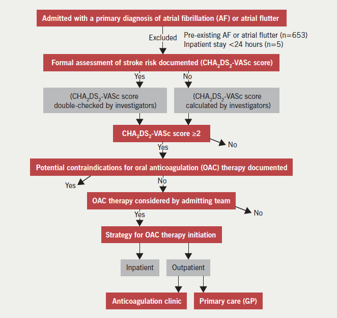
Figure 1. The data collection process

Clinical information was obtained from paper and electronic health records. The following baseline clinical characteristics were recorded: age, sex, hypertension, diabetes mellitus, congestive heart failure (CHF), ischaemic heart disease (IHD), peripheral vascular disease (PVD), and previous stroke. CHF was defined by prior history or imaging evidence of a left ventricular ejection fraction <40%. IHD was defined by a history of angina, myocardial infarction, myocardial revascularisation, or by use of anti-anginals. PVD was defined by self-reported lower limb claudication or a history of lower limb arterial angioplasty or bypass surgery.

Statistical analysis

Continuous variables with a normal distribution were compared using the student t-test, and the Mann Whitney U-test was used to compare non-normal data. Categorical variables were compared using the Z-test. Odds ratios and relative risks were also calculated. All analyses were conducted using SPSS statistical software. A p value <0.05 was considered to be significant. This study was approved as an audit/quality improvement project.

Results

In the one-year study period, 234 patients were newly diagnosed with AF or atrial flutter during a hospital admission >24 hours. Their baseline characteristics are shown in table 1. Two hundred and one (86%) patients had a CHA2DS2-VASc score ≥2, eight patients had a CHA2DS2-VASc score of 1 and 25 patients had a CHA2DS2-VASc score of 0.
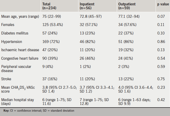
Table 1. Baseline patient cohort characteristics

Formal assessment of stroke risk

CHA2DS2-VASc scores were documented in the medical records of 202 (86%) of 234 patients. Among the 201 patients in whom we determined that OAC was clearly indicated, CHA2DS2-VASc scores were documented in 179. In the remaining 22 patients, there was no documentation to suggest that stroke risk had been assessed or that OAC therapy had been considered. Our retrospective assessment of stroke risk showed that these patients had a mean CHA2DS2-VASc score of 3.8. None of these patients were commenced on OAC therapy. Neither were any of the 33 patients with a CHA2DS2-VASc score ≤1.

Initiation of oral anticoagulation
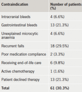
Table 2. Contraindications to oral anticoagulation

Among the 179 patients who were considered for OAC therapy, 61 had documented contraindications or declined therapy (table 2). Three further patients were not anticoagulated (inappropriately) because of the paroxysmal nature of their AF. The remaining 115 patients were intended to receive OAC: 56 were initiated as an inpatient (49%) and 59 (51%) were referred for outpatient initiation (table 1).

Those with CHA2DS2-VASc scores above the mean (≥4) were not more likely to have inpatient OAC compared with those below the mean (odds ratio [OR] 0.72; 95% confidence interval [CI] 0.39–1.34; p=0.31). Cardiologist compared with non-cardiologist was not more likely to use inpatient initiation (OR 1.08; 95% CI 0.58–2.00; p=0.81), however, they were more likely to utilise anticoagulation clinics rather than GPs for outpatient OAC (OR 13.67; 95% CI 6.14–30.38; p<0.0001).

Initiation of OAC as an outpatient was associated with poor uptake, with 18 patients failing to commence on therapy, and mean delays of 33 days as a whole, and 93 days in the primary care arm (table 3). Those that failed to be initiated on OAC had a mean and maximum CHA2DS2-VASc score of 4.6 (range 2–6; standard deviation [SD] 1.4) and 6, respectively. Those patients with a scheduled (n=25) versus non-scheduled (n=4) anticoagulant clinic appointment were commenced on OAC within a mean of six and 37 days, respectively.
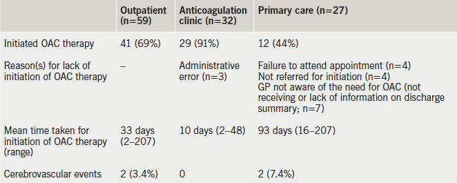
Table 3. The number of patients that were initiated on oral anticoagulant (OAC) therapy as an outpatient, the time taken, the reason(s) for lack of initiation and the number of cerebrovascular events during follow-up

Clinical events during mean follow-up of 194 days (range 76–365, table 3)

In the two patients (CHA2DS2-VASc score of 4) that suffered a stroke, OAC therapy was planned but was not started due to, either the GP not receiving the discharge summary, or failing to refer the patient for initiation of OAC therapy. There were no reported episodes of major bleeding among the patients who were commenced on OAC.

Discussion

Stroke is a potentially devastating condition, which is associated with rates of death and major disability of 7–10%11 and 10–12%,12 respectively. The efficacy of OAC in preventing AF-related strokes is well established. Despite this, only 82% of these patients in our cohort with CHA2DS2-VASc score ≥2, and no contraindications, were referred for OAC, and still fewer (69% of those in whom OAC was indicated) were initiated on such therapy.

These data add to the considerable weight of evidence, which shows that OAC is underused in patients with AF,11 irrespective of the way in which they present. Patients are only likely to receive OAC if their increased risk for stroke is recognised. In this study, 32 (14%) of 234 patients did not have a CHA2DS2-VASc score documented in their medical records. Among the 201 patients in whom OAC was clearly indicated, 22 (12%) patients did not undergo formal risk stratification for stroke. This emphasises the importance of education.

Among the patients who were considered for OAC, more than one-third were felt to have contraindications to therapy, with recurrent falls or gastrointestinal bleeding being most common, or the patient declined therapy. Many of the contraindications were relative ones, which do not preclude OAC if the stroke risk is high.13-15 A study has demonstrated that the calculated risk of subdural haematoma from falling was such that a patient with a 5% annual stroke risk for AF would need to fall 295 times in a year for the fall risk to outweigh the stroke reduction benefit of OAC.16 Among patients who are at increased risk of bleeding, clinicians appear more ready to prescribe aspirin than OAC therapy despite its limited efficacy for stroke prevention and a similar rate of major bleeding compared with OAC.8,17,18

Outpatient OAC therapy was associated with a high failure rate, and the main explanation for this, in more than 56% of cases, was poor discharge communication from secondary to primary care and administrative errors. Outpatient initiation of OAC therapy was also associated with considerable delays to initiation of treatment (mean 33 days). Patients who were dependent upon their GP to organise initiation of OAC therapy waited an average of three months before they were initiated on OAC therapy. By contrast, those who left hospital with a scheduled anticoagulation clinic appointment waited a significantly shorter time to start therapy (six days). It is difficult to see the rationale for introducing an additional step in the clinical pathway by asking the GP to organise OAC therapy. It is, thereby, the discharging team’s responsibility to organise outpatient anticoagulation. Interestingly, patients who had a CHA2DS2-VASc score above the mean were no more likely to be initiated on OAC as an inpatient than other patients (OR 0.72; 95% 0.39–1.34; p=0.31).

In our cohort, two patients that were intended to initiate OAC therapy but did not, suffered an ischaemic stroke. Inpatient OAC therapy initiation negates the possibility that it will not be started later, but there is a fear that the time to achieve therapeutic international normalised ratios (INRs) will prolong hospital stay. However, we have demonstrated that this approach does not cause longer hospital stays compared with outpatient initiation (median 6 vs. 7 days; p=0.42) and it has been reported that therapeutic INRs can be achieved within this timeline.19,20

Three NOACs are approved by the National Institute for Health and Care Excellence (NICE) for the prevention of non-valvular AF-related strokes.21 Despite faster onset of action compared with warfarin, in our centre, their use is restricted to those either allergic to warfarin or who have erratic INRs, and they are not widely prescribed. Indeed, in our cohort, only three patients were commenced on a NOAC. Their wider use might reduce the length of stay through obviating the need to achieve therapeutic INRs. A third approach, which was not used in this study, is to use a low molecular weight heparin in discharged patients until a therapeutic INR is achieved.

Limitations

The retrospective design of this study makes it more prone to bias than prospective studies. The composition of the study population, for example, was dependent upon the accuracy of hospital discharge coding data. Its accuracy for the positively identified cases was validated by a review of the medical records of a random subgroup of 80 patients among whom the discharge code I48 was correct in 79 (99%) cases. However, it is not known how many hospitalised patients with AF were not given the correct discharge code and were, consequently, not included in the study population.

In this study, we described physician-reported contraindications to OAC but we did not record whether bleeding risk was formally assessed using a tool such as the HAS-BLED score.

Conclusion

In this single-centre study of hospitalised patients with newly diagnosed AF, only 82% of patients with a CHA2DS2-VASc score ≥2 were referred for initiation of OAC, and still fewer actually received treatment. Outpatient referral for OAC therapy was associated with poor uptake and significant delays. Patient pathways that maximise treatment uptake and minimise delays are required.

Conflict of interest

None declared.

Editors’ note

See also the editorial in this issue.

Key messages

  • Formal risk stratification for stroke is still not performed in all patients with atrial fibrillation (AF)
  • A large group of patients with AF where oral anticoagulation (OAC) is indicated still fail to receive such therapy
  • Inpatient OAC does not result in longer hospital stays
  • Outpatient OAC is associated with poor uptake and significant delays

References

1. Chugh SS, Havmoeller R, Narayanan K et al. Worldwide epidemiology of atrial fibrillation: a global burden of disease 2010 study. Circulation 2014;129:837–47. http://dx.doi.org/10.1161/CIRCULATIONAHA.113.005119

2. Wolf PA, Abbott RD, Kannel WB. Atrial fibrillation as an independent risk factor for stroke: the Framingham study. Stroke 1991;22:983–8. http://dx.doi.org/10.1161/01.STR.22.8.983

3. Benn Christiansen C, Gislason G, Torp-Pedersen C et al. Primary prevention of ischemic stroke: implication of multiple risk factors in patients with and without atrial fibrillation. European Society of Cardiology 2013 Congress, Amsterdam, the Netherlands; 31 August 2013.

4. Marini C, De Santis F, Sacco S et al. Contribution of atrial fibrillation to incidence and outcome of ischaemic stroke: results from a population-based study. Stroke 2005;36:1115–19. http://dx.doi.org/10.1161/01.STR.0000166053.83476.4a

5. Lin HJ, Wolf PA, Kelly-Hayes M et al. Stroke severity in atrial fibrillation. The Framingham Study. Stroke 1996;27:1760–4. http://dx.doi.org/10.1161/01.STR.27.10.1760

6. Jorgensen HS, Nakayama H, Reith J. Acute stroke with atrial fibrillation. The Copenhagen stroke study. Stroke 1996;27:1765–9. http://dx.doi.org/10.1161/01.STR.27.10.1765

7. Atrial Fibrillation Association. Why the UK must address the personal, clinical and economic impact of atrial fibrillation. June 2010. Available from: http://www.atrialfibrillation.org.uk/files/file/af-afa-keeping-your-finger-on-the-pulse-report.pdf

8. Andersen LV, Vestergaard P, Deichgraeber P et al. Warfarin for the prevention of systemic embolism in patients with non-valvular atrial fibrillation: a meta-analysis. Heart 2008;94:1607–13. http://dx.doi.org/10.1136/hrt.2007.135657

9. Capodanno D, Capranzano P, Giacchi G et al. Novel oral anticoagulants versus warfarin in non-valvular atrial fibrillation: a meta-analysis of 50, 578 patients. Int J Cardiol 2013;167:1237–41. http://dx.doi.org/10.1016/j.ijcard.2012.03.148

10. The Task Force for the Management of Atrial Fibrillation of the European Society of Cardiology (ESC). Guidelines for the management of atrial fibrillation. Eur Heart J 2010;31:2369–429. http://dx.doi.org/10.1093/eurheartj/ehq278

11. Cowan C, Healicon R, Robson I et al. The use of anticoagulants in the management of atrial fibrillation among general practices in England. Heart 2013;99:1166–72. http://dx.doi.org/10.1136/heartjnl-2012-303472

12. Royal College of Physicians National Sentinel Stroke Clinical Audit 2010 Round 7. Public report for England, Wales and Northern Ireland, prepared on behalf of the Intercollegiate Stroke Working Party 2011. Available from: https://www.rcplondon.ac.uk/sites/default/files/national-sentinel-stroke-audit-2010-public-report-and-appendices_0.pdf

13. O’Brien EC, Holmes DN, Ansell JE et al. Physician practices regarding contraindications to oral anticoagulation in atrial fibrillation: findings from the Outcomes Registry for Better Informed Treatment of Atrial Fibrillation (ORBIT-AF) registry. Am Heart J 2014;167:601–09. http://dx.doi.org/10.1016/j.ahj.2013.12.014

14. Rosenman MB, Simon TA, Teal E et al. Perceived or actual barriers to warfarin use in atrial fibrillation based on electronic medical records. Am J Ther 2012;19:330–7. http://dx.doi.org/10.1097/MJT.0b013e3182546840

15. Garwood CL, Corbett TL. Use of anticoagulation in elderly patients with atrial fibrillation who are at risk for falls. Ann Pharmacother 2008;42:523–32. http://dx.doi.org/10.1345/aph.1K498

16. Man-Son-Hing M, Nichol G, Lau A et al. Choosing antithrombotic therapy for elderly patients with atrial fibrillation who are at risk for falls. Arch Intern Med 1999;159:677–85. http://dx.doi.org/10.1001/archinte.159.7.677

17. Assiri A, Al-Majzoub O, Kanaan AO et al. Mixed treatment comparison meta-analysis of aspirin, warfarin, and new anticoagulants for stroke prevention in patients with nonvalvular atrial fibrillation. Clin Ther 2013;35:967–84. http://dx.doi.org/10.1016/j.clinthera.2013.05.011

18. Mant J, Hobbs FD, Fletcher K et al. BAFTA investigators; Midland Research Practices Network (MidReC). Warfarin versus aspirin for stroke prevention in an elderly community population with atrial fibrillation (the Birmingham Atrial Fibrillation Treatment of the Aged Study, BAFTA): a randomised controlled trial. Lancet 2007;370:493–503. http://dx.doi.org/10.1016/S0140-6736(07)61233-1

19. Crowther MA, Ginsberg JB, Kearon C et al. A randomized trial comparing 5mg and 10mg warfarin loading disease. Arch Intern Med 1999;159:46–8. http://dx.doi.org/10.1001/archinte.159.1.46

20. Harrison L, Johnston M, Massicotte MP, Crowther M, Moffat K, Hirsh I. Comparison of 5mg and 10mg loading doses in initiation of warfarin therapy. Ann Intern Med 1997;126:133–6. http://dx.doi.org/10.7326/0003-4819-126-2-199701150-00006

21. NICE Implementation Collaborative. Consensus. Supporting local implementation of NICE guidance on use of the novel (non-Vitamin K antagonist) oral anticoagulants in non-valvular atrial fibrillation. London: NICE, June 2014. Available from: https://www.nice.org.uk/guidance/cg180/resources/cg180-atrial-fibrillation-nic-consensus-statement-on-the-use-of-noacs2
UK experience of conversion of radial to femoral access in coronary interventions

Br J Cardiol 2015;22:118doi:10.5837/bjc.2015.031 Leave a comment
Click any image to enlarge
Authors:

Transradial access for coronary procedures is associated with less vascular access site complications. Occasionally, radial access fails and makes conversion to a transfemoral route inevitable. In this paper, which updates UK radial experience, we report the outcomes in a single UK centre in developing a transradial access programme.

We analysed 3,225 consecutive patients who underwent transradial coronary procedures over a five-year period. The primary outcome measure was rate of conversion from transradial to transfemoral access route. Of 3,225 radially approached cases, conversion from radial to femoral access route occurred in 148 patients (4.6%). With experience after the learning curve, the conversion rate fell to 2.0%.

In conclusion, after an initial learning curve, procedural success rate is high with low cross-over rate from radial to femoral entry site.

Introduction

Arterial access through the transradial approach was described in the late 1980s.1 Radial approach in coronary interventions has advantages over the femoral approach with less vascular complications, less access site bleeding and earlier discharge post-procedure.2-4 In the PREVAIL (Prospective REgistry of Vascular Access in Interventions in Lazio region) study, patients who underwent coronary procedures through the radial artery had a statistically significant reduction in both major and minor bleeding (4.2% vs. 1.96%, p=0.03) and death or myocardial infarction (3.1% vs. 0.6%, p=0.005).5

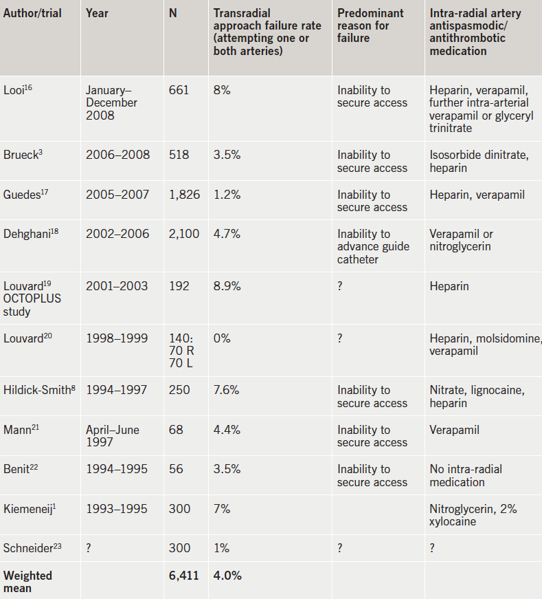
Despite the multiple advantages of the transradial access route, many cardiologists remain unconvinced and unwilling to change practice, as the radial access procedure is technically more challenging, with a steep learning curve. Multiple studies have reported various rates of transradial approach failure, varying from 0 to 8.9% (table 1).

Table 1. International trials that had radial approach failure requiring femoral access

The number of coronary procedures performed via the radial artery access route is increasing, but unfortunately, radial access still accounts for less than 10% of procedures worldwide. In the US, the overall rate of radial use remains at 10–16% of percutaneous coronary intervention (PCI) procedures.6,7

UK literature on this subject is limited. The purpose of this retrospective study is to report a UK single-centre experience developing a transradial approach. High success rate with low radial to femoral cross-over rate are described.

To our knowledge, since a discouraging paper published in 1998,8 this is the first large single-centre study that has assessed conversion rate from radial to femoral approach in a UK centre.

Methods

We studied all consecutive emergency, urgent and elective patients who underwent diagnostic coronary angiography and/or PCI procedures using an initial radial artery vascular access at Watford General Hospital, over five years from April 2009 to March 2014. From these data we identified all patients in whom the procedure was initially started using the radial artery vascular access, but were converted during the procedure to a femoral artery access route. The initial selection of the arterial access route was decided by the operator and the patients’ preference. The radial access site was routinely obtained using local anaesthetic, a 21-gauge needle, 0.018 inch guide wire and 11 cm 5Fr or 6Fr sheaths for diagnostic and PCI procedures, respectively. All cases who had an initial radial artery vascular access approach had routine pre-procedure sublingual glyceryl trinitate 400–800 µg. After introduction of the sheath, all patients also routinely received 50 µg/kg heparin, 200 µg isosorbide dinitrate and 5 mg verapamil by intra-arterial administration through the sheath.

Cardiac procedures in the catheterisation laboratory were reported by the primary operator using the McKesson Horizon Cardiology Medcon reporting module. Cardiac clinical physiologists also separately reported all procedures using the Siemens Sensis angiography reporting module. We reviewed all clinical and procedural data and the outcome of the procedures where more than one vascular access site was used from each database. Indication for the procedure, the type of procedure, operator (diagnostician or interventionalist, consultant or trainee), reason for conversion and any complications during conversion to another arterial access site were documented.

The outcome of this retrospective study was to analyse the conversion rate from radial to femoral arterial access, at our centre over a five-year period of time and over consecutive serial six-month periods. We also documented the reason for conversion and the relationship to the operators’ experience. Analysis of the data was performed using IBM SPSS statistics software. Our data contain categorical variables, which are reported as absolute values and actual percentages. Comparison between categorical variables was performed using Pearson Chi-square test.

Results

A total of 8,912 consecutive diagnostic and interventional coronary angiographic procedures were performed at Watford General Hospital over a five-year period, from April 2009 to March 2014. There were 3,225 cases (36%) done using the radial access route. Of these, 1,374 procedures (43%) were diagnostic angiography and 1,851 (57%) were PCI or potential PCI.

The primary operator in 2,422 (75%) cases was a consultant cardiologist. In 803 procedures (25%) the primary operator was a specialist registrar (trainee), directly supervised by a consultant cardiologist.

In 148 patients (4.6% of the radial cases), the initial vascular access site was changed from radial artery to femoral artery during the procedure. Of these cases, 89 had diagnostic angiography and 59 were interventional cases.

The conversion rate from radial access site to femoral access site was 4.3% in procedures performed by consultants, whereas this rate was 5.6% in procedures in which specialist trainees were the primary operators. This difference was not statistically significant (p=0.11).

The conversion rate from radial access site to femoral access site was 3.8% in procedures performed by interventionalists, and 6.1% in procedures performed by diagnosticians. This difference was not statistically significant (p=0.11).

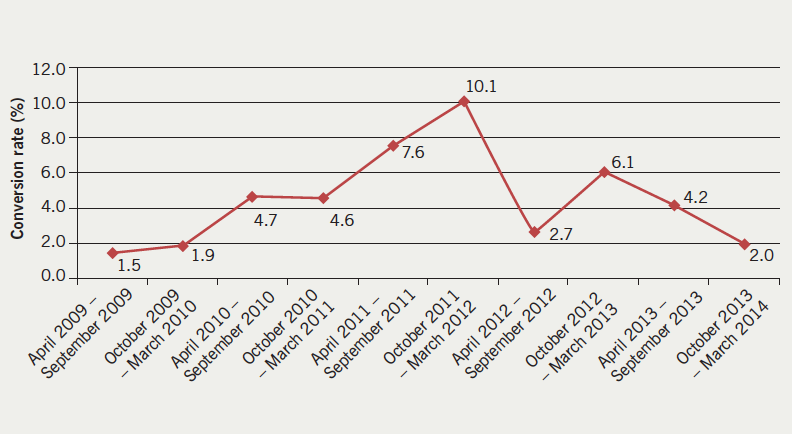
The conversion rate was also assessed over consecutive six-monthly periods (figure 1). The conversion rate was 1.5% (two of 133 cases) during the initial six months of starting the radial approach technique in our laboratories in 2009. The conversion rate increased to a maximum of 10.1% (27 of 267 cases) more than two years later. The most recent conversion rate was 2.0% (9 of 435 cases) between October 2013 and March 2014.

Figure 1. The conversion rate from radial to femoral access route over five years at Watford General Hospital

Causes of failed radial attempt

Table 2. Reason for conversion from radial artery access to femoral access route

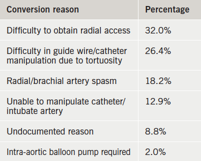
The reasons for conversion from radial to femoral access site in our laboratory are categorised in six main groups and listed in table 2. The most common reason was difficulty in securing radial access (31.8%). This includes inability to puncture the radial artery due to small size, inability to advance the wire through the needle into the artery, and inability to advance the sheath.

The second most common reason of failure was inability to advance the guide wire or catheter due to tortuosity of brachial or axillary artery (26.4%). Inability to advance or manipulate the guide wire and catheter due to radial or brachial spasm accounted for 18.2% of failures. In 12.9% of cases, intubation of the left or right coronary artery was not possible due to difficulty in advancing or manipulating the catheter. In 13 cases (8.8%), no reason for conversion was documented. A further 2% of patients required intra-aortic balloon pump (IABP).

There were no statistically significant differences between various groups of operators (consultants, trainees, interventionists, diagnosticians) in the reasons for conversion from radial access to a femoral one (p=0.76).

Complications

Complications occurred in six of the 148 conversion cases. Five patients developed a vasovagal reaction (3.4%). Dissection of radial artery, which required surgical intervention, occurred in one patient.

Discussion

In 2009, Watford General Hospital commenced a transradial vascular access programme. During early phases of adopting this technique, a relatively small proportion of patients were chosen for this approach. Over the first 12 months of initiating a radial vascular access technique the conversion rate was the lowest of these five years (1.5–1.9%). This high number of successful procedures with low conversion rate may well reflect that less complicated cases were chosen by operators, who were less experienced at the beginning of adopting the radial technique. With increasing experience, the volume of coronary procedures performed via the radial access route increased, including more difficult cases; the conversion rate increased for up to two years.

We believe the operators’ experience and proficiency increased with higher volumes of radial procedures. The concept of a learning curve, in which operators’ skills improve with increased number of procedures, was demonstrated in this observational study. With more experienced operators, the success rate has become more consistent. The conversion rate has fallen to about 2.0% in recent months.

Table 1 lists the conversion rates reported in the medical literature. The overall reported weighted conversion rate was 4.0%. We suggest that, as a clinical standard, an appropriate conversion rate for experienced operators should be approximately 2%, and less than 4%.

In 2013, a common consensus view on the technical, clinical and organisational implications of using the radial approach for coronary angiography and interventions was developed by the European Association of Percutaneous Cardiovascular Interventions (EAPCI).9 The goal of the association was to define the role of the radial approach in modern practice, and to provide a consensus on different aspects of the transradial procedure, such as learning curve and conversion rate. According to this consensus, the conversion rate in recent registries has been as low as 1.5%, which is due to improved technique and materials. In the published literature, however, it varies between zero and 8.9%. The learning curve for radially performed procedures is steep, with no significant difference in failure rate after 50 cases compared with that of experienced operators.7,10 The relationship between volume of procedures and failures demonstrates a high failure rate, with wide inter-operator variability for the first one to 50 radial cases, and a consistent reduction in failure rate with higher transradial volumes. It is agreed that a case-volume threshold of 50–80 transradial procedures is required to overcome the learning-curve threshold.11 This number, however, can be individual and vary from operator to operator.

One study, that has specifically analysed learning curve to assess radially approached coronary procedures, is based on single-centre data from Canada.10 In this study, authors showed that a case-volume threshold of at least 50 transradial procedures was required for new radial operators to achieve similar procedural outcomes as experienced radial performers.

The learning curve experience at our centre was more than 200 cases per operator, considerably more than the literature suggests. Difficulty in obtaining access is one of the main concerns in radial technique. Compared with the transfemoral route, the radial artery is small in size with a tendency to develop spasm. Older, female, and shorter patients more often exhibit anatomic anomalies in the arms,12 as well as tortuous brachiocephalic arteries,13 small radial arteries,12 and radial spasm.14 The most difficult anatomical variations for transradial approach are the radiolunar loop and the presence of a lusoria subclavian artery.15

Difficulty in obtaining access was the main cause of failure of radial attempts in our centre. Difficult arterial puncture and radial artery spasm account for most failures during the first 50 cases.

With increasing transradial experience, operators can select more complex cases without sacrificing procedural success.

Strengths

This study is a large analysis with a low chance of selection bias. It provides contemporaneous data of radial access. Since 1998, when Hildick-Smith et al.8 published a UK experience of coronary angiography from the radial artery – and discouraged this technique due to its limitations – no further studies have assessed radial to femoral conversion rate in a UK centre.

Study limitations

This is a retrospective analysis of a large data set. In our department, consultant cardiologists supervise trainees very closely. This might reflect why any differences between the two groups of operators were not statistically significant.

Conclusion

This single-centre, large, retrospective analysis of all radially performed diagnostic coronary angiography and interventional procedures shows that after an initial learning curve, there is a high success rate with low cross-cover rate from radial to femoral entry site. This low failure rate matches the world literature.

Conflict of interest

None declared.

Key messages

  • Transradial access is advantageous over transfemoral access, with fewer vascular complications
  • The transradial approach is preferred in ST-elevation myocardial infarction (STEMI)
  • After 50–80 cases, the learning-curve threshold will be overcome
  • All radial-proficient team members should aim to maintain transfemoral competency
  • Training in the procedure is safe when undertaken by trainees supervised by consultants

References

1. Kiemeneij F, Laarman GJ. Percutaneous transradial artery approach for coronary stent implantation. Cathet Cardiovasc Diagn 1993;30:173–8. http://dx.doi.org/10.1002/ccd.1810300220

2. Jolly SS, Yusuf S, Cairns J et al. Radial versus femoral access for coronary angiography and intervention in patients with acute coronary syndromes (RIVAL): a randomised, parallel group, multicentre trial. Lancet 2011;377:1409–20. http://dx.doi.org/10.1016/S0140-6736(11)60404-2

3. Brueck M, Bandorski D, Kramer W, Wieczorek M, Holtgen R, Tillmanns H. A randomized comparison of transradial versus transfemoral approach for coronary angiography and angioplasty. JACC Cardiovasc Interv 2009;2:1047–54. http://dx.doi.org/10.1016/j.jcin.2009.07.016

4. Jolly SS, Amlani S, Hamon M, Yusuf S, Mehta SR. Radial versus femoral access for coronary angiography or intervention and the impact on major bleeding and ischemic events: a systematic review and meta-analysis of randomized trials. Am Heart J 2009;157:132–40. http://dx.doi.org/10.1016/j.ahj.2008.08.023

5. Pristipino C, Trani C, Nazzaro MS et al. Major improvement of percutaneous cardiovascular procedure outcomes with radial artery catheterisation: results from the PREVAIL study. Heart 2009;95:476–82. http://dx.doi.org/10.1136/hrt.2008.150714

6. Feldman DN, Swaminathan RV, Kaltenbach LA et al. Adoption of radial access and comparison of outcomes to femoral access in percutaneous coronary intervention: an updated report from the national cardiovascular data registry (2007–2012). Circulation 2013;127:2295–306. http://dx.doi.org/10.1161/CIRCULATIONAHA.112.000536

7. Hess CN, Peterson ED, Neely ML et al. The learning curve for transradial percutaneous coronary intervention among operators in the United States: a study from the national cardiovascular data registry. Circulation 2014;129:2277–86. http://dx.doi.org/10.1161/CIRCULATIONAHA.113.006356

8. Hildick-Smith DJ, Lowe MD, Walsh JT et al. Coronary angiography from the radial artery – experience, complications and limitations. Int J Cardiol 1998;64:231–9. http://dx.doi.org/10.1016/S0167-5273(98)00074-6

9. Hamon M, Pristipino C, Di MC et al. Consensus document on the radial approach in percutaneous cardiovascular interventions: position paper by the European Association of Percutaneous Cardiovascular Interventions and Working Groups on Acute Cardiac Care and Thrombosis of the European Society of Cardiology. EuroIntervention 2013;8:1242–51. http://dx.doi.org/10.4244/EIJV8I11A192

10. Ball WT, Sharieff W, Jolly SS et al. Characterization of operator learning curve for transradial coronary interventions. Circ Cardiovasc Interv 2011;4:336–41. http://dx.doi.org/10.1161/CIRCINTERVENTIONS.110.960864

11. Burzotta F, Trani C, Mazzari MA et al. Vascular complications and access crossover in 10,676 transradial percutaneous coronary procedures. Am Heart J 2012;163:230–8. http://dx.doi.org/10.1016/j.ahj.2011.10.019

12. Yoo BS, Yoon J, Ko JY et al. Anatomical consideration of the radial artery for transradial coronary procedures: arterial diameter, branching anomaly and vessel tortuosity. Int J Cardiol 2005;101:421–7. http://dx.doi.org/10.1016/j.ijcard.2004.03.061

13. Cha KS, Kim MH, Kim HJ. Prevalence and clinical predictors of severe tortuosity of right subclavian artery in patients undergoing transradial coronary angiography. Am J Cardiol 2003;92:1220–2. http://dx.doi.org/10.1016/j.amjcard.2003.07.038

14. Jia DA, Zhou YJ, Shi DM et al. Incidence and predictors of radial artery spasm during transradial coronary angiography and intervention. Chin Med J (Engl) 2010;123:843–7.

15. Valsecchi O, Vassileva A, Musumeci G et al. Failure of transradial approach during coronary interventions: anatomic considerations. Catheter Cardiovasc Interv 2006;67:870–8. http://dx.doi.org/10.1002/ccd.20732

16. Looi JL, Cave A, El-Jack S. Learning curve in transradial coronary angiography. Am J Cardiol 2011;108:1092–5. http://dx.doi.org/10.1016/j.amjcard.2011.06.009

17. Guedes A, Dangoisse V, Gabriel L et al. Low rate of conversion to transfemoral approach when attempting both radial arteries for coronary angiography and percutaneous coronary intervention: a study of 1,826 consecutive procedures. J Invasive Cardiol 2010;22:391–7.

18. Dehghani P, Mohammad A, Bajaj R et al. Mechanism and predictors of failed transradial approach for percutaneous coronary interventions. JACC Cardiovasc Interv 2009;2:1057–64. http://dx.doi.org/10.1016/j.jcin.2009.07.014

19. Louvard Y, Benamer H, Garot P et al. Comparison of transradial and transfemoral approaches for coronary angiography and angioplasty in octogenarians (the OCTOPLUS study). Am J Cardiol 2004;94:1177–80. http://dx.doi.org/10.1016/j.amjcard.2004.07.089

20. Louvard Y, Lefevre T, Allain A, Morice M. Coronary angiography through the radial or the femoral approach: the CARAFE study. Catheter Cardiovasc Interv 2001;52:181–7. http://dx.doi.org/10.1002/1522-726X(200102)52:2<181::AID-CCD1044>3.0.CO;2-G

21. Mann T, Cubeddu G, Bowen J et al. Stenting in acute coronary syndromes: a comparison of radial versus femoral access sites. J Am Coll Cardiol 1998;32:572–6. http://dx.doi.org/10.1016/S0735-1097(98)00288-5

22. Benit E, Missault L, Eeman T et al. Brachial, radial, or femoral approach for elective Palmaz-Schatz stent implantation: a randomized comparison. Cathet Cardiovasc Diagn 1997;41:124–30. http://dx.doi.org/10.1002/(SICI)1097-0304(199706)41:2<124::AID-CCD3>3.0.CO;2-9

23. Schneider JE, Mann T, Cubeddu MG, Arrowood ME. Transradial coronary stenting: a United States experience. J Invasive Cardiol 1997;9:569–74.

News from the 17th International Symposium of Atherosclerosis

Br J Cardiol 2015;22:(3) Leave a comment
Click any image to enlarge
Authors:

The latest in lipids medicine was presented at the 17th International Symposium of Atherosclerosis (ISA), which took place in Amsterdam, The Netherlands, from 23rd–26th May 2015. This triannual meeting of the International Atherosclerosis Society (IAS) attracted delegates from 78 countries. Familial hypercholesterolaemia (FH) was very much a hot topic, with the launch at the meeting of FH-CONNECT*, a global initiative aimed at bringing together all stakeholders to improve the care of FH patients. Novel treatments, including the PCSK9 inhibitors and antisense therapies targeting apolipoprotein CIII, were also in the news.

FH and myocardial infarction

A report from EUROASPIRE IV, a survey of secondary prevention management in 24 European countries, highlighted the need to consider FH in patients with a myocardial infarction (MI).1 In this cohort of 7,044 patients, 8.3% of MI patients – one in 12 – had a likely FH diagnosis, based on adapted Dutch Lipid Clinic Network Criteria. This rose to about one in five patients had likely FH in those aged less than 50 years.

According to Dr Joost Besseling (Academic Medical Center, Amsterdam, The Netherlands): “This study indicates that clinicians should consider the possibility of FH diagnosis in patients with a first coronary event, especially young patients. The identification of FH patients also acts as a starting point for family-based cascade screening”.

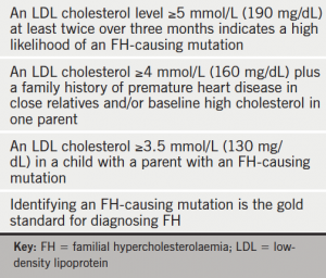
Table 1. Criteria for identifying FH in children

ISA 2015 coincided with publication of the European Atherosclerosis Society (EAS) Consensus Paper on Paediatric FH.2 Heterozygous FH is common, affecting about one in 200–250 people; identifying and treating children with FH early will undoubtedly improve life expectancy and have socioeconomic benefits. “Early detection and treatment of children with FH will save lives and money by preventing early heart attacks in early middle age,” said Professor Gerald Watts (University of Western Australia, Perth, Australia), EAS Consensus Panel Writing Group and lead author of the International FH Foundation guidelines.3

Criteria for diagnosis of FH in children are summarised in the table 1. The EAS Consensus Panel recommends diet, lifestyle and early initiation of a statin, age depending on prescribing information, as the fundamentals of management. This approach can significantly reduce atherosclerosis and improve event-free survival.4,5 Identifying children with FH early also allows for reverse cascade screening of other family members.

New therapies

PCSK9 monoclonal antibody therapy continued to be a hot topic. Reports showed that these treatments were also effective in individuals with mixed dyslipidaemia, i.e. elevated low-density lipoprotein (LDL) cholesterol and elevated triglycerides with or without low high-density lipoprotein (HDL) cholesterol.6,7 Although concerns have been raised about the acceptability of these injectable therapies, a report at ISA 2015 countered this. Provided adequate training is given, the majority of patients (65−72% compared with 11−26% before training, p<0.05) were very willing to consider using self-injectable alirocumab treatment.8

There was also much interest in novel therapies aimed at addressing elevated triglycerides, building on genetic studies showing causality with the individual components of triglyceride-rich lipoproteins (remnant and apolipoprotein [apo] CIII).9−11 Various approaches were discussed, including K-877, a potent and selective peroxisome proliferator-activated receptor alpha agonist, which reduces plasma levels of apoCIII and remnant cholesterol in patients with elevated triglycerides with or without low HDL cholesterol.12 Reduction in apoCIII may also explain the triglyceride-lowering effects of omega-3 carboxylic acids, according to an analysis from the EVOLVE (Evaluation of Cinacalcet Hydrochloride Therapy to Lower Cardiovascular Events) study.13 ApoCIII antisense therapy has been tested in early clinical studies in a broad range of patient groups with moderately to severely elevated triglyceride levels. Although treatment was generally well tolerated, further investigation of effects on liver fat is needed, given the theoretical possibility of hepatic triglyceride accumulation. Looking forward: will treatments targeting triglycerides be the next therapeutic advance after PCSK9 inhibitors?

References

1. De Backer G, Besseling J, Chapman J et al. Prevalence and management of familial hypercholesterolaemia in coronary patients: an analysis of EUROASPIRE IV, a study of the European Society of Cardiology. Atherosclerosis 2015;241:169−75. http://dx.doi.org/10.1016/j.atherosclerosis.2015.04.809

2. Wiegman A, Gidding SS, Watts GF et al. Familial hypercholesterolaemia in children and adolescents: gaining decades of life by optimising detection and treatment. Eur Heart J 2015. http://dx.doi.org/10.1093/eurheartj/ehv157

3. Watts GF, Gidding S, Wierzbicki AS et al. Integrated guidance on the care of familial hypercholesterolaemia from the International FH Foundation. Eur J Prev Cardiol 2015;22:849−54. http://dx.doi.org/10.1177/2047487314533218

4. Kusters DM, Avis HJ, de Groot E et al. Ten-year follow-up after initiation of statin therapy in children with familial hypercholesterolemia. J Am Med Assoc 2014;312:1055–7. http://dx.doi.org/10.1001/jama.2014.8892

5. Braamskamp MJ, Kusters DM, Avis HJ et al. Patients with familial hypercholesterolemia who initiated statin treatment in childhood are at lower risk for CHD than their affected parents. Circulation 2013;128:A17837.

6. Hovingh K, Louie M, Minini P et al. Alirocumab treatment effect did not differ between patients with/without low HDL-C or high triglyceride baseline levels in Phase 3 trials. ISA 2015, Abstract 326.

7. Rosenson RS, Jacobson T, Preiss D et al. Efficacy and safety of the PCSK9 inhibitor evolocumab in patients with mixed dyslipidemia. ISA 2015, Abstract 252.

8. Cariou B, Roth E, Bujas-Bobanovic M et al. Patient and physician perspectives on administration of of the PCVSK9 monoclonal antibody alirocumab, an injectable medication to lower LDL-C levels. ISA 2015, Abstract 1039.

9. Varbo A, Benn M, Tybjærg-Hansen A et al. Remnant cholesterol as a causal risk factor for ischemic heart disease. J Am Coll Cardiol 2013;61:427−36. http://dx.doi.org/10.1016/j.jacc.2012.08.1026

10. Crosby J, Peloso GM, Auer PL et al. TG and HDL Working Group of the Exome Sequencing Project, National Heart, Lung, and Blood Institute. Loss-of-function mutations in APOC3, triglycerides, and coronary disease. N Engl J Med 2014;371:22–31. http://dx.doi.org/10.1056/NEJMoa1307095

11. Jørgensen AB, Frikke-Schmidt R, Nordestgaard BG, Tybjærg-Hansen A. Loss-of-function mutations in APOC3 and risk of ischemic vascular disease. N Engl J Med 2014;371:32−41. http://dx.doi.org/10.1056/NEJMoa1308027

12. Nordestgaard BG. K-877, a SPPARMalpha, improves the proatherogenic lipid profile of hypertriglyceridemic patients, even when on stable statin therapy. ISA 2015, Abstract 1041.

13. Morton A, Furtado JD, Sacks F et al. The effect of omega-3 carboxylic acids on apolipoprotein CIII- containing lipoproteins in moderate to severe hypertriglyceridemia. ISA 2015, Abstract 621.

*Readers wanting more information about FH-CONNECT should see http://www.isa-2015.com/familial-hypercholesterolaemia-getting-global-connection/).

Correspondence: influences on novel oral anticoagulant prescribing – findings of a NICE scholarship project

Br J Cardiol 2015;22:(3) Leave a comment
Click any image to enlarge
Authors:

Correspondence from the world of cardiology

We read with interest Diana Gorog’s recent article on the uptake of non-vitamin K oral anticoagulants (NOACS) in the UK.1 She drew attention to the slow uptake of these agents in the UK as opposed to many countries in Europe, and certainly the USA, and to the role that local medicines management committees (MMCs) may play in this. While the National Institute for Health and Care Excellence (NICE) guidance regarding all three NOACs available in the UK (apixaban, dabigatran and rivaroxaban) is that they should be available as an option for stroke prevention in non-valvular atrial fibrillation (AF), many MMCs in the UK have sought to control prescriptions of the new agents by issuing policies which keep warfarin first line, and reserve NOACs for situations where warfarin is unsuitable.

As part of a NICE scholarship project, we sought to collect further information on the influence of these restrictive local prescribing policies on the prescribing patterns of doctors. The focus of the project was on the Surrey/Hampshire area: this area was chosen as particularly illuminating, as the two neighbouring counties had different anticoagulant prescribing policies. Clinical Commissioning Groups (CCGs) in Surrey followed the policy of the Surrey Prescribing Clinical Network, which states: “Patients with a CHA2DS2-VASc score ≥2 or CHA2DS2-VASc score 1 and considered high risk should be initiated on warfarin in the first instance, unless contraindicated. Warfarin remains the agent of choice for the prevention of stroke and systemic embolism in AF”.

In Hampshire, on the other hand, CCGs largely followed the policy of Basingstoke, Southampton and Winchester District Prescribing Committee. This did not restrict clinician choice, but simply stated NICE’s position that new anticoagulants should be considered as an option. Particularly significantly, it stated: “Prescribers may wish to consider if newly diagnosed patients are suitable for the new OACs in the first instance”.

Table 1. Clinical Commissioning Groups (CCGs) prescribingdabigatran
Table 1. Clinical Commissioning Groups (CCGs) prescribing
dabigatran

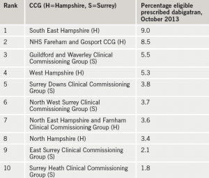
We focused on one of the NOACs – dabigatran – and compared prescribing rates in the five Hampshire and five Surrey CCGs. Data on number of prescriptions was obtained from dabigatran’s manufacturer, Boehringer Ingelheim; this was combined with CCG population numbers (Office for National Statistics data2) and AF prevalence (Health and Social Care Information Centre data3) to give an estimate of the population eligible for anticoagulation. Significantly, the top two CCGs for dabigatran prescribing were both in Hampshire, and the bottom two both in Surrey (see table 1).


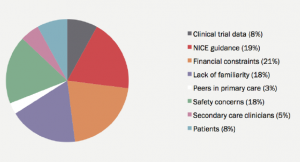
Figure 1. Most important influence on dabigatran prescribing, asselected by Surrey and Hampshire primary care prescribers
Figure 1. Most important influence on dabigatran prescribing, as selected by Surrey and Hampshire primary care prescribers

We then sought further evidence for the influence of local prescribing policies by surveying 120 primary care prescribers in the Surrey/Hampshire area. 33% responded; two thirds of respondents felt that their prescribing of dabigatran was influenced by financial pressure from their local CCG. Interestingly, however, there was no difference between Surrey and Hampshire prescribers in their answers to this question. We also asked respondents to pick the factor which had the strongest influence on their decision making regarding dabigatran prescribing. Replies showed an even split between NICE guidance, financial constraints, safety concerns and lack of familiarity (see figure 1).

We concluded that local prescribing policies do have an effect on prescribing habits, but that they are only one of a number of factors which influence prescribers when they are selecting an anticoagulant. NICE guidance clearly ranks among the most important of these factors, which is in agreement with data published recently in this journal by Campbell et al, suggesting that NOAC prescribing in the UK has increased since the publication of the revised NICE guidance on atrial fibrillation in 2014.4 This guidance emphasises the importance of anticoagulation for all but the lowest risk patients, and indicates that vitamin K antagonists and NOACs should be considered as equally valid options.5

As an aside, we also collected data on prescription rates for all anticoagulants, including warfarin, over the year from November 2012 to October 2013; we found a steady increase over time in prescriptions for all the anticoagulants. This confirms the encouraging data from registries such as PREFER in AF,6 which suggest that, regardless of anticoagulant used, increasing numbers of patients in the UK with AF who are at risk of stroke are being offered the protection of anticoagulation.

Acknowledgements

Dr C Hendry, Royal Surrey County Hospital (Project Supervisor); Mr B White, Nottingham University Hospital (NICE Mentor); and Mr F Ratcliff, Boehringer Ingelheim

Sources of support

This work was undertaken as part of National Institute for Health and Care Excellence Scholarship scheme 2013–2014.

Conflict of interest

None declared.

Dr Matthew Rogers
Haematology SpR
Royal Marsden Hospital, Sutton, Surrey SM2 5PT

References

1. Gorog D. Rivaroxaban in non-valvular AF – UK experience in perspective. Br J Cardiol 2014;21 (suppl 1):S1–S11.

2. Annual Mid-year Population Estimates for Clinical Commissioning Groups, 2011. Office for National Statistics, August 2013. http://www.ons.gov.uk/ons/publications/re-reference-tables.html?edition=tcm%3A77-318167#tab-all-tables. Accessed July 2015.

3. Quality and Outcomes Framework, 2011–12, PCT level. Health and social care information centre, published October 2012. http://www.hscic.gov.uk/catalogue/PUB08722. Accessed July 2015.

4. Cowan C, Fay M, Maskrey N. The new NICE AF guideline and NOACs: a response. Br J Cardiol 2015;22:53–5. http://dx.doi.org/10.5837/bjc.2015.019

5. National Institute for Health and Care Excellence. Atrial fibrillation: the management of atrial fibrillation. CG180. London: NICE, June 2014. Available from: http://www.nice.org.uk/guidance/cg180

6. Kirchhof P, Ammentorp B, Darius H et al. Management of atrial fibrillation in seven European countries after the publication of the 2010 ESC Guidelines on atrial fibrillation: primary results of the PREvention oF thromboemolic events – European Registry in Atrial Fibrillation (PREFER in AF). Europace 2014;16:6–14. http://dx.doi.org/10.1093/europace/eut263

Book review

Br J Cardiol 2015;22:(3) Leave a comment
Click any image to enlarge
Authors:

The ESC textbook of preventive cardiology

Screen shot 2015-07-15 at 13.01.09
Publisher: Oxford University Press, 2015
ISBN: 978-0-19-965665-3
Price: £115 (print), £29 (online, 1-year subscription)

It is widely accepted knowledge among health professionals and the general public that premature cardiovascular disease can be prevented. However the evidence and guidelines on prevention are typically scattered, making integration into clinical practice problematic. The ESC textbook of preventive cardiology aims to collate all the aspects of prevention into one textbook.

While the title may give the initial impression that the editors are going to be providing the old rhetoric on hypertension, smoking and lipid control, a quick glance at the preface and contents reveal that they are aiming far beyond these basics.

The editors correctly point out that “there is no book that unites” the principles and practice of preventive cardiology. They are promising “a single resource” with a “practical hands-on approach”, that will help the reader integrate the knowledge in this book into their clinical practice. In the preface it is stated that this book is not only aimed at doctors but any health professional that has to deal with cardiovascular disease. It’s available in both print and digital formats with the online version to be updated when new evidence becomes available.

The initial chapter on epidemiology swiftly brings the reader up to date with the past 60 years of research into the prevention of cardiovascular disease. While it is clearly outlined that mortality has significantly improved, the reader is presented with evidence for a potential “new phase of the epidemic” of cardiovascular disease mainly related to the meteoric rise of obesity and diabetes. This is an overarching theme that appears time and time again throughout the book.

Chapters dedicated to modification of risk factors are expertly written and provide the reader with the current evidence and guidelines for management of patients. Clinical application is further supported by accompanying case studies for which a solution is provided at the end of each chapter. The cases are well thought out and thoroughly explained making them a valuable addition to the textbook and well worth reading.

Of particular note to me was the section on imaging in cardiovascular prevention. This is an area I’ve previously found a minefield with both understanding the indications, relative advantages and disadvantages and interpretation of the numerous imaging modalities available within cardiovascular radiology. Extensive, but clear, tables outlining the various properties of each modality are of great reference to anyone regularly ordering and interpreting cardiovascular radiology.

The editors admit in the preface that this textbook “will not be perfect in all aspects”, and there are areas in which this is true. Risk assessment is a fundamental backbone of prevention, but the section on this mainly pertains to use of the SCORE risk estimation system. Writing from a UK perspective, where QRISK and the more recent JBS3 guidelines (which are notably completely absent from the comparative table), makes the risk assessment section less applicable than it could be. Also, focussing on the SCORE system and its unique use of 10-year risk of mortality rather than risk of events, which it admits is not “universally popular”, I feel adds confusion rather than simplifying the issue.

I found the delivery of preventive cardiology sections not hugely useful. They seem squarely aimed at how to set up a service. Tables outlining the minimum equipment requirements, personnel and organisational issues could be better laid out in books covering cardiac rehabilitation already available.

Overall, I feel the book succeeds in its aim to provide a valuable single resource for the health professional faced with assessing and managing patients and their cardiovascular risk. The evidence and subsequent guidance offered brings the reader up to date and also gives a glimpse of what trends and developments may be around the corner. Not all sections will be applicable to everyone but with such a wide proposed audience base that’s to be expected.

The fervent tone of the book promises to update the digital version and the simple fact that this one of the first books (and the only one in the past five years) dedicated to this subject make this a valuable resource to any health professional that is involved in cardiovascular prevention.

Jonty Bennett
FY2 Doctor, London

FY1 in heart failure: the good, the bad and the ugly! Reflections by the FY1 doctors in heart failure and their supervisor on the first year of a new post

Br J Cardiol 2015;22:(3)doi:10.5837/bjc.2015.023 Leave a comment
Click any image to enlarge
Authors:
First published online July 16th, 2015

With the expansion of the heart failure services to meet the rise in demand, we established, in Sheffield, a new training post for the junior medical staff in their first year of training. This is a four-month post for the Foundation Year one (FY1) doctors in heart failure. The post differs from the classic FY1 posts in that it is based in the heart failure multi-disciplinary team (HF-MDT) rather than being ward-based. Thus, the trainee works under the supervision of a consultant cardiologist with an interest in heart failure, and works alongside a group of heart failure specialist nurses screening new admissions for heart failure, and offering advice and follow-up of patients with heart failure who are not under the care of the cardiologists. The trainee attends the heart failure diagnostic clinic along with the consultant cardiologist, and participates in the work of the HF-MDT ward round. These are the collective personal views of the first three trainees who have worked in this post in the year 2013–2014; with a footnote from their supervisor.

The three trainees and their supervisor. From left to right: Dr Sarah Soar, Dr Philippe Wheeler,Dr Laura Styles and Dr Abdallah Al-Mohammad
The three trainees and their supervisor. From left to right: Dr Sarah Soar, Dr Philippe Wheeler,
Dr Laura Styles and Dr Abdallah Al-Mohammad

Introduction

For newly qualified doctors, the Foundation Programme provides a stimulating and exciting entry into a career in medicine. As the name suggests, doctors work within a range of specialties and environments in order to build on the knowledge learnt at medical school, and develop as a clinician in preparation for specialty training. We had the privilege of being the first to work as foundation doctors in a new role – FY1 in heart failure – and, in this article, we hope to outline some of the joys and challenges we have encountered.

The heart failure (HF) service at Sheffield Teaching Hospitals1 was run by a consultant cardiologist,* heart failure specialist nurses, a dedicated administration team, and from summer 2013, an FY1 doctor. The HF multi-disciplinary team (MDT) cares for inpatients and outpatients, and is linked to a community team of specialist nurses. Inpatients with HF on non-cardiology wards are referred directly to the HF service by the parent team using single point of access, and are seen by the specialist nurses (or the FY1 doctor). They offer the parent medical team advice on HF management, and provide the patient with advice and support during their stay. These patients are seen on the weekly consultant cardiologist-led MDT ward round, and plans for further investigation and management are suggested. For HF outpatients, an MDT meeting is held where their cases are discussed and management plans are drawn up and communicated to the community nurses and general practitioners.

*From September 2014, three consultant cardiologists are working in the HF service.

Experience

It has been immensely rewarding to work as part of a well-developed MDT, in which vast experience and knowledge is pooled to achieve good outcomes for each patient. We have valued being able to work with and alongside such a team, which works efficiently and holds the patient as the centre of concern.

As an FY1, a large proportion of our time was spent seeing inpatients – either those newly referred to the HF service, or to review those already under the care of the team. As the year has progressed, we have each been able to contribute towards the remit of the FY1’s role, and have been encouraged to engage in discussions on how the service is running. Through seeing the needs of patients from a fresh perspective, we’ve been able to impact on quality improvement in the service and see through those changes at a personal level. Throughout the rotation, we gathered information for the National Heart Failure Audit, and were also able to carry out audit projects of our own.

There have been plenty of opportunities for learning; through shadowing community HF nurses on home visits, attending HF clinics and during the HF MDT ward rounds and meetings. Learning has been one of the key positives of this role. In many junior doctor jobs, despite officially being in training post, the pressures of ward work mean that, in fact, there is little opportunity for learning. In this job, the FY1 is made to feel that they can take any opportunities that they like to further their education. With regard to HF specifically, while working in general medicine provides a basic overview of how to manage the disease acutely, there is little focus on management of the chronic disease, optimising medications or thinking about how the patient is going to be managed in the community. This job gave us an excellent insight into all aspects of the condition, and enabled us to see the importance of the input of many different healthcare professionals, both in the hospital and in the community.

Furthermore, we were given the opportunity to spend a few sessions with the community HF nurses and GPs with special interest in HF, the number of sessions in the community varied according to whether the FY1 doctor has a planned attachment to a GP practice in their rotation or not. This time helped us to see management plans made at our HF MDT meetings coming to fruition in the community, and we were given a real sense of the interface between primary and secondary care. Seeing patients in their own homes with the community nurses gave us a different insight to HF. We were able to see how a patient’s care is optimised in the community to prevent them from becoming acutely unwell, and also how the specialist nurses work with the GPs to tailor care to individual needs.

In the hospital, we have had the opportunity to sit in on consultant-led clinics where new patients are seen with suspected HF. This was very useful in exposing us to a doctor–patient interaction, which most FY1s do not routinely get the chance to experience. It also allowed us to practice patient examination with good clinical signs, as well as get constructive feedback.

Issues

Much of the job involved mirroring the work of HF specialist nurses, but without that title. This could prove awkward at times. The most difficult part of the job was working out where you stand as an FY1 in HF, offering advice to other teams, as we didn’t have any of the experience or reputation of the HF specialist nurses. However, by the end of the job, we certainly felt comfortable reviewing patients with HF and making suggestions regarding their management even though it could feel quite uncomfortable, when, as the most junior of doctors, we were offering advice to other medical teams. In areas where our role and the HF team were well known, we were met with respect and encouragement, and, in general, our advice would be well received. In other areas, however, medical staff would appear confused and a little hostile, when an FY1 doctor was sent to review a patient who they had referred to the specialist HF service. In these cases, we would seek advice from specialist nursing or cardiology colleagues, who would be able to back up our ideas. We felt that four months of working as an FY1 in HF alone was perhaps a little too much, especially as, when we had initially applied for the job, we were expecting to work in general cardiology. We were able to vary how we spent our time a little, by covering the cardiology teams when they were short-staffed, and spending time with the on-call cardiology team. However, being unbanded, we did miss out on the experience (and pay!) that other FY1 doctors in cardiology gained through doing general medical on-calls. Perhaps rotating through the HF team on two-monthly cycles with general cardiology would give more doctors the learning opportunities that we had in HF, while also maintaining acute ward experience.

Overall, however, we will look back on this post as being a positive experience. We were welcomed into the team and given every possible opportunity to further our education as junior doctors. It has served as a strong and unforgettable element of our ‘foundations’ as young doctors, and the skills and knowledge we have gained from this post will be transferrable wherever our careers take us.

Footnote

By Dr Abdallah Al-Mohammad

Several reasons drove me to propose this new post to our deanery (Yorkshire and Humber). First, there is a need to provide young doctors with the opportunity to learn the management of complex chronic illnesses associated frequently with multiple comorbidities and requiring input from a MDT. This is best exemplified by HF. The skills gained in the process are usually acquired much later in the medical career, but there is no reason why that couldn’t be brought forward. Those skills are transferrable irrespective of their future career aspirations. Second, I wanted to provide them with the opportunity of working in both primary and secondary care even though the post is mainly in a secondary-care setting. Third, HF is increasing in prevalence for many reasons, and is likely to rise due to the pending expansion of the octogenarian population in this country, who have almost 10 times the prevalence of HF in the younger age groups, and I wanted to spread the seeds early on in the career of some medical trainees, so that they will be ready to face the changes that are on the horizon. Some of them may take up medicine as a career, either in primary or secondary care, and would hopefully benefit from this early experience.

The feedback from these three trainees has certainly been very helpful in guiding further development of the post. The potential feedback from the BJC’s readership would not only help us locally in developing the post, but may create a helpful debate into the shape of medical training and its delivery.

Conflict of interest

None declared.

Key messages

  • The interface between community and hospital care is important in chronic disease management, and can be approached early in training
  • A role in a specialist heart failure team helps trainees to fulfil learning requirements in managing chronic disease and its acute presentations
  • Working within a specialist heart failure multi-disciplinary team (MDT) gives foundation trainees a broader perspective on disease management

Reference

1. Sheffield Teaching Hospitals NHS Foundation Trust. Sheffield’s Heart Failure MDT (Outreach service into non-cardiology wards). Shared Learning Databases on the NICE.org website, November 2014. Available from: http://www.nice.org.uk/savingsandproductivityandlocalpracticeresource?ci=http%3A%2F%2Fsearch.nice.org.uk%2Fsl_808

The use of EKOS-catheter-directed thrombolysis in the management of extensive thromboembolism

Br J Cardiol 2015;22:(3)doi:10.5837/bjc.2015.024 Leave a comment
Click any image to enlarge
Authors:
First published online July 16th, 2015

This case report invites discussion on the challenges of the management of extensive thromboembolism despite standard anticoagulation.

A previously healthy 49-year-old male had an acute pulmonary embolism (PE) and was managed with rivaroxaban anticoagulation and an inferior vena cava (IVC) filter implantation. This patient re-presented with occlusion of his IVC filter with extensive thrombus extending down to his femoral veins bilaterally.

We performed catheter-directed thrombolysis using the EKOSonic Endovascular system. The patient had invasive monitoring alongside use of peri-operative cardiac imaging (TOE). A valvuloplasty balloon was used to prevent upward migration of thrombus.

There is potential for the wider use of the EKOSonic Endovascular system and catheter-directed thrombolysis in centres where there is surgical support available, and in selected patients where there is extensive thrombotic burden with risk of recurrence or long-term complications.

Continue reading The use of EKOS-catheter-directed thrombolysis in the management of extensive thromboembolism

Calling the cardioverted: an audit of long-term anticoagulation in patients attending for DCCV

Br J Cardiol 2015;22:(3)doi:10.5837/bjc.2015.025 Leave a comment
Click any image to enlarge
Authors:
First published online July 16th, 2015

Direct current cardioversion (DCCV) is a method to restore sinus rhythm in patients diagnosed with atrial fibrillation (AF). Despite having high initial efficacy, the long-term success rate of this procedure is lower. Consequently, the European Society of Cardiology (ESC) guidelines recommend indefinite anticoagulation in patients with a high risk of recurrence. We sought to establish whether these guidelines had been adhered to in a district general hospital.

Anticoagulation data were provided by GP practices for 208 patients who had undergone a DCCV for AF between 2008 and 2010. One hundred and sixty-five patients (79%) were prescribed warfarin. The remaining 43 patients were invited to a screening clinic with 21 subsequently attending (49%). Eleven of the patients were in AF (p=0.0002) and in five of the 11 patients this had not previously been documented (p=0.035). Nine of the 11 patients in AF (82%) met ESC criteria for anticoagulation with a mean CHA2DS2‑VASc score =2.18 ± 1.48.

Our findings suggest that nearly half of patients not on anticoagulation following DCCV have recurrence of AF warranting antithrombotic therapy. We propose similar screening is adopted in other centres in order to ensure that ESC guidelines are being met.

Introduction

Atrial fibrillation (AF) is the most common arrhythmia in the UK and is estimated to affect 2% of the general population, rising to affect 8% of individuals aged over 75 years.1 Without appropriate antithrombotic therapy, non-valvular AF confers a five-fold risk of stroke and thromboembolism.2 Oral anticoagulation effectively reduces stroke risk by two-thirds.3

Screen shot 2015-07-14 at 17.45.59

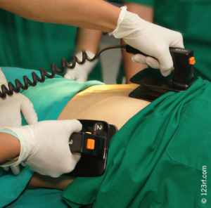
Direct current (DC) cardioversion (DCCV) is one strategy to restore sinus rhythm in patients with AF (see figure 1), and has been used in clinical practice since the 1960s. This procedure has relatively high initial success rates, however, it has become increasingly evident that the procedure has a high failure rate in the longer term. Previous studies have shown that between 20% and 47% of patients remain in sinus rhythm at one year.4,5 Accordingly, recent guidelines recommended that anticoagulation should be continued indefinitely in those with a high risk of AF recurrence. In 2010, the European Society of Cardiology (ESC) stated that “in patients with risk factors for stroke or AF recurrence, VKA (vitamin K antagonist) treatment should be continued lifelong irrespective of apparent maintenance of sinus rhythm following cardioversion”.6 Updated guidelines in 2012 also suggested a more aggressive approach to anticoagulation, with consideration of antithrombotics in those patients with a CHA2DS2-VASc risk score ≥1.7 Current literature suggests that a significant proportion of AF patients do not receive appropriate anticoagulation despite these measures.8

Screen shot 2015-07-16 at 11.48.16
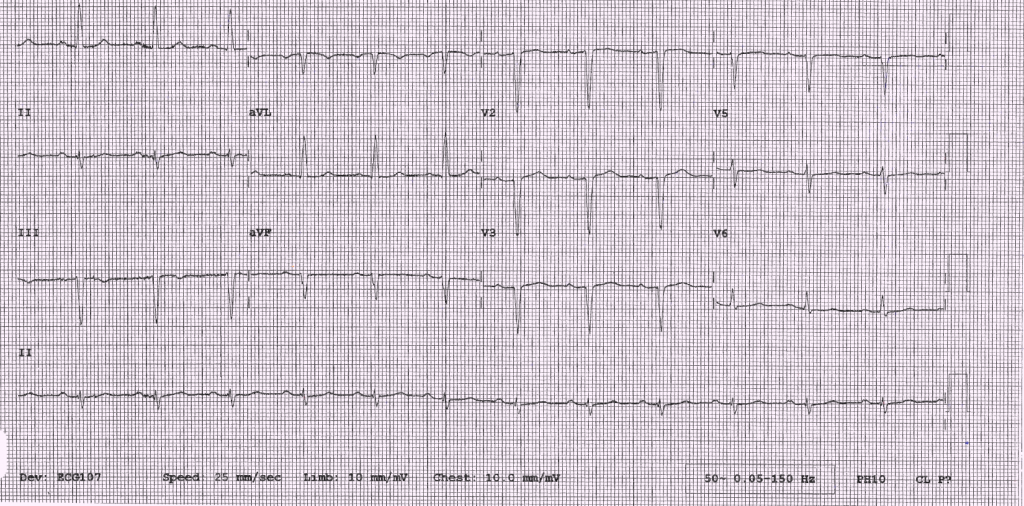
Figure 1. ECG showing atrial fibrillation and successful restoration to sinus rhythm following direct current cardioversion (DCCV)

Aims

  • To determine the total number of patients not on warfarin, or alternative anticoagulant therapy, having attended for an elective DCCV for AF at a district general hospital three to six years previously.
  • To identify patients with recurrent AF who warranted anticoagulation but were not on appropriate treatment.
  • To establish the number of patients with a recurrence of AF, which had not been documented previously, since the DCCV.
  • To assess clinical variables that predict maintenance of sinus rhythm in the long-term following DCCV.

Materials and methods

Patients who attended for a DCCV between 2008 and 2010 were identified using hospital IT records. Local general practitioners (GPs) were subsequently contacted to establish which of these patients were not currently prescribed warfarin. These patients were invited to attend a clinic between March and October 2014. During attendance, a cardiologist took a brief medical history and performed a clinical examination. A resting 12-lead electrocardiogram (ECG) was also performed. The findings from this consultation were audited against ESC guidelines.

Results

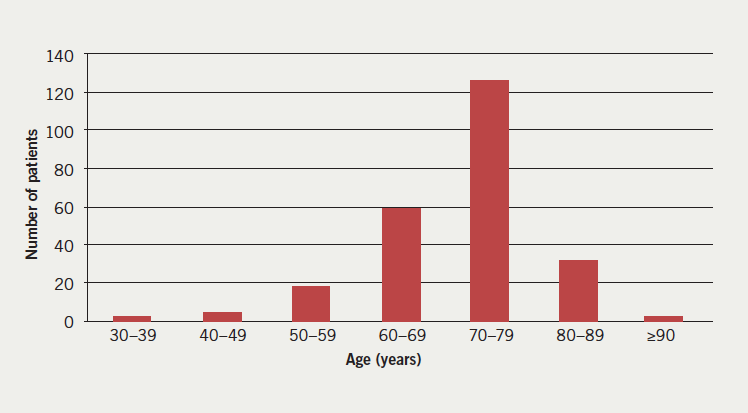
Two hundred and sixty DCCVs were performed over a three-year period between 2008 and 2010 for patients registered with local practices: 19 of these were repeat attempts representing 241 patients in total. At the time of the cardioversion the patients had a median age of 71 years (figure 2).

Figure 1. A graph to show the number of patients in different age groups at first attendance for direct current cardioversion (DCCV)
Figure 2. A graph to show the number of patients in different age groups at first attendance for direct current cardioversion (DCCV)

Number of patients

Figure 2
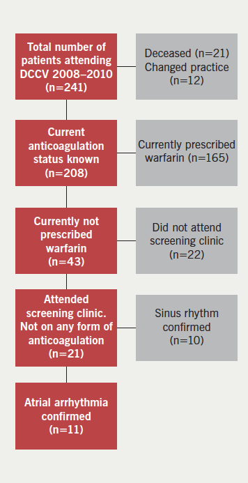
Figure 3. A diagram illustrating patient selection and clinic attendance

By 2014, 21 patients (9%) were identified to be deceased. We were able to establish a cause of death for 16 of these patients; none were classified as a stroke or bleeding event. Twelve patients (5%) had changed practice, hence, data regarding their anticoagulation status was unavailable. Therefore, anticoagulation status was provided for 208 patients. One hundred and sixty-five patients (79%) were prescribed warfarin, and 43 (21%) were not, according to their current GP prescription records. The latter 43 patients were invited to a designated cardiology clinic with, overall, 21 patients attending (49% uptake) (figure 3).

The mean time to follow-up was 4.7 ± 0.7 years. On attendance, it was confirmed that none of the patients were prescribed warfarin or a novel oral anticoagulant. In total, 11 (52%) of the 21 patients had a recurrence of an atrial arrhythmia (52% vs. 0% in an ‘ideal’ setting; p=0.0002, Fisher’s exact test). Specifically, 10 were diagnosed with AF and one with atrial flutter. Five of the 11 cases were newly identified at the clinic (45% vs. 0%; p=0.035, Fisher’s exact test), whereas six (55%) had previously been documented. Of those 11 patients attending the clinic with known or newly documented AF, nine (82%) met ESC criteria for oral anticoagulation with a mean CHA2DS2-VASc score =2.18 ± 1.48. The same group had a mean HAS-BLED score =1.73 ± 1.01.

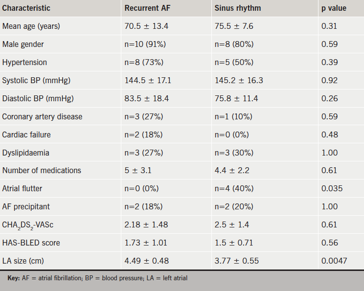
Of the 10 patients found to be in sinus rhythm at the screening clinic, 40% had a previous diagnosis of atrial flutter, contrasting with 0% in the group with recurrent AF (p=0.035). Left atrial (LA) diameter was significantly increased in those with recurrent AF (4.49 ± 0.48 vs. 3.77 ± 0.55 cm; p=0.0047). Other baseline characteristics did not differ significantly between the groups (table 1).

Table 1. Patients’ baseline characteristics

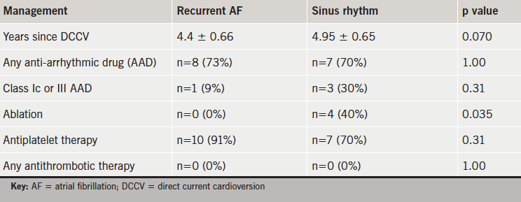
An ablation procedure following the DCCV significantly favoured maintenance of sinus rhythm (40% vs. 0%; p=0.035). There was also a tendency for the patients in sinus rhythm to be prescribed class Ic or III anti-arrhythmic drugs (30% vs. 9%; p=0.31), although this did not meet statistical significance (table 2).

Table 2. Management of atrial arrhythmia

Discussion

This audit shows that the majority of patients, three to six years following a DCCV, were prescribed anticoagulation appropriately. However, a significant number who were not on anticoagulation therapy had a recurrence of atrial arrhythmia, yet met current ESC criteria for treatment. Furthermore, a large proportion of those patients with recurrent AF had formerly not been identified. Overall, in the long term, few patients appear to remain both in sinus rhythm and without anticoagulant therapy. LA dilatation appears to predispose to AF recurrence and, conversely, a diagnosis of atrial flutter, and ablation therapy, confers a reduction in risk of arrhythmia recurrence. This is in keeping with previously reported literature.9

Admittedly, our findings are limited by a small sample size, and are also prone to selection bias; the patients attending the clinic were likely to exhibit health-seeking behaviour. Alternatively, it is possible that non-responders were already under the care of an appropriate physician.Despite these potential weaknesses, these results suggest that nearly half of patients not on any form of anticoagulation following DCCV in the long term have a recurrence of atrial arrhythmia warranting antithrombotic therapy. We propose that similar screening strategies are adopted either in primary or secondary care. Prospectively, we also suggest that, if a patient does not remain on long-term anticoagulation following DCCV, clear instruction with regards to ongoing rhythm monitoring should be given both to the GP and patient. We believe that these measures should ensure appropriate uptake of anticoagulation therapy and reduce subsequent thromboembolic risk in these individuals.

Acknowledgements

We are very grateful to Jennie Morritt, Laura Greswell, Noemi Findlay and our local general practices for their help with the audit.

Conflict of interest

None declared.

Key messages

  • There is a high rate of recurrence of atrial fibrillation (AF) in the long term following direct current cardioversion (DCCV)
  • Accordingly, European Society of Cardiology (ESC) guidance recommends indefinite anticoagulation if there is a high risk of recurrence
  • A significant proportion of patients seen in our clinic, and not on anticoagulation after DCCV three to six years previously, had recurrence of AF and warranted anticoagulation
  • We suggest long-term, routine screening if patients do not remain on anticoagulation therapy following DCCV for AF

References

1. Davis R, Hobbs R, Kenkre J et al. Prevalence of atrial fibrillation in the general population and in high-risk groups: the ECHOES study. Europace 2012;14:1553–9. http://dx.doi.org/10.1093/europace/eus087

2. Dulli D, Stanko H, Levine R. Atrial fibrillation is associated with severe acute ischemic stroke. Neuroepidemiology 2003;22:118–23. http://dx.doi.org/10.1159/000068743

3. Hart R, Pearce L. Meta-analysis: antithrombotic therapy to prevent stroke in patients who have non-valvular atrial fibrillation. Ann Int Med 2007;146:857–67. http://dx.doi.org/10.7326/0003-4819-146-12-200706190-00007

4. Sandler A. Whatever happens to the cardioverted? An audit of the success of direct current cardioversion for atrial fibrillation in a district general hospital over a period of four years. Br J Cardiol 2010;17:86–8. Available from: https://bjcardio.co.uk/2010/03/whatever-happens-to-the-cardioverted-an-audit-of-the-success-of-direct-current-cardioversion-for-atrial-fibrillation-in-a-district-general-hospital-over-a-period-of-four-years/

5. Kuppahally S, Foster E, Shoor S
et al. Short-term and long-term success of electrical cardioversion in atrial fibrillation in a managed care system. Int Arch Med 2009;2:39. http://dx.doi.org/10.1186/1755-7682-2-39

6. Camm J, Kirchof P, Lip G et al. Guidelines for the management of atrial fibrillation. The Task Force for the Management of Atrial Fibrillation of the European Society of Cardiology (ESC). Eur Heart J 2010;31:2369–429. http://dx.doi.org/10.1093/eurheartj/ehq278

7. Camm J, Lip G, Caterina R et al. 2012 focused update of the ESC guidelines for the management of atrial fibrillation. Eur Heart J 2012;33:2719–47. http://dx.doi.org/10.1093/eurheartj/ehs253

8. Cowan C, Healicon R, Robson I et al. The use of anticoagulation in the management of atrial fibrillation among general practices in England. Heart 2013;99:1166–72. http://dx.doi.org/10.1136/heartjnl-2012-303472

9. Marches P, Bursi F, Delle Donne G et al. Indexed left atrial volume predicts the recurrence of non-valvular atrial fibrillation after successful cardioversion. Eur J Echocardiogr 2011;12:214–21. http://dx.doi.org/10.1093/ejechocard/jeq176

Single-dimensional estimation of LV size using echo and MRI: effect of measurement location

Br J Cardiol 2015;22:(3)doi:10.5837/bjc.2015.026 Leave a comment
Click any image to enlarge
Authors:
First published online July 16th, 2015

Typical echocardiographic assessment of left ventricle (LV) size is based on single-dimensional measurements at mitral valve leaflet tips. In ischaemic and non-ischaemic cardiomyopathy (ICM and NICM) and aortic regurgitation (AR) where spherical remodelling is observed, this single-dimensional measurement at the LV base may underestimate LV volume. We hypothesised the maximum diameter would provide a closer approximation. A retrospective analysis of 1,680 consecutive cardiovascular magnetic resonance (CMR) examinations identified 82 patients with substantial LV dilation (LVEDVi >130 ml/m2) and 23 controls. LV end-diastolic and end-systolic diameters were measured using echocardiography and CMR at the standard level (EDDMV and ESDMV) and the maximum diameter (EDDmax and ESDmax). Indexed diameters were fitted to indexed volumes using cubic regressions. Maximum diameters had higher R2 values in fitting LV volume, and improved categorisation of subjects with chamber enlargement without substantially increasing the false-positive rate. Standard measurements may underestimate LV volume in cases of spherical remodelling, use of the maximum dimension may be a straightforward approach to improve assessment of LV volume and remodelling.

Introduction

In volume overload states, such as aortic regurgitation (AR), ischaemic cardiomyopathy (ICM), and non-ischaemic cardiomyopathy (NICM), spherical dilation occurs.1 Khouri et al. described four remodelling patterns based on the indexed left ventricle (LV) end-diastolic volume and LV concentricity.2

Standard LV chamber quantification, according to the American Society of Echocardiography (ASE) guidelines, involves obtaining a single-dimensional measurement of the LV diameter from the parasternal long-axis acoustic window at the level of mitral valve leaflet tips. Although it is the combination of qualitative and quantitative measurements that guides the final clinical management, this measurement has several management implications.3,4 We hypothesised that in cases of spherical dilation, this measurement may underestimate the extent of remodelling compared with the measurement at the widest level, due to altered geometry.

Materials and methods

We retrospectively analysed 1,680 patients who underwent cardiovascular magnetic resonance (CMR) and echocardiograms at our institution between January 2007 and April 2010. The study was Institutional Review Board approved, with waiver of individual informed consent. The time interval between CMR and echocardiography was within a month in 74% of patients (median interval: one day). CMR reports were reviewed for subjects with marked LV dilation (indexed end-diastolic volume [EDV] ≥130 ml/m2) in the absence of significant atrioventricular valve insufficiency on echocardiography. Subjects where the exam indication was AR and with regurgitant fraction >10% (mean=32%) on CMR were classified as AR. Subjects where the exam indication was a viability study with qualitatively mild-moderate regurgitation or less on CMR were classified as cardiomyopathy. They were subcategorised into ICD or NICD based on ischaemic damage on delayed gadolinium-enhanced imaging. Control subjects were obtained by selecting patients from the same institutional records as above during the same time period that had normal indexed EDV on CMR (63–98 ml/m2 for males, 57–92 ml/m2 for females),5 an ejection fraction ≥55%, <5% regurgitant fraction by CMR, and no evidence of ischaemic damage. Further, these patients had normal blood pressure and no significant abnormal findings on other cardiovascular testing (e.g. catheterisation, nuclear imaging).

Figure 1. Measurements of left ventricle (LV) geometry on cardiac magnetic resonance (CMR). Volumes and masses were computed using the steady-state free precession short-axis stack by drawing endocardial (red) and epicardial (green) contours
Figure 1. Measurements of left ventricle (LV) geometry on cardiac magnetic resonance
(CMR). Volumes and masses were computed using the steady-state free precession short-axis stack by drawing endocardial (red) and epicardial (green) contours

All patients underwent CMR on 1.5 T magnetic resonance scanners. After initial scout images were obtained, turbo spin echo and gradient echo imaging was performed for anatomic definition. Dynamic cine imaging (steady-state free precession) was performed to analyse cardiac chamber anatomy, wall-motion, and qualitative valvular function, and velocity-encoded phase contrast flow quantification sequences were obtained to assess haemodynamics. Viability studies included 0.2 mmol/kg of gadolinium-chelate contrast intravenously (Magnevist) to assess ischaemic damage. Transthoracic echocardiography was performed by experienced sonographers using standardised protocols.

EDV, end-systolic volume (ESV), and myocardial mass were measured on the steady-state free precession short-axis stack by drawing epicardial and endocardial contours excluding papillary muscles (figure 1). Aortic flow and regurgitant fraction was computed using phase-contrast flow quantification. Using the long-axis parasternal view, end-diastolic diameter and end-systolic diameter were measured on echocardiographic images at the level of the mitral valve tips (EDDMV and ESVMV) using 2D calliper,3 and at the level perceived to yield the largest diameter (EDDmax and ESVmax). These diameters were also measured using CMR using an equivalent (three-chamber) view.

The EDDmax/EDDMV and ESDmax/ESDMV ratios were computed for both CMR and echocardiography. The EDDmax/EDDMV and ESDmax/ESDMV ratios were compared between groups using the Kruskal-Wallis test. Among subjects with remodelling, the correlation between the percentage increase of EDDmax from EDDMV measured on echocardiography and indexed LV volumes measured on magnetic resonance imaging (MRI) was calculated.

Diameters measured at mitral valve tips and at the maximum diameter during systole and diastole on echocardiography were indexed by the subject’s height. End-diastolic and systolic volumes were indexed using body surface area (BSA).2,6,7 Indexed diameters were fitted to indexed volumes by cubic polynomial regression and R2 values were computed. Absolute and indexed end-diastolic diameters were used to predict the presence of LV dilation (LVEDV/BSA >98 ml/m2 for males, >92 ml/m2 for females).

Statistical analyses were performed using JMP 9.0 software (SAS Institute, Cary, NC) and R version 3.0.1 (GNU Public License). A significance level of 0.05 was used for all statistical tests.

Results

There were 82 subjects that fit criteria for AR, ICM, and NICM, and 23 control subjects were included in the analysis. Mean age was 53.2 ± 16.8 years. Subjects with ICM and NICM had lower ejection fraction (EF) than controls or subjects with AR. Patients in the AR group were younger and had significantly higher regurgitant fraction than the other groups (table 1). LVEDV was much larger in subjects with spherical remodelling compared with controls. LVESV was smaller in subjects with AR compared to ICM and NICM.

Table 1
Table 1. Characteristics of the population by group. Means ± standard deviation (SD) are shown; p values are shown for statistical testing across all groups (ANOVA)

The difference between maximum LV diameter and the diameter measured at mitral valve tips was greater in subjects with LV dilation compared with controls (table 2). Comparing the ratio of EDDmax/EDDMV across all groups using the Kruskal-Wallis test, a significant difference was observed on echocardiography (p=0.0466) and CMR (p=0.0024). For ESDmax/ESDMV, a significant difference was present for CMR (p=0.001), but not for echocardiography (p=0.17). Among patients with LV remodelling, EDDmax was 9.8% greater than EDDMV on average on echocardiography. The correlation coefficient between this percentage difference and indexed LV volume was 0.134 with no apparent relationship.

Table 2. Table of diameters (in cm) measured on echocardiography and cardiac magnetic resonance (CMR) in diastole and systole. Means ± SD are shown

Indexed diameters at the maximum diameter exhibited better fits with LVEDV indexed by BSA when fitted to a cubic relationship. The R2 value for EDD at the maximum was 0.528, compared with 0.491 for EDD at the valve tips. For ESD, the maximum diameter showed a better fit as well (0.686 vs. 0.537) (figure 2).

Figure 2. Cubic fit of end-diastolic diameter (EDD)/height and end-systolic diameter (ESD)/height at mitral valve tips and maximum measurement measured on echocardiography to left ventricle end-diastolic (or systolic) volume (LVEDV or LVESV)/body surface area (BSA) measured on CMR. Data points and regression curves are shown

When using standard cut-offs for absolute end-diastolic diameters on echocardiography (EDD >5.9 cm for males, >5.3 for females),3 diameters at mitral valve tips correctly identified 57.3% (47/82) of abnormal subjects, while incorrectly classifying 4.3% (1/23) of control subjects as abnormal. Absolute measurements at the maximum diameter identified 78.0% (64/82) of abnormal subjects and incorrectly classified 13.0% (3/23) controls. Using indexed end-diastolic diameters on echocardiography (EDDi >3.2 cm/m for males, >3.1 cm/m for females),3 diameters at mitral valve tips correctly identified 72.0% (59/82) of abnormal subjects and incorrectly classified 4.3% (1/23) of control subjects. Indexed measurements at the maximum diameter identified 87.8% of abnormal subjects and incorrectly classified 13.0% (3/23) of control subjects.

Discussion

In volume overload states such as AR, ICM, and NICM, CMR depicts spherical remodelling as a common remodelling pathway, an adaptation that maintains normal net forward stroke volume. These geometric alterations affect the assumed relationship between the standard LV dimensions measured at mitral valve leaflet tips and true LV volume. The results in this investigation demonstrate that EDDmax/ EDDMV and ESDmax/ESDMV ratios are larger in subjects with AR, ICM and NICM, where spherical dilation occurs. The percentage difference among these subjects did not vary with indexed LV volumes, indicating that the standard measurement at mitral valve tips underestimates LV size in spherical dilation regardless of the extent of remodelling. The maximum diameter on echocardiography showed a better fit to the presumed cubic relationship between diameter and volume than the diameter at mitral valve tips during diastole and systole. Although abnormal cut-offs for the maximum diameter are undefined, applying the same thresholds used for standard measurements to the maximum diameter improved sensitivity for detecting abnormal chamber volumes without substantially increasing the false-positive rate. Indexed dimensions demonstrated better classification of the presence or absence of LV dilation compared with absolute dimensions.

Limitations of the study include its retrospective nature, subjects being from a single tertiary care centre, and selection bias. The majority of cardiomyopathy patients had ischaemic damage, limiting the characterisation of remodelling in NICM, and there were relatively few control subjects for analysis. However, this is typical in a population where CMR is clinically indicated.

Spherical dilation is observed in AR, ICM and NICM. Indexed dimensions demonstrated better classification accuracy compared with absolute dimensions. Despite guidelines that refer to measurements at mitral valve leaflet tips, these results suggest that measuring the LV maximum dimension may better estimate LV volume in subjects with remodelling. A combination of both measurements may more accurately characterise LV volume and geometry, and the ratio of these measurements may serve as a straightforward method of quantifying the extent of spherical dilation.

Further investigations could verify the use of the maximum diameter as a stronger predictor of LV volume than the standard measurement in a larger cohort of patients with a greater cohort of control subjects. In addition, characterisation of the typical ranges of maximum diameters in relation to the standard measurement in various disease states both on CMR and echocardiography could be performed to determine the use of the ratio of these two measurements to determine the extent of remodelling, and the feasibility of incorporating the maximum diameter into clinical management guidelines.

Conflict of interest

All authors have no conflicts of interest to disclose.

Funding information

This work was supported by a National Institutes of Health training grant (T35 HL082544) for C.W.H.

Key messages

  • In spherical remodelling, a single-dimensional measurement at the mitral valve leaflet tips may underestimate ventricular volume due to altered geometry
  • Diameters measured at the widest dimension were more predictive of left ventricle (LV) volume than the standard measurements
  • When using standard cut-offs, maximum diameters also improved categorisation of subjects with chamber enlargement without substantially increasing the false-positive rate
  • Measuring the maximum diameter may be a straightforward approach to improve assessment of LV volume

References

1. American College of Cardiology Foundation Task Force on Expert Consensus Documents, Hundley WG, Bluemke DA et al. ACCF/ACR/AHA/NASCI/SCMR 2010 expert consensus document on cardiovascular magnetic resonance: a report of the American College of Cardiology Foundation Task Force on Expert Consensus Documents. J Am Coll Cardiol 2010;55:2614–62. http://dx.doi.org/10.1016/j.jacc.2009.11.011

2. Khouri MG, Peshock RM, Ayers CR, de Lemos JA, Drazner MH. A 4-tiered classification of left ventricular hypertrophy based on left ventricular geometry: the Dallas heart study. Circ Cardiovasc Imaging 2010;3:164–71. http://dx.doi.org/10.1161/CIRCIMAGING.109.883652

3. Lang RM, Bierig M, Devereux RB et al. Recommendations for chamber quantification: a report from the American Society of Echocardiography’s Guidelines and Standards Committee and the Chamber Quantification Writing Group, developed in conjunction with the European Association of Echocardiography, a branch of the European Society of Cardiology. J Am Soc Echocardiogr 2005;18:1440–63. http://dx.doi.org/10.1016/j.echo.2005.10.005

4. Bonow RO, Carabello BA, Chatterjee K et al. 2008 focused update incorporated into the ACC/AHA 2006 guidelines for the management of patients with valvular heart disease: a report of the American College of Cardiology/American Heart Association Task Force on Practice Guidelines (Writing Committee to revise the 1998 guidelines for the management of patients with valvular heart disease). Endorsed by the Society of Cardiovascular Anesthesiologists, Society for Cardiovascular Angiography and Interventions, and Society of Thoracic Surgeons. J Am Coll Cardiol 2008;52:e1–e142. http://dx.doi.org/10.1016/j.jacc.2008.05.007

5. Maceira AM, Prasad SK, Khan M, Pennell DJ. Normalized left ventricular systolic and diastolic function by steady state free precession cardiovascular magnetic resonance. J Cardiovasc Magn Reson 2006;8:417–26. Available from: http://www.scmr.org/assets/files/members/documents/JCMR/008/LCMR_i_008_03_tfja/LCMR_i_8_03_O/LCMR_A_157271_O.pdf

6. Grothues F, Smith GC, Moon JC et al. Comparison of interstudy reproducibility of cardiovascular magnetic resonance with two-dimensional echocardiography in normal subjects and in patients with heart failure or left ventricular hypertrophy. Am J Cardiol 2002;90:29–34. http://dx.doi.org/10.1016/S0002-9149(02)02381-0

7. Lauer MS, Anderson KM, Larson MG, Levy D. A new method for indexing left ventricular mass for differences in body size. Am J Cardiol 1994;74:487–91. http://dx.doi.org/10.1016/0002-9149(94)90909-1

Syncope in a patient with cardiac sarcoidosis

Br J Cardiol 2015;22:(3)doi:10.5837/bjc.2015.027 Leave a comment
Click any image to enlarge
Authors:
First published online July 16th, 2015

A 46-year-old man with cutaneous sarcoidosis and pulmonary involvement was referred to a cardiologist for possible pulmonary hypertension in view of increasing shortness of breath. Echocardiogram findings and electrocardiogram (ECG) changes prompted the need for coronary angiography, which was subsequently normal.

Cardiac sarcoidosis was one of the differential diagnoses. The patient was booked for a treadmill test. Unfortunately, in the interim, the patient had an episode of collapse while playing football. The local district hospital discharged the patient after finding normal computed tomography (CT) brain scan and negative troponin. He was re-admitted with a pre-syncopal episode and ambulatory ECG monitoring revealed non-sustained ventricular tachycardia. A cardiac magnetic resonance imaging (MRI) scan was arranged followed by insertion of an implantable cardioverter defibrillator (ICD).

This case report and literature review highlights the importance of cardiac screening in patients deemed ‘high risk’ for sudden cardiac death, and the need for immediate investigation and treatment. Current guidelines are yet to be universally accepted, so such cases are important in highlighting current methods of investigation and treatment.

Introduction

Cardiac sarcoidosis can present in a broad spectrum of entities ranging from a benign condition, which is diagnosed incidentally, to a potentially serious disease leading to sudden cardiac death, which only becomes apparent at autopsy, as is the case in 5% of the affected population.1 Due to its subtle, but also sometimes fatal presentation, cardiac sarcoid is hugely underdiagnosed, and awareness of such cases should be brought to light whenever possible.

This case report highlights the importance of being aware of the potential presentations of cardiac involvement in patients with sarcoidosis and the general investigations and management that are common place in current practice.

Case report

A 46-year-old man, with a previous biopsy-proven diagnosis of cutaneous sarcoidosis with pulmonary involvement, was referred two years after his first diagnosis of cutaneous sarcoidosis to a cardiologist to investigate further for possible pulmonary hypertension in view of increasing shortness of breath. He had an echocardiogram, which showed severely impaired left ventricular function with a hypokinetic anterior septum. Together with the electrocardiogram (ECG) findings (figure 1) both coronary artery disease and cardiac sarcoid were within the list of differential diagnoses. Therefore, a coronary angiogram was undertaken, which was deemed normal. In the interim period, the patient had an episode of syncope while playing football. His local hospital was unaware of the background and current investigations, and so, after a normal computed tomography (CT) brain scan and observation period, the patient was discharged.

Figure 1. Electrocardiogram (ECG) findings showing Q-waves in antero-septal leads

A cardiac magnetic resonance imaging (MRI) was booked on the patient’s subsequent appointment to his cardiologist, and an exercise-tolerance test was arranged to see if he had any evidence of inducible ventricular arrhythmias. This showed a left bundle branch block (LBBB) at peak of stress and the patient remained asymptomatic throughout.

Figure 2. Polymorphic non-sustained ventricular tachycardia was seen on monitoring the patient (X)
Figure 3. Cardiac magnetic resonance (MRI) showing diffuse cardiomyopathy with punctate areas of increased uptake, predominantly in the left ventricle

The patient was re-admitted with pre-syncope six months later. Ambulatory ECG recordings (figure 2) showed non-sustained ventricular tachycardia (VT) prompting urgent MRI (figure 3) and subsequent implantable cardioverter debfibrillator (ICD) insertion.

The patient was commenced on prednisolone. At 12 months follow-up, a gallium scan with single-photon emission computed tomography (SPECT) was arranged to determine disease activity. This showed active inflammation in the parotid glands and also in the left ventricular apex. This led to the initiation of methotrexate, with subsequent scans showing a decline in disease activity.

Discussion

Cardiac sarcoid is largely underdiagnosed in the population. At present, there is no clear consensus on the detection, monitoring and treatment process, meaning the physician must manage each case on a more individual basis.

The symptoms and signs of cardiac sarcoid are dependent upon the extent and location of the granulomatous formation. Within the heart, it may involve pericardium, myocardium and endocardium. Aetiology is unclear: genetic, environmental and immune phenomena may contribute.

The most common conduction abnormality in cardiac sarcoidosis is third-degree atrioventricular (AV) block or complete heart block. This is followed by ventricular tachycardia. Among bundle branch blocks, right is more frequent than the left. Heart failure is the most common cause of death in sarcoidosis, and studies show this to be secondary to papillary infiltration in most of the cases.

The diagnosis of cardiac sarcoid can be challenging, and a high index of suspicion should be kept. The patients in whom sarcoidosis should be suspected include those with: sustained ventricular tachycardia, young patients ( <55 years) with unexplained heart block and extra cardiac sarcoidosis.

The 1999 Joint Statement of the American Thoracic Society, the European Respiratory Society and the World Association for Sarcoidosis and Other Granulomatous Disorders considers cardiac dysfunction, ECG abnormalities, and thallium-201 imaging defects, with or without endomyocardial biopsy, as evidence of cardiac sarcoidosis.

Several radionuclide techniques are available to image inflammation. 18F-flurodeoxyglucose (18F-FDG) positron emission tomography (PET) scan shows more sensitivity than thallium-201 and gallium-67 scans in detecting cardiac sarcoidosis active tissue and a scar, but with less specificity.2,3 It can also be used to follow treatment response.4

The mainstay of treatment for cardiac sarcoid is that of corticosteroids, in order to halt or regress the inflammation and fibrosis associated with this pathology. However, it is impossible to predict which patients will respond to such therapy, and, thus, the implantation of a primary preventative ICD is largely recommended in both national and international guidelines with regards to device therapy in such cases. The role of secondary agents, such as methotrexate, in cases where glucocorticoids are unsuccessful, has a limited evidence base, but has been shown in patient cohort studies to have a role.

Evidence to date suggests cardiac involvement in sarcoidosis carries a higher morbidity and mortality than in those patients exempt of cardiac involvement.

Conflict of interest

None declared.

References

1. Johns CJ, Michele TM. The clinical management of sarcoidosis. A 50-year experience at the Johns Hopkins Hospital. Medicine (Baltimore) 1999;78:65–111. http://dx.doi.org/10.1097/00005792-199903000-00001

2. Okayama K, Kurata C, Tawarahara K, Wakabayashi Y, Chida K, Sato A. Diagnostic and prognostic value of myocardial scintigraphy with thallium-201 and gallium-67 in cardiac sarcoidosis. Chest 1995;107:330–4. http://dx.doi.org/10.1378/chest.107.2.330

3. Abdul RD, Byron RW. Cardiac sarcoidosis. Heart 2006;92:282–8. http://dx.doi.org/10.1136/hrt.2005.080481

4. Youssef G, Beanlands RSB,
Birnie DH, Nery PB. Systemic disorders in heart disease. Cardiac sarcoidosis: applications of imaging in diagnosis and directing treatment. Heart 2011;97:2078–87. http://dx.doi.org/10.1136/hrt.2011.226076