A wide array of investigations is available to the heart failure specialist. Their proper utilisation requires knowledge of their indications, and proficiency in interpretation. This was the focus of the second morning session, entitled “Different tests”, of the 8th British Society for Heart Failure (BSH) Training and Revalidation Day. Held on 3rd March 2016, at the Golden Jubilee National Hospital, Clydebank, this session included excellent presentations on cardiopulmonary exercise testing (CPET), cardiac catheterisation in heart failure, assessment of haemodynamics using echocardiography, and the use of cardiac CT in heart failure. Dr Simon Beggs reports on some of the highlights from a lively and thought-provoking day.
CPET: an overview of “the cardiac cycle”
 The breathless patient with heart failure and comorbidity can pose a diagnostic conundrum: is the dyspnoea cardiac or respiratory (or something else entirely)? Dr Christopher Boos (Poole Hospital NHS Foundation Trust) outlined the role of cardiopulmonary exercise testing (CPET) in such situations.
CPET integrates a broad range of variables related to cardiorespiratory function, including oxygen uptake and expiratory ventilation, along with blood pressure and electrocardiogram (ECG) tracing. It provides objective information on physiological performance under stress: key output data include peak VO2 (a measure of maximal aerobic capacity), anaerobic threshold (AT), respiratory exchange ratio (RER) and breathing reserve (BR).
Determination of the primary aetiology in unresolved cases of dyspnoea is aided by proper interpretation of these data. Dr Boos illustrated this with two case examples.
First, he presented a 54-year old male with a left ventricular ejection fraction (LVEF) of 25%, BMI of 33 and exertional dyspnoea, in whom a relatively normal AT, moderately impaired VO2 and normal BR indicated deconditioning as the primary cause of his presentation. Second, a 49-year old female with severe interstitial lung disease and moderate-to-severe mitral stenosis, in whom a low AT and high BR suggested valve disease as the principal aetiology of her dyspnoea. In both cases, CPET facilitated a confident diagnosis and altered patient management.
Wither the cardiac catheter?
Invasive assessment of the coronary anatomy and cardiac haemodynamics are oft- used tools in the cardiologist’s armoury. The précis of Professor Andrew Clark’s (University of Hull) talk was that the former adds little value in the assessment of patients with heart failure, whilst the latter’s worth is underappreciated.
The STICH1 and HEART2 trials failed to show significant mortality benefit from coronary revascularisation in patients with heart failure. The benefit of routine coronary angiography in these patients is dubious.
Meanwhile, right heart catheterisation (RHC) may provide valuable diagnostic and prognostic data. Both procedural technique and knowledge of typical haemodynamic waveforms are vital. In cardiac transplant candidates, assessment of right heart data including pulmonary artery pressure, transpulmonary gradient and pulmonary vascular resistance is essential: unacceptable parameters contraindicate transplantation. RHC also provides a route by which to perform endomyocardial biopsy when indicated, such as in suspected infiltrative cardiac disorders.
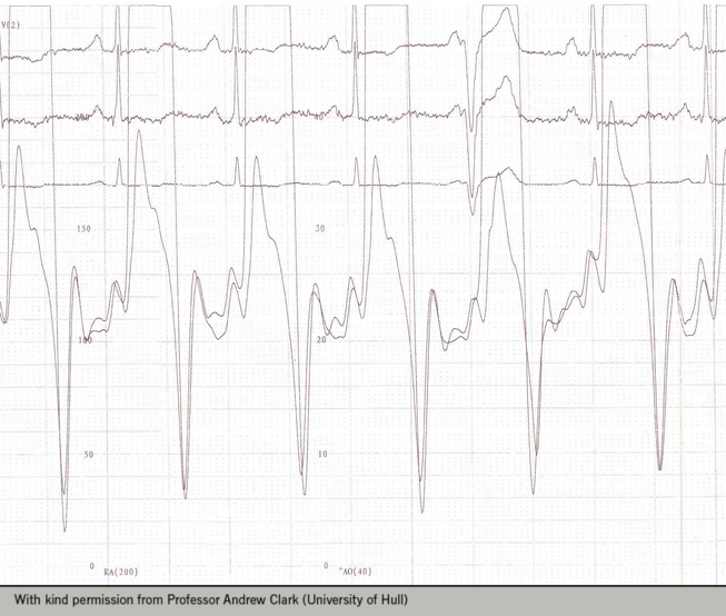
Simultaneous left and right heart catheterisation may help discern the aetiology of heart failure. For example, the classic “dip and plateau” sign resulting from diastolic equalisation of cardiac pressures in constrictive pericarditis (see figure 1). Similarly, RHC occasionally detects unsuspected congenital anomalies, such as atrial septal defects (ASDs), via inappropriate “step-up” in venous oxygen saturations.
Professor Clark concluded that assessment of cardiac haemodynamics is a vital competency for the heart failure specialist, and one requiring both technical and analytical rigour. A recent review of the subject is available.3
Assessment of haemodynamics with echocardiography
Assessment of cardiac haemodynamics is also feasible non-invasively. Dr Alison Duncan (Royal Brompton Hospital, London) summarised advantages and pitfalls of echocardiography used for this purpose.
Echocardiographic estimation of intra-cardiac pressure gradients and valve areas using the Bernoulli and Continuity Equations is common practice. Mean aortic valve gradient by echocardiography correlates with catheter-based measurements, although operators must recall that measurements by these modalities are not equivalent. Cross-sectional area calculations can be extended to non-valvular orifices, such as ASDs.
Other echo-derived haemodynamic parameters are clinically relevant: cardiac output and systemic vascular resistance, for example. Left atrial pressure (LAP) may be estimated via velocity quantification of the mitral regurgitant jet.
Dr Duncan further demonstrated how left ventricular filling patterns are readily understood from physiology. The intraventricular relaxation time (IVRT) lengthens with impaired LV relaxation. Eventually this curtails the mitral inflow E wave (passive filling), necessitating a compensatory dominant A wave (active filling), thus producing the classic reversed E:A ratio of LV diastolic dysfunction. Conversely, prolonged restrictive physiology and a high LAP results in a dominant, “spiky” E wave, producing a “pseudonormal” pattern.
The clinician must be aware that LV loading can alter echocardiographic filling patterns in such situations. For example, markedly different results may be obtained in the same patients before and after renal dialysis. This, as Dr Duncan pointed out, is a reminder that clinical assessment of the patient remains essential when utilising echocardiography.
Cardiac CT: what role in heart failure?
The emergence of 64-slice multi-detector computed tomography (MDCT) over the last decade has delivered high spatial and temporal resolution; an entire heart can be imaged in less than 15 seconds. In a visually spectacular epic, Dr Ronak Rajani (Guy’s and St Thomas’ Hospitals, London) predicted an increasing role for MDCT in the management of patients with heart failure.
Ischaemic cardiomyopathy remains a leading cause of heart failure. MDCT has a growing role in the definition of anatomic coronary stenosis, detection of high-risk coronary plaque features, and non-invasive functional assessment of coronary disease via FFRct, enabled by the application of computational fluid dynamics.
There is advocacy of MDCT for evaluation of aetiology and severity of left ventricular dysfunction,4,5 and a recent meta-analysis concluded that, along with 3D echocardiography, MDCT had the narrowest limits of agreement in assessment of LVEF when compared to cardiac MRI.6 Looking forward, new Spectral and Dual Energy CT technologies promise to revolutionise myocardial characterisation.
Dr Rajani proposed that MDCT may soon provide a “one stop shop” for cardiac resynchronisation therapy patients, via assessment of scar burden, ventricular function, mechanical dyssynchrony via endocardial strain and deformation, and anatomical mapping of the coronary venous system. In patients with left ventricular assist device (LVADs), MDCT can already provide assessment of device alignment and presence of thrombus. The application of computational fluid dynamics might in the future enable sophisticated MDCT-based analyses of LVAD function. Dr Rajani’s message was clear: the capabilities of MDCT are widening and deepening, and its application to patients with heart failure looks set to grow.
Dr Simon Beggs
Clinical fellow, Scottish National Advanced Heart Failure Service
Golden Jubilee National Hospital, Agamemnon St, Clydebank, Dunbartonshire G81 4DY
Acknowledgements
The BSH gratefully acknowledges the support provided by the Friends of BSH:
Bayer HealthCare, Biotronik, Boston Scientific, Medtronic, Novartis, Roche Diagnostics, Servier Laboratories, St. Jude Medical and Vifor Pharma.
Diary Dates
19th BSH Annual Autumn Meeting, 24–25 November 2016, QE II Centre, London
9th BSH Heart Failure Day for Revalidation and Training, 2 March 2017, London
7th BSH Heart Failure Nurse Study Day, 3 March 2017, London
Contact
British Society for Heart Failure
E-mail: [email protected]
References
1. Velazquez EJ, Lee KL, Deja MA, et al. Coronary-artery bypass surgery in patients with left ventricular dysfunction. N Engl J Med 2011;364:1607–1. http://dx.doi.org/10.1056/NEJMoa1100356
2. Cleland JG, Calvert M, Freemantle N, et al. The Heart Failure Revascularisation Trial (HEART). Eur J Heart Fail 2011;13:227–33. http://dx.doi.org/10.1093/eurjhf/hfq230
3. Callan P, Clark AL. Right heart catheterisation: indications and interpretation. Heart 2016;102:147–57. http://dx.doi.org/10.1136/heartjnl-2015-307786
4. Tummala LS, Young RK, Singh T, et al. Role of non-invasive imaging in the work-up of cardiomyopathies. Curr Atheroscler Rep 2015;17:486. http://dx.doi.org/10.1007/s11883-014-0486-1
5. Taylor AJ, Cerqueira M, Hodgson JM, et al. ACCF/SCCT/ACR/AHA/ASE/ASNC/NASCI/SCAI/SCMR 2010 Appropriate use criteria for cardiac computed tomography. Circulation 2010;122:e525–e555. http://dx.doi.org/10.1161/CIR.0b013e3181fcae66
6. Pickett CA, Cheezum MK, Kassop D, et al. Accuracy of cardiac CT, radionucleotide and invasive ventriculography, two- and three-dimensional echocardiography, and SPECT for left and right ventricular ejection fraction compared with cardiac MRI: a meta-analysis. Eur Heart J Cardiovasc Imaging 2015;16:848–52. http://dx.doi.org/10.1093/ehjci/jeu313
