Syncope, as a symptom, remains poorly understood and handled in the UK. Personal involvement stemmed from successful abolition of syncope by cardiac pacing, initially in atrioventricular block then other bradycardias; UK facilities were sparse in the late 1970s. The aim was to broaden distribution and encourage understanding. To achieve this required abandoning the typical cardiology mindset and re-engaging with internal medicine. Syncope is poorly taught in medical schools, which implies that few physicians understand it and, therefore, cannot practise it to benefit patients. Syncope patients presenting in emergency departments (>1% of all) are frequently admitted to hospital to undergo numerous non-diagnostic tests, then discharged without diagnosis. Syncope has life-threatening causes in >1% of patients; given its prevalence, this represents a large number. Treatment is now possible.
Syncope science has notably advanced, offering valuable diagnostic and therapeutic strategies. Ubiquitous delivery in the UK requires a new approach, which has been applied in the Netherlands. Now, more than ever, is the time to take syncope seriously, with action now to address this critical situation.
Introduction
This is a personal story of the first author, in the form of a review, who has had a career in a medical specialty that does not officially exist, even its coding is unreliable. My co-author brings a different perspective, being much younger, not British and in his early career, with great ambition directed to advancing this nebulous specialty. The Netherlands is a less conservative country where syncope is taken more seriously than in the UK.
Syncope is a common symptom but is not a diagnosis
Syncope is transient loss of consciousness due to reduced cerebral perfusion with rapid onset, brief duration and full prompt recovery.1 Thus, it is a symptom that requires a cause to be ascertained – a diagnosis. Syncope occurs in nearly half the population during life.2 Its very commonness demands attention in the medical student’s curriculum, which it does not receive. While the dominant cause of syncope is a reflex, vasovagal syncope, which may be correctly considered neurological, but its main manifestations are cardiovascular, implying that often cardiologists, rather than neurologists, take care of the patients. This is the pair of stools between which many patients fall. Neither specialty has great interest in accepting this burden, to the detriment of patient care. This has profound implications, displayed by a nationwide lack of physicians who understand and are willing to take the required time to help.
Syncope aetiology
Coming to syncope as a severe symptom of atrioventricular block, with its almost miraculous abolition by permanent cardiac pacing, thinking was directed to broadening indications for pacing, resulting in success in sinoatrial node disease3 and carotid sinus syndrome,4 but limited success in vasovagal syncope (VVS).5 The latter has taken 35 years to be refined to a point where there is agreement on which VVS patients benefit, and the guideline recommendation is class 1A.6
Comprehending the latter difficulty required much study of VVS, which was facilitated by the serendipitous discovery that tilt-testing could induce VVS in the laboratory.7 Many workers subsequently contributed to the identification of both cardioinhibition (vagal bradycardia-asystole) and vasodepression (fall in blood pressure closely related to redistribution of blood to abdomen, pelvis and lower limbs by the erect posture).8 Vasodepression was what pacing could not readily address, despite successful treatment of cardioinhibition.
These studies led to the realisation that syncope requires an approach that is neither typical cardiological nor typical neurological. First, the patient’s history is the most important part of the diagnosis, usually requiring a witness, in addition to what the patient can reveal. All of this takes time, but time is what physicians are no longer allowed to take; they must be productive, seeing more patients in less time. This has swept the history out of medicine, rendering physicians to be test selectors. Sadly, most select the wrong tests, for example brain scans, electroencephalograms (EEGs) and Holter monitors, which tell us nothing about syncope aetiology. Even worse is what happens in emergency departments, where there is little time for history taking and huge pressure on processing patients, with the result that a high percentage are admitted as inpatients to receive tests that carry no possibility of diagnosis and occupy time, which costs much money. Patients often then leave hospital with no diagnosis and no referral to any follow-up that might make a diagnosis and prevent recurrence.
Beyond the cardiovascular aspects of syncope, it is necessary to be a physician encompassing cardiology, neurology, endocrinology, paediatrics and geriatrics, at least, and integrating them appropriately into the care of each patient. The image of a cardiologist as aggressive and, possibly, lifesaving, must be substituted by a broad and thoughtful approach. When practised, there is appreciation that syncope is very common, with only very few sufferers benefitting from pacing, but all require, and can improve with, help. VVS can be described as constitutional, meaning that it cannot be cured. It is often genetically determined, although precise identification of the genetic abnormalities is not yet available.9
How the science of syncope has developed
In the early 1990s, a group of like-minded individuals formed the Vasovagal International Study (VASIS), which initially tried to classify tilt-test responses in the belief that the different patterns would guide therapy. The VASIS classification was born,10 but it only really delivered as a classification without contributing much to selection of therapy. It was improved in 2000,11 and continues to be used, but mostly as a classification.
The group moved on to a study of pacing in VVS, taking eight years to completion.12 Its strong positivity was based on a misconception that a paced patient could be compared legitimately with an unpaced patient. The leader of the group, Michele Brignole, has made a massive contribution to the science and delivery of care of syncope patients. This began with studies on carotid sinus syndrome (CSS) and drug control of VVS, progressing to guideline creation, with the first presented in 2001, and followed by updates in 2004, 2009, and 2018.1 These have provided a basis for improvement of syncope care; however, their implementation in clinical practice is limited.
The quest for finding a successful way of pacing VVS continued, culminating in very tight refinement of the candidates from two powerful studies, both of which demonstrated the efficacy of pacing in VVS in older patients (>40 years) who had failed general measures (increased fluids and salt, avoidance of triggers for attacks, employment of physical counter-measures13 and medication) and documented severe cardioinhibition on tilt.14,15 These trials allowed pacing guidelines to be changed to clear advice – class 1A.6
Drugs
Drugs have been much studied, but so far there has been little success. This may be explained by the need for a continuous drug effect in attempting to control a paroxysmal event, which recurs quite rarely, on a background of normality. Benefit can be shown in trials to a limited extent, but the patient needs to be persistent in taking the medication. Perhaps, the greatest benefit is the breaking of a cycle of repeated attacks, which has been little studied.
Physical counter-measures
Physical counter-measures have proved very valuable.13 Muscle tensing is easily done and works well to abort an attack, provided that a prodrome of VVS is recognised by the patient. However, prodromes are not always present, or appreciated, by patients, as they may have amnesia for the whole event – a tendency that appears increasingly in older people.16
Tilt-test methodology
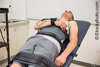
Tilt-testing remains an important investigation in syncope, as the challenge of upright posture without the benefit of the leg-muscle pump tends to precipitate syncope under clinical staff observation. Methodology of tilt-testing has advanced. The so-called Italian protocol has been shortened, without inferior results. It is now five minutes supine, 10 minutes passive tilt to 70º head-up, followed by a dose of 300–400 µg sublingual nitroglycerin, and tilt continued for 10 minutes, if syncope occurs the test ceases.17 The patient is returned to supine, which is followed by recovery. In another report from Naples, it has been emphasised that full loss of consciousness must occur before tilt-down, as important bradycardias will otherwise be missed.18 These two studies combined offer a shorter laboratory time, without prejudicing results and learning more of the bradycardia potential of the patient, which is very relevant to consideration of invasive therapy by pacing or cardioneuroablation (CNA). Patients without severe cardioinhibition (asystole) will not be considered for these therapies. Tilt has other attributes; it may be used to provoke a vasovagal syncope episode to confirm the diagnosis, also to allow patients to learn their individual prodrome, and demonstration to the patient of the blood pressure benefit by physical counter-measures, yielding improvement in, or termination, of symptoms.13,19,20
Low blood pressure phenotype
Another approach was adopted by an international group led by Brignole, which was able to demonstrate that people with low blood pressure (BP) (low BP phenotype) are more prone to VVS than those with normal BP.21 Cooperation persisted between members of that group to modify the investigation protocol to include more cardiovascular autonomic tests and 24-hour ambulatory BP monitoring (ABPM),22 resulting in a diagnosis of >80% of referrals with unexplained syncope.23,24
Life-threatening syncope
Syncope has a small percentage of life-threatening causes, estimated at >1%, these include ventricular tachyarrhythmias complicating myocardial ischaemia and cardiomyopathy, or one of the channelopathies, of which the best known is long QT syndrome. These conditions are well covered in the guidelines.1 This small percentage translates into a substantial number, due to the common nature of syncope. Most of these conditions can be diagnosed or suspected on a standard 12-lead ECG, which forms an essential part of every investigation. Once diagnosed, all these patients can be treated. There are also rare structural cardiovascular causes of syncope, which carry high mortality and may offer diagnostic and therapeutic difficulties. Once identified, specific therapy is required, for example, aortic stenosis.
Falls and syncope
Falls in older people, in some cases (~15%), can be explained by syncope, but teasing out these two presentations can be challenging. The influence of amnesia for the event raises particular problems. A fall tends not to be a benign event, with injury being frequent. Too often the syncopal cause is ignored. A career of work by Kenny and her colleagues in Dublin has brought the matter to general attention and offered solutions.25 Pertinent to this age group is that both syncope and falls prejudice quality of life, often confining patients to their homes.
Cardioneuroablation
CNA was invented by Pachon in 2004.26 It was very slowly adopted at first, but in the last five years has seen great increase in use. The principle of the therapy is to achieve, by intracardiac radiofrequency ablation, extracardiac detachment of the heart from its vagal nerve input at the level of the ganglionic plexi. The technique is similar to that employed for atrial fibrillation ablation, which has been practised for more than 20 years with fair success and few complications. CNA in many countries yields notable financial benefit to the operator, which may explain the sudden rise in numbers of cases treated. Destroying the heart’s vagal input has in the past been seen in patients after myocardial infarction, with ventricular tachyarrhythmias resulting.27 Further, we know little about the long-term effects of cardiac vagal denervation. A recent scientific statement on this therapy has been published.28
Dutch syncope research and practice
In The Netherlands, a tradition for careful physiological study has developed over many years. A leader in the field of such studies in syncope is JG van Dijk, a neurophysiologist, in Leiden. Cooperative projects, including many units across the country, are frequently undertaken. F de Lange has assumed the lead of the syncope unit of the Amsterdam Academic Medical Center, where the Dutch tradition for active research is followed.
History-building
An important aspect of syncope is that, when the patient attends for evaluation, there are no symptoms. This is why history-building is required, a level above history-taking, which includes that from a witness and a description of each and every syncope.29 The guideline-directed initial evaluation was carefully implemented in a tertiary syncope unit, and found to be the most important tool to achieve certain/highly likely or possible syncope diagnoses.30 Additional tilt-table testing, combined with autonomic function assessment, did not change the diagnosis after the initial evaluation, but increased confidence in the diagnosis, without overlooking structural cardiovascular disease.
Critical follow-up as gold standard for diagnosis
Follow-up is considered to be the gold standard to assess diagnostic accuracy. Long-term critical follow-up is best achieved by an expert committee reviewing all diagnoses, initially established at a syncope unit. To our knowledge, only four studies have performed such critical follow-up to determine diagnostic accuracy, these are FAST1 and FAST2 – performed in a tertiary syncope unit – and two acute studies in emergency departments.30–33 These studies could imply that some diagnoses assumed in other studies have not been correct. It is suggested, that to avoid inaccurate diagnoses, long-term follow-up by a multi-disciplinary committee is of critical value.34 Moreover, by using the guideline-based initial syncope evaluation, combined with critical follow-up, it was found that diagnoses missed, mostly in secondary care, were reflex syncope, initial orthostatic hypotension and psychogenic pseudosyncope (PPS).1 A new classification of syncope, refined in 2024,24 which reflects broader understanding of syncope, allows improved and more personalised therapy. In reflex syncope patients, asymptomatic systolic blood pressure drops to <90 mmHg and <100 mmHg occurring on 24-hour ABPM were very helpful to identify hypotensive susceptibility.35,36 Prevention of these systolic drops is a treatment target in these recurrent syncope patients.37,38 Along the above lines, a new diagnostic management strategy has been proposed by including 24-hour ABPM and short cardiovascular autonomic testing to increase diagnostic yield, permitting mechanism-specific therapy.22
Syncope units
The role of specialised syncope units (SU), their rationale and requirements, were described in the European Heart Rhythm Association (EHRA) consensus position paper.39 A SU is a facility featuring:
- A standardised approach to diagnosis and management of transient loss of consciousness and related symptoms
- Dedicated staff
- Access to appropriate diagnostics and therapies
- Should take the lead in training and education.
The network of all SUs in the Netherlands was founded on the contents of this paper, which prompted implementation of guideline-based syncope care.20 For this, the SU-19 score for best practice was developed, focusing on the first three above items (training and education omitted). The Netherlands has a network of 20 SUs, of which five are considered tertiary, all located in academic medical centres.40 Forty-five per cent fully met the SU-19 score (mean 18.0 ± 1.1) and only slight variety existed in protocols for autonomic function tests. Furthermore, in most units, neurology and cardiology were both involved in syncope management. Nurses and technical staff play an important and increasing role in syncope care. This programme is a triumph of organisation and care, well deserving its replication in the UK, where the current situation is very poor with only a handful of syncope units in the four nations. Syncope pathways, based on European Society of Cardiology (ESC) or National Institute for Health and Care Excellence (NICE) guidance, are followed in only about half the NHS trusts in England, and some trusts have no special service at all for these patients.
Conclusion
Syncope is very common. While most patients have the relatively benign VVS. There are some potentially mortal causes. Syncope is poorly taught in medical schools, making physicians uneducated in its care and not appreciating its importance. Even the benign causes may imprison patients in their own homes, unwilling to go outside and sustain syncope. Syncope and falls in older people can be difficult to unravel. Understanding care is required at all levels. The history of attacks is crucial. Investigation has improved, leading to more diagnoses, but availability is very limited. Treatment in most cases is very successful, despite the inevitable possibility of syncope recurrence.
Needed now is to take syncope seriously and take advantage of the valuable research performed, largely abroad, in recent times, to give effective care to our patients. It is imperative to act now, as the UK is already far behind the leaders.
Key messages
- Syncope is poorly understood despite being very common
- Syncope is not well represented in medical school curricula
- Syncope can be well managed with good outcomes
- Syncope care has improved in many countries. Dedicated facilities are required
Conflicts of interest
None declared.
Funding
None.
References
1. Brignole M, Moya A, de Lange FJ et al. 2018 ESC guidelines for the diagnosis and management of syncope. Eur Heart J 2018;39:1883–948. https://doi.org/10.1093/eurheartj/ehy037
2. Ganzeboom KS, Mairuhu G, Reitsma JB, Linzer M, Wieling W, van Dijk N. Lifetime cumulative incidence of syncope in the general population: a study of 549 Dutch subjects aged 35-60 years. J Cardiovasc Electrophysiol 2006;17:1172–6. https://doi.org/10.1111/j.1540-8167.2006.00595.x
3. Mitsuoka T, Kenny RA, Au Yeung T, Chan SL, Perrins JE, Sutton R. Benefits of dual chamber pacing in sick sinus syndrome. Heart 1988;60:338–47. https://doi.org/10.1136/hrt.60.4.338
4. Morley CA, Perrins EJ, Grant P, Chan SL, McBrien DJ, Sutton R. Carotid sinus syncope treated by pacing. Analysis of persistent symptoms and role of atrioventricular sequential pacing. Heart 1982;47:411–18. https://doi.org/10.1136/hrt.47.5.411
5. Petersen MEV, Chamberlain-Webber R, Fitzpatrick AP, Ingram A, Williams T, Sutton R. Permanent pacing for cardio-inhibitory malignant vasovagal syndrome. Heart 1994;71:274–81. https://doi.org/10.1136/hrt.71.3.274
6. Glikson M, Nielsen JC, Kronborg MB et al. ESC Scientific Document Group 2021 ESC guidelines on cardiac pacing and cardiac resynchronization therapy. Europace 2022;24:71–164. https://doi.org/10.1093/europace/euab232
7. Kenny RA, Bayliss J, Ingram A, Sutton R. Head-up tilt: a useful test for investigating unexplained syncope. Lancet 1986;327:1352–5. https://doi.org/10.1016/S0140-6736(86)91665-X
8. Jardine DL, Wieling W, Brignole M, Lenders JWM, Sutton R, Stewart J. The pathophysiological mechanism of the vasovagal response. Heart Rhythm 2018;15:921–9. https://doi.org/10.1016/j.hrthm.2017.12.013
9. Sheldon RS, Gerull B. Genetic markers of vasovagal syncope. Auton Neurosci 2021;235:102871. https://doi.org/10.1016/j.autneu.2021.102871
10. Sutton R, Petersen M, Brignole M, Raviele A, Menozzi C, Gianni P. Proposed classification for tilt induced vasovagal syncope. Eur J Card Pacing Electrophysiol 1992;2:180–3.
11. Bartoletti A, Alboni P, Ammirati F et al. ‘The Italian Protocol’: a simplified head-up tilt testing potentiated with oral nitroglycerin to assess patients with unexplained syncope. Europace 2000;2:339–42. https://doi.org/10.1053/eupc.2000.0125
12. Sutton R, Brignole M, Menozzi C et al. Dual-chamber pacing in the treatment of neurally mediated tilt-positive cardioinhibitory syncope: pacemaker versus no therapy: a multicenter randomized study. The Vasovagal Syncope International Study (VASIS) Investigators. Circulation 2000;102:294–9. https://doi.org/10.1161/01.CIR.102.3.294
13. van Dijk N, Quartieri F, Blanc JJ et al.; PC-Trial Investigators. Effectiveness of physical counterpressure maneuvers in preventing vasovagal syncope: the Physical Counterpressure Manoeuvres Trial (PC-Trial). J Am Coll Cardiol 2006;48:1652–7. https://doi.org/10.1016/j.jacc.2006.06.059
14. Baron-Esquivias G, Morillo CA, Moya-Mitjans A et al. Dual-chamber pacing with closed loop stimulation in recurrent reflex vasovagal syncope: the SPAIN study. J Am Coll Cardiol 2017;70:1720–8. https://doi.org/10.1016/j.jacc.2017.08.026
15. Brignole M, Russo V, Arabia F et al.; BioSync CSL trial investigators. Cardiac pacing in severe recurrent reflex syncope and tilt-induced asystole. Eur Heart J 2020;42:508516. https://doi.org/10.1093/eurheartj/ehaa936
16. O’Dwyer C, Bennett K, Langan Y, Fan CW, Kenny R. Amnesia for loss of consciousness is common in vasovagal syncope. Europace 2011;13:1040–5. https://doi.org/10.1093/europace/eur069
17. Russo V, Parente E, Tomaino M et al. Short-duration head-up tilt test potentiated with sublingual nitroglycerin in suspected vasovagal syncope: the fast Italian protocol. Eur Heart J 2023;44:2473–9. https://doi.org/10.1093/eurheartj/ehad322
18. Russo V, Parente E, Groppelli A et al. Prevalence of asystole during tilt test-induced vasovagal syncope may depend on test methodology. Europace 2023;25:263–9. https://doi.org/10.1093/europace/euac154
19. Thijs RD, Brignole M, Falup-Pecurariu C et al. Rethinking neurological attitudes towards vasovagal syncope: the European Federation of Autonomic Societies (EFAS) recommendations regarding tilt table testing. Eur J Neurol 2021;28:e69–e70. https://doi.org/10.1111/ene.14963
20. van Zanten S, Sutton R, Hultman M, Hamrefors V, Fedorowski A, de Lange FJ. Tilt table testing, methodology and practical insights for the clinic. Clin Physio Funct Imaging 2024;44:119–30. https://doi.org/10.1111/cpf.12859
21. Brignole M, Rivasi G, Sutton R et al. Low blood pressure phenotype underpins the tendency to reflex syncope. J Hypertens 2021;39:1319–25. https://doi.org/10.1097/HJH.0000000000002800
22. Brignole M, Rivasi G. New insights in diagnostics and therapies in syncope: a novel approach to non-cardiac syncope. Heart 2021;107:864–73. https://doi.org/10.1136/heartjnl-2020-318261
23. Torabi P, Hamrefors V, Sutton R, Brignole M, Fedorowski A. Definitive aetiology of unexplained syncope after cardiovascular autonomic tests in a tertiary syncope unit. Europace 2023;25:euad247. https://doi.org/10.1093/europace/euad247
24. Brignole M, Rivasi G, Fedorowski A. Mechanism-based therapy of non-cardiac syncope: a practical guide. Europace 2024;26:euae073. https://doi.org/10.1093/europace/euae073
25. Jusmanova K, Rice C, Bourke R et al. Impact of a specialist service in the emergency department on admission, length of stay and readmission of patients presenting with falls, syncope and dizziness. QJM 2021;114:32–8. https://doi.org/10.1093/qjmed/hcaa261
26. Pachon M JC, Pachon M EI, Pachon M JC et al. A new treatment for atrial fibrillation based on spectral analysis to guide the catheter RF-ablation. Europace 2004;6:590–601. https://doi.org/10.1016/j.eupc.2004.08.005
27. Schwartz PJ, La Rovere MT. ATRAMI: a mark in the quest for the prognostic value of autonomic markers. Autonomic tone and reflexes after myocardial infarction. Eur Heart J 1998;19:1593–5. https://doi.org/10.1053/euhj.1998.1292
28. Aksu T, Brignole M, Calo L et al. Cardioneuroablation for the treatment of reflex syncope and functional bradyarrhythmias. A scientific statement of the European Heart Rhythm Association (EHRA) of the ESC, the Heart Rhythm Society (HRS), the Asia Pacific Heart Rhythm Society (APHRS) and the Latin American Heart Rhythm Society (LAHRS). Europace 2024;26:euae206. https://doi.org/10.1093/europace/euae206
29. Wieling W, Thijs RD, Linzer M et al. Great expectations: what patients with unexplained syncope desire. J Intern Med 2016;279:259–64. https://doi.org/10.1111/joim.12450
30. de Jong JSY, Blok MRS, Thijs RD et al. Diagnostic yield and accuracy in a tertiary referral syncope unit validating the ESC guideline on syncope: a prospective cohort study. Europace 2021;23:797–805. https://doi.org/10.1093/europace/euaa345
31. van Dijk N, Boer KR, Colman N et al. High diagnostic yield and accuracy of history, physical examination, and ECG in patients with transient loss of consciousness in FAST: the Fainting Assessment study. J Cardiovasc Electrophysiol 2008;19:48–55. https://doi.org/10.1111/j.1540-8167.2007.00984.x
32. van Wijnen VK, Gans ROB, Wieling W, Ter Maaten JC, Harms MPM. Diagnostic accuracy of evaluation of suspected syncope in the emergency department: usual practice vs. ESC guidelines. BMC Emerg Med 2020;20:59. https://doi.org/10.1186/s12873-020-00344-9
33. Ghariq M, van den Hout WB, Dekkers OM et al.; SYNERGY Consortium. Diagnostic and societal impact of implementing the syncope guidelines of the European Society of Cardiology (SYNERGY study). BMC Med 2023;21:365. https://doi.org/10.1186/s12916-023-03056-6
34. de Jong JSY, Van Zanten S, Thijs RD et al. Syncope diagnosis at referral to a tertiary syncope unit: in-depth analysis of the FAST II. J Clin Med 2023;12:2562. https://doi.org/10.3390/jcm12072562
35. Rivasi G, Groppelli A, Brignole M et al. Association between hypotension during 24-hour ambulatory blood pressure monitoring and reflex syncope: the SynABPM study. Eur Heart J 2022;43:3765–76. https://doi.org/10.1093/eurheartj/ehac347
36. Sharad B, Rivasi G, Hamrefors V et al. 24-hour ambulatory blood pressure profile in patients with reflex syncope and matched controls. J Am Heart Assoc 2023;12:e028704. https://doi.org/10.1161/JAHA.122.028704
37. Groppelli A, Rivasi G, Fedorowski A et al. Interventions aimed to increase average 24-h systolic blood pressure reduce blood pressure drops in patients with reflex syncope and orthostatic intolerance. Europace 2024;26:euae026. https://doi.org/10.1093/europace/euae026
38. Groppelli A, Rivasi G, Fedorowski A et al. Targets for deprescribing in patients with hypertension and reflex syncope. Eur J Intern Med 2024;128:40–44. https://doi.org/10.1016/j.ejim.2024.05.014
39. Kenny RA, Brignole M, Dan G-A et al. Syncope unit: rationale and requirement – the European Heart Rhythm Association position statement endorsed by the Heart Rhythm Society. Europace 2015;17:1325–40. https://doi.org/10.1093/europace/euv115
40. van Zanten S, de Jong JSY, Scheffer MG, Kaal ECA, de Groot JR, de Lange FJ. A cross-sectional nationwide survey of guideline-based syncope units in the Netherlands: the SU-19 score – a novel validation for best practices. Europace 2024;26:euae002. https://doi.org/10.1093/europace/euae002
