A 53-year-old woman presented with history of exertional chest pain. A coronary angiogram subsequently showed an unusual and rare coronary artery anatomy: all of her coronary arteries originate from the right coronary cusp, with separate ostia. In addition, the left anterior descending (LAD) artery was hypoplastic resulting in ischaemia.

Introduction
Coronary anomalies are congenital abnormalities in the coronary anatomy of the heart. They are found in approximately 1% of the population undergoing coronary angiography,1 and are often associated with other structural heart disease. Coronary artery anomalies are a cause of sudden death in the young athlete in the absence of additional heart abnormalities. The aim of this report is to revise this important but often neglected topic, its clinical implications, and to discuss a rare case that was recently encountered in our hospital.
Case presentation
A 53-year-old woman, previously fit and well, presented with a history of exertional chest discomfort radiating to left breast and left arm, with risk factors for coronary artery disease (hypertension and smoking).

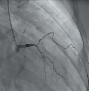
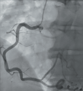
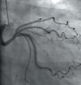
A stress echocardiogram showed a small area of reversible ischaemia in the apex and apical lateral wall. Coronary angiography subsequently showed an interesting coronary artery anatomy: all of her coronary arteries originate from the right coronary cusp with separate ostia. The left anterior descending artery (LAD) was hypoplastic (see figure 1), which is a very rare coronary artery anomaly (less than 0.03%). The right coronary artery (RCA) was a large calibre, tortuous vessel wrapping around the left ventricular apex (see figure 2). The left circumflex artery (Cx) was moderate in size, unobstructed and took a retro-aortic course (see figure 3).
The case was discussed in our regional cardiology and cardiothoracic multi-disciplinary team (MDT) meeting. As she was asymptomatic with only a small area of reversible ischaemia and the LAD (culprit vessel) was hypoplastic, it was not attractive for any sort of intervention or surgery. It was decided that medical management was the best strategy for her.

The patient was followed up in the cardiology clinic six months after medical treatment was commenced and she remained asymptomatic.
Discussion
Anomalous origin of coronary arteries are associated with sudden cardiac death, myocardial ischaemia,2,3 volume overload (coronary fistula), aortic root distortion (large coronary fistula or aneurysm), complication during aortic root surgery or angioplasty, and misdiagnosis (missing coronary artery).4 Depending on the number of coronary ostia present, this anomaly may be classified as type I (one coronary ostia), type II (two coronary ostia) and type III (three coronary ostia).2 Type III is the rarest and the one described in this case report.
Unlike effort-related ischaemia typical of fixed obstructive lesions, ischaemia associated with coronary anomalies is not always reproducible with stress testing.2 Ischaemia in these cases only occurs under inconsistent or extreme conditions.
During cardiac catheterisation, if the coronary arteries are difficult to cannulate in their normal positions, we should keep in mind the presence of coronary anomalies. An aortogram is commonly helpful to locate the coronary artery ostia. The success rate is variable, depending upon the operator experience. Non-standard catheters, like a multi-purpose one, are commonly helpful.
In figure 1, there is a faint opacification of the RCA when the LAD was selectively cannulated, suggesting there may be a common stem. However, on selective cannulation of the RCA in figure 2, there does not appear to be a common stem. Other non-invasive techniques like cardiac computed tomography (CT) coronary angiography and cardiac magnetic resonance imaging (MRI) or transoesophageal echocardiography may help if there remains uncertainty.
Medical management includes the use of various anti-anginal medications such as beta blockers, calcium-channel blockers, and the use of diuretics if heart failure is present.5 Specific surgical treatments vary depending upon the type of coronary anomalies, presence of symptoms, and individual circumstances. Common procedures used are: coronary bypass grafting, re-implantation of an anomalous coronary artery from the pulmonary artery into the aorta, reconstruction of proximal or distal coronary arteries, and fistula ligation.
Conflict of interest
None declared.
Key messages
- Anomalous coronary artery is a rare and diverse group of congenital disorder present in approximately 1% of the population
- The vast majority are benign and just an incidental finding, but care should be given to identify the types with a malignant course, as this can cause sudden cardiac death
- Further imaging using computed tomography (CT) coronary angiography or cardiac magnetic resonance imaging (MRI) may be helpful
References
1. Paolo A. Congenital heart disease for the adult cardiologist. Circulation 2007;115:1296–305. http://dx.doi.org/10.1161/CIRCULATIONAHA.106.618082
2. Paolo A, José AV, Scott F. Coronary anomalies: incidence, pathophysiology, and clinical relevance. Circulation 2002;105:2449–54. http://dx.doi.org/10.1161/01.CIR.0000016175.49835.57
3. Michael H. Congenital anomalies of the coronary arteries. Heart 2005;91:1240–5. http://dx.doi.org/10.1136/hrt.2004.057299
4. Dollar AL, Roberts WC. Retroaortic epicardial course of the left circumflex coronary artery and anteroaortic intramyocardial (ventricular septum) course of the left anterior descending coronary artery: an unusual coronary anomaly and proposed classification based on the number of coronary ostia in the aorta. Am J Cardiol 1989;64:828–9. http://dx.doi.org/10.1016/0002-9149(89)90780-7
5. Reul RM, Cooley DA, Hallman GL, Reul GJ. Surgical treatment of coronary artery anomalies: report of a 37 ½-year experience at the Texas Heart Institute. Tex Heart Inst J 2002;29:299–307. Available from http://www.ncbi.nlm.nih.gov/pmc/articles/PMC140292/
Further reading
Pollock BD, Belkin RN, Lazar S, Pucillo A, Cohen MB, Weiss MB. Origin of all three coronary arteries from separate ostia in the right sinus of Valsalva: a rarely reported coronary artery anomaly. Cathet Cardiovasc Diagn 1992;26:26–30. http://dx.doi.org/10.1002/ccd.1810260107
Kapoor A, Kumar S, Sinha N. Coronary angioplasty in a case of quadriostial origin of coronary arteries from right aortic sinus. Indian Heart J 2004;56:143–6.
