

Coronary artery anomalies (CAAs) are typically discovered by accident during imaging. We describe a 57-year-old man who presented with chest pain suggestive of acute coronary syndrome. Invasive angiography revealed unobstructed coronaries with an anomalous left anterior descending (LAD) artery originating from the right coronary sinus. Computed tomography coronary angiography confirmed its anomalous nature and revealed a trans-septal (intramuscular) course between the aortic root and right ventricular outflow tract. This configuration is regarded as benign, unlike inter-arterial forms associated with ischaemia or sudden cardiac death. The patient remained asymptomatic on follow up. This case highlights the value of multimodal imaging in accurately assessing coronary anatomy, avoiding misdiagnosis and directing the appropriate course of treatment.
Introduction
Congenital coronary artery anomalies (CAAs) are rare structural variations affecting the origin and course of coronary vessels, reported in 0.2–1.3% of invasive angiographies and up to 5.6% of computed tomography (CT) coronary angiography studies.1 Although most variants are benign, those involving an inter-arterial course between the great arteries may result in myocardial ischaemia, arrhythmias or sudden cardiac death.2 Anomalous origin of the left anterior descending (LAD) artery from the right coronary sinus is extremely uncommon, with clinical implications depending largely on its anatomical trajectory.3 Herein, we present a case of a middle-aged man with chest pain, in whom CT coronary angiography revealed an LAD arising from the right coronary sinus and coursing trans-septally (intramuscularly) through the interventricular septum. This finding confirmed the anomaly and demonstrated its benign nature, allowing safe patient management.
Case presentation
A 57-year-old man presented to the emergency department reporting central chest heaviness radiating to his right shoulder. The discomfort subsided following administration of glyceryl trinitrate and morphine by paramedics. He had a medical history of hypertension, hypercholesterolaemia, non-alcoholic fatty liver disease and pre-diabetes. His medications included ramipril 5 mg, felodipine 2.5 mg, atorvastatin 20 mg, and metformin 500 mg daily. He was a non-smoker and there was no preceding history of exertional angina. On examination, his vital signs were stable, with a blood pressure of 143/88 mmHg, a heart rate of 66 beats per minute, a respiratory rate of 16 breaths per minute, a temperature of 36.5°C, and an oxygen saturation of 100% in room air. The systemic examination was unremarkable. His electrocardiogram (ECG) showed a sinus rhythm with a left anterior fascicular block and transient lateral biphasic T-wave inversions. Additionally, his laboratory investigations revealed a normal troponin I and mild leukocytosis. Transthoracic echocardiography showed preserved left ventricular systolic function with mild concentric hypertrophy. Given the typical ischaemic pain and transient ECG changes, a provisional diagnosis of non-ST-elevation acute coronary syndrome was made. Coronary angiography via the right radial approach demonstrated unobstructed arteries, with an anomalous LAD arising from the right coronary cusp (Figure 1).
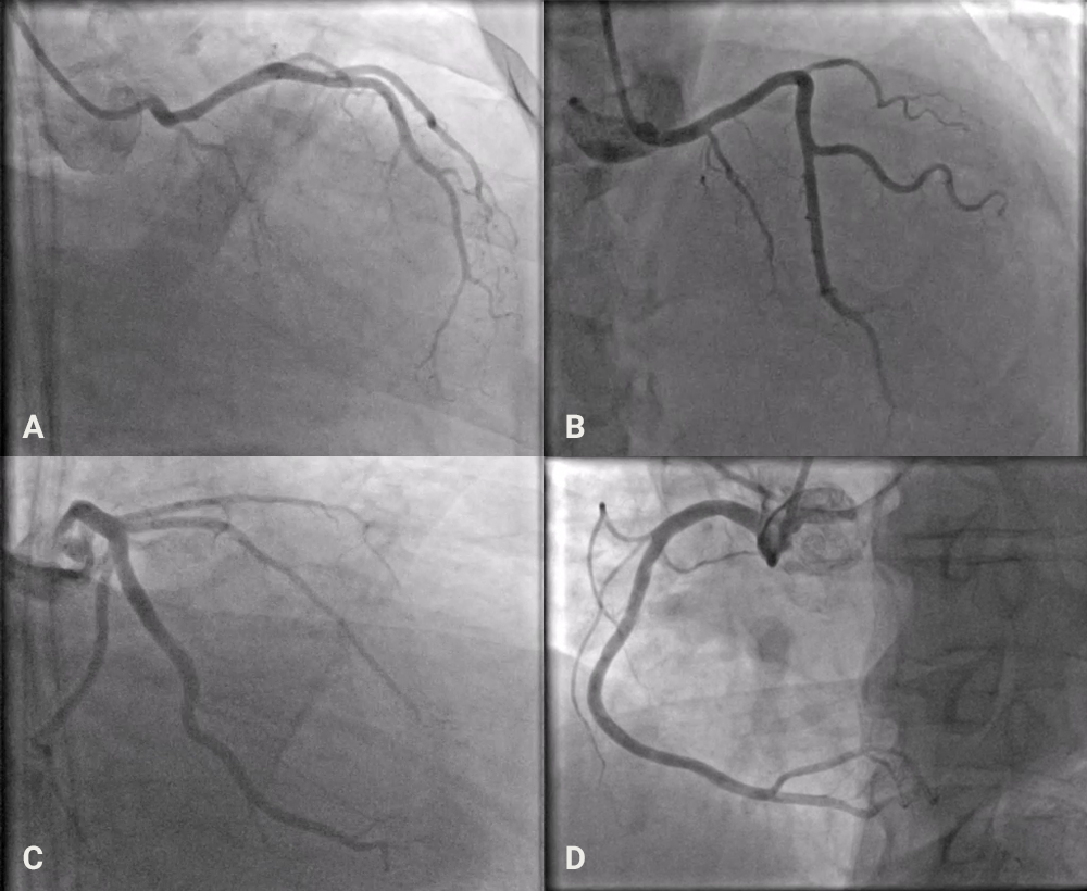
| Key: LAD = left anterior descending artery; LAO = left anterior oblique; LCX = left circumflex artery; LMCA = left main coronary artery; RCA = right coronary artery; RCC = right coronary cusp; RI = ramus intermedius artery |
ECG-gated CT coronary angiography subsequently confirmed a trans-septal (intramuscular) course of the LAD between the aortic root and right ventricular outflow tract, with no evidence of coronary compression or systolic stenosis (Figure 2). The patient remained asymptomatic and was discharged with medical therapy and follow up.
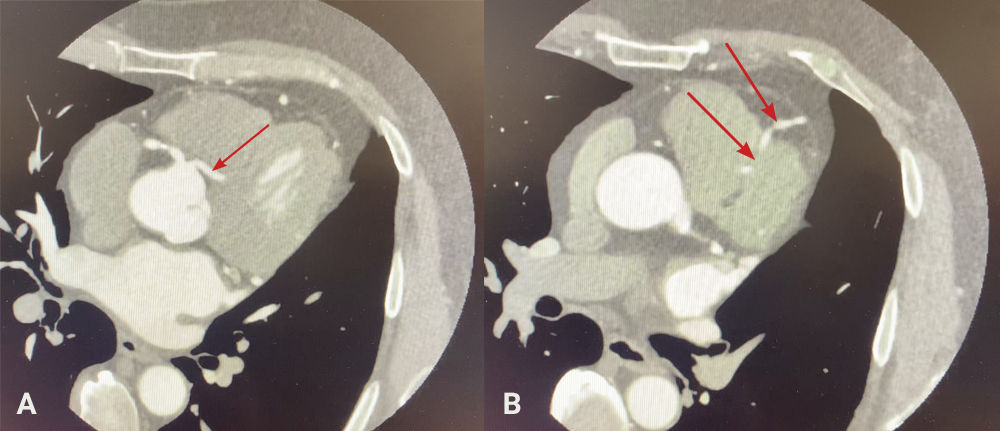
| Key: CAA = coronary artery anomaly; LAD = left anterior descending artery; MPR = multiplanar reformation or reconstruction; RVOT = right ventricular outflow tract |
Discussion
CAAs represent a spectrum of structural coronary variants with diverse clinical implications. Although most are discovered incidentally, accurate identification is essential to distinguish benign from potentially malignant courses.4 The trans-septal (intramuscular) trajectory, as observed in this case, is generally benign and does not involve compression between major vessels.5 Classification systems, such as those by Lipton and Yamanaka, categorise anomalies based on the sinus of origin and arterial course.6 In this context, the present case corresponds to a right sinus origin with a trans-septal trajectory.
CT coronary angiography provides excellent spatial resolution and is the modality of choice for delineating coronary anatomy when anomalies are suspected.7 Invasive angiography alone might only partially demonstrate the spatial relation of aberrant vessels to adjacent structures. The combination of anatomical and functional assessment ensures accurate risk stratification and avoids unnecessary intervention. Previous reports have described similar trans-septal LAD variants presenting with chest pain but showing no haemodynamic compromise, aligning with our findings.8
Conclusion
This report describes a rare case of an anomalous LAD originating from the right coronary sinus with a trans-septal course. Recognition of this variant is crucial to avoid misinterpretation as acute coronary syndrome. Multimodal imaging, particularly CT coronary angiography, plays a key role in accurately defining anatomy, excluding malignant variants, and guiding safe, evidence-based management. Follow up remains important despite the benign nature of this anomaly.
Conflicts of interest
None declared.
Funding
None.
Patient consent
Written informed consent was obtained from the patient for publication of this case report, including accompanying images.
References
1. Yildiz A, Okcun B, Peker T et al. Prevalence of coronary artery anomalies in 12,457 adult patients undergoing coronary angiography. Am J Cardiol 2010;106:1627–33. https://doi.org/10.1002/clc.20588
2. Cheezum MK, Liberthson RR, Shah NR et al. Anomalous aortic origin of a coronary artery from the inappropriate sinus of Valsalva. J Am Coll Cardiol 2017;69:1592–608. https://pubmed.ncbi.nlm.nih.gov/28335843/
3. Maller VV, Asriyan A. Transseptal coronary artery – a pictorial review. Cardiovasc Diagn Ther 2024;14:1234–44. https://doi.org/10.1007/s00247-024-05911-x
4. Angelini P. Coronary artery anomalies: an entity in search of an identity. Circulation 2007;115:1296–305. https://doi.org/10.1161/CIRCULATIONAHA.106.618082
5. Yamanaka O, Hobbs RE. Coronary artery anomalies in 126,595 patients undergoing coronary arteriography. Cathet Cardiovasc Diagn 1990;21:28–40. https://doi.org/10.1002/ccd.1810210110
6. Baz RO, Marinho A, Nieman K. Coronary artery anomalies: A computed tomography perspective. J Clin Med 2024;13:3920. https://doi.org/10.3390/jcm13133920
7. Cavallo AU, Muscogiuri E, Forcina M et al. Trans-septal course of anomalous left main coronary artery originating from single right coronary ostium presenting with atrial fibrillation in a severely obese patient: a case report. Egypt Heart J 2020;72:61. https://doi.org/10.1186/s43044-020-00093-x
8. Maller VV, Sieniewicz BJ, Al-Shaibi KF, Singh R, Chalil S. Transseptal coronary artery — a pictorial review. Egypt Heart J 2024;76:12. https://doi.org/10.1007/s00247-024-05911-x


