Percutaneous coronary intervention in the very elderly (≥85 years): trends and outcomes

Br J Cardiol 2013;20:27–31doi:10.5837/bjc.2013.006 Leave a comment
Click any image to enlarge
Authors:

This single-centre, retrospective, cohort study aims to provide insight into the long-term survival of patients ≥85 years old undergoing percutaneous coronary intervention (PCI) over a four-year observational period in a high-volume PCI centre. Between 2006 and 2010, 294 patients (mean age 88 ± 2 years, 56% male) underwent PCI at our institute. A total of 180 patients (61.2%) had an acute coronary syndrome (ACS) defined as unstable angina, non-ST elevation myocardial infarction (NSTEMI) or ST-elevation myocardial infarction (STEMI). One hundred and fourteen patients underwent PCI electively (38.8%). 

The primary outcome was all-cause 30-day and one-year mortality rates. In-hospital, 30-day and one-year mortality rates were 2.4% (7 patients), 4.4% (13 patients) and 17.7% (52 patients), respectively, in the entire cohort. In addition, 30-day (5.6% vs. 3.4%, p=0.24) and one-year (20.0% vs. 14.0%, p=0.19) mortality rates were similar between the ACS and elective patients, respectively. Following multi-variable analysis, age (hazard ratio [HR] 1.14, 95% confidence interval [CI] 1.04 to 1.26), male sex (HR 1.85, 95% CI 1.01 to 3.42), previous PCI (HR 2.74, 95% CI 1.36 to 5.56) and the presence of shock (HR 15.39, 95% CI 6.67 to 35.50) emerged as independent predictors of one-year mortality rates.

We conclude that PCI appears to be a safe treatment option in very elderly patients with good one-year survival rates. Future randomised-controlled trials should specifically include this age group to guide interventional cardiologists in making decisions when faced with this very challenging cohort.

Introduction

istockOver the last several years, the UK has witnessed a gradual ageing of its population.1 Moreover, the proportion of the very elderly (≥85 years old) in the general population is expected to rise fastest with a three-fold increase by the year 2035.1 Advancing age is perhaps the strongest predictor of de novo cardiovascular disease (CVD).2 As a consequence, cardiovascular (CV) mortality rates demonstrate a linear association with increasing age beyond the seventh decade. For example, octogenarians have a 10-fold greater risk of developing CVD in comparison with patients <50 years of age.2 Furthermore, mortality rates from CVD are higher for the very elderly, irrespective of clinical presentation.

Following an acute coronary syndrome (ACS) with either ST elevation or non-ST elevation myocardial infarction (STEMI or NSTEMI), 30-day mortality rates in the very elderly have been shown to be 10-fold higher compared with patients <65 years old.3,4 In the setting of chronic stable angina, the Trial of Invasive versus Medica therapy in Elderly patients with chronic angina (TIME) demonstrated that patients managed medically had a two-fold increased risk of an adverse CV outcome compared with those undergoing myocardial revascularisation.5

Although the very elderly are at an increased CV mortality risk, evidence suggests that the proportion of patients who receive standard secondary prevention or revascularisation declines as they age beyond the eighth decade, despite both therapies being of proven prognostic benefit in this age group.6 Consequently, the American Heart Association (AHA) has developed guidelines to highlight the importance of these treatment options in the very elderly.4 As a result, new emerging evidence suggests that the proportion of very elderly patients undergoing percutaneous coronary intervention (PCI) is increasing.3,7 For example, two very large US registries have observed that up to one in nine patients undergoing PCI can be expected to be ≥85 years old.6,8 Paradoxically, there is clearly a paucity of very elderly patients included in major landmark PCI trials, which have historically underrepresented this age group (comprising ≤2% of the study population).4 Furthermore, the very elderly patients that have been included in randomised-controlled trials have traditionally had fewer comorbidities in comparison with their counterparts from registries and retrospective studies.4

In the absence of robust randomised clinical data on PCI treatment strategies for the very elderly cohort, observational studies remain valuable in providing insights to outcome and mortality trends. The purpose of this four-year retrospective analysis of patients ≥85 years treated with PCI at our centre was to demonstrate safety and efficacy of therapies and examine for predictors of mortality.

Methods

Study cohort

Between 1 July 2006 and 30 June 2010, a total of 6,398 patients were treated with PCI at our institute, currently the highest volume non-surgical PCI centre in the UK. Two hundred and ninety-four patients were aged 85 years or over (4.6%). This group was subdivided by mode of clinical presentation.

The ACS group comprised those patients with STEMI, NSTEMI or unstable angina. Patients were diagnosed with STEMI in the presence of new ST-segment elevation ≥1 mm seen in any location or new left bundle branch block (on index or subsequent electrocardiograms [ECGs]) with at least one elevated biochemical marker of myocardial necrosis (including troponin measurements). Patients with NSTEMI were diagnosed with ECG evidence of myocardial ischaemia (without ST-segment elevation) and biochemical evidence of cardiac necrosis. Finally, unstable angina was diagnosed with a history consistent with ACS and normal biochemical markers of myocardial necrosis. The elective group comprised patients with chronic stable angina who had symptoms despite two anti-anginal agents and, in addition, had demonstrable ischaemia with either fractional flow reserve (FFR) <0.80 or a positive non-invasive test (adenosine-stress magnetic resonance imaging [MRI], stress echocardiography or myocardial perfusion scintigraphy).

Cardiogenic shock patients were included in the ACS group and were defined according to the criteria used in the SHould we emergently revascularise Occluded Coronaries for cardiogenic shocK (SHOCK) trial.9 Standardised international definitions of all patient-related clinical diagnoses, complications and outcomes were adhered to. All patients undergoing PCI received heparin, aspirin and clopidogrel in accordance with current guidelines.10,11 Procedural decisions including access site, device selection, use of adjunctive pharmacotherapy and type of stent were left at the operator’s discretion. For patients undergoing ‘staged’ procedures, whereby they returned for an elective PCI having had the culprit vessel previously treated, only the index episode was included. All baseline clinical, angiographic and procedural data were entered into a dedicated electronic database by trained cardiac catheterisation laboratory physiologists and interventional cardiologists. All patients were established on standard secondary prevention therapy as recommended by guidelines at discharge.10,11 All patients were prospectively followed up for a minimum of one year through a specialised cardiac rehabilitation service and post-PCI clinics.

Primary outcome

The primary outcome measure for the present study was all-cause 30-day and one-year mortality rates.

Statistical analyses

All statistical analyses were performed using SPSS statistical software, version 19.0 (IBM, Armonk, NY, USA). Categorial variables are presented as number and percentage while continuous variables are presented as mean ± standard deviation (SD). An independent t-test was used to compare baseline characteristics between the ACS and elective groups with a significance level of p<0.05. Cumulative event rates of all-cause mortality were analysed using the Kaplan-Meier method. We used Cox proportional hazards regression to identify independent predictors of mortality for the entire cohort. Variables were presented with hazard ratios (HR) and 95% confidence intervals (CI). Following uni-variable analyses, we identified potential predictors of mortality (variables with a p value <0.1), which were subsequently jointly included in a multi-variable analysis to identify independent predictors of mortality presented as HR and 95% CI. A p value <0.05 was considered significant.

Results

Baseline patient characteristics

Baseline clinical and angiographic data are presented in table 1. The ACS group comprised 180 patients (61.2%) of which nine patients presented with cardiogenic shock. The elective group contained 114 patients (38.8%). There were no statistically significant differences in the baseline characteristics of the two groups other than a slightly higher proportion of the elective group having a history of elevated cholesterol and peripheral vascular disease.

Table 1. Baseline characteristics of study population. Data are presented as percentages or mean ± standard deviation (SD)

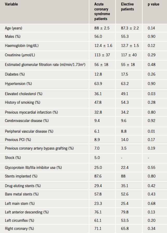
Of the whole cohort, mean age was 88 ± 2 years, with the oldest patient treated aged 98 years. Within the ACS group, the percentage of patients that underwent an angiogram following STEMI and NSTEMI (or unstable angina) was 85% and 63%, respectively, according to the Myocardial Ischaemia National Audit Project (MINAP) admission data of our institute. Subsequently, the rate of progression to PCI was 91% and 71% for the STEMI and NSTEMI (or unstable angina) patients, respectively. Eighteen patients (6.1%) had advanced chronic kidney disease (estimated glomerular filtration rate [eGFR] <30 ml/min/1.73m2) and 49 patients (17%) had a history of previous coronary artery bypass graft (CABG) or PCI. Furthermore, transradial PCI was performed in 150 patients (51%). In addition, 282 patients (96%) had ≤2 vessels treated, 250 patients (85%) had ≤2 lesions treated and the number of patients receiving 1, 2 or ≥3 stents was 102 (34.7%), 82 (28%) and 72 (24.5%), respectively. Finally, 164 patients (55.8%) and 93 patients (31.6%) received bare metal stents (BMS) and drug-eluting stents (DES), respectively.

Clinical outcome

Figure 1. Overall one-year survival rates between acute coronary syndrome (ACS)group and elective group

One-year follow-up was complete for all 294 patients. In-hospital, 30-day and one-year mortality rates were 2.4% (seven patients all presenting in cardiogenic shock), 4.4% (13 patients) and 17.7% (52 patients), respectively, for the entire cohort (table 2). Furthermore, in-hospital, 30-day and one-year mortality rates for the ACS and elective groups were 3.9% vs. 0% (p=0.033), 5.6% vs. 3.4% (p=0.24), and 20.0% vs. 14.0% (p=0.19), respectively. The in-hospital mortality rate for patients presenting with shock was 78%. In fact, there was zero mortality in the ACS group when the shock patients were excluded. The Kaplan–Meier curve representing one-year mortality rates in the ACS and elective groups is shown in figure 1. Furthermore, there was no significant statistical difference in one-year target vessel revascularisation (TVR) rates, which were 6.1% and 3.5% in the ACS and elective groups, respectively (p=0.33).

Table 2. One-year clinical outcomes in patients post-percutaneous coronary intervention (PCI). Data are presented as percentages

Subsequently, significant uni-variable predictors of one-year mortality were per year increase in age, male sex, shock and previous PCI. In fact, multi-variable analysis using Cox proportional hazard model revealed that the same parameters, i.e. age (HR 1.14, 95% CI 1.04 to 1.26), male sex (HR 1.86, 95% CI 1.01 to 3.43), previous PCI (HR 2.62, 95% CI 1.30 to 5.31) and the presence of shock (HR 15.81, 95% CI 6.86 to 36.50) emerged as independent predictors of one-year mortality rate as shown in table 3.

Table 3. Uni-variable and multi-variable predictors of one-year mortality rates

Discussion

To our knowledge, this paper represents the largest UK-based single-centre study examining the efficacy and safety of PCI in a large cohort of patients ≥85 years. Our data indicate that PCI is safe in patients ≥85 years with low in-hospital, 30-day and one-year mortality rates, irrespective of whether the PCI is performed in the setting of an ACS or electively. Patients ≥85 years presenting with cardiogenic shock recorded extremely high in-hospital mortality rates. In addition, per year incremental increase in age and being male increased the risk of one-year all-cause mortality by 14% and 85%, respectively.

Patients ≥85 years represent around 12% of all ACS admissions to UK hospitals according to a recent MINAP report.12 Furthermore, this cohort recorded the longest length of hospital stay and highest in-hospital mortality rate. Despite these observations, this age group was least likely to receive revascularisation, with only 10% of patients undergoing a diagnostic coronary angiogram. This age group, however, recorded the largest decrease in in-hospital mortality rates in comparison with patients <55 years (45% vs. 20%, respectively) over a seven-year period. We strongly agree with the observation of the authors of this report that “the elderly hospitalised with an ACS continue to be disadvantaged”.

The Global Registry of Acute Coronary Syndromes (GRACE) recently demonstrated that, following an ACS, octogenarians were less likely to receive standard secondary prevention treatment, in comparison with younger patients, despite recording the highest in-hospital mortality rate.13 The very elderly patients that underwent revascularisation during the index admission recorded the greatest absolute reduction in in-hospital death in comparison with other age groups. For example, the in-hospital mortality rates in patients <70 years old decreased from 2.9% to 1.6% following revascularisation in comparison with a decline from 11% to 7% in the very elderly. Subsequently, revascularisation in the very elderly translated to a 7% absolute reduction in all-cause mortality at six months as compared with a 1.8% reduction in the younger cohort. The in-hospital and six-month mortality rates in our study are comparable with those of the GRACE registry patients that received revascularisation, 3.9% vs. 7.0% and 11.7% vs. 12.0%, respectively.

Approximately, 39% of patients in our study underwent PCI for refractory stable angina, despite being on two or more anti-anginal agents. To date, no trial has examined the role of revascularisation in patients with stable angina ≥85 years old. The TIME trial was the first randomised trial to compare an invasive strategy versus medical therapy in 301 patients aged ≥75 years with stable angina.5 At six months, there was no difference in the all-cause mortality rate between the two groups. However, 50% of patients in the medical therapy group suffered an ACS and 75% of these underwent revascularisation. Subsequently, in a four-year follow-up of the TIME study, the sustained beneficial effect of revascularisation on the quality of life and angina status was noted.14 More importantly, revascularisation within one year of randomisation decreased the risk of long-term cardiac mortality by 56%. Finally, an invasive strategy was cost-effective in comparison with medical therapy.15

In concordance with our results, a large US-based registry demonstrated an in-hospital mortality rate of 5.4% in patients ≥85 years old, which was significantly higher in comparison with younger age groups.6 This age group recorded the most rapidly declining PCI rate over a four-year period. In contrast, patients ≥85 years old undergoing PCI represented the only group with a reduction in repeat revascularisation rates. The study also reported TVR rates of 7.0%, which are comparable with our data.6

Our data demonstrate that cardiogenic shock emerged as the strongest predictor of one-year all-cause mortality with an in-hospital mortality rate of 78%. These results are consistent with existing data.16 Furthermore, recent studies have also confirmed our findings of increasing age and male sex as independent predictors of one-year mortality rates.15,17 Previous PCI emerged as an independent predictor of one-year all-cause mortality in our study. Very elderly patients are at a greater risk of stent thrombosis.18 This is a consequence of several age-related biological changes, including endothelial dysfunction, higher levels of clotting factors and increased platelet reactivity.19 In the absence of any post-mortem data and relatively low event rates in our study it
was not possible to make this link.

Randomised trials have conventionally underrepresented the very elderly, comprising up to 2% of the study population.4 For example, the Clinical Outcomes Utilising Revascularisation and Aggressive Drug Evaluation (COURAGE) trial did not include any patients >75 years.20 In contrast, the proportion of patients ≥85 years in large registries has been five-fold higher.8 We feel that the evidence base to treat this age group can be strengthened substantially with future randomised-controlled trials.

Limitations

This is a single-centre retrospective study and is prone to inherent bias. Statistical tools were utilised to minimise this bias. Our findings are ‘hypothesis-generating’ and support the need for a randomised-controlled trial to examine the role, safety and efficacy of PCI in patients ≥85 years old. We were unable to ascertain the cognitive or frailty status for our cohort, which are important comorbidities in this age group and can influence management decisions and treatment options.

Conclusion

PCI is a safe and efficacious treatment option in very elderly patients with good one-year survival rates. There appears to be no difference in one-year survival rates between patients aged ≥85 years requiring acute or elective PCI. Future randomised-controlled trials should specifically include this age group to guide interventional cardiologists in making decisions when faced with this very challenging cohort.

Funding

This study was supported by the research fund of The Dorset Heart Centre, Royal Bournemouth and Christchurch Hospitals NHS Foundation Trust.

Conflict of interest

None declared.

Editors’ note

See also the editorial by Knight et al.

Key messages

  • There are very limited data examining the role of percutaneous coronary intervention (PCI) in the very elderly
    (>85 years), who suffer from refractory angina or acute coronary syndrome (ACS)
  • Registry data suggest that the very elderly are less likely to receive PCI in comparison with younger patients and yet derive the greatest benefit in terms of survival and re-hospitalisation rates
  • Our data indicate that PCI is a safe and acceptable treatment strategy for very elderly patients who present with ACS or refractory angina
  • There is a need for randomised-controlled trials to examine the role of PCI in the very elderly

References

  1. National Population Projections 2010-based Statistical Bulletin. London: Office for National Statistics, 2011. Available from: http://www.ons.gov.uk/ons/dcp171778_235886.pdf
  2. Driver JA, Djoussé L, Logroscino G et al. Incidence of cardiovascular disease and cancer in advanced age: a prospective cohort study. BMJ 2008;337:a2467. http://dx.doi.org/10.1136/bmj.a2467
  3. Claessen BEPM, Kikkert WJ, Engstrom AE et al. Primary percutaneous coronary intervention for ST elevation myocardial infarction in octogenarians: trends and outcomes. Heart 2010;96:843–7. http://dx.doi.org/10.1136/hrt.2009.185678
  4. Alexander KP, Newby K, Cannon CP et al. Acute coronary care in the elderly, part 1 non-ST-segment-elevation acute coronary syndromes: a scientific statement for healthcare professionals from the American Heart Association Council on Clinical Cardiology in collaboration with the Society of Geriatric Cardiology. Circulation 2007;115:2549–69. http://dx.doi.org/10.1161/CIRCULATIONAHA.107.182615
  5. Pfisterer M, Bertel O, Erne P et al. Trial of invasive versus medical therapy in elderly patients with chronic symptomatic coronary artery disease (TIME): a randomised trial. Lancet 2001;358:951–7. http://dx.doi.org/10.1016/S0140-6736(01)06100-1
  6. Wang TY, Masoudi FA, Messenger JC et al. Percutaneous coronary intervention and drug-eluting stent use among patients ≥85 years of age in the United States. J Am Coll Cardiol 2012;59:105–12. http://dx.doi.org/10.1016/j.jacc.2011.10.853
  7. Johnman C, Oldroyd KG, Mackay DF et al. Percutaneous coronary intervention in the elderly: changes in case-mix and periprocedural outcomes in 31,758 patients treated between 2000 and 2007. Circ Cardiovasc Interv 2010;3:341–5. http://dx.doi.org/10.1161/CIRCINTERVENTIONS.109.928705
  8. Alexander KP, Roe MT, Chen AY et al. Evolution in cardiovascular care for elderly patients with non-ST-segment elevation acute coronary syndromes: results from the CRUSADE national quality improvement initiative. J Am Coll Cardiol 2005;46:1479–87. http://dx.doi.org/10.1016/j.jacc.2005.05.084
  9. Hochman JS, Sleeper LA, Webb JG et al. Early revascularization in acute myocardial infarction complicated by cardiogenic shock. SHOCK investigators. Should we emergently revascularize occluded coronaries for cardiogenic shock. N Engl J Med 1999;341:625–34. http://dx.doi.org/10.1056/NEJM199908263410901
  10. Van de Werf F, Bax J, Betriu A et al. Management of acute myocardial infarction in patients presenting with persistent ST-segment elevation. Eur Heart J 2008;29:2909–45. http://dx.doi.org/10.1093/eurheartj/ehn416
  11. Hamm CW, Bassand J-P, Agewall S et al. ESC guidelines for the management of acute coronary syndromes in patients presenting without persistent ST-segment elevation. Eur Heart J 2011;32:2999–3054. http://dx.doi.org/10.1093/eurheartj/ehr236
  12. Gale CP, Cattle BA, Woolston A et al. Resolving inequalities in care? Reduced mortality in the elderly after acute coronary syndromes. The Myocardial Ischaemia National Audit Project 2003–2010. Eur Heart J 2012;33:630–9. http://dx.doi.org/10.1093/eurheartj/ehr381
  13. Devlin G, Gore JM, Elliott J et al. Management and 6-month outcomes in elderly and very elderly patients with high-risk non-ST-elevation acute coronary syndromes: the Global Registry of Acute Coronary Events. Eur Heart J 2008;29:1275–82. http://dx.doi.org/10.1093/eurheartj/ehn124
  14. Pfisterer M. Long-term outcome in elderly patients with chronic angina managed invasively versus by optimized medical therapy: four year follow-up of the randomized trial of invasive versus medical therapy in elderly patients (TIME). Circulation 2004;110:1213–18. http://dx.doi.org/10.1161/01.CIR.0000140983.69571.BA
  15. Claude J, Schindler C, Kuster GM et al. Cost-effectiveness of invasive versus medical management of elderly patients with chronic symptomatic coronary artery disease: findings of the randomized trial of invasive versus medical therapy in elderly patients with chronic angina (TIME). Eur Heart J 2004;25:2195–203. http://dx.doi.org/10.1016/j.ehj.2004.09.013
  16. Zeymer U, Vogt A, Zahn R et al. Predictors of in-hospital mortality in 1333 patients with acute myocardial infarction complicated by cardiogenic shock treated with primary percutaneous coronary intervention (PCI). Eur Heart J 2004;25:322–8. http://dx.doi.org/10.1016/j.ehj.2003.12.008
  17. Roe MT, Chen AY, Thomas L et al. Predicting long-term mortality in older patients after non-ST-segment elevation myocardial infarction: the CRUSADE long-term mortality model and risk score. Am Heart J 2011;162:875–83. http://dx.doi.org/10.1016/j.ahj.2011.08.010
  18. Klein LW. Is patient frailty the unmeasured confounder that connects subacute stent thrombosis with increased periprocedural bleeding and increased mortality? J Am Coll Cardiol 2012;59:1760–2. http://dx.doi.org/10.1016/j.jacc.2012.01.042
  19. Wang TY, Gutierrez A, Peterson ED. Percutaneous coronary intervention in the elderly. Nat Rev Cardiol 2011;8:79–90. http://dx.doi.org/10.1038/nrcardio.2010.184
  20. Boden WE, O’Rourke RA, Teo KK. Optimal medical therapy with or without PCI for stable coronary disease. N Engl J Med 2007;356:1503–16. http://dx.doi.org/10.1056/NEJMoa070829

Primary angioplasty for acute STEMI in secondary care: feasibility, outcomes and potential advantages

Br J Cardiol 2013;20:32–7doi:10.5837/bjc.2013.007 Leave a comment
Click any image to enlarge
Authors:

Primary percutaneous coronary intervention (PCI) has become treatment of choice for ST-elevation myocardial infarction (STEMI) in England. The assumption in most trials, and in their translation into clinical practice, is that centralisation of primary PCI for STEMI into large facilities is inevitable and essential. We feel that a successful and preferable primary PCI service can be delivered in a medium-sized district general hospital (DGH).

We performed a retrospective analysis of the first 18 months of primary PCI for STEMI in our unit. We compared our results with standards set out in the National Infarct Angioplasty Project report. Our median call-to-balloon time was 95 minutes and median door-to-balloon time was 50 minutes. Door-to-balloon times were shorter during working hours than out of hours, and were shorter for patients taken directly to the catheterisation laboratory than those admitted via A&E. During the period of assessment, 14% of patients experienced a major adverse cardiovascular and cerebrovascular event (MACCE). The overall mortality rate was 6.7%. No patients were transferred to our surgical centre for emergency treatment. 

We believe our data demonstrate that a system utilising regional Heart Attack Centres is not essential for satisfactory clinical outcomes, and may not be the preferred choice for patients and their ongoing post-myocardial infarction care. Expansion of PCI services in well-organised DGHs may be an equally good solution.

Introduction

Screen shot 2013-03-04 at 13.30.02On inference from a range of randomised clinical trials, timely primary percutaneous coronary intervention (PPCI) has become the optimal strategy for the treatment of ST-segment elevation myocardial infarction (STEMI).1-8 Despite the logistic complexity and potential for delay compared with fibrinolytic treatment, the standard outcomes of safety and effectiveness of PPCI are superior to fibrinolysis unless the time delay is substantial.9-12 These data have led to the decision that, not only should PPCI be the treatment of choice for STEMI in England, it must be available 24 hours per day, seven days per week.13 This generates logistical implications with regards to the timely delivery of PPCI, such as appropriate training of paramedic services, adequate staffing, and maintaining sufficient operator and centre volumes. Guidelines suggest that an individual operator should be performing at least 75 PCI procedures per annum14 within a facility performing at least 200 PCIs per annum,14,15 with 36 of these being PPCI for STEMI.14 These recommendations correlate to shorter door-to-balloon times and more use of evidence-based therapies, but not with in-hospital mortality or inpatient length of stay.16 The assumption in most trials, and in their translation into clinical practice, is that centralisation of PPCI for STEMI into large facilities, usually tertiary care surgical centres, is both inevitable and necessary despite any problems of access that may result.13,15,17 Access problems in the UK are of two types:

  • Increased travel time/distance for patients associated with lack of infrastructure, and, thus, delay.
  • Exclusion of some patients for a variety of reasons (age, frailty, comorbidity, diagnostic uncertainty, acute severe haemodynamic instability, etc.).

In England, PPCI for patients in their local secondary care hospital (usually a District General Hospital [DGH]) could reduce these access problems. However, it is not clear that such services can be provided safely and effectively in a sustainable way. Even in DGHs that provide elective coronary angioplasty services, it is, at present, unusual for them to provide 24-hour access to PPCI. There are two reasons:

  • Concern that activity is below the minimum for safety and effectiveness to be assured.
  • Inadequate facilities and staff numbers to maintain the service cost-effectively.

Context

Coronary angioplasty has been available at Basingstoke and North Hampshire Foundation Trust (BNHFT), a medium-sized DGH without on-site cardiac surgery, for seven years. When it began this was an unusual service model, and there were questions about safety, effectiveness and sustainability of the service. However, over this period there have been satisfactory clinical outcomes and the service has been uninterrupted with no emergency transfers for surgery. Only a handful of cases have been referred for PCI in other centres. PPCI has been available from the beginning of the service, but only since 2 January 2009 has it been available 24 hours per day, seven days per week. Equitable access has developed during this period in other facilities for populations to the north, east and south of the normal catchment population, and while there has been some increase in the referral footprint of the service to the west, the experience is substantially that of a sole comprehensive DGH provider to a defined and stable population. This experience is, therefore, well placed to answer questions about safety, effectiveness and sustainability in real life, and to examine whether there are advantages or disadvantages to this model of care, against the prevailing strategy of centralisation into Heart Attack Centres.

Possible advantages include:

  • Comprehensive access regardless of comorbidity and acute instability.
  • Early access for patients with and without a pre-hospital diagnosis.
  • Better integration of the STEMI episode into more long-term cardiac and non-cardiac primary and secondary care.
  • Superior patient experience as a result of local rather than remote care.

We report the first 18 months of the service with regard to safety, effectiveness and sustainability, and provide information about its contribution to realising these possible advantages.

Methods

BNHFT is a medium-sized DGH providing secondary care cardiac services to a population of approximately 300,000 people. The population is part rural, part urban with a young but rising age profile. The standardised mortality ratio (SMR) for coronary heart disease is 1.0.

PPCI takes place in a dedicated cardiac catheterisation laboratory that is available 24 hours per day, seven days per week. This is adjacent to the eight-bedded coronary care unit. A non-dedicated angiography suite, located within the radiology department, is occasionally used for cardiac work. The elective and non-elective invasive cardiac programme is integrated, running five days per week, with out-of-hours on-call for non-elective work; the service is never suspended. The cardiac catheterisation laboratory is staffed with nurses, cardiac physiologists and dedicated radiographers in and out of hours; there is no residential on-call team. Our facility performs >500 elective or urgent PCI procedures per year.

Patients with STEMI access PPCI in three ways:

  • Pre-hospital diagnosis by paramedics with pre-alert through telemedicine and direct admission to the catheterisation laboratory.
  • Presentation to Accident & Emergency department (A&E) and direct transfer to the catheterisation laboratory.
  • Diagnosis as current inpatients with direct transfer to the catheterisation laboratory.

There are no set exclusion criteria and a senior cardiologist is always available for triage and discussion. Diagnosis of STEMI is by standard electrocardiogram (ECG) criteria (including [presumed] new left bundle branch block) and clinical features.18 Diagnostic coronary angiography is the default care pathway in uncertain cases. Individual patient care records are kept in standard hospital notes, and all PPCI activity is also recorded in a dedicated cardiac database (DataCam™), for internal use and submission to national databases (Central Cardiac Audit Database [CCAD] and Myocardial Infarction National Audit Project [MINAP]).

Patients in this report were identified from our in-house cardiac database (DataCam™). Individual patient details were retrieved from the cardiac database and individual hospital records, and follow-up information was cross-checked with patients’ general practitioners.

Standard definitions for call-to-balloon times, door-to-balloon times, and major adverse cardiovascular and cerebrovascular events (MACCE) were used. For patients who self-presented to A&E, their time of arrival was taken as first medical contact (and call) time. Out of hours is defined as outside the hours 0800–1800 Monday to Friday, and all bank holidays.

BNHFT benefits from having a dedicated team of cardiac rehabilitation nurses who identify and offer to enrol all STEMI patients into our nationally recognised cardiac rehabilitation programme.

Results

Data between the dates 02/01/2009 and 21/08/2010 were available for analysis. A total of 165 PPCI procedures for STEMI were performed in 163 patients (male=135, female=28) during this time. There were 29 ‘false activations’ of the service; patients who underwent emergency angiography, had no evidence of acute coronary occlusion, and, therefore, no PCI was performed. Nine of these procedures were undertaken out of hours. All procedures were carried out via a transfemoral approach. Glycoprotein IIb/IIIa inhibitor was used in 30% of cases. Two of these patients (both male) underwent a second procedure during the same admission for STEMI due to stent thrombosis (ST) and are included in the analyses (one acute and one sub-acute). One patient (male) went on to have inpatient coronary artery bypass grafting (CABG) in view of severe complex coronary artery disease (not following a complication of the PPCI procedure). A total of 152 patients (male=126, female=26) were treated with PPCI and stenting, while 11 patients (male=9, female=2) underwent balloon angioplasty only. Of these 11 patients, four had presented with late ST, one had acute ST, one had sub-acute ST, one had an embolic coronary artery occlusion, which resolved with antithrombotic medication, in two patients the vessel was heavily calcified and felt to be not amenable to stenting, one case had in-stent re-stenosis (ISR) treated with a drug-eluting balloon, and, in the final patient, the occlusion was in a distal small calibre vessel felt to be unsuitable for stenting.

Median patient age was 62 years (range 34–93) for the total population; males median age 61 years (range 38–93) and females median age 72 years (range 34–91). Route of admission was via A&E for 115 (70%) patients, direct admission from ambulance to catheter laboratory for 45 (27%) patients, and transfer to catheter laboratory from current inpatient ward in five (3%) patients. These demographic data in our population were similar to those presented in the National Infarct Angioplasty Project (NIAP) report.13

Overall median call-to-balloon (CTB) time was 95 minutes (range 11–645), while overall median door-to-balloon (DTB) time was 50 minutes (range 8–578). Median first medical contact-to-balloon time (FTB) was 84 minutes (range 11–632). For patients admitted via A&E, median DTB was 58 minutes (range 8–578), while for those with direct admission to the catheter lab median DTB time was 35 minutes (range 11–459). DTB times were shorter during working hours than out of hours, and were shorter for patients taken directly to the catheterisation laboratory than those admitted via A&E. Interestingly, both CTB and DTB times were longer in women than men (figure 1). The percentages of patients in whom PPCI reperfusion was achieved within the recommended CTB and DTB time points are shown in table 1.

Figure 1. Median time intervals to delivery of primary percutaneous coronary intervention (PPCI) for ST-elevation myocardial infarction (STEMI)
Table 1. Percentage of patients treated by recommended time points

For 11 patients ultimately treated with PPCI, there was diagnostic uncertainty due to the initial ECG being determined as non-diagnostic for STEMI leading to a delay in DTB times. An additional five patients, who presented after suffering an out-of-hospital cardiac arrest, were admitted and stabilised in the A&E department following successful resuscitation before a decision to proceed to coronary angiography with or without PPCI was taken. In these cases, the time to delivery of PPCI was longer than for the straightforward STEMI presentations. Our finding of longer CTB and DTB times in females compared with males has been reported previously. We are not able to ascertain the cause for this, or whether this gender-related delay affects outcome from our data.

In our data, 37 of 165 (22%) patients treated with PPCI were aged 75 years or older (75–93 years), while 19 patients (12%) were aged 80 years or older. Of those aged 75 years and above, 26 patients (70%) presented via the A&E, 15 of which presented outside of office hours. In our subgroup of patients aged 75 years or older, the median DTB time was 68 minutes (range 18–578) while the median CTB time was 150 minutes (range 90–656).

In 160 of the cases, single-vessel PPCI was undertaken to the infarct-related artery (IRA) only. In the majority of cases of multi-vessel disease, the culprit IRA was treated and significant stenoses in non-IRAs were electively addressed four to six weeks later. However, in five cases, critical stenoses were identified in a non-IRA that were felt by the operator to be clinically important and were treated by PCI during the index PPCI procedure.

The mean number of stents implanted was 1.2 (range 0–3, median 1), while mean number of drug-eluting stents (DES) used was 0.5 (range 0–3, median 0). Two patients with left main stem occlusion received DES.

Mean length of stay (LOS) for the total population was 3.2 days (range 1–29, median 2 days). For men, the mean LOS was 3.3 days (range 1–29, median 2 days), and for women, mean LOS was 2.7 days (range 1–23, median 2 days).

During the observational period, 23 (14%) patients experienced a MACCE (including death). There were four cases of definite stent thrombosis (one acute, one sub-acute and two late) and one case of possible late stent thrombosis. One patient suffered an ischaemic stroke and three patients experienced a transient ischaemic attack. Two patients had significant gastrointestinal bleeds requiring hospital admission. There were two cases of in-stent re-stenosis presenting as recurrent angina.

Over the course of the study period, 11 patients (male=8, female=3) died, giving an overall mortality rate of 6.7%. Nine of these patients died during the index hospital admission equating to a 30-day mortality rate of 5.5%. The other two patients died within six months.

Discussion

PPCI is now considered the gold standard of treatment for STEMI in both the UK and around the world. This is reflected in the Department of Health’s (DoH) Treatment of Heart Attack National Guidance report.13 In order to achieve the best clinical outcomes, it is essential that PPCI for STEMI is delivered as quickly after patient presentation as possible. In guidelines, standards for minimum acceptable call- and door-to-balloon times have been set at 120 and 90 minutes, respectively,14,19 times that are also reflected in DoH guidance for the UK.13 Several randomised-controlled trials have shown that these times, and good clinical outcomes, can be achieved, both in patients presenting directly to a PPCI centre, and those transferred from a non-PCI centre to a facility capable of performing PPCI.9-16 However, in order to achieve these results, it is imperative that all teams involved in the process, such as general practitioners, paramedic services, non-PCI hospitals, and A&Es have received sufficient education and training. Furthermore, excellent infrastructure and logistics is required for an efficient PPCI service. Finally, the cardiology team in the PPCI facility should perform a minimum number of PCI procedures per annum to ensure the most favourable outcomes.14-16,19

Hub and spoke system

In England, it has been suggested that a ‘hub-and-spoke’ system be developed to provide PPCI for STEMI, whereby in each region there would be a specialised Heart Attack Centre (often the Cardiology/Cardiothoracic surgical referral centre) that would provide all PPCI. Although this system has been shown to be clinically effective in Europe and North America,9-12 this has not been replicated in any English studies to date. While it may seem reasonable that the same outcomes would be achieved in England, this should be proven by randomised-controlled trial, given that our geography, road networks and infrastructure differ.

We believe that a ‘hub-and-spoke’ system is not necessary, and that safe and effective PPCI can be delivered in a secondary care hospital (with off-site surgical cover that is performing a medium-to-high number of PCI procedures per year). We present the findings from the first 18 months of PPCI for STEMI from one such facility, Basingstoke & North Hampshire Hospital. In our facility, PPCI was delivered within the recommended DTB time for 82% of our patients, with a median DTB time of 50 minutes. However, the proportion of patients receiving PPCI within a CTB time of <120 minutes was less good, especially out of hours and in the elderly, which we feel reflects patient delays in presentation, poor pre-hospital diagnosis, and inefficient transport to hospital. The pre-hospital delays occur despite the attending paramedic team making a diagnosis of STEMI in many cases and utilisation of a telephonic transfer system for ECGs to be relayed from the ambulance to our cardiac care unit to improve pre-hospital diagnosis for STEMI. A total of 115 (70%) of our patients came via our A&E department, where the diagnosis of STEMI was formalised and the PPCI team alerted. This figure is higher than we would like, given that admission via A&E leads to prolonged CTB and DTB times. This may reflect a need for either improved education of paramedic services, including increased awareness of the availability of direct catheter lab admission, or improvement of the logistics of patient admission, or both.

Our 30-day and overall mortality rates of 5.5% and 6.7%, respectively are similar to those published by NIAP (5.6% at 30 days, 8.7% at one year, and 9.9% at 18 months13) and no patients had to be transferred to our surgical centre for emergency CABG or salvage PCI.

Our data show that we are able to provide an effective and safe PPCI service. In our facility, a consultant cardiologist is available 24 hours per day for individual case discussion, and, if required, to clinically assess such patients. We feel such decisions would be more difficult for interventional operators based in regional Heart Attack Centres, who are reliant on accurate assessment by staff reviewing a patient with possible STEMI at a distant location. A process that, at best, undoubtedly introduces a time delay and at worst could lead to either inappropriate admission to regional centres for unsuitable patients, or non-transfer of potentially suitable (albeit higher risk) patients who may then receive suboptimal treatment.

Similar issues exist for patients who are successfully resuscitated following an out-of-hospital cardiac arrest. In these patients, it is not always immediately clear that the cause of the cardiac arrest is STEMI, and such patients often require a period of stabilisation and assessment in hospital prior to consideration for PPCI.

We believe that, while PPCI services at Heart Attack Centres are likely to be very good, these centres may be located at significant distances from the patient’s home and family, making visiting financially onerous and time-consuming. In addition, it needs to be determined whether the regional Heart Attack Centre has the capacity to cope with all STEMI for PPCI in a designated region. The average length of stay after PPCI for STEMI in our data was three days. It is not clear from the ‘hub-and-spoke’ proposal whether STEMI patients would remain in the Heart Attack Centre until time of discharge or be transferred back to the local DGH immediately after PPCI (depending on clinical stability). If the latter, how would the local DGHs feel about this design, given they were not providing the treatment in the first instance? Further, this also raises the issue of payment for PPCI; it is likely that payment be made to the regional Heart Attack Centre from the Health Authority relating to the patient’s locality. Would the payers feel comfortable that care was being provided in the most cost-effective way, as the care providers are likely to be employed within a different Health Authority. Many of these issues would be avoided in a system utilising appropriate local DGHs for PPCI.

As we provide our own PPCI service we can comprehensively enrol all STEMI patients into our cardiac rehabilitation programme to ensure good recovery and optimal ongoing care. There is a concern that, in a ‘hub-and-spoke’ system, some patients from our catchment area may receive PPCI and post-myocardial infarction (MI) management in the Heart Attack Centre but then, following discharge, not be readily identified to us, and, therefore, miss their rehabilitation and follow-up.

The BNHFT PPCI programme detailed in this paper was delivered by a dedicated team who operated a 24-hour-per-day, seven-day-per-week, on-call rota system: 87 (53%) of the PPCI procedures performed in this study were undertaken out of hours, which impacts on the daily responsibilities of all the specialties represented in the PPCI team, as well as having a financial impact for the hospital in the form of out-of-hours payment to staff. However, these out-of-hours payments would need to be compared with the costs involved in the transfer of patients to and from regional Heart Attack Centres, added to the tariff charge in such centres for the PPCI procedure, should a regional model be established. We would hope that our data would reassure the DoH that PPCI for STEMI can be delivered safely and effectively in a DGH, and, therefore, the strategy of Heart Attack Centres may not be required. Rather, service expansion within existing DGHs with PPCI facilities could be considered to ensure sustainability.

Conclusion

In conclusion, we present data from the first 18 months of 24/7 PPCI for STEMI at a medium-sized, high-volume PCI DGH in England, illustrating that a good service can be provided in a dedicated secondary care facility. We believe that these data demonstrate that a ‘hub-and-spoke’ system utilising regional Heart Attack Centres is not essential for satisfactory clinical outcomes, and may not be the preferred choice for patients and their longer-term post-MI care. Expansion of PCI services in well-organised DGHs may be an equally good solution.

Conflict of interest

This research received no specific grant from any funding agency in the public, commercial or not-for-profit sectors. The authors declare: no support from any organisation for the submitted work; no financial relationships with any organisations that might have an interest in the submitted work in the previous three years; no other relationships or activities that could appear to have influenced the submitted work.

Key messages

  • Primary angioplasty (PPCI) for STEMI can be efficiently and safely delivered in a medium-sized secondary care facility with off-site cardiac surgical cover
  • A hub-and-spoke Heart Attack Centre design will likely deliver high-quality PPCI, but logistical issues may limit service delivery; expansion of PPCI services in secondary care facilities presents an equally good option
  • Patient symptom recognition and pre-hospital diagnosis remain an important determinant of call-to-balloon times

References

  1. Grines CL, Browne KF, Marco J et al. A comparison of immediate angioplasty with thrombolytic therapy for acute myocardial infarction. The Primary Angioplasty in Myocardial Infarction Study Group. N Engl J Med 1993;328:673–9. http://dx.doi.org/10.1056/NEJM199303113281001
  2. Gibbons RJ, Holmes DR, Reeder GS, Bailey KR, Hopfenspirger MR, Gersh BJ. Immediate angioplasty compared with administration of a thrombolytic agent followed by conservative treatment for myocardial infarction. The Mayo Coronary Care Unit and Catheterisation Laboratory Groups. N Engl J Med 1993;328:685–91. http://dx.doi.org/10.1056/NEJM199303113281003
  3. Zijlstra F, de Boer MJ, Hoorntje JC, Reiffers S, Reiber JH, Suryapranata H. A comparison of immediate coronary angioplasty with intravenous streptokinase in acute myocardial infarction. N Engl J Med 1993;328:680–4. http://dx.doi.org/10.1056/NEJM199303113281002
  4. GUSTO IIb Angioplasty Substudy Investigators. A clinical trial comparing primary coronary angioplasty with tissue plasminogen activator for acute myocardial infarction. The Global Use of Strategies to Open Occluded Coronary Arteries in Acute Coronary Syndromes (GUSTO IIb) Angioplasty Substudy Investigators. N Engl J Med 1997;336:1621–8. http://dx.doi.org/10.1056/NEJM199706053362301
  5. Weaver WD, Simes RJ, Betriu A et al. Comparison of primary coronary angioplasty and intravenous thrombolytic therapy for acute myocardial infarction: a quantitative review. JAMA 1997;278:2093–8. http://dx.doi.org/10.1001/jama.1997.03550230069040
  6. Zijlstra F, Hoorntje JC, de Boer MJ et al. Long-term benefit of primary angioplasty as compared with thrombolytic therapy for acute myocardial infarction. N Engl J Med 1999;341:1413–19. http://dx.doi.org/10.1056/NEJM199911043411901
  7. Schomig A, Kastrati A, Dirschinger J et al. Coronary stenting plus platelet glycoprotein IIb/IIIa blockade compared with tissue plasminogen activator in acute myocardial infarction. Stent versus Thrombolysis for Occluded Coronary Arteries in Patients with Acute Myocardial Infarction Study Investigators. N Engl J Med 2000;343:385–91. http://dx.doi.org/10.1056/NEJM200008103430602
  8. Keeley EC, Boura JA, Grines CL. Primary angioplasty versus intravenous thrombolytic therapy for acute myocardial infarction: a quantitative review of 23 randomised trials. Lancet 2003;361:13–20. http://dx.doi.org/10.1016/S0140-6736(03)12113-7
  9. Widimsky P, Groch L, Zelizko M, Aschermann M, Bednár F, Suryapranata H. Multicentre randomised trial comparing transport to primary angioplasty vs immediate thrombolysis vs combined strategy for patients with acute myocardial infarction presenting to a community hospital without a cardiac catheterisation laboratory. The PRAGUE study. Eur Heart J 2000;21:823–31. http://dx.doi.org/10.1053/euhj.1999.1993
  10. Widimsky P, Budesinsky T, Vorac D et al. Long distance transport for primary angioplasty vs immediate thrombolysis in acute myocardial infarction. Final results of the randomised national multicentre trial – PRAGUE 2. Eur Heart J 2003;24:94–104. http://dx.doi.org/10.1016/S0195-668X(02)00468-2
  11. Grines CL, Westerhausen DR Jr, Grines LL et al.; Air PAMI Study Group. A randomised trial of transfer for primary angioplasty versus on-site thrombolysis in patients with high-risk myocardial infarction: The Air Primary Angioplasty in Myocardial Infarction study. J Am Coll Cardiol 2002;39:1713–19. http://dx.doi.org/10.1016/S0735-1097(02)01870-3
  12. Moon JC, Kalra PR, Coats AJ. DANAMI-2: Is primary angioplasty superior to thrombolysis in acute MI when the patient has to be transferred to an invasive centre? Int J Cardiol 2002;85:199–201. http://dx.doi.org/10.1016/S0167-5273(02)00183-3
  13. DH Vascular Programme Team. Treatment of Heart Attack National Guidance, Final Report of the National Infarct Angioplasty Project (NIAP). Available from: http://www.dh.gov.uk/en/Publicationsandstatistics/Publications/PublicationsPolicyAnd
    Guidance/DH_089455
  14. Kushner FG, Hand M, Smith SC Jr et al. ACC/AHA Guidelines for the management of patients with ST-elevation myocardial infarction and ACC/AHA/SCAI guidelines on percutaneous coronary intervention. A report of the American College of Cardiology Foundation/American Heart Association Task Force on Practice Guidelines. J Am Coll Cardiol 2009;54:2205–41. http://dx.doi.org/10.1016/j.jacc.2009.10.015
  15. Dawkins KD, Gerschlick T, de Belder M et al.; Joint Working Group on Percutaneous Coronary Intervention of the British Cardiovascular Intervention Society and the British Cardiac Society. Percutaneous coronary intervention: recommendations for good practice and training. Heart 2005;91(suppl VI):1–27. http://dx.doi.org/10.1136/hrt.2005.061457
  16. Kumbhani DJ, Cannon CP, Fonarow GC et al.; Get With the Guidelines Steering Committee and Investigators. Association of hospital primary angioplasty volume in ST-segment elevation myocardial infarction with quality and outcomes. J Am Coll Cardiol 2009;302:2207–13. http://dx.doi.org/10.1001/jama.2009.1715
  17. Boyle R. Mending Hearts and Brains. London: Department of Health, 2006. Available from: http://www.dh.gov.uk/en/Publicationsandstatistics/Publications/PublicationsPolicyAnd
    Guidance/DH_063282
  18. Van de Wurf F, Bax J, Betriu A et al.; The Task Force on the management of ST-segment elevation acute myocardial infarction of the European Society of Cardiology. Management of acute myocardial infarction in patients with persistent ST-segment elevation. Eur Heart J 2008;29:2909–45. http://dx.doi.org/10.1093/eurheartj/ehn416
  19. Wijns W, Kolh P, Danchin N et al. Guidelines on myocardial revascularisation. Eur Heart J 2010;31:2501–55. http://dx.doi.org/10.1093/eurheartj/ehq277

Book reviews

Br J Cardiol 2013;20:37 Leave a comment
Click any image to enlarge

Hypertension

Editors: Bakris G, Baliga RR
Publisher: Oxford University Press USA, USA, 2012
ISBN: 978-0-19-975490-8
Price: £22.50

BJC_book_reviewMcCormack COVERRather than a structured textbook this is a collection of individual essays, some of which are very useful and interesting, and some not quite so good. There are 38 contributors for 18 chapters.

Compiled for doctors ranging from the internist to the specialist, it is very much an American text. Outside of the US it will arouse the curiosity of those of us interested in hypertension, more in terms of an insight into the current thoughts of our American cousins. One particular insight is the recommendation to do urine spot tests for sodium and potassium levels, to check adherence to the Dietary Approaches to Stop Hypertension (DASH) diet, with the slightly cheesy line: “you can inform patients if they are not DASHing”!

Interestingly, they do not make a distinction between thiazide and thiazide-like diuretics. As in the UK they are only advocating beta blockers for hypertension control in patients with coronary artery disease, not that they mention the ASCOT trial even once. Almost every chapter starts with lots of epidemiological data, much of which is repeated and eventually leads the reader to develop PDFF – population disease fact fatigue, a new disorder for which book editors everywhere should be on the lookout.

A large part of the book concentrates on special populations, the best chapter being ‘Hypertension in pregnancy’. The adherence section spends a lot of time discussing the problems of drug affordability and insurance company support. This is an amazing contrast to the NHS where, for most of our patients, the drugs are free and they still do not take them. I was especially taken by the point that with the expense of hair styling these days, patients will be disinclined to take any exercise advice, which will risk their coiffure. The biggest surprise in an American textbook published in 2012 is the total absence of any mention, whatsoever, of renal denervation.

Terry McCormack
General Practitioner and Hospital Practitioner in Anaesthetics Whitby, North Yorkshire, YO21 1SD

Type 2 diabetes, 2nd edition

Editor: Barnett A
Publisher: Oxford University Press, Oxford, 2012
ISBN: 978-0-19-959617-1
Price: £19.99

bk1This pocket-sized textbook of diabetes was a pleasure to read. It is a whirlwind tour of almost all the aspects that one comes to associate with this complex multi-system disorder. Written by a collection of experts from the UK and elsewhere, this ensures that a wide range of subjects are covered and the knowledge imparted is of sufficient depth to keep the interested reader well informed. The book is very well referenced and would make an excellent addition to the libraries of both primary care practitioners and junior medical staff.

The chapters include: the epidemiological aspect of diabetes; complications and costs; pathophysiology of type 2 diabetes; the pharmacology of drugs used to treat the condition; and the evidence base behind diet and exercise. There is also a section on multiple cardiovascular risk intervention. As with all textbooks, it does suffer a little by being out of date, but has referenced most of the up-to-date evidence. Importantly, there is also a chapter looking at the most recent trials done to assess the effects of tight glycaemic control on cardiovascular risk. The book from such a highly respected author, is a succinct summary of the available data.

In covering the several different aspects of diabetes management, the authors have gathered together information from international guidelines, while retaining a UK focus with the inclusion of relevant NICE (National Institute for Health and Clinical Excellence) guidelines.

Overall, this book will help healthcare teams looking to increase their background knowledge about not only how to manage the patient with diabetes, but why they are doing what they are asked to do. It should sit comfortably on many shelves.

Ketan Dhatariya
Consultant Physician/Honorary Senior Lecturer
Norfolk and Norwich University Hospital, Norwich, NR4 7UY

How to untie a transfemoral catheter knot with a transradial Lasso

Br J Cardiol 2013;20:38doi:10.5837/bjc.2013.008 Leave a comment
Click any image to enlarge
Authors:

Diagnostic coronary angiography (CA) remains the gold-standard assessment of coronary artery disease (CAD). Transfemoral access remains a commonly used approach. Arterial tortuosity can lead to difficulties in coronary engagement, particularly when intubating the right coronary artery (RCA). Excessive catheter manipulation may result in knotting. 

Gentle application of counter-rotational torque may permit guidewire passage to release the knot, but usually distal catheter fixation is required. Previous reports describe ‘grabbing forceps’ or snare delivery from the contra-lateral femoral approach.1,2

We present three transfemoral CA cases, complicated by catheter knotting resolved using transradial snare delivery for distal fixation (figure 1).

Figure 1. Angiographic images demonstrating a series of irreducible knotted catheters (panels Ai, Bi, Ci; white arrows), with distal catheter capture using a gooseneck snare (panels Aii, Bii, Cii) allowing catheter fixation, knot reduction (panels Aiii, Biii, Ciii) and catheter removal

Case 1: A man with known CAD presented for repeat angiography. During RCA intubation the Judkins right 4 (JR4) catheter knotted in the iliac artery (figure 1A).

Case 2: A man with previous coronary artery bypass surgery required CA. Femoral tortuosity and over manipulation of a JR4 resulted in a catheter knot forming in the iliac artery (figure 1B).

Case 3: A man attended for RCA percutaneous coronary intervention (PCI). An 8F Amplatz left 1 (AL1) guide catheter knot developed at the secondary curve during intubation and remained despite retracting into descending aorta (figure 1C).

Discussion

In each case, the catheter knot was undone by distal catheter fixation with an Amplatz gooseneck snare (ev3 endovascular, Plymouth, USA) delivered transradially (figure 1). While the use of transfemoral snares has been described,2,3 this is the first report utilising a transradial approach.

The snare catheter is delivered to the aortic root on a 0.035″ guidewire. The snare is introduced and the distal knotted catheter is manoeuvred into the open lasso. Tightening the loop around the distal catheter permits fixation and, with effective counter-torque on the proximal catheter, it is removed.

This technique, through a 6F sheath, avoids further femoral puncture so reducing vascular
access complications.

Conflict of interest

None declared.

References

  1. Tanner MA, Ward D. Percutaneous technique for the reduction of knotted catheters. Heart 2003;89:1132–3. http://dx.doi.org/10.1136/heart.89.10.1132
  2. Rafie IM, Viswanathan G, Penny WJ. Transfemoral contralateral technique to retrieve knotted coronary artery catheter using Amplatz Goose Neck snare catheter. BMJ Case Reports 2010. http://dx.doi.org/10.1136/bcr.12.2009.2598
  3. Khoubyari R, Arsanjani R, Habibzadeh MR, Echeverri J, Movahed MR. Successful removal of an entrapped and kinked catheter during right transradial cardiac catheterization by snaring and unwinding the catheter via femoral access. Cardiovasc Revasc Med 2012;13:202.e1–202.e3. http://dx.doi.org/10.1016/j.carrev.2012.01.001

Eliminate non-cardiac chest pain

Br J Cardiol 2013;20:40doi:10.5837/bjc.2013.009 1 Comment
Click any image to enlarge
Authors:

Eliminate non-cardiac chest pain. Or rather, eliminate the expression. Cumulative irritation over several years leads me to comment on the readiness of doctors to use this, and ‘atypical chest pain’, as a diagnosis, and even carry out trials to assess treatment. To study a condition defined by what it is not seems weird; perhaps some people also describe non-brain head pain or atypical abdominal pain? 

Findings on examination

I looked at 75 consecutive referrals to a chest pain clinic; I noted the following distinguishing features.

For angina:

  1. Predictable on exertion.
  2. Goes with rest.
  3. Rarely at rest.
  4. Same as previous proved ischaemic discomfort.

Mechanical:

  1. Effected by movement or inspiration.
  2. Reproduced by movement, pressure or percussion on examination.
  3. Can last hours or days.
  4. Located to a specific structure or nerve root distribution.
  5. Could be severe and prolonged with normal electrocardiogram (ECG)/troponin.

The most important finding on examination was reproduction of the symptoms by using passive movements of the cervical or thoracic spine. Movements used were thoracic rotation in both directions, in flexion and extension, and cervical flexion, extension, rotation and lateral flexion. In many of the patients dubbed mechanical, referred pain along an intercostal nerve or in a cervical nerve root distribution could be reproduced and confirmed as the presenting symptom. These movements could also cause pallor, sweating and dyspnoea.

All patients underwent either an exercise test, achieving at least a rate pressure product of 200 x 100, or a myocardial perfusion imaging study, or both.

The results were that 44 patients had clinically mechanical chest pain referred from cervical or thoracic spine or, in a few, clearly referred from a costo-chondral or chondro-sternal joint, and two of these patients had positive stress tests; 26 of the patients clinically had angina and five of them had negative exercise tests and went on to myocardial perfusion imaging. Five patients clinically had oesophageal symptoms as suggested by exacerbation on lying flat, the presence of acid reflux and improvement with antacids or proton-pump inhibitors (PPIs).

Examination of the spine

The crucial thing seems to be proper examination of the spine. I asked 25 consecutive doctors in training how they would demonstrate that chest pain was coming from the spine, and not one of them knew about passive spinal movements. None of the patients referred to the chest pain clinic had had their spines examined. The findings are very similar to a study I did in the same context about 12 years ago. I cannot believe that the pathology in different parts of the country varies that much from West Yorkshire. But in virtually none of the articles on ‘non-cardiac chest pain’ does musculoskeletal examination merit more than a passing reference. The following comments from various articles are of interest, I think:

  • “Non-cardiac chest pain is also classed as a functional gastrointestinal disorder” (a report of hypnosis as treatment).
  • “They also continue to seek medical advice and it has been shown that they consult even more than individuals with demonstrable coronary artery disease.”1
  • “Patients frequently comment on a need for explanation for their symptoms in addition to reassurance that they do not have heart disease.”2 Hardly surprising if people are told what they haven’t got rather than what they have. Patients do not know that they haven’t got a serious disease just because cardiologists reassure them that their heart tests are normal. This study led to a recommendation for intensive individual psychological treatment for those with enduring psychological problems.
  • “Patients discharged with non-cardiac chest pain do not seem to have excess mortality risk; yet they make up a substantial part of re-admissions.”3 After attending the clinic, levels of anxiety were significantly higher among those with non-cardiac chest pain. Again they had no positive diagnosis.
  • “Musculoskeletal disease… this diagnostic category swelled during follow-up mainly as a result of clinical assessment. A search for areas of anterior chest wall tenderness, pressure over which reproduces symptoms, was particularly helpful. Additionally, examination of the cervical and thoracic spine yielded positive findings.” At last! From a rheumatology department.4
  • “In our experience patients with chest pain require a cause or explanation for their symptoms.” We all agree. This was an article looking at oesophageal function.5

I wonder how many of the patients described with non-cardiac chest pain and who “have a high prevalence of anxiety, depression, re-admission and unemployment” would acquire a diagnosis by a more detailed history and by examination of their spine. (The same also applies to undiagnosed headache and abdominal pain, but that’s a different story.) I appreciate that with grossly overloaded emergency departments and admissions units, the first thing is often a knee-jerk exclusion of myocardial infarction. But what is said to the patient on an initial visit with a new symptom is very important, and to be sent home ‘reassured’ without a diagnosis, but often with a nitroglycerin spray and aspirin, can initiate a long sequel of anxiety, which can be hard to unravel •

Conflict of interest

None declared.

Key messages

  • The terms ‘non-cardiac chest pain’ or ‘atypical chest pain’ should not be used as a diagnosis
  • Sending patients home without a proper diagnosis can cause long-term anxiety
  • In many cases a detailed history and musculoskeletal examination would lead to diagnosis

References

1. Jones H, Cooper P, Miller V, Brooks N, Whorwell PJ. Treatment of non-cardiac chest pain: a controlled trial of hypnotherapy. Gut 2006;55:1403–08. http://dx.doi.org/10.1136/gut.2005.086694

2. Mayou RA, Bass CM, Bryant BM. Management of non-cardiac chest pain: from research to clinical practice. Heart 1999;81:387–92. http://dx.doi.org/10.1136/hrt.81.4.387

3. Rosengren A. Psychology in chest pain. Heart 2008;94:266–7. http://dx.doi.org/10.1136/hrt.2006.108126

4. Spalding L, Reay E, Kelly C. Cause and outcome of atypical chest pain in patients admitted to hospital. J R Soc Med 2003;96:122–5. http://dx.doi.org/10.1258/jrsm.96.3.122

5. Heatley M, Rose K, Weston C. The heart and the oesophagus: intimate relations. Postgrad Med J 2005;81:515–18. http://dx.doi.org/10.1136/pgmj.2004.029074

Acute heart failure – a call to action

Br J Cardiol 2013;20(suppl 2):S1–S11doi:10.5837/bjc.2013.s02 Leave a comment
Click any image to enlarge
Authors:
Sponsorship Statement: This supplement is based on the proceedings of a one-day consensus meeting on acute heart failure held at the Royal College of Physicians, London, on 19th December 2012. The meeting was convened by Professor Martin Cowie and Logos Communications, who approached Novartis Pharmaceuticals for support. Novartis had no input into the content of the meeting or in the selection of the participants but provided an educational grant to cover the meeting costs, honoraria and travel expenses, and the services of medical writer, Jenny Bryan, who wrote up the meeting for this supplement. The authors all reviewed and approved this supplement before publication.  Novartis also reviewed the supplement for technical accuracy and ABPI compliance, and sponsored the development of this supplement in the BJC and its distribution.

Insights from the world of cardiology

Continue reading Acute heart failure – a call to action

Disclaimer: Medinews Cardiology Limited advises healthcare professionals to consult up-to-date Prescribing Information and the full Summary of Product Characteristics available from the manufacturers before prescribing any product. Medinews Cardiology Limited cannot accept responsibility for any errors in prescribing which may occur.

Optimising hypertension treatment: NICE/BHS guideline implementation and audit for best practice

Br J Cardiol 2013;20(suppl 1): S1–S16doi:10.5837/bjc.2013.s01 5 Comments
Click any image to enlarge
Authors:
Sponsorship Statement: The costs for this meeting and the development of this report were met by a financial grant from Takeda UK Limited. The costs of the faculty’s honoraria and travel expenses were also met by Takeda UK Limited. The supplement was written by a medical writer commissioned by BJC, Sue Lyon, and the faculty all reviewed the supplement before publication. Takeda UK Ltd also reviewed this supplement for technical accuracy and compliance with the ABPI Code of Practice before publication. Takeda UK Ltd had no editorial control over the production of the supplement and no input into selection of attendees or the discussions during the meeting itself. The topics to be discussed during the meeting were determined by the BJC but shared and agreed by Takeda.

Insights from the world of cardiology

Continue reading Optimising hypertension treatment: NICE/BHS guideline implementation and audit for best practice

Disclaimer: Medinews Cardiology Limited advises healthcare professionals to consult up-to-date Prescribing Information and the full Summary of Product Characteristics available from the manufacturers before prescribing any product. Medinews Cardiology Limited cannot accept responsibility for any errors in prescribing which may occur.

Cardiology training in the UK – an observational study based on the 2012 BJCA survey

Br J Cardiol 2013;20:22-4 Leave a comment
Click any image to enlarge
Authors:
First published online February 1st 2013

Demographics

The sample size was 261, constituting a 35% response rate (denominator: 745 trainees enrolled in cardiology with the Joint Royal Colleges of Physicians Training Board [JRCPTB]). Of respondents, 21% were female, though still a small proportion, this is the highest in eight years (for comparison, 13% female in 2004). Of the sample, 44% described themselves as white: white British (41%) or other white (3%). This continues a trend towards greater ethnic diversity. An increasing proportion of trainees (32%) originate in the Indian subcontinent (India 23%, Pakistan 7%, Sri Lanka and Bangladesh 1% each). The total in 2004 was 19%. The median age is 33 (range 27–44) years. Finally, 78% of trainees trained in the UK and 22% overseas.

Training intentions

A total of 51% of trainees are currently intending to dual accredit in cardiology and general medicine. This continues a downward trend since the first survey (2004: 89%, 2005: 75%, 2007: 68% intending to dual accredit). This reflects a decreasing number of consultant cardiologists on acute general medicine rotas.

As in previous surveys, we asked trainees to state their preferred primary subspecialty (figure 1). These responses have been previously discussed.4 They demonstrate a general trend (2004–2012) for reduction in coronary intervention and general cardiology as subspecialty choices, a stable proportion preferring electrophysiology (EP) and increasing preference for imaging (especially cardiac magnetic resonance [CMR] and cardiac computed tomography [CCT]), heart failure and complex device implantation. The result is a more balanced distribution of trainees across an increasing range of cardiology subspecialties.

Figure 1. Intended primary subspeciality

We asked those who selected non-invasive imaging as a primary subspecialty choice to indicate their preferred imaging modality. CMR (45%) and specialist echocardiography (40%) were clear favourites, with 15% selecting CCT. In this sample, no one selected nuclear cardiology as a preferred primary imaging modality (figure 2).

Figure 2. Preferred imaging modality

Challenges in training

We asked trainees to describe which subspecialty areas they find most difficult to access training in (figure 3). Imaging (CCT, 37% of trainees, and CMR, 34%) was consistently the most commonly cited. As in previous surveys, adult congenital heart disease (31%) and paediatric cardiology (30%) were also challenging to access for a significant minority.

Figure 3. Number of trainees encountering challenges in accessing training
Figure 4. Greatest obstacle to subspecialty training

Of trainees, 61% confirmed that their training had been negatively affected by an insufficient number of sessions available to them. In this group, the most commonly cited reasons for this were on-call commitments (66%) with competition from other local trainees (19%) and clinical fellows (10%) constituting the majority of the remainder (figure 4).

When asked about the nature of disruption to training, 68% of trainees reported that hours limitation from the EWTD restricted their access to training, 57% cited cardiology on-call commitments and 45% general medicine on-calls as restricting training.

Working patterns

Figure 5. Comparison of contracted hours and weekly hours worked

The majority (82%) of trainees are employed to work 48 hours or fewer (as directed by the EWTD: averaged over six months, weekly hours must not exceed 48), though 15% describe being employed to work 49–56 hours per week. The reported hours worked are much higher (figure 5). Only 21% of all trainees describe working within a 48-hour weekly restriction: 40% of responders report working more than 57 hours per week, with a small proportion (3.5%) working between 70 and 90 hours.

We then asked whether trainees chose to work on days off or after nights (figure 6). Three quarters do so commonly (32%) or occasionally (43%), while only 7% report never doing so. It was clear from ‘free text’ comments that many trainees did not resent this but saw it as a necessity to ensure adequate training exposure and/or procedure numbers. Although, clinical workload (43%) was the most common single reason for working on days off, working to achieve stipulated training requirements (15%) and sufficient clinical exposure (38%) accounted for 53% of responses (figure 7).

Figure 7. Reason for working during days off
Figure 6. Working during days off

Procedure numbers

Table 1. Median procedure numbers by year of training 2012

Data on procedure numbers are available, for the first time, from the 2009 and 2012 surveys. Tables 1 and 2 show the median procedure numbers for trainees from each year of training (2012). Transthoracic echocardiography (TTE), transoesophageal echocardiography (TOE), diagnostic angiography, percutaneous coronary intervention (PCI) and pacemaker device insertion are displayed in more detail in figures 8–12 and median value changes in these procedures between 2009 and 2012 are compared in figures 13–17.

Table 2. Median procedure numbers by year of training (minimum–maximum) 2012
Figure 9. Transoesophageal studies 2012
Figure 8. Transthoracic echocardiography studies 2012
Figure 11. Percutaneous coronary intervention procedures 2012
Figure 10. Diagnostic angiography 2012
Figure 13. Transthoracic echocardiography studies
Figure 12. Pacemaker procedures 2012
Figure 15. Diagnostic angiography
Figure 14. Transoesophageal echocardiography studies
Figure 17. Pacemaker procedures
Figure 16. Percutaneous coronary intervention procedures

Trainees were asked to self-allocate to year of training. Though training is now restricted to five years only, the first ‘run-through’ trainees will not reach year 5 (ST 7) until summer 2013. The 2009 and 2012 cohorts include specialist registrar (SpR) trainees completing six years of specialist training. These data suggest that the cohort of year 5 and 6 trainees of 2009 have achieved significantly higher procedure numbers in TOE, PCI and pacemaker implantation than the 2012 cohort. The trend is less marked in diagnostic angiograms and numbers appear equivalent in terms of TTE numbers. This may reflect the impact of EWTD and service provision on reducing training time. In addition, it is also possible that a group of trainees are emerging who have both commenced their training sooner after graduation, and are also progressing through specialist training more rapidly. The preservation of median TTE numbers may reflect the increasing number of trainees pursuing an imaging subspecialty choice.

ARCP and representation

We asked trainees if the annual review of competence progression (ARCP) format “successfully addresses training issues?”: 52% of trainees said no; 66% did not feel that the ARCP process reflected their clinical ability.

Only 64% felt that their views were adequately collected and expressed to the British Cardiovascular Society – a spur to the BJCA to do better in this area. However, a higher proportion, 83%, felt well informed about forthcoming regional and national cardiology courses and events.

Study leave

The majority (72%) of trainees attend between one and three courses or meetings per year (figure 18).

Figure 18. Course/conferences attended per year

Trust study budgets fund 33% of educational events, while 50% of events are self-funded. Research (7%) and Industry (10%) account for a small proportion of educational events (figure 19).

Figure 19. Source of educational funding

Fellowships, research and academic medicine

Of trainees, 10% have already completed a clinical fellowship, with a further 56% planning to do so. Only one in three trainees (34%) do not plan to complete a fellowship programme. The majority of trainees intend to undertake their fellowship in the UK (18%) or other EU nations (39%). The most popular destination outside Europe is Canada (15%), with Australia and New Zealand jointly accounting for 9%.

Three-quarters (73%) of trainees anticipate completing a fellowship during training with the remainder (27%) planning a post-certificate of completion of training programme. Increased clinical experience is cited by 78% as their principal reason to complete a fellowship, with 20% stating securing a consultant position as their main motivation.

Of trainees, 59% have already completed a period of research (42% PhD, 58% MD; approximately half prior to obtaining national training number [NTN] and half during training). Of the 41% who have not yet undertaken research, 66% plan to do so. Consequently, only 8% of trainees do not plan to do any formal postgraduate research. In spite of this, only 29% feel that postgraduate research should be a prerequisite for obtaining certificate of completion of training.

Of trainees, 32% wish to pursue an academic career. For the remainder, who do not, they cite lack of interest (28%), difficulty balancing academic and clinical workload (27%) and the pressure to publish (20%) as the main reasons.

Conclusion

This year’s survey of UK cardiology trainees is the first to report since the implementation of a new cardiology curriculum; the Modernising of Medical Careers (MMC)-directed change to a shortened specialty training, comprising a core-cardiology component of three years followed by a modular subspecialty period of two years, and the 48-hour working week restriction of the EWTD.

The survey reveals a predominantly male group of trainees, albeit with a steadily growing female minority, which is more ethnically diverse than at the start of the survey eight years ago. One fifth of trainees completed their undergraduate medical training overseas.

Trainee subspecialty choices reflect the continuing super-specialisation and subdivision of cardiology – into an increasing number of popular diagnostic and interventional subspecialty areas. Imaging, in particular, is an area of rapid growth, as measured by trainee indication of subspecialty preference. This is also cited as the area of greatest challenge in terms of access to training.

It is clear that the majority of trainees do not feel able to work and train within the hours-limit directed by EWTD. The majority exceed these hours and also work unpaid, on days off, or after nights, to complete clinical work, further their exposure and to fulfil training requirements. It is also clear that most trainees do not begrudge this activity, but regard it as a necessity. In spite of this, a comparison of procedure numbers by senior trainees between 2009 and 2012 shows that median procedure numbers have fallen considerably. An exception to this trend is TTE, in which median study numbers are maintained.

Perhaps reflecting the perceived dearth of consultant jobs in prospect and the level of competition to secure a post, the vast majority of cardiology trainees incorporate both a research degree and fellowship (mostly in Europe) into their training plan.

This survey underlines some of the key challenges inherent in the reduction of trainee hours, increased demands of general medicine service provision and increased array of diagnostic and therapeutic techniques in which cardiologists must be trained. The BJCA continue to liaise closely with the cardiology specialty advisory committee (SAC), informed by these data, to attempt to provide optimum, standardised, national cardiology training, in the face of a dynamically developing specialty within a hard-pressed health service.

Acknowledgement

The author would like to thank Azeem Ahmad and Dill Hussain, at the British Cardiovascular Society, for their generous help in running the survey.

Conflict of interest

None declared

David Holdsworth

Specialty trainee year 5 in cardiology and general medicine, Oxford Deanery, President of the BJCA.

([email protected])

Editors’ note

Also see Editorial by Niall Keenan
(doi: 10.5837/bjc.2013.001)

References

  1. Kelly D, Gale C. 2007 BJCA survey of cardiology trainees. Br J Cardiol 2008;15:134–6.
  2. Myerson S. 2005 BJCA survey of cardiology trainees. Br J Cardiol 2006;13:102–04.
  3. Greenwood J, Myerson S. National survey gives unique picture of trainee cardiologists. Br J Cardiol 2004;11:440–2.
  4. Holdsworth D. Changes and challenges in cardiology training. BMJ Careers. Available from: http://careers.bmj.com/careers/advice/view-article.html?id=20009602

2012 BJCA trainee survey

Br J Cardiol 2013;20:8-9doi:10.5837/bjc.2013.001 Leave a comment
Click any image to enlarge
Authors:
First published online February 1st 2013

The 2012 British Junior Cardiologists Association (BJCA) survey of cardiology trainees gives an important insight into what is happening in cardiology training in the UK.1,2 Conducted six times since 2004, it was most recently performed in 2009. The authors should be congratulated on the effort that has clearly been involved. Several important issues emerge from these data, which, if the survey is truly representative of all UK trainees, necessitate some radical thinking. The issues that I shall discuss are: working hours and the role of general medicine, imaging training, and the percentage of female trainees. 

Response rate and working hours

Although typical of similar surveys, the response rate was disappointingly poor at 35% (261 of a total of 745 trainees enrolled with the Joint Royal Colleges Physicians Training Board [JRCPTB]). This limits, partially, the conclusions that can be drawn from the data as the sample may not be representative. However, given that important workforce planning decisions are made from these data, trainees should be strongly encouraged to take part, and it has even been suggested that the survey should be made compulsory through the Annual Review of Clinical Performance (ARCP) process.

A majority (66%) of respondents say that on-call commitments are the greatest obstacle to subspecialty training, and the vast majority of trainees (93%) have worked on days off or post nights, over half of whom did this to achieve training requirements or adequate clinical exposure. Only a fifth of trainees are compliant with the European Working Time Directive (EWTD – 48 hours a week averaged over six months) and 40% of trainees report working more than the contractual maximum of 56 hours. These findings are in keeping with the Royal Colleges of Physicians (RCP) 2010 Census (page 251).3

Combining the relevant statistics like this makes for uncomfortable reading. Whatever one may think of EWTD, it is a legal requirement. And doing clinical work on days off can be problematic too – for instance it has even been suggested that a hospital is not obliged to indemnify a doctor if he or she is working beyond their hours and an error takes place – perhaps if a trainee stays on to do a list in the cath lab after a night shift. It may be that many respondents are counting other non-clinical activities, such as research, in their working week, but, if the survey results are correct, then urgent action is required as most trainees are working way in excess of their contracted hours.

Are the 48 working hours of the week being spent efficiently? Are they mainly being spent doing activities that can be considered as training, or not? And if not, what radical steps can be taken to ensure that time is spent training? For example, if 66% of respondents say that on-call commitments get in the way of training, does this mean that 66% of respondents would like to have a reduced on-call commitment to improve opportunities for specialty training? Do all trainees need to do on-call for all five years of the programme? Would trainees be willing to come off the on-call rota (and be unbanded with an ensuing pay cut) to improve training? Is this a case of ‘be careful what you wish for?’

While cardiology on-call is likely to remain an important part of cardiology training, the role of general medicine is more debatable. Only 51% of trainees wish to accredit in general medicine (a steady decline: 2004, 89%; 2005, 75%; 2007, 68%). So why are trainees who do not wish to accredit in general medicine spending two or sometimes three years on the general medical on-call rota, given the large time requirement and the subsequent loss in training opportunities (e.g. a week of nights followed by a statutory week off meaning two weeks away from cardiology)? True, there is much acute cardiology on the general medical take, but the respondents to the survey have identified these on-calls as an obstacle to training, not an opportunity for training. Currently, apart from the out-of-hours supplement, cardiology trainees’ salaries come from the deaneries, and it is not the primary role of cardiology trainees to ensure that the general medicine rota is covered. Again, radical thinking is required.

Imaging training

One of the main findings of the 2012 survey is that 23% of trainees have identified imaging as their primary speciality (up from 12% in 2004). This is now equivalent to intervention (25% in 2012, 19% in 2009, but 41% in 2004). Electrophysiology is unchanged at 12% (perhaps reflecting that atrial fibrillation ablation remains a relatively specialised procedure). The increase in non-invasive imaging should be welcomed: over the last 50 years there has undoubtedly been a shift from more invasive to less invasive. Broadly, non-invasive testing is safer, more acceptable to patients,4 and often cheaper. Recent developments in non-invasive imaging have been dramatic, in particular in computed tomography coronary angiography (CTCA) where we have come from the first commercial scanner in 1971, to four-slice (1998), 16-slice (2002), 64-slice (2005) and 320-slice (2007) technology, and along with rapid hardware development, radiation doses have dropped from about 20 mSv to single figures, and the sub 1 mSv scan is currently a reality.5 It is highly likely that within one to two decades the majority of coronary assessment will be performed non-invasively. Having a significant cohort of cardiologists fully trained in imaging is, therefore, vital.

When asked about their primary imaging modality the results were: nuclear 0%, echo 40%, and cardiovascular magnetic resonance (CMR) 45%. These findings are surprising. Although nuclear assessment is perhaps less fashionable in the UK, it is widely used, and it is a cause for alarm that no respondent is planning a career in nuclear cardiology. For CMR the concern is the opposite. If the survey is representative, then the number of trainees in the UK whose main interest is CMR would be 77 (45% of 23% of 745). As of 2010, there were 30 CMR centres in the UK, with a target of 42 (i.e. all cardiothoracic centres) and a suggested cap of 50 according to the combined British Society of Cardiovascular Magnetic Resonance (BSCMR)/British Society of Cardiovascular Imaging (BSCI) working group.6 This may of course change, but it is still a cause for concern that there are perhaps over two trainees currently in training for every CMR unit!

It is vital that CMR training is of high quality, yet in the survey poor access to CMR is identified as the second most common complaint of all trainees (34%), after poor access to CT (37%). Subspecialty imaging training is clearly stratified with British and European accreditation processes for echocardiography (transthoracic [TTE] and transoesophageal [TOE]) and level 1, 2 and 3 accreditation in both CT and CMR. But according to the 2010 working group report, for level 3 accreditation the trainee needs to have reported 300 scans (including performing 100),6 and the BSCMR training guidelines state that CMR trainees require a minimum of one-year full-time training.7 I suggest that within subspecialty imaging training, dedicated CMR programmes/fellowships should be developed to ensure that we have an appropriate number of individuals trained to a high level in CMR, rather than a larger number of individuals trained to a level where they may not achieve independent status.

Female trainees

Women only represent 21% of survey respondents. If this is representative of the national situation then action is required. Women have accounted for over 51% of medical students since 1991,8 and so must be regarded as significantly underrepresented in cardiology. Why are more female senior house officers (SHOs) not considering careers in cardiology? What are their concerns? What is being done to make part-time training and job-sharing easier for trainees? What provision is being made to assist with childcare out of hours? And what is being done to encourage female SHOs to seriously consider cardiology? The British Cardiac Society had a working group to investigate this issue back in 2004, when women represented 16.8% of trainees.9 And they commented in the 2010 RCP Census that “The BCS is attempting to address the gender imbalance by promoting the specialty to female trainees” (page 92),3 but these efforts seem to be taking time to bear fruit; perhaps a more radical approach is required?

Conclusion

The BJCA survey is a considerable achievement, but the findings need to be taken seriously if it is to be worth carrying out. Some of the results are surprising and identify issues in working practices, subspecialty training, and gender inequality that need to be addressed. The main priority should be that high-quality training is delivered within an appropriate working week. And when it is repeated, participation needs to be much higher!

Conflict of interest

None declared.

Editors’ note

See also the news article by David Holdsworth giving the results from this survey.

References

  1. Holdsworth D. Cardiology training in the UK – an observational study based on the 2012 BJCA survey. Br J Cardiol 2013;20:(1)
  2. Holdsworth D. Changes and challenges in cardiology training. BMJ Careers. Available from: http://careers.bmj.com/careers/advice/view-article.html?id=20009602
  3. Federation of the Royal Colleges of Physicians of the UK. Census of consultant physicians and medical registrars in the UK, 2010: data and commentary. London: Royal College of Physicians, 2011.
  4. Schönenberger E, Schnapauff D, Teige F, Laule M, Hamm B, Dewey M. Patient acceptance of noninvasive and invasive coronary angiography. PLoS One 2007;2:e246. http://dx.doi.org/10.1371/journal.pone.0000246
  5. Schuhbäck A, Marwan M, Gauss S et al. Interobserver agreement for the detection of atherosclerotic plaque in coronary CT angiography: comparison of two low-dose image acquisition protocols with standard retrospectively ECG-gated reconstruction. Eur Radiol 2012;22:1529–36. http://dx.doi.org/10.1007/s00330-012-2389-2
  6. BSCMR/BSCI. Delivering cardiovascular magnetic resonance in the UK. BSCMR/BSCI guidelines. 2010. Available from: http://www.bscmr.org/assets/files/CMR_service/BSCMR+BSCI_CMR_standards_2010.doc
  7. BSCMR. BSCMR guidance for CMR training (cardiology). Available from: http://bscmr.org/assets/files/bscmr%20guidance%20for%20cmr%20training.doc
  8. Department of Health. Women doctors: making a difference. London: DoH, 2009; pp. 11. Available from: http://www.dh.gov.uk/prod_consum_dh/groups/dh_digitalassets/@dh/@en/@ps/documents/digitalasset/dh_115374.pdf
  9. Timmis AD, Baker C, Banerjee S et al.; Working Group of the British Cardiac Society. Women in UK cardiology: report of a Working Group of the British Cardiac Society. Heart 2005;91:283–9. http://dx.doi.org/10.1136/hrt.2004.047340

Feasibility of using CTCA in patients with acute low-to-intermediate likelihood chest pain in a DGH

Br J Cardiol 2013;20:39doi:10.5837/bjc.2013.002 Leave a comment
Click any image to enlarge
Authors:
First published online February 1st 2013

Current European Society of Cardiology guidelines state that in troponin-negative acute coronary syndrome with no ST-segment change on electrocardiogram (ECG), a stress test is recommended. In the UK, exercise tolerance testing (ETT) is currently the most common first-line test. The high proportion of false-positive and inconclusive results often mandates second-line tests. We compared the diagnostic accuracy and cost implication
of computed tomography coronary angiography (CTCA) as first-line investigation compared with ETT. We hypothesised that CTCA would outperform ETT because of its excellent negative-predictive value. 

Our results suggest that it is feasible to use CTCA to investigate patients with acute low-to-intermediate likelihood chest pain in place of ETT at no extra cost. Moreover, this cost analysis only took into consideration the actual cost of investigation. Three US clinical trials have shown that CTCA in the emergency room can substantially reduce patient length of stay, reducing overall cost further. CTCA also recognises non-obstructive coronary atheroma, which, combined with clinical risk factors, may prompt the physician to initiate secondary prevention medication earlier.

Introduction

Multi-detector computed tomography coronary angiography (CTCA) is becoming increasingly available in UK Hospitals. The National Institute for Health and Clinical Excellence (NICE) clinical guideline 95, released in 2010, recommended the use of calcium score ± CTCA in patients with low likelihood chest pain of recent onset.1 American College of Cardiology (ACC)/American Heart Association (AHA) appropriateness criteria for CTCA recommend its use in patients with low or intermediate likelihood chest pain.2 The rationale for the recommendations of CTCA is its excellent negative-predictive value.3 A further important point is that functional imaging tests may exclude the presence of ischaemia but not the presence of coronary artery disease (CAD). CTCA will rule out significant CAD (<50%) reliably, but may demonstrate the presence of milder degrees of CAD in some of the patients. Recent data from the Coronary CT Angiography Evaluation for Clinical Outcomes: An International Multicenter Registry (CONFIRM) registry demonstrated that these patients are at an increased risk of future events and, hence, should benefit from secondary prevention.4

ctca

Current European Society of Cardiology (ESC) guidelines state that in troponin-negative acute coronary syndrome with no ST-segment change on electrocardiogram (ECG), a stress test is recommended. In patients with significant ischaemia during the stress test, invasive coronary angiography (ICA) and subsequent revascularisation is recommended. The guidelines also recommend CTCA as an alternative to functional testing.5 In the UK, exercise tolerance testing (ETT) is the most common first-line test in patients presenting
with troponin-negative acute chest pain. The high proportion of false-positive and inconclusive results often mandates second-line tests. We evaluated the diagnostic accuracy and cost implications of CTCA as a first-line investigation in a cohort of patients admitted to our district general hospital (DGH) emergency medical service, with troponin-negative acute onset chest pain, and low-to-intermediate risk as defined by Thrombolysis in Myocardial Infarction (TIMI) score. We compared them with a similar cohort of patients who were investigated with ETT prior to the introduction of CTCA in our DGH. We hypothesised that CTCA would outperform ETT because of its excellent negative-predictive value.

Methods

First cohort (patients)

Fifty-four consecutive patients (mean age 50.7 ± 13.1 years; 74% male) presenting with chest pain to our emergency medical service were referred for ETT as a first-line investigation following a clinical assessment, a negative troponin and a low-to-intermediate TIMI score, as per standard of care at the time. Patients were assessed by acute medical teams, led by consultant general physicians.

The ETT

Bruce protocol was employed according to our rapid access chest pain clinic (RACPC) guidelines. The target heart rate was calculated as 85 x (220 – age). The test was terminated when the target heart rate was reached or the patient developed persistent chest pain and/or a shift in the ST segment
of 1 mm in one or more leads.

Second-line investigations were decided based on the clinical judgement of the admitting consultant general physician. Patients with a positive ETT are usually referred for ICA. Patients with inconclusive ETT, or in whom target heart rate was not achieved, are usually considered for an imaging functional test (stress echocardiography or nuclear myocardial perfusion stress test) or ICA.

Second cohort (patients)

After introduction of CTCA in our hospital, a further 55 consecutive patients (mean age 50.1 ± 8.6 years; 65.5% male) were referred for CTCA as a first-line investigation. Patients presenting with acute chest pain, who were admitted via the emergency department, were assessed by acute medical teams led by consultant general physicians and referred for CTCA based on a low-to-intermediate likelihood of having CAD based on clinical judgement and TIMI score.

CTCA

Patients were given oral beta blockers by the referring clinician (atenolol 50 mg) and/or were given intravenous beta blockers (intravenous metoprolol 5–30 mg) with the aim of achieving a heart rate of <60 bpm. All patients received two 400 µg doses of sublingual glyceryl trinitrate. For the contrast part of the scan, 100 ml of ivoersol (Optiray™ 350 mg/ml, Covidien UK), at a flow rate of 5 ml/s followed by 100 ml of saline solution, were injected into an antecubital vein via an 18-gauge cannula. Bolus tracking was used with a region of interest placed into the ascending aorta.

All CTCAs were performed with a 64-slice LightSpeed VCT XTe™ scanner (GE Healthcare) and prospective gating. We used the commercially available protocol (SnapShot Pulse, GE Healthcare) and the following scanning parameters: slice acquisition 64 x 0.625 mm, smallest X-ray window, Z-coverage value of 20 mm with an increment of 20 mm, and gantry rotation time of 350 ms. The patient’s size was judged visually for both the adapted tube voltage and effective tube current. Padding with 65% to 85% phases of the RR-cycle was used for the first 20 patients, after which only the 75% phase or the RR-cycle was used. The effective radiation dose of CTCA was calculated as the product of the dose–length product (DLP) times a conversion factor coefficient for the chest (K=0.014 mSv/mGy cm).

Second-line investigations were carried out as follows. Patients in whom CTCA demonstrated a severe stenosis (defined as >70% luminal narrowing), were referred for ICA with a view to proceeding to percutaneous coronary intervention (PCI) since the CTCA service in our hospital is cardiologist led. Patients with moderate-to-severe stenosis (defined as 50–70% luminal narrowing) were referred for functional testing (stress echocardiography or nuclear myocardial perfusion stress test).

Statistical analysis

This was performed using SPSS version 20.0.0 (IBM). All analyses of categorical data were performed using the Fisher exact test. The cost comparison was assessed using the two-tailed student t-test.

Cost analysis

The total cost of investigation to reach diagnosis was calculated for each patient based on the UK Payment by Results tariffs. As there is currently no agreed UK national tariff for CTCA, we calculated a cost of £150 based on similar computed tomography (CT) examinations, specifically CT pulmonary angiography. The tariff for ETT was £117.

Results

The main results are summarised in figure 1.

Figure 1. A comparison of the clinical performance and cost of computed tomography coronary angiography (CTCA) and exercise tolerance testing (ETT) procedures in the study

The ETT cohort

Fifty-four patients admitted to hospital with troponin-negative chest pain underwent ETT. Of those, 40 patients had a low TIMI score (≤2) and 14 patients had an intermediate TIMI score (3–4). Thirty-six (66.6%) of the whole cohort had a negative ETT, nine (16.7%) had a positive ETT, and nine (16.7%) had an inconclusive ETT.

A positive ETT led to ICA in 66.7% of patients or further functional testing in 11.1% of patients, while the remaining 22.2% were medically managed for CAD. Of the six patients (66.7%) with a positive ETT who were referred for ICA, two (33%) had severe coronary stenoses requiring PCI.

An inconclusive ETT led to ICA in 33.3% of patients, while the remaining 66.6% did not undergo any further investigations. Of the three patients (33%) with an inconclusive ETT who were referred for ICA, two (66.7%) had severe stenoses requiring PCI.

In the negative ETT group, 2.8% of patients were referred for ICA, 13.9% were referred
for further functional testing, while the remaining 83.3% of patients were reassured and discharged.

The CTCA cohort

Fifty-five patients with TIMI score ≤4 underwent CTCA. 50 had a low TIMI score (≤2) and five patients had an intermediate TIMI score (3–4). Thirty-two (58%) received a clear diagnosis of normal coronary arteries, were reassured and discharged. Eleven (20.0%) were found to have mild CAD and secondary prevention was recommended. These two groups did not require any further tests. Ten (18%) patients had significant stenosis (lumen diameter reduction ≥50%). Two patients had inconclusive studies, one because of inadequately controlled heart rate and the other because of a run of ectopics during the acquisition.

Seven patients had severe stenoses on CTCA and were referred for ICA with a view to proceeding to PCI. Of these, five (71%) had obstructive CAD disease that warranted revascularisation. The remaining two patients were deemed to have moderate stenoses only on ICA, and did not require revascularisation, so they were managed medically.

Three patients were found to have moderate stenosis on CTCA. Of these, two were referred for functional tests and both had no inducible ischaemia. The third patient was concurrently diagnosed with a pulmonary embolus from the CTCA, and he was treated with anticoagulants. The mean radiation dose for the CTCA cohort was 3.1 ± 0.9 mSv per patient.

CTCA versus ETT

CTCA ruled out obstructive CAD in a higher proportion of patients compared with ETT (78.2% vs. 61.1%, p=0.06), had a lower false-positive rate (5.4% vs. 11.1%, p=0.32) and led to fewer referrals for second-line testing (10.9% vs. 24.1%, p=0.08).

Looking at costs, CTCA had a higher cost compared with ETT. The overall cost per patient was £375 with CTCA vs. £309 with ETT, but this was not statistically significant (p=0.28).

Discussion

A recent study by Maffei et al. compared ETT with CTCA, against ICA as the gold standard, in a cohort of patients with chest pain and low-to-intermediate risk score (all patients underwent all three tests). The sensitivity, specificity, positive-predictive value and negative-predictive values for ETT were 46.2%, 16.6%, 8.7% and 64.1%, respectively, compared with CTCA which were 100%, 98.7%, 92.9% and 100%, respectively. The prevalence of significant CAD (defined as >50% stenoses) was 14.7%, which is similar to our data. In Maffei et al., however, the ETT did not perform well, with 50% of the patients found to have a positive ETT, 22% an equivocal ETT and only 28% a clear negative ETT.

In our study, however, ETT performed better than expected compared with other published data, with two thirds of patients having a clear negative test. Of these, 16.7% still went on to have a further test. Interestingly, only 16.7% of our ETT cohort had an inconclusive test, with only a third of these patients going on to have further investigations in the form of ICA. Since these patients do need further investigation, usually in the form of imaging functional tests or ICA, this will increase the overall cost of patients who have ETT as a first test.

Of the patients with a positive ETT who had an ICA, only 33% were found to have significant coronary stenoses demonstrating the known low sensitivity of ETT. The numbers in our ETT cohort are small but the sensitivity in the similar study by Maffei et al. was similarly low at 46%.6 This means that a large number of patients with false-positive ETT would have to undergo an ICA with considerable cost and a degree of risk (0.1% mortality). Recent data from a large US registry shows that 39% of all patients undergoing ICA have normal coronary arteries.7 NICE recommended, in their recent guidance, that the ETT is no longer used as a diagnostic test for CAD.1

For the CTCA cohort, 71% of the patients referred for ICA had their severe stenoses confirmed and were revascularised, which is significantly superior to ETT. Otherwise CTCA ruled out significant CAD in 78% of the cohort, giving clear diagnoses of either normal coronaries or mild CAD. In our DGH, the CTCA service is cardiologist led and the reporting is done immediately after acquisition. The cardiologist is able to speak to the patient immediately after reporting to reassure and discharge, recommend secondary prevention as per the recent evidence from the CONFIRM registry,4 or plan further investigations. Using this CTCA strategy, the vast majority of patients were immediately discharged from hospital, shortening costly hospital stay.
Our findings are in keeping with three recent US clinical trials, which demonstrated that CTCA can lead to early discharge from the emergency room, substantially reducing patient length of stay, reducing overall cost, compared with standard of care and single-photon emission CT (SPECT). CTCA also had a higher rate of detection of CAD.8-10

CTCA is a safe and non-invasive diagnostic tool. Prospective gating, which is available on all modern 64-slice CT scanners, reduces the radiation dose to 2–3 mSv. This is a fraction of the radiation dose of SPECT perfusion. Used in patients with a low-to-moderate likelihood of having CAD, CTCA has been shown to be efficient and clinically effective to categorically exclude the presence of CAD in the majority of these patients. A UK multi-centre clinical trial is needed to evaluate this relatively new and important technology in a UK context. In the interim, NICE are likely to look favourably on CTCA following the recent US clinical trials.

Conflict of interest

None declared.

Key messages

  • Computed tomography coronary angiography (CTCA) has excellent negative-predictive value in patients with low-to-moderate likelihood chest pain
  • CTCA has been shown to lead to early discharge reducing patient stay and cost
  • A multi-centre clinical trial is needed to evaluate the clinical efficacy of CTCA in a UK context
  • Prospective gating reduces the radiation dose to 2–3 mSv

References

  1. National Institute for Health and Clinical Excellence. NICE Clinical Guideline 95. Chest pain of recent onset. London: NICE, 2010. Available from http://www.nice.org.uk/CG95
  2. Taylor AJ, Cerqueira M, Hodgson JM et al. ACCF/SCCT/ACR/AHA/ASE/ASNC/SCAI/SCMR 2010: appropriate use criteria for cardiac computed tomography. J Am Coll Cardiol 2010;56:1864–94. http://dx.doi.org/10.1016/j.jacc.2010.07.005 
  3. Budoff MJ, Dowe D, Jollis JG et al. Diagnostic performance of 64-multidetector row coronary computed tomographic angiography for the evaluation of coronary artery stenosis in individuals without known coronary artery disease. Results from the prospective multicentre ACCURACY trial. J Am Coll Cardiol 2008;52:1724–32. http://dx.doi.org/10.1016/j.jacc.2008.07.031
  4. Min JK, Dunning A, Lin FY et al. Age- and sex-related differences in all-cause mortality risk based on coronary computed tomography angiography findings results from the International Multicenter CONFIRM (Coronary CT Angiography Evaluation for Clinical Outcomes: An International Multicenter Registry) of 23,854 patients without known coronary artery disease. J Am Coll Cardiol 2011;58:849–60. http://dx.doi.org/10.1016/j.jacc.2011.02.074
  5. Hamm CW, Bassand JP, Agewall S et al. ESC Guidelines for the management of acute coronary syndromes in patients presenting without persistent ST-segment elevation: the task force for the management of acute coronary syndromes (ACS) in patients presenting without persistent ST-segment elevation of the European Society of Cardiology (ESC). Eur Heart J 2011;32:2999–3054. http://dx.doi.org/10.1093/eurheartj/ehr236
  6. Maffei E, Seitun S, Martini C et al. CT coronary angiography and exercise ECG in a population with chest pain and a low to intermediate pre-test likelihood of coronary artery disease. Heart 2010;96:1973–9. http://dx.doi.org/10.1136/hrt.2009.191361
  7. Patel RP, Peterson ED, Dai D et al. Low diagnostic yield of elective coronary angiography. N Engl J Med 2010;362:886–95. http://dx.doi.org/10.1056/NEJMoa0907272
  8. Litt HI, Gatsonis C, Snyder B et al. CT angiography for safe discharge of patients with possible acute coronary syndromes. N Engl J Med 2012;366:1393–403. http://dx.doi.org/10.1056/NEJMoa1201163
  9. Hoffmann U, Truong QA, Schoenfeld DA et al. Coronary CT angiography versus standard evaluation in acute chest pain. N Engl J Med 2012;367:299–308. http://dx.doi.org/10.1056/NEJMoa1201161
  10. Goldstein JA, Chinnaiyan KM, Abidov A et al. The CT-STAT (Coronary Computed Tomographic Angiography for Systematic Triage of Acute Chest Pain Patients to Treatment) trial. J Am Coll Cardiol 2011;58:1414–22. http://dx.doi.org/10.1016/j.jacc.2011.03.068