Are the current guidelines for performing sports with an ICD too restrictive?

Br J Cardiol 2016;23:16–20doi:10.5837/bjc.2016.008 Leave a comment
Click any image to enlarge
Authors:

Current guidelines recommend against vigorous sports for all patients with an implantable cardioverter defibrillator (ICD). In this study, we established the risk of life-threatening arrhythmias and shocks in patients with an ICD participating in sports. 

In this single-centre, cohort survey with 71 patients (59% male) ≤40 years old at ICD implantation and with a left ventricular ejection fraction (LVEF) ≥35%, 16 patients were defined as athlete (exercise ≥5 hours per week). Sports-related and clinical data were obtained using questionnaires and medical records. Median age was 38 years (19–53 years). Median follow-up period was 67 months (11–249 months). Idiopathic ventricular fibrillation (VF) was the most frequent indication (20%) for implantation. There were 22 patients (31%) who experienced 127 shock episodes, of which 112 were appropriate: 15% of shocks occurred during physical exercise. Shocks did not occur more frequently in athletes (25%) compared with non-athletes (33%, p=0.760). Intensity of exercise and appropriateness of shocks were not associated. 

In conclusion, we found no evidence that participation in sports contributed to the risk of life-threatening arrhythmias and (in)appropriate ICD shocks in patients with an ICD. In individual cases, the advice to participate in sports could be more lenient compared with current guidelines.

Introduction

Screen shot 2016-02-25 at 17.23.00

An implantable cardioverter defibrillator (ICD) is used for primary and secondary prophylaxis in the treatment of life-threatening arrhythmia. Guidelines for ICD patients, originally published in 2005, advise against any competitive sports more vigorous than ‘Class IA’ activities such as bowling or golf.1 American College of Cardiology (ACC)/American Heart Association (AHA)/European Society of Cardiology (ESC) embraced this advice stating “for legal and ethical reasons athletes receiving cardiovascular drugs and devices such as pacemakers and ICDs are generally not allowed to participate in high-grade competition.”2 For leisure-time sports, Heidbuchel et al. allow exercise from six weeks after an ICD implantation with assessment of expected maximal sinus rate and/or preponderance for atrial fibrillation with prophylactic institution of antiarrhythmic or bradycardic therapy.3

The postulated risks on which these restrictions are based, are, first, increased risk of defibrillator shocks, both appropriate and inappropriate, and, second, potential failure of a shock to convert a life-threatening arrhythmia. Competing athletes have an increased adrenergic tone, which may reduce the efficacy of defibrillator shocks. Third, in some sports, such as weightlifting and golf, repetitive arm-motion can induce excessive stress on lead systems. Contact sports have a risk of device and lead damage.4 Finally, adverse events have been reported due to arrhythmia or shock during sports participation, for instance falling from a bicycle while having an arrhythmia treated by an ICD shock.4

Young athletes with an ICD and preserved left ventricular function are often caught between the passion to continue their sport and the guidelines, which are felt to be too restrictive. At the heart of this dilemma is a lack of evidence about the natural history of athletes with an ICD participating in (competitive) sports. As there are no prospective data in elite athletes with ICDs, the current guidelines are based on realistic, albeit largely theoretical, considerations without the essential level-of-evidence classifications. Evidence is needed to guide these patients, their families and physicians to make an informed decision about sports participation.

To address this issue, we performed a retrospective single-centre cohort survey in which the primary end points were to establish:

  1. Whether participating in sports for patients with an ICD is related to an increased risk of untreatable ventricular arrhythmias.
  2. Whether athletes with an ICD received more frequently appropriate shocks compared with non-athletes.

Additionally, the following secondary end points were addressed:

  1. Is the intensity level of the sport related to the initiation of ICD shocks?
  2. Is there an association between the indication for ICD implantation (diagnosis, primary or secondary prophylaxis) and the occurrence of (in)appropriate shocks?
  3. Are certain sport activities more likely to induce life-threatening arrhythmias?
  4. What is the risk of damage to the cardioverter device during physical exercise and does this result in an increased risk of receiving (in)appropriate shocks?

Materials and methods

For this cohort-survey we analysed all patients from our ICD database over the period of 1992 to 2010. We included all patients younger than 40 years of age with a preserved left ventricular ejection fraction (LVEF >35%) at implantation of the device. Ninety-five patients with an ICD were included. LVEF was assessed by ultrasound, radionuclide heart scan or magnetic resonance imaging (MRI). All participants were interviewed via telephone using a questionnaire (available on request). Sports performed by the participants before and after implantation of the device were classified using the Bethesda Classification of Sports.1 Other descriptive variables obtained from the questionnaires, such as prevalence of both appropriate and inappropriate ICD shocks, were categorised in groups and compared with a similar group of patients who did not experience shocks.

To define ‘sport’ we used the World Health Organization’s definition: “Physical activity with a described functional purpose, e.g. competition, practicing for competition, improving physical health. This includes practice and training as well as pre-event (e.g. taping, dressing), warm-up, cool down, and post-event (e.g. showering, dressing) activities. Travel to and from the event or activity is not included.”5

As an athlete we classified those participants who exercised ≥5 hours per week on a structural basis, in analogy of Drezner et al.6

An ‘appropriate shock’ was considered to be a shock delivered by the ICD because of a life-threatening arrhythmia (ventricular tachycardia [VT] or ventricular fibrillation [VF]). An ‘inappropriate shock’ was considered to be a shock delivered by the ICD because of any other reason, for instance supraventricular tachycardia or malsensing. To decide whether the shock was appropriate, the treating physician’s diagnosis after interrogation of the ICD was used.

Table 1. Demographics of study population

All data were entered into a computerised database. Descriptive statistics were provided, and cross-tabulation and subsequent Chi-square tests were performed for categorical and dichotomous variables. The study population was categorised in groups: athletes versus non-athletes. Variables were untreatable arrhythmias and appropriate shocks for analysis of primary end points. To analyse whether the intensity level of the sport relates to the initiation of ICD shocks, we categorised our study population in groups using the Bethesda classification of sports.1 To analyse whether there is an association between the indication for implantation of an ICD and the occurrence of shocks, we categorised our study population in groups using International Classification of Diseases, version 10. The same test was performed categorising the study population in groups according to primary or secondary prophylaxis. To analyse whether certain sport activities are more likely to induce life-threatening arrhythmias we categorised the study population in groups according to sport: contact sports, arm sports, swimming, fitness, endurance sports, horseback riding, other sports. The variable in this case was untreatable arrhythmias. To assess the risk of damage to the ICD device we used the same groups as in the third question, variable in this case was damage to ICD device.

Data were analysed using SPSS version 20.0 and a p value <0.05 was considered as significant.

Results

A total of 95 patients fulfilled the inclusion criteria, of whom, 19 were lost to follow-up and two patients did not want to participate. The patients who were lost to follow-up came to our hospital for ICD implantation only. After the operation they went back to their own cardiologist’s practice for follow-up. These patients must have moved in the meantime, since the registered contact details no longer worked. Three patients died during the follow-up period because of non-related causes. We did not include these three patients in our analysis because it was not possible to establish whether they would have met the criteria for an athlete.

The median age of the 71 remaining participants was 38 years (range 19–53 years). The median age at ICD implantation was 30 years (range 14–40 years). Median follow-up after implantation was 67 months (range 11–249 months). Demographic and clinical characteristics of our population are shown in table 1.

The most frequent diagnosis for ICD implantation in our study population was idiopathic VF (20%). In 62% of participants the ICD was implanted as secondary prophylaxis. Before ICD implantation the most common sports in our population were equally divided over fitness (18%), endurance (18%) and contact sports (17%). A total of 10 patients did not participate in any sport before ICD implantation. After implantation a considerable part of our population became non-athletic (29 patients) or even quit exercising (26 patients). Time spent exercising decreased after ICD implantation, from an average of 4.1 hours per week to 2.7 hours per week (table 1). The most common sports remained endurance sports and fitness. Patients participating in swimming sports before ICD implantation (n=4) all quit after implantation.

Of our study population, 70% stated that their cardiologist advised them to either quit their sport or to choose another, less vigorous type of sport according to Bethesda classification. Prudence enforced by restrictive current guidelines was the basis for this advice. Interestingly, 89% of our study population said they were confident to exercise, even after receiving a shock, although only 63% actually participated in sports in one form or another.

Primary end points

There were 22 patients (31% of study population) who experienced 127 shock episodes. Eventually, 112 (88%) of these shock episodes were shown to be appropriate (figure 1). In our study population, six individuals experienced an electrical storm, i.e. ≥3 distinct episodes of VT/VF in 24 hours (8% of our study population). In the present survey, electrical storms occurred in patients with idiopathic VF (n=3), hypertrophic cardiomyopathy (n=1), long QT syndrome (n=1) and arrhythmogenic cardiomyopathy (n=1). None of our patients with catecholaminergic polymorphic VT experienced an electrical storm, even though electrical storms are common in this cardiac condition.7 Two of the six persons who experienced an electrical storm had it during exercise, i.e. walking and delivering a baby. Only one of these six persons experiencing an electrical storm was classified to be an athlete.

Figure 1. Study population

Overall, 15% of both appropriate and inappropriate shocks occurred during exercise. Athletes did not receive shocks more frequently than non-athletes (p=0.760). ICD shocks in athletes were not more frequently appropriate nor inappropriate compared with non-athletes (p=0.501).

Secondary end points

We could not establish a relationship between receiving either appropriate or inappropriate shocks and the level of physical exercise or the kind of sports. This is due to small sample size; we didn’t perform a power analysis on secondary end points. Eleven patients received a shock during physical exercise. When classified in groups according to the Bethesda classification,1 three patients received a shock during exercise at low intensity (VO2max <40%) and six patients received a shock during exercise at high intensity (VO2max >70%). One patient in this group received a shock when having sexual intercourse and one patient during delivery of a baby.

Shocks during or after high-intensity physical activity occurred in patients with idiopathic VF (n=3), hypertrophic cardiomyopathy (n=1), catecholaminergic polymorphic VT (n=1) and arrhythmia not specified (n=1). Shocks occurred during low-intensity physical activity in individuals with Brugada syndrome (n=1), arrhythmogenic cardiomyopathy (n=2) and idiopathic VF (n=2). Specific details on the 20 patients receiving either an appropriate or inappropriate shock are available on request. In this analysis, more non-athletes than athletes received a shock, both in rest and during exercise. The athletes in our study population did not experience more damage to their leads or devices than the non-athletes (p=1.000).

In a subgroup analysis, we found that receiving ICD shocks was not associated with the cardiac diagnosis (p=0.224). This was also the case for receiving appropriate shocks in relation to the cardiac diagnosis (p=0.505). Moreover, the indication (primary or secondary) for implanting the device was not associated with receiving shocks (p=0.075) nor with receiving appropriate shocks (p=0.153).

Discussion

The guidelines for cardiac patients with an ICD restrict physical activity to the level IA (meaning a low dynamic and low static component) sports, e.g. playing golf or billiards, the rationale being the fear for intractable VT/VF during more demanding sports. However, no risk stratification based on, for instance, underlying cardiac disease or LVEF has ever been made, and so one wonders whether there are subsets of patients with an ICD for which the restrictions can be eased.

In this study, we could not establish that in patients under 40 years with an ICD and a LVEF >35%, performing vigorous sports, were at greater risk of appropriate or inappropriate shocks compared with patients who did not perform these sports. This was true for all the indications (underlying cardiac disease, primary or secondary indication). So far, our study included the largest population. Due to the lack of consensus on clinically important event rates, a sample size calculation cannot be performed. The way this study was designed, the possibility of a selection bias was minimised. The population represents a cross-section of a population with an ICD not restricted by further selection of a population with a certain type or brand of cardioverter device.

The number of both appropriate and inappropriate shocks are in line with the numbers found in the literature,8 although it concerned patients with a reduced LVEF (<35%) after myocardial infarction. We note that our follow-up time was longer.

There are only limited data on the safety of sports in ICD patients. Lampert et al. found in 372 patients engaged in organised or high-risk sports (meaning that loss of consciousness could result in severe injury or death) that 10% received a shock, of which about half were appropriate.9 In a subset of 60 young patients (<21 years), from university teams engaging in competitive sports, 28% received 25 shocks, of which only half were appropriate and only two occurred during competition or practice, and six during other physical activity. There was no statistical difference in shock frequency between the competitive and the non-competitive groups.

The follow-up period of Lampert’s study was 31 months (median) with a slightly younger population (average age 33 years, median).9 In our study, the follow-up time was 67 months (median) with an average age of 38 years (median): our estimates are, therefore, more precise. The incidence of shocks in our study group and in the young competitive group of Lampert et al. was similar, thus, indicating a low a priori risk.9 Although it is tempting to speculate that the incidence of ICD shocks may not change over time in this subpopulation, this has not been established yet.

From this study, it cannot be inferred whether the underlying diagnosis must influence a decision to engage as an athlete or not, since a wide diversity of diagnoses for which the ICD was implanted was present in the cohort. However, it concerned relatively young patients with a preserved LVEF and there were no reasons, other than the ICD implantation itself and the concomitant fear of a life-threatening arrhythmia provoking an ICD shock, not to engage in sports. A reason not to engage as an athlete could be the evidence that sport indeed lowers the arrhythmia threshold. This has been described in a study with patients with arrhythmogenic right ventricular cardiomyopathy.10 A subset of patients with hypertrophic cardiomyopathy and VT/VF during exercise testing are also more at risk of VT/VF during exercise.11 An association of VT/VF with exercise was found in polymorphic catecholaminergic VT and long QT syndrome type I, but not in Brugada syndrome nor idiopathic VF.12 Although it may be inferred that exercise can provoke arrhythmias in some patients with the above-mentioned diagnoses, an athletic lifestyle does not apparently increase the risk of ICD in these categories.

We realise that the ICD implantation itself has an effect on performing sports and on the type of sports performed (figure 2). ICD patients tend to give up sports, especially contact sports or arm sports. Swimming was given up altogether. However, a lot of participants admit that they would like to do some sort of exercise but that they were advised against. Another explanation to give up sport or to choose another sport can be found in psychological factors, such as the fear to provoke an ICD shock. After ICD implantation, the most frequently performed sports were fitness and endurance sports. Some athletes participated in moderate contact sports like soccer and hockey; none played more aggressive contact sports, such as rugby or ice hockey. It is possible that damage to the ICD system would have occurred in these types of exercise. Therefore, the results from this study must be restricted to this category.

Figure 2. Main sport before and after implantable cardioverter defibrillator (ICD) implantation

A limitation of this survey is its retrospective design. Three possible sources of bias are recognised: the 19 subjects, lost to follow-up, are possibly a deviating subgroup; differences in athlete/non-athlete group characteristics as the groups were not randomised; and recall bias. To deal with the first of these we checked the underlying diagnosis and the demographics of these 19 subjects: there were no relevant differences in baseline characteristics, e.g. age, LVEF or underlying disease. We, thus, conclude that they were ‘missing at random’. When subjects are deemed to be more prone to shocks than others, the advice to stop exercising will/may be more stringent. These subjects will easily slip into the non-athlete group and show possibly a higher shock rate: this ‘increased probability’ was comparable with the shock rate in athletes, which is then also ‘higher’ as expected. At the moment, however, there are no clear objective reasons and/or diagnosis to advise stronger against sports and we judge this mechanism as improbable. The third source, recall bias, must be assumed to be present, while realising the profound effect of a shock on the subjects’ psyche. Recall bias, however, to invalidate the outcome must be unequally distributed over the athlete/non-athlete groups: when both groups suffer from the same amount of recall bias, the effects are similar and do not influence the comparison. There is no reason to assume that one group suffers from stronger recall bias.

Another possible limitation of this survey is the length of the follow-up period. Five and a half years of follow-up, on average, is a long time, however, we cannot predict whether this period is long enough to make recommendations for a patient’s lifetime. On the other hand, there is currently no study with a longer follow-up period.

Conclusion

In young patients with an ICD and with a preserved LVEF, we found no evidence that participation in sports (mainly endurance sport and fitness) contributed to the risk of life-threatening arrhythmias and
(in)appropriate ICD shocks.

The current guidelines for performing sports with an ICD may be too restrictive for some patients. Patient-tailored advice is recommended for patients who want to continue their sport activities after ICD implantation, which might include lifting the restrictions to sport beyond the IA category.

Sources of support for research

None.

Conflict of interest

None declared.

Editors’ note

Additional information showing the number of appropriate and inappropriate shock events and the diagnosis for ICD implantation is available on request. Email: [email protected]

Key messages

  • There is no increased risk of untreatable ventricular arrhythmias for young patients participating in sports with an implantable cardioverter defibrillator (ICD) (≤40 years old) and with a preserved left ventricular ejection fraction (LVEF)
  • Athletes with an ICD do not receive more frequent appropriate shocks compared with non-athletes
  • The intensity level of sport is not related to the occurrence of shocks
  • In individual cases, the advice to participate in sports could be more lenient compared with current guidelines

References

1. Maron BJ, Zipes DP. 36th Bethesda Conference: eligibility recommendations for competitive athletes with cardiovascular abnormalities. J Am Coll Cardiol 2005;45:1313–75. http://dx.doi.org/10.1016/j.jacc.2005.02.006

2. Zipes DP, Camm AJ, Borggrefe M et al. ACC/AHA/ESC 2006 guidelines for management of patients with ventricular arrhythmias and the prevention of sudden cardiac death. Europace 2006;8:746–837. http://dx.doi.org/10.1093/europace/eul108

3. Heidbuchel H, Corrado D, Biffi A et al. Recommendations for participation in leisure-time physical activity and competitive sports of patients with arrhythmias and potentially arrhythmogenic conditions. Part II: ventricular arrhythmias, channelopathies and implantable defibrillators. Eur J Cardiovasc Prev Rehabil 2006;13:676–86. http://dx.doi.org/10.1097/01.hjr.0000239465.26132.29

4. Lampert R, Philip Saul J. Chapter 18. Athlete with a device: implantable cardioverter defibrillators and pacemakers. In: Lawless CE, ed. Sports Cardiology Essentials. Evaluation, Management and Case Studies. New York, NY: Springer, 2011. http://dx.doi.org/10.1007/978-0-387-92775-6_18

5. Valkenberg H, Schoots W, Van Nunen M et al. Handboek Epidemiologie Sportblessures, versie 4.1. Stichting Consument & Veiligheid, maart 2010.

6. Drezner JA, Fischbach P, Froelicher V et al. Normal electrocardiographic findings: recognising physiological adaptations in athletes. Br J Sports Med 2013;47:125–36. http://dx.doi.org/10.1136/bjsports-2012-092068

7. Miyake CY, Webster G, Czosek RJ et al. Efficacy of implantable cardioverter defibrillators in young patients with catecholaminergic polymorphic ventricular tachycardia: success depends on substrate. J Circ Arrhythm Electrophysiol 2013;3:579–87. http://dx.doi.org/10.1161/CIRCEP.113.000170

8. Daubert JP, Zareba W, Cannom DS et al. Inappropriate implantable cardioverter-defibrillator shocks in MADIT-II: frequency, mechanisms, predictors and survival impact. J Am Coll Cardiol 2008;51:1357–65. http://dx.doi.org/10.1016/j.jacc.2007.09.073

9. Lampert R, Olshansky B, Heidbuchel H et al. Safety of sports for athletes with implantable cardioverter-defibrillators: a prospective, multinational registry. Circulation 2013;127:2021–30. http://dx.doi.org/10.1161/CIRCULATIONAHA.112.000447

10. James CA, Bhonsale A, Tichnell C et al. Exercise increases age-related penetrance and arrhythmic risk in arrhythmogenic right ventricular dysplasia/cardiomyopathy-associated desmosomal mutation carriers. J Am Coll Cardiol 2013;62:1290–7. http://dx.doi.org/10.1016/j.jacc.2013.06.033

11. Gimeno JR, Tome-Esteban M, Lofiego C et al. Exercise-induced ventricular arrhythmias and risk of sudden cardiac death in patients with hypertrophic cardiomyopathy. Eur Heart J 2009;30:2599–605. http://dx.doi.org/10.1093/eurheartj/ehp327

12. Prystowsky EN, Padanilam BJ, Joshi S, Fogel RI. Ventricular arrhythmias in the absence of structural heart disease. J Am Coll Cardiol 2012;59:1733–44. http://dx.doi.org/10.1016/j.jacc.2012.01.036

Heart valve disease module 5: medical therapies for treatment of valvular heart disease

Released 1 March 2016     Expires: 01 March 2018      Programme: Heart valve disease 1 CPD/CME credit

Sponsorship Statement: This module has been sponsored by Edwards Lifesciences with an unrestricted educational grant

Designed to give healthcare professionals a clear understanding medical therapies for the treatment of valvular heart disease. It is one of several modules in our heart valve disease programme.

1 CPD/CME credit

Module originally published July 2013. Revised module released March 2016

Learning objectives

Upon completing this module, participants should be better able to:

  • Understand the role of disease-modifying pharmacotherapy in valvular heart disease (VHD)
  • Understand the role of supportive pharmacotherapy in VHD
  • Recognise the indications for anticoagulation in VHD
  • Be aware of the patient groups at high risk of developing infective endocarditis
  • Realise the reasons for recent changes in international guidelines which restrict antibiotic prophylaxis of endocarditis
  • Compare and contrast the differing recommendations for antibiotic prophylaxis of the European Society of Cardiology and the UK National Institute for Health and Clinical Excellence.

Faculty

Dr Sean Coffey, Research Fellow, Oxford Biomedical Research Centre, John Radcliffe Hospital, Oxford
Dr Bernard D Prendergast, Consultant Cardiologist, Guy’s and St Thomas’s NHS Trust, London

Accreditation

1 CPD/CME credit, 1 hour
BJC Learning has assigned one hour of CPD/CME credit to this module
The European Board for Accreditation in Cardiology (EBAC) has assigned one CME credit to this module. German participants should contact EBAC to receive a German VNR code for this course.
email: [email protected]
website: http://www.ebac-cme.org/

Produced in collaboration with:

You need to login to take this module

You need to be a registered member to view this page. It's quick, free and offers you a host of other benefits, including the facility to print and download articles and supplements, access our archived issues and receive email updates when new issues and other content are online.

Advances in transcatheter options in the management of mitral valve disease

Br J Cardiol 2016;23:21–6doi:10.5837/bjc.2016.009 Leave a comment
Click any image to enlarge
Authors:
Sponsorship Statement:

This article was commissioned by the British Journal of Cardiology on behalf of Abbott Laboratories Ltd. who provided the funding. BJC engaged Dr Mamta Buch, who received an honorarium for its writing. Abbott Laboratories had no input into the content of the article written by Dr Buch, but has reviewed the article for regulatory compliance before publication. 

MitraClip® is a trademark of the Abbott Group of Companies. The product is subject to prior training requirement as per the Instructions for Use. The product is intended for use by or under the direction of an appropriately qualified medical practitioner. Prior to use, it is important to read the package insert thoroughly for instructions for use, warnings and potential complications associated with use of this device.

Current transcatheter mitral valve techniques are at the beginning of an era of innovation before their full potential is realised. The broadening of available options for mitral regurgitation (MR) reduction is welcome and transcatheter mitral valve interventions provide complementary strategies in the drive for more safe and effective therapies for patients. In this article, the evidence and indications for MitraClip® are reviewed.

Introduction

Dr Mamta Buch, University Hospital of South Manchester NHS Foundation Trust

Mitral regurgitation (MR) is increasingly prevalent in developed countries and represents a significant cause of morbidity and mortality. It affects 24% of adults with valvular heart disease and is present in 7% of the population over the age of 75 years.1,2 Significant MR is a complex condition and, left untreated, it leads to slow progressive deterioration. Up to 50% of patients with criteria for surgical intervention are not referred for surgery, largely due to advanced age, significant comorbidities and the presence of left ventricular (LV) dysfunction.3,4 This unmet clinical need has fuelled innovation in transcatheter approaches for the mitral valve.

Mitral valve anatomy and function

The mitral valve structure is complex. The dynamic, saddle-shaped annulus provides fibrous continuity between the anterior mitral valve leaflet and aortic valve cusps. The leaflets are supported by a subvalvular apparatus (including the chordae tendinae and papillary muscles) and are in proximity to the aortic valve, circumflex coronary artery, coronary sinus and the conduction system. The central position of the mitral valve, with valvular and subvalvular connections in the left ventricle, helps maintain left ventricular shape and chamber contractility.5 Mitral valve therapy requires attention to the ‘functional anatomy’ of the mitral valve complex,6,7 in order to avoid injury and complications.

Classification of MR and mitral valve pathology

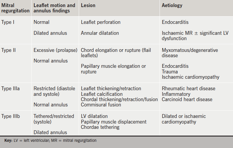
Screen shot 2016-02-12 at 17.09.31MR is broadly classified into two main categories: primary valvular disease or organic MR (intrinsic valve lesions) and secondary or functional MR (a structurally normal mitral valve but ventricular remodelling causes downward papillary muscle displacement, leaflet tethering and annular dilatation). The Carpentier classification8 describes MR based on leaflet movement and provides a unifying language to describe mitral valve pathology based on echocardiographic findings (table 1). The posterior leaflet is divided into three segments or scallops: lateral P1, middle P2 and medial P3. The opposing A1, A2 and A3 segments of the larger anterior leaflet correspond to the posterior leaflet segments.

Table 1. Carpentier classification of mitral regurgitation. This functional classification originally described by Carpentier is based on the surgical aim to restore normal valve function rather than normal valve anatomy. It is categorised by the opening and closing motion of the mitral valve leaflets. The lesion abnormalities and aetiologies in relation to this classification are described. It should be noted that more than one Carpentier type may co-exist in any one individual

Indications for mitral valve surgery

Guidelines recommend surgical repair or replacement for symptomatic patients (class I) with severe MR due to a primary valvular abnormality or asymptomatic patients with severe MR and LV dysfunction or enlargement.9,10 Surgery may also be considered an option for symptomatic patients (class IIb) with secondary (functional) MR.9,10 Mitral valve repair, when feasible, is almost universally regarded as the preferred method of MR correction (in non-rheumatic valves) over mitral valve replacement due to the advantages of even partial preservation of subvalvular chordae.11-14 Surgical repair techniques are defined by the patho-anatomy and physiology of MR,15,16 and provide a tailored approach to restoring normal mitral valve function.

Surgical repair results for primary or degenerative MR are excellent in experienced centres with procedural volume.17 Secondary or functional MR (FMR) remains a surgical challenge. Data have failed to clearly demonstrate survival benefit in these patients,18 and reported recurrence rates range from 15% to 60%.19,20 The pathophysiology of FMR is complex and the more contentious role of surgery reflects the underlying ventricular nature of the problem in a high-risk population. Only a third of patients with FMR are referred for surgery, and FMR is present in 90% of patients who are denied surgery.21,22

Challenges of transcatheter mitral valve techniques

The first case of percutaneous mitral valve repair was performed in 2003,23 and, yet, the clinical application of this approach has been more challenging compared with aortic valve stenosis.24 The greater complexity of mitral regurgitation, and the patient heterogeneity,25 presents greater challenges for the transcatheter approach, device development and, indeed, trial design.

MitraClip®

The MitraClip® (Abbott Vascular, Santa Clara, California, US) percutaneous repair system is the only transcatheter mitral valve therapy to undergo a pivotal randomised-controlled trial. It is based on the surgical procedure pioneered by Ottavio Alfieri in the early 1990s.26 Over 30,000 patients have been treated worldwide. This technique creates a competent double orifice valve by suturing the free edges of the middle portion of the anterior leaflet (A2) to the middle portion of the posterior leaflet (P2). To improve the durability of results, the Alfieri repair is typically combined with implantation of a partial band or complete annuloplasty ring, except in cases with a severely calcified mitral annulus.27

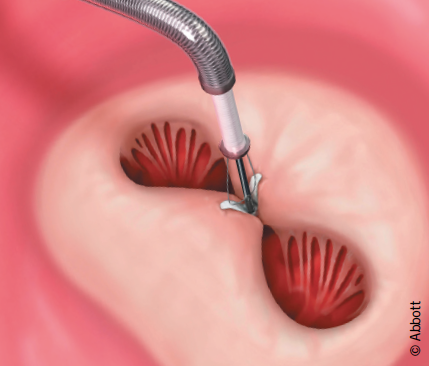
The MitraClip® is made of a cobalt–chromium alloy and covered with polypropylene fabric to promote tissue in-growth.28,29 It is a single-size clip device that has been used to treat patients with functional, mixed and degenerative MR. The Clip has a dual-arm structure, with grippers above the arms to assist with capture of the mitral valve leaflets and their approximation while the heart is beating.

The procedure

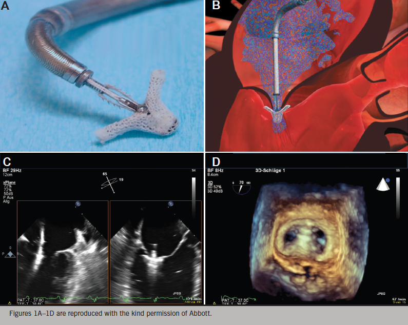
The procedure is approached via the femoral vein, and trans-septal access permits delivery of the catheter-based MitraClip® device into the left atrium (figure 1). All manoeuvres are performed under transoesophageal echocardiographic (TOE) visualisation. The device is positioned directly above the regurgitant jet and advanced across the mitral valve into the left ventricle. The Clip is retracted toward the mitral valve leaflets to engage the appropriate segments of the mitral valve. The grippers are dropped and arms of the Clip are closed. If the leaflet insertion visualised on TOE is acceptable, the degree of residual MR is assessed with the Clip fully closed. If reduction is inadequate, the Clip may be released and repositioned or a second clip may be implanted. The addition of Clips is guided by, among other factors, transmitral valve diastolic gradient, as a surrogate measure for potential development of mitral stenosis. After the Clip(s) are deployed, and the delivery catheter is removed from the patient, manual compression, use of a temporary subcutaneous suture, or placement of a percutaneous suture may be used to close the femoral vein access site.

Figure 1. MitraClip® percutaneous edge-to-edge repair for mitral regurgitation. A. Mitraclip® clip at the end of the steerable delivery system. B. Illustration of the clip just below leaflets prior to grasping. C. Echocardiographic view of the open clip positioned just below leaflets. D. Three-dimensional echocardiographic view of double orifice valve

The unique features of this procedure are:

  • the clip is repositionable (if a decision is made not to deploy the Clip, it may be removed)
  • real-time echocardiographic assessment of MR reduction is obtained
  • it produces vertical coaptation of leaflets and is not simply an edge-to-edge repair
  • surgical options are preserved.

Evidence base for MitraClip®

EVEREST II

MitraClip® is the first percutaneous device to be compared with surgery in a randomised-controlled trial (RCT). The pivotal EVEREST II (Endovascular Valve Edge-to-Edge REpair Study) phase II RCT comprising 279 patients was completed in 2008.30 It included patients with both degenerative and functional MR, although predominantly degenerative MR (73%). Surgery was superior for the primary outcome of freedom from death, surgery for mitral valve dysfunction, or ≥3+ MR. MitraClip® was safe with only 15% of patients experiencing a major adverse event compared with 48% of surgical patients (largely composed of transfusion ≥2 units). The degree of MR reduction was greater in the surgical patients, however, important clinical indicators, including heart failure functional status (New York Heart Association, NYHA), ejection fraction, LV dimensions and quality of life (QoL), improved in both groups. Nearly 80% of patients were free from 3+ or 4+ MR after Clip placement and did not require surgery in 12-month follow-up. The majority of MitraClip®-treated patients who required an additional procedure did so within the first six months after initial treatment.30 Clinically significant cases of mitral stenosis in the MitraClip® group was not observed.

Five-year follow-up of patients in the EVEREST II trial reported no increase in late MR recurrence compared with surgery.31 A good immediate result with MitraClip® showed excellent durability of the repair (at five years). The improved NYHA class was sustained and mortality rates were not different between the treatment arms at years one or five.

The rates of re-operation, or additional MitraClip® procedures, were no different between the two treatment groups after the first year. There did not appear to be a significant change in MR grade, ventricular function or dimension in follow-up, however, longer-term follow-up of the subset of patients with MR grade 2+ is ongoing.

Registry data

In Europe, CE mark was obtained in March 2008. ACCESS-EU (ACCESS-Europe A Two-Phase Observational Study of the MitraClip System in Europe) is a post-marketing registry of MitraClip® patients comprising 567 patients.32 These patients were more elderly and higher surgical risk candidates than those evaluated in the EVEREST II trial. More patients had functional MR, and the anatomical characteristics of the mitral valve in 70–80% of these patients were outside the inclusion criteria stated in the EVEREST II trial.33 Positive clinical outcomes with the Clip were still demonstrated at one year with improvements in degree of MR reduction and NYHA heart failure classification, QoL and six-minute-walk test results. Outside the USA, where Food and Drug Administration (FDA) approval has been limited to degenerative MR, and at least in Europe, this is currently the group of patients most commonly receiving MitraClip® repair.

Systematic reviews of MitraClip® versus surgery confirm that, while MitraClip® is not as effective as surgery in reducing MR, it can provide clinical benefits. Munkholm-Larsen et al.34 identified MitraClip® implantation as an option in managing select high surgical risk patients with severe MR, but noted a lack of mid- to long-term data in high-risk groups. In a meta-analysis by Wan et al.35 the clinical outcomes were similar despite a higher risk profile in the MitraClip® patients compared with surgical intervention, although surgery was more effective in reducing MR in the early post-procedure period. Buzzatti et al. reported over 5.5 years of a single centre’s experience comparing MitraClip® with surgery in octogenarians with degenerative MR.36 MitraClip® was safer, despite being applied in an older and more symptomatic population with higher burden of comorbidities. MR reduction was not as optimal as surgery, but provided reduction in symptoms. This improvement in QoL, with greater procedural safety and quicker recovery compared with surgery, as well as reduction in burden on health services, is of key relevance in managing the elderly population.

Guidelines

The European Society of Cardiology (ESC) guidelines for heart failure 201237 provide a class IIb (level of evidence C) indication for MitraClip® in patients with both degenerative and functional MR in order to improve symptoms. Patients must be judged inoperable or at unacceptably high surgical risk, and have a life-expectancy of greater than one year. Primary MR (degenerative MR) and secondary MR (FMR) reflect two very distinct clinical groups. Patients with FMR must be on optimal medical therapy including cardiac resynchronisation therapy where appropriate.

Functional MR – an emergent indication?

There remains some skepticism, particularly with respect to heart failure patients with FMR, due to the limited data from the EVEREST II RCT,30 and extrapolation from uncontrolled registry data.38 MR in heart failure confers a worse prognosis,39-41 but it has not been clear whether it is simply a marker of underlying LV muscle disease or a target for intervention.

Currently, four trials directly comparing MitraClip® to medical therapy in patients with heart failure and MR are underway to fill the current knowledge gap; one in the USA – COAPT (Clinical Outcomes Assessment of the MitraClip Percutaneous Therapy for High Surgical Risk Patients Trial),42 and three in Europe – RESHAPE-HF (A Randomised Study of the MitraClip Device in Heart Failure Patients with Clinically Significant Functional Mitral Regurgitation), MITRA-FR (Multi-centre Study of Percutaneous Mitral Valve Repair MitraClip Device in Patients With Severe Secondary Mitral Regurgitation) and MATTERHORN (A Multicenter, Randomised, Controlled Study to Assess Mitral vAlve reconsTrucTion for advancEd Insufficiency of Functional or iscHaemic ORigiN).43-45 These contemporary prospective, randomised trials will be instructive in guiding MitraClip® implantation in FMR, or indeed, mitral valve intervention of any kind.

Patient selection

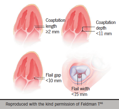
Figure 2. EVEREST II (Endovascular Valve Edge-to-Edge REpair Study) trial anatomic eligibility criteria. These criteria describe characteristics to ensure sufficient leaflet tissue for mechanical coaptation when the MitraClip® device is used. The coaptation length must be at least 2 mm. Coaptation depth must be <11 mm. If a flail leaflet exists, the flail gap must be ≤10 mm, and the flail width must be ≤15 mm

A heart team comprised of cardiac surgeon, heart failure specialist, echocardiologist and interventional cardiologist is critical to determining the most appropriate patients for intervention with MitraClip® therapy.

Patients are assessed for clinical indication, which broadly reflects the ESC guidelines.37 Evaluation may include stress echocardiography to assess for dynamic MR and cardiac magnetic resonance (CMR) imaging for viability and ischaemia testing. Technical suitability is determined by assessing the anatomical characteristics of the valve and pathology with TOE. Approximately 25% of patients referred for consideration of MitraClip® are considered suitable, which reflects the complex nature of MR. Moderate or severe tricuspid regurgitation and pulmonary hypertension are poor predictors of outcome with respect to NYHA functional class and re-hospitalisation with heart failure.

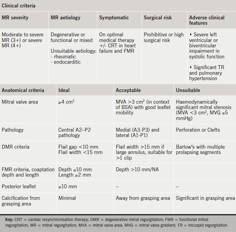
The EVEREST trial included stringent anatomical criteria that focused on the middle, A2–P2, segment of the valve.30 The anatomic eligibility criteria are shown in figure 2.46 The European experience suggests technical feasibility in a more complex group of patients can be achieved with an increase in the learning curve.32 Nevertheless, adverse anatomical features exist and recognition of these limitations is important to ensure optimal outcomes.

Table 2 provides a summary of the clinical and anatomical criteria.

Table 2. Clinical and anatomical selection criteria for MitraClip® therapy. The EVEREST (Endovascular Valve Edge-to-Edge REpair Study) data were based on ‘ideal’, rigorous anatomic criteria. Registry experience suggest more complex pathology may be treated, and are described here as ‘acceptable’ and ‘unsuitable’ criteria

MitraClip® in England

Although the technology has been commercially available in Europe since 2008, National Health Service (NHS) England only commissioned this technology for evaluation in 2014. The Commissioning through Evaluation (CtE) programme47 supports a small number of procedures to be funded within a limited number of selected centres and within a limited time frame. Three centres in the country were selected to deliver this therapy:

  • Royal Brompton and Harefield Hospitals (RBHT), London
  • University Hospital of Bristol (UHB)
  • University Hospital of South Manchester (UHSM).

Each was commissioned to treat 40 patients per year. Associated data collection will provide evidence on the relative clinical- and cost-effectiveness of the procedure, which will be used to inform a commissioning policy decision. The MitraClip® registry is managed by NICOR (National Institute for Cardiovascular Outcomes Research), with the National Institute for Health and Care Excellence (NICE) supporting NHS England in the evaluation of the scheme.

Links to the three centres can be found below. These will direct you to dedicated proformas (which function as stand-alone referral letters or may be used to guide referral details) with coordinator contact details for the three centres:

Summary

MitraClip® offers a safe and effective percutaneous strategy for high surgical risk or inoperable patients. Comorbidity, such as frailty, severe lung disease or multi-organ decline, should be recognised as key limitations in achieving positive outcomes. Primary MR and secondary MR should be recognised as two distinct patient populations with specific approaches to diagnosis and treatment. Secondary MR reflects a ventricular problem that should primarily be managed via optimisation of medical therapy and cardiac resynchronisation therapy (CRT), where indicated. Registry data, however, suggest FMR as a target for intervention. RCTs will be crucial to informing whether this expansion in indication is supported for mitral valve therapies of any kind. The importance of timely intervention, before an irreversible decline in LV remodelling and pulmonary hypertension ensues, may focus attention on earlier intervention, if safe and effective outcomes are shown to be durable beyond five years.

Percutaneous strategies presently limit repair options to a single point of intervention. The MR reduction and clinical improvements observed with MitraClip® without annuloplasty may reflect the advantages of the vertical coaptation produced with the MitraClip®. Nevertheless, longer follow-up of patients with residual grade 2 MR in particular, will be important. Development of percutaneous direct annuloplasty technologies might offer the potential to enhance long-term outcomes.

Conclusion

Current transcatheter mitral valve techniques are at the beginning of an era of innovation before their full potential is realised. The broadening of available options for MR reduction is welcome and transcatheter mitral valve interventions provide complementary strategies in the drive for more safe and effective therapies for patients. Surgical repair will be the standard for many years, certainly in low risk DMR. Randomised trials that challenge and enhance our understanding of new technologies are vital, and will guide the risk-benefit analysis of different approaches. This will permit more tailored care according to individual patient profile. Multi-disciplinary and collaborative practice is essential to achieving optimal standard of care.

Funding

This article was commissioned by the British Journal of Cardiology on behalf of Abbott Laboratories Ltd. who provided the funding. BJC engaged Dr Mamta Buch, who received an honorarium for its writing. Abbott Laboratories had no input into the content of the article written by Dr Buch, but has reviewed the article for regulatory compliance before publication.

MitraClip® is a trademark of the Abbott Group of Companies. The product is subject to prior training requirement as per the Instructions for Use. The product is intended for use by or under the direction of an appropriately qualified medical practitioner. Prior to use, it is important to read the package insert thoroughly for instructions for use, warnings and potential complications associated with use of this device.

Conflict of interest

MHB has received a speaker’s honorarium from Abbott.

Key messages

  • Mitral regurgitation (MR) is a complex and heterogeneous disease
  • MitraClip® is a safe, less invasive option for patients with degenerative MR who are high risk or inoperable
  • MitraClip® may provide benefits in patients with heart failure and severe functional MR who are symptomatic despite optimal medical therapy (including cardiac resynchronisation therapy)
  • The NHS England MitraClip® Commissioning through Evaluation programme will determine whether patients in England will have access to MitraClip® therapy.

If you would like to learn more about heart valve disease and earn CPD points at the same time, click here to visit our modular BJC Learning programme on this topic.

References

1. Nkomo VT, Gardin JM, Skelton TN, Gottdiener JS, Scott CG, Enriquez-Sarano M. Burden of valvular heart disease: a population-based study. Lancet 2006;368:1005–11. http://dx.doi.org/10.1016/S0140-6736(06)69208-8

2. Iung B, Baron G, Butchart EG et al. A prospective survey of patients with valvular heart disease in Europe: the Euro Heart Survey on Valvular Heart Diseasae. Eur Heart J 2003;24:1231–43. http://dx.doi.org/10.1016/S0195-668X(03)00201-X

3. Mirabel M, Iung B, Baron G et al. What are the characteristics of patients with severe, symptomatic, mitral regurgitation who are denied surgery? Eur Heart J 2007;28:1358–65. http://dx.doi.org/10.1093/eurheartj/ehm001

4. Iung B, Baron G, Butchart EG et al. A prospective survey of patients with valvular heart disease in Europe: The Euro Heart Survey on Valvular Heart Disease. Eur Heart J 2003;24:1231–43. http://dx.doi.org/10.1016/S0195-668X(03)00201-X

5. Lillehei CW. New ideas and their acceptance: as it has related to preservation of chordate tendinea and certain other discoveries. J Heart Valve Dis 1995;4(suppl2):S106–S114.

6. Perloff JK, Roberts WC. The mitral apparatus: functional anatomy of mitral regurgitation. Circulation 1972;46:227–39. http://dx.doi.org/10.1161/01.CIR.46.2.227

7. Otto CM. Clinical practice: evaluation and management of chronic mitral regurgitation. N Engl J Med 2001;345:740–6. http://dx.doi.org/10.1056/NEJMcp003331

8. Carpentier A. Cardiac valve surgery: the „French correction”. J Thorac Cardiovasc Surg 1983;86:323–37.

9. Nishimura RA, Otto CM, Bonow RO et al. 2014 AHA/ACC guideline for the management of patients with valvular heart disease: a report of the American College of Cardiology/American Heart Association Task Force on practice guidelines. Circulation 2014;129:e521–e643. http://dx.doi.org/10.1161/CIR.0000000000000031

10. Vahanian A, Alfieri O, Andreotti F et al. Guidelines on the management of valvular heart disease (version 2012): The Joint Task Force on the Management of Valvular Heart Disease of the European Society of Cardiology (ESC) and the European Association for Cardio-Thoracic Surgery (EACTS). Eur Heart J 2012;33:2451–96. http://dx.doi.org/10.1093/eurheartj/ehs109

11. Jokinen JJ, Hippelainen MJ, Pitkanen OA et al. Mitral valve replacement versus repair: propensity-adjusted survival and quality-of-life analysis. Ann Thorac Surg 2007;84:451–8. http://dx.doi.org/10.1016/j.athoracsur.2007.03.058

12. Enriquez-Sarano M, Schaff HV, Orszulak TA et al. Valve repair improves the outcome of surgery for mitral regurgitation: a multivariate analysis. Circulation 1995;91:1022–8. http://dx.doi.org/10.1161/01.CIR.91.4.1022

13. Horskotte D, Schulte HD, Bircks W et al. The effect of chordal preservation on late outcome after mitral valve replacement: a randomized study. J Heart Valve Dis 1993;2:150–8.

14. David TE, Uden DE, Strauss HD. The importance of the mitral apparatus in left ventricular function after correction of mitral regurgitation. Circulation 1983;68(suppl II):II-17–II-82.

15. De Bonis M, Lorusso R, Lapenna E et al. Similar long-term results of mitral valve repair for anterior compared with posterior leaflet prolapsed. J Thorac Cardiovasc Surg 2006;131:364–70. http://dx.doi.org/10.1016/j.jtcvs.2005.09.040

16. Gillinov AM, Cosgrove DM, Blackstone EH et al. Durability of mitral valve repair for degenerative disease. J Thorac Cardiovasc Surg 1998;116:734–43. http://dx.doi.org/10.1016/S0022-5223(98)00450-4

17. Gillinov AM, Cosgrove DM. Mitral valve repair for degenerative disease. J Heart Valve Dis 2002;11(suppl 1):S15–S20.

18. Wu AH, Aaronson KD, Bolling SF, Pagani FD, Welch K, Koelling TM. Impact of mitral valve annuloplasty on mortality risk in patients with mitral regurgitation and left ventricular systolic dysfunction. J Am Coll Cardiol 2005;45:381–7. http://dx.doi.org/10.1016/j.jacc.2004.09.073

19. Lee AP, Acker M, Kubo SH et al. Mechanisms of recurrent mitral regurgitation after mitral valve repair in nonischemic dilated cardiomyopathy. Circulation 2009;119;2606–14. http://dx.doi.org/10.1161/CIRCULATIONAHA.108.796151

20. McGee EC, Gillinov AM, Blackstone EH et al. Recurrent mitral regurgitation after annuloplasty for functional ischemic mitral regurgitation. J Thorac Cardiovasc Surg 2004;128:916–24. http://dx.doi.org/10.1016/j.jtcvs.2004.07.037

21. Mirabel M, Iung B, Baron G et al. What are the characteristics of patients with severe, symptomatic, mitral regurgitation who are denied surgery? Eur Heart J 2007;28:1358–65. http://dx.doi.org/10.1093/eurheartj/ehm001

22. O’Brien SM, Shahian DM, Filardo G et al. The Society of Thoracic Surgeons 2008 cardiac surgery risk models: part 2 – isolated valve surgery. Ann Thorac Surg 2009;88(suppl 1):S23–S42. http://dx.doi.org/10.1016/j.athoracsur.2009.05.056

23. Webb JG, Harnek J, Munt BI et al. Percutaneous transvenous mitral annuloplasty: initial human experience with device implantation in the coronary sinus. Circulation 2006;113:851–5. http://dx.doi.org/10.1161/CIRCULATIONAHA.105.591602

24. Leon MB, Smith CR, Mack M et al. PARTNER Trial Investigators. Transcatheter aortic-valve implantation for aortic stenosis in patients who cannot undergo surgery. N Engl J Med 2010;363:1597–1607. http://dx.doi.org/10.1056/NEJMoa1008232

25. Quill JL, Hill AJ, Laske TG, Alfieri O, Iaizzo PA. Mitral leaflet anatomy revisited. J Thorac Cardiovasc Surg 2009;137:1077–81. http://dx.doi.org/10.1016/j.jtcvs.2008.10.008

26. Alfieri O, Maisano F, De Bonis M et al. The double-orifice technique in mitral valve repair. A simple solution for complex problem. J Thorac Cardiovasc Surg 2001;122:674–81. http://dx.doi.org/10.1067/mtc.2001.117277

27. De Bonis M, Lapenna E, La Canna G et al. Mitral valve repair for functional mitral regurgitation in end-stage dilated cardiomyopathy: role of the “edge-to-edge” technique. Circulation 2005;112(9 suppl):I402–I408.

28. St Goar FG, Fann JI, Komtebedde J et al. Endovascular edge-to-edge mitral valve repair: short-term results in a porcine model. Circulation 2003;108:1990–3. http://dx.doi.org/10.1161/01.CIR.0000096052.78331.CA

29. Herrmann HC, Feldman T. Percutaneous mitral valve edge-to-edge repair with the Evalve Mitraclip system: rationale and phase I results. EuroIntervention 2006;1(suppl A):A36–A39.

30. Feldman T, Foster E, Glower DD et al. Percutaneous repair or surgery for mitral regurgitation. N Engl J Med 2011;364:1395–1406. http://dx.doi.org/10.1056/NEJMoa1009355

31. Feldman T, Kar S, Elmirah S, et al. Randomised comparison of percutaneous repair and surgery for mitral regurgitation. J Am Coll Cardiol 2015;66:2844–54. http://dx.doi.org/10.1016/j.jacc.2015.10.018

32. Maisano F, Franzen O, Baldus S et al. Percutaneous mitral valve interventions in the real world. Early and 1-year results from the ACCESS-EU, a prospective, multicenter, nonrandomized post-approval study of the MitraClip® therapy in Europe. J Am Coll Cardiol 2013;62:1052–61. http://dx.doi.org/10.1016/j.jacc.2013.02.094

33. Schillinger W, Athanasiou T, Weicken N et al. Impact of the learning curve on outcomes after percutaneous mitral valve repair with MitraClip® and lessons learned after the first 75 consecutive patients. Eur J Heart Fail 2011;13:1331–9. http://dx.doi.org/10.1093/eurjhf/hfr141

34. Munkholm-Larsen S, Wan B, Tian DH et al. A systematic review on the safety and efficacy of percutaneous edge-to-edge mitral valve repair with the MitraClip system for high surgical risk candidates. Heart 2014;100:473–8. http://dx.doi.org/10.1136/heartjnl-2013-304049

35. Wan B, Rahnavardi M, Tian DH et al. A meta-analysis of MitraClip system versus surgery for treatment of severe mitral regurgitation. Ann Cardiothorac Surg 2013;2:683–92. http://dx.doi.org/10.3978/j.issn.2225-319X.2013.11.02

36. Buzzatti N, Maisano F, Latib A et al. Comparison of outcomes of percutaneous MitraClip versus surgical repair or replacement for degenerative mitral regurgitation in octogenarians. Am J Cardiol 2015;115:487–92. http://dx.doi.org/10.1016/j.amjcard.2014.11.031

37. McMurray JJ, Adamopoulos S, Anker SD et al. ESC Guidelines for the diagnosis and treatment of acute and chronic heart failure 2012: The Task Force for the Diagnosis and Treatment of Acute and Chronic Heart Failure 2012 of the European Society of Cardiology. Developed in collaboration with the Heart Failure Association (HFA) of the ESC. Eur Heart J 2012;33:1787–847. http://dx.doi.org/10.1093/eurheartj/ehs104

38. Coats AJ, Shewan LG. Inconsistencies in the development of the ESC Clinical Practice Guidelines for Heart Failure. Int J Cardiol 2013;168:1724–7. http://dx.doi.org/10.1016/j.ijcard.2013.05.045

39. Junker A, Thayssen P, Nielsen B et al. The hemodynamic and prognostic significance of echo-Doppler-proven mitral regurgitation in patients with dilated cardiomyopathy. Cardiology 1993;83:14–20. http://dx.doi.org/10.1159/000175942

40. Conti JB, Mills RM Jr. Mitral regurgitation and death while awaiting cardiac transplantation. Am J Cardiol 1993;71:617–18. http://dx.doi.org/10.1016/0002-9149(93)90526-I

41. Blondheim DS, Jacobs LE, Kotler MN et al. Dilated cardiomyopathy with mitral regurgitation: decreased survival despite a low frequency of left ventricular thrombus. Am Heart J 1991;122:763–71. http://dx.doi.org/10.1016/0002-8703(91)90523-K

42. Clinicaltrials.gov. Cardiovascular Outcomes Assessment of the MitraClip Percutaneous Therapy For High Surgical Risk Patients (COAPT). Available at: http://www.clinicaltrials.gov/ct2/show/NCT01626079

43. Clinicaltrials.gov. A Randomised Study of the MitraClip Device in Heart Failure Patients with Clinically Significant Mitral Regurgitation (RESHAPE-HF). Available at: http://www.clinicaltrials.gov/ct2/show/NCT01772108

44. Clinicaltrials.gov. Multicentre Study of Percutaneous Mitral Valve Repair MitraClip Device in Patients With Severe Secondary Mitral Regurgitation (MITRA-FR). Available at: http://www.clinicaltrials.gov/ct2/show/NCT01920698

45. Clinicaltrials.gov. A Multicenter, Randomized, Controlled Study to Assess Mitral vAlve reconsTrucTion for advancEd Insufficiency of Functional or iscHemic ORigiN (MATTERHORN). Available at: http://www.clinicaltrials.gov/ct2/show/NCT02371512

46.  Feldman T, Kar S, Rinaldi M, et al. Percutaneous mitral repair with the MitraClip system: safety and midterm durability in the initial EVEREST (Endovascular Valve Edge-to-Edge REpair Study) cohort. J Am Coll Cardiol 2009;54:686–94. http://dx.doi.org.10.1016/j.jacc.2009.03.077

47. NHS England. Commissioning through Evaluation. Available at: https://www.england.nhs.uk/commissioning/spec-services/npc-crg/comm-eval/

A profile of patients with postural tachycardia syndrome and their experience of healthcare in the UK

Br J Cardiol 2016;23:33doi:10.5837/bjc.2016.010 Leave a comment
Click any image to enlarge
Authors:

Postural tachycardia syndrome (PoTS) is a recently recognised condition that usually affects younger women, who develop symptoms of orthostatic intolerance and a persistent tachycardia on standing upright. Healthcare professionals, patients and the national patient support group (PoTS UK) together created a survey, and the responses of 779 UK PoTS patients were analysed. The most common symptoms of PoTS at presentation were the triad of fatigue, lightheadedness and palpitations. Mobility, ability to work or attend education, and quality of life were significantly restricted. Cardiologists, followed by patients, were most likely to be the first to suggest the diagnosis of PoTS. Patients waited a mean of almost four years from presentation to obtain their diagnosis and, meantime, psychiatric mislabeling was common. Advice given to patients regarding lifestyle changes was variable, and those referred to specialist practitioners for help, found practitioners had limited knowledge about management of PoTS. Increased education of healthcare professionals and improved services for patients are recommended. 

Continue reading A profile of patients with postural tachycardia syndrome and their experience of healthcare in the UK

Heart valve disease module 4: diagnosis

Released 1 March 2016     Expires: 01 March 2018      Programme: Heart valve disease 1 CPD/CME credit

Sponsorship Statement: This module has been sponsored by Edwards Lifesciences with an unrestricted educational grant

Designed to give healthcare professionals an understanding of diagnosis in heart valve disease, with an introduction to various imaging strategies. It is one of several modules in our heart valve disease programme.

1 CPD/CME credit

Module originally published May 2013. Revised module released March 2016

Learning objectives

Upon completing this module, participants should be better able to:

  • Understand the routine echocardiographic methods of quantifying valve disease
  • Appreciate the additional information obtained from advanced echocardiographic techniques including stress and 3D
  • Understand the complementary roles of computed tomography and cardiac magnetic imaging
  • Recognise the cut-off values for grading valve disease
  • Understand how imaging relates to criteria for surgical intervention.

Faculty

Dr Ronak Rajani, St Thomas’ Hospital, London
Dr Rajdeep Khattar, Royal Brompton Hospital, London
Professor John Chambers, St Thomas’ Hospital, London

Accreditation

1 CPD/CME credit, 1 hour
BJC Learning has assigned one hour of CPD/CME credit to this module
The European Board for Accreditation in Cardiology (EBAC) has assigned one CME credit to this module. German participants should contact EBAC to receive a German VNR code for this course.
email: [email protected]
website: http://www.ebac-cme.org/

Produced in collaboration with:

You need to login to take this module

You need to be a registered member to view this page. It's quick, free and offers you a host of other benefits, including the facility to print and download articles and supplements, access our archived issues and receive email updates when new issues and other content are online.

Heart valve disease module 3: disease presentation

Released 1 March 2016     Expires: 01 March 2018      Programme: Heart valve disease 1 CPD/CME credit

Sponsorship Statement: This module has been sponsored by Edwards Lifesciences with an unrestricted educational grant

Designed to give healthcare professionals an understanding of the presentation of heart valve disease. It is one of several modules in our heart valve disease programme.

1 CPD/CME credit

Module originally published May 2013. Revised module released March 2016

Learning objectives

Upon completing this module, participants should be better able to:

  • Appreciate the variable and unpredictable natural history of valve lesions
  • Recognise common symptoms of the individual valve lesions
  • Be aware of physical signs commonly associated with valve lesions .

Faculty

Dr Joanna d’Arcy, John Radcliffe Hospital, Oxford
Dr Marzia Rigolli, John Radcliffe Hospital, Oxford

Accreditation

1 CPD/CME credit, 1 hour
BJC Learning has assigned one hour of CPD/CME credit to this module
The European Board for Accreditation in Cardiology (EBAC) has assigned one CME credit to this module. German participants should contact EBAC to receive a German VNR code for this course.
email: [email protected]
website: http://www.ebac-cme.org/

Produced in collaboration with:

You need to login to take this module

You need to be a registered member to view this page. It's quick, free and offers you a host of other benefits, including the facility to print and download articles and supplements, access our archived issues and receive email updates when new issues and other content are online.

The clinical and cost impact of implementing NICE guidance on chest pain of recent onset in a DGH

Br J Cardiol 2016;23:37doi:10.5837/bjc.2016.011 Leave a comment
Click any image to enlarge
Authors:

In 2010, the National Institute for Health and Care Excellence (NICE) introduced new guidelines for the assessment of people with recent-onset chest pain, recommending investigations based upon one’s pre-test likelihood of having coronary artery disease. We aim to determine the impact these guidelines have made on the numbers of patients being discharged and referred for further investigations. We retrospectively analysed a database of 337 consecutive patients seen in the rapid access chest pain clinic: 162 patients were seen in the three months preceding, and 175 were seen in the three months following implementation of the new guidelines. We found that after implementation of the new guidelines, fewer patients (25% vs. 37%, p=0.018) were discharged at the first visit, and a greater number of patients were referred for an angiogram (20% vs. 6%, p=0.0001). The number of referrals for stress imaging significantly reduced from 57% to 37%. According to the new guidelines, 18% of patients were referred for coronary calcium scoring. This reflects a definite change in clinical practice with reduced direct discharges from the chest pain clinic, reduced reliance on functional imaging and increased direct referrals for invasive coronary angiography, resulting in higher investigational costs of the chest pain service.

Introduction

There are 2.3 million people living with coronary heart disease in the UK, which results in a healthcare burden of 1% of all GP and 40% of all accident and emergency (A&E) visits.1

It is estimated that 20–40% of the general population will experience chest pain during their life. Chest pain caused by coronary artery disease has a potentially poor prognosis, emphasising the importance of prompt and accurate diagnosis. Treatments are available to improve symptoms and prolong life, hence, the need for the development of the National Institute for Health and Care Excellence (NICE) guidelines for the diagnosis of chest pain.1

NICE updated guidelines in March 2010 for the assessment and diagnosis of people with recent-onset chest pain. Previously, in the rapid access chest pain clinic (RACPC), patients referred by the GP with recent-onset chest pain underwent a clinical assessment, after which, an exercise tolerance test (ETT) was the first diagnostic test performed to diagnose myocardial ischaemia, generally during the same visit. Further functional imaging, in the form of a myocardial perfusion scan or a dobutamine stress echocardiogram were recommended if the test was inconclusive.2 A patient was referred directly for a diagnostic coronary angiogram if the test was positive, which may have progressed to angioplasty, if necessary, or, if the patient was unable to undergo exercise electrocardiogram (ECG). A negative ETT would usually result in a patient being discharged from the RACPC.

In the 2010 NICE guidelines, the Diamond–Forrester algorithm is employed to measure the ‘pre-test probability’ (PTP) of coronary artery disease based on age, gender and typicality of symptoms. The presence of all three features of: constricting chest pain, which is exacerbated by exertion, and relieved with rest or glyceryl trinitrate (GTN), is defined as typical angina, while the presence of only two or one feature is defined as atypical angina and non-cardiac chest pain, respectively. This probability estimate of coronary artery disease has been modified by taking into account additional risk factors, such as smoking, diabetes and hypercholesterolaemia, giving rise to a range of probabilities. NICE recommends further investigations depending on the PTP of coronary artery disease for each individual case. If the probability is <10%, the patient is discharged without any further investigation. For a probability of 10–29%, computerised tomography (CT) calcium scoring is recommended for an anatomical assessment of the coronary arteries, which might include a 64-slice CT angiogram, if the CT calcium score is between 1 to 400. For a PTP of 30–60%, functional testing is recommended, such as a stress echocardiogram, stress cardiac magnetic resonance imaging (CMR) or myocardial perfusion scan (MPS). A probability of >60% would warrant a coronary angiogram.3

While there have been data published addressing some of the specific issues in implementing NICE guidance 95,4 we sought to investigate the overall anticipated impact on clinical practice, service provision and cost-effectiveness in a district general hospital (DGH), where most patients with new-onset chest pain are seen. We have carried out this study to assess the actual impact of these updated guidelines on the rapid access chest pain service by comparing a similar number of patients being seen before and after the current guidelines were introduced.

Methods

We retrospectively analysed a database of 337 consecutive patients seen in the RACPC between June and November 2010: 162 of these were seen before implementation of the current guidelines, between June and August 2010; 175 patients were seen after implementation of current guidelines, between September and November 2010.

Risk factors for coronary artery disease, including diabetes and hypertension, were documented on the referral from the GP or information provided by the patients themselves. Hypertension was not included in the Diamond–Forrester algorithm of the new guidance. A fasting blood cholesterol level of 6.4 mmol/L was applied to define hypercholesterolaemia in both populations. Any patient already on statins, prior to attending the RACPC, was also considered to have hypercholesterolaemia, regardless of their current fasting cholesterol level. Patients were considered to be smokers if they were still smoking at the time of consultation or had stopped within the previous year.

Patients who were seen between June to August 2010, underwent an ETT according to Bruce Protocol5 after a clinical assessment. Patients whose ETTs were negative were discharged at the first visit. Patients who were unable to use the treadmill, or had an inconclusive ETT, were referred for a dobutamine stress echocardiogram or MPS. Patients who had a positive ETT were referred for a coronary angiogram.

The new NICE guidelines for chest pain were implemented in September 2010 in our trust. Clinical practices strictly followed the new NICE guidance based upon the PTP of each patient, with the exception of a few high-risk patients who were apprehensive of the invasive procedure that is coronary angiography, opting for functional imaging instead.

We made comparison between patients who were seen before and those who were seen after the implementation of new NICE guidelines. Chi-square test was used for non-continuous parameters. A p value of <0.05 was considered as statistically significant.

Results

The two patient groups who were managed according to the 2003 and 2010 NICE guidelines were similar in age, gender and numbers of cardiovascular risk factors.

Following the implementation of the 2010 guidelines, fewer patients were discharged at first visit (25% vs. 37%, p=0.018), and more patients were referred for coronary angiogram (20% vs. 6%, p=0.0001). The number of referrals for stress imaging reduced from 57% to 37% after implementation of the 2010 guidelines (p=0.008). Since the new guidelines, 18% of patients were referred for CT calcium scoring (table 1).

Table 1. Different investigations, before and after implementation of the 2010 National Institute for Health and Care Excellence (NICE) guidelines

The estimated cost according to NHS tariff was increased by £50 per patient after implementation of the new guidance (table 2).

Table 2. Estimated cost of cardiac tests in the rapid access chest pain service

Discussion

Following implementation of the 2010 NICE guidelines, we report a statistically significant reduction in the number of patient discharges and functional imaging studies performed, but an increase in the number of coronary angiograms. In addition, 18% of all patients were referred for CT calcium scoring, but none had ETT. This reflects a definite trend towards more specialised imaging techniques and has implications for the RACPC, particularly those held in DGHs.

Impact on service provision

Specialised cardiac investigations are still unavailable in many DGHs, where patients with chest pain are first seen, causing a delay in establishing a diagnosis as patients await referral to a tertiary centre.

Although fewer patients were discharged at first visit, they were discharged on clinical grounds alone, without any investigations. While PTP scoring is considered to be credible in identifying patients who would not benefit from further testing for the diagnosis of coronary artery disease,3 many physicians would hesitate to discharge a patient without investigations. Likewise, many patients will not feel reassured after being informed by the referring physician that they are at risk of heart disease.

Impact on cost-effectiveness

Table 3. Comparison between predicted and observed incidences
Table 3. Comparison between predicted
and observed incidences

The cost-effectiveness model of NICE is based upon their predictions of the proportion of patients within each category of likelihood of coronary artery disease.6 Table 3 demonstrates that NICE’s predictions appear to underestimate the incidence of the 30–60% PTP group, while over-estimating the >60% PTP group. Using the recommended NHS tariffs to compare the investigational cost of running the RACPC service, we find the average costs per patient were significantly higher following implementation of the new guidelines (table 2). Similar findings have been reported by other centres.7,8 Our costings do not take into consideration subsequent patient visits, which are expected to increase with the lower patient discharge rate.

Our calculations will have their limitations, and may not be representative for the whole UK, given the generally higher risk of coronary artery disease in the London inner city area. Second, the individual doctors who deal with patients in the chest pain clinic may have personal preferences to cardiac tests, despite the recommendation of the guidelines. Finally, one could argue that the cost is only a snap shot of the lifelong healthcare of the patients, and is too simple and short-term a measure.

Of interest, the European Society of Cardiology (ESC) has more recently published guidelines, which differ from NICE by advising that patients with <15% or >85% PTP should generally not receive stress testing, as it is more likely to produce a falsely positive or negative result in patients at extremes of probability when the specificity of most functional testing techniques are estimated to be 85%.9 Implementation of the ESC guidelines will probably reduce the number of invasive coronary angiography referrals by our local centre.10

Conclusion

Implementation of NICE chest pain guidelines at our DGH has significantly reduced direct discharges from the chest pain clinic, reduced our reliance on functional imaging and increased direct referrals for invasive coronary angiography. This has resulted in higher investigational costs of the chest pain service, and further investigations may well be fruitful in addressing how to reduce cost, while fulfilling the merits of the new guidelines.

Conflict of interest

None declared.

Key messages

  • Strict application of National Institute for Health and Care Excellence (NICE) chest pain guidelines has reduced direct discharges, reduced reliance on functional imaging, and increased coronary angiography referrals
  • Reliance on specialised cardiac investigations may delay establishing a diagnosis, as patients await referral to a tertiary centre
  • The short-term investigational cost of the rapid access chest pain service has increased

If you would like to learn more about chest pain and earn CPD points at the same time, click here to visit our modular BJC Learning programme on angina.

References

1. National Institute for Health and Care Excellence. CG95. Chest pain of recent onset: assessment and diagnosis of recent onset chest pain or discomfort of suspected cardiac origin. London: NICE, 2010. Available from: https://www.nice.org.uk/guidance/cg95/chapter/guidance

2. National Institute for Health and Care Excellence. TA73. Myocardial perfusion scintigraphy for the diagnosis and management of angina and myocardial infarction. London: NICE, 2003. Available from: https://www.nice.org.uk/guidance/ta73/chapter/1-guidance

3. Pryor B, Shaw L, McCants C et al. Value of the history and physical in identifying patients at increased risk for coronary artery disease. Ann Intern Med 1993;118:81-90. http://dx.doi.org/10.7326/0003-4819-118-2-199301150-00001

4. Ormerod JO, Wretham C, Beale A et al. Implementation of NICE clinical guideline 95 on chest pain of recent onset: experience in a district general hospital. Clin Med 2015;15:225–8. http://dx.doi.org/10.7861/clinmedicine.15-3-225

5. Bruce R. Exercise testing of patients with coronary heart disease. Principles and normal standards for evaluation. Ann Clin Res 1971;3:323–32.

6. National Institute for Health and Care Excellence. Chest pain of recent onset. Costing report. London: NICE, 2011. Available from: https://www.nice.org.uk/guidance/cg95/resources

7. Ghosh A, Qasim A, Woollcombe K et al. Cost implications of implementing NICE guideline on chest pain in rapid access chest pain clinics: an audit and cost analysis. J Public Health 2012;34:397–402. http://dx.doi.org/10.1093/pubmed/fdr118

8. Rogers T, Dowd R, Yap H et al. Strict application of NICE Clinical Guideline 95 ‘chest pain of recent onset’ leads to over 90% increase in cost of investigation. Int J Cardiol 2013;166:740–2. http://dx.doi.org/10.1016/j.ijcard.2012.09.180

9. Monalescot G, Sechtem U, Achenbach S et al. 2013 ESC guidelines on the management of stable coronary artery disease: the Task Force on the management of stable coronary artery disease of the European Society of Cardiology. Eur Heart J 2013;34:2949–3003. http://dx.doi.org/10.1093/eurheartj/eht296.

10. Demir OM, Dobson P, Papamichael ND et al. Comparison of ESC and NICE guidelines for patients with suspected coronary artery disease: evaluation of the pre-test probability risk scores in clinical practice. Clin Med 2015;15:234–8. http://dx.doi.org/10.7861/clinmedicine.15-3-234

Heart valve disease module 2: pathophysiology

Released 1 March 2016     Expires: 01 March 2018      Programme: Heart valve disease 1 CPD/CME credit

Sponsorship Statement: This module has been sponsored by Edwards Lifesciences with an unrestricted educational grant

Designed to give healthcare professionals an understanding of the structure and pathophysiology of heart valve disease, with an introduction to novel therapeutic strategies. It is one of several modules in our heart valve disease programme.

1 CPD/CME credit

Module originally published May 2013. Revised module released March 2016

Learning objectives

Upon completing this module, participants should be better able to:

  • Understand the basic structure of cardiac valves
  • Understand the pathophysiology underlying the most common heart valve disorders including:
    – degenerative heart valve disease
    – myxomatous heart valve degeneration
    – rheumatic heart disease
    – functional heart valve disease
    – endocarditis
    – systemic inflammatory diseases
  • Discuss novel therapeutic strategies and how these might relate to the underlying pathophysiology.

Faculty

Dr Marc R Dweck, University of Edinburgh and Edinburgh Heart Centre
Dr Russell J Everett, University of Edinburgh and Edinburgh Heart Centre
Professor David E Newby, University of Edinburgh and Edinburgh Heart Centre

Accreditation

1 CPD/CME credit, 1 hour
BJC Learning has assigned one hour of CPD/CME credit to this module
The European Board for Accreditation in Cardiology (EBAC) has assigned one CME credit to this module. German participants should contact EBAC to receive a German VNR code for this course.
email: [email protected]
website: http://www.ebac-cme.org/

Produced in collaboration with:

You need to login to take this module

You need to be a registered member to view this page. It's quick, free and offers you a host of other benefits, including the facility to print and download articles and supplements, access our archived issues and receive email updates when new issues and other content are online.

A challenging case of collapse with tri-fascicular block: from permanent pacemaker to thrombolysis

Br J Cardiol 2016;23:39doi:10.5837/bjc.2016.012 Leave a comment
Click any image to enlarge
Authors:

A 90-year-old man with a history of prostate cancer was admitted with haematuria and mild normocytic anaemia on routine blood tests. Baseline observations were normal and chest X-ray was unremarkable. Electrocardiogram (ECG) showed tri-fascicular block. He underwent successful bladder irrigation. Prior to discharge, he suffered a syncopal episode: ECG confirmed tri-fascicular block, for which he was discussed with the cardiology team for consideration of permanent pacemaker implantation. Pre-procedural transthoracic echocardiogram (TTE) revealed a large mobile thrombus attached to the tricuspid valve (TV) and extending to the right ventricle (RV), significant RV impairment and severe TV regurgitation (figure 1A–B). Following discussion between urology and cardiology teams and, in view of the significant risk of massive pulmonary embolism (PE), the patient underwent urgent thrombolysis. This was not complicated by significant haematuria. Post-intervention TTE demonstrated complete dissolution of the right-sided thrombus and mild TV regurgitation only (figure 1C–D). Warfarin was started and no further haematuria or syncope was reported on subsequent follow-up.

Figure 1. A. Pre-thrombolysis transthoracic echocardiogram. Technically difficult study: large mobile thrombus extending from the tricuspid valve into the right ventricle; dilated right heart chambers (with impaired right ventricular function). B. Pre-thrombolysis transthoracic echocardiogram: in addition to the findings described in panel A, colour flow shows a jet of severe tricuspid valve regurgitation. C. Post-thrombolysis transthoracic echocardiogram: complete dissolution of the right heart thrombus (with improved right ventricular function). D. Postthrombolysis transthoracic echocardiogram: colour flow shows mild regurgitation only
Figure 1. A. Pre-thrombolysis transthoracic echocardiogram. Technically difficult study: large mobile thrombus extending from the tricuspid valve into the right ventricle; dilated right heart chambers (with impaired right ventricular function). B. Pre-thrombolysis transthoracic echocardiogram: in addition to the findings described in panel A, colour flow shows a jet of severe tricuspid valve regurgitation. C. Post-thrombolysis transthoracic echocardiogram: complete dissolution of the right heart thrombus (with improved right ventricular function). D. Postthrombolysis
transthoracic echocardiogram: colour flow shows mild regurgitation only

A 90-year-old man with a history of prostate cancer was admitted with haematuria and mild normocytic anaemia on routine blood tests. Baseline observations were normal and chest X-ray was unremarkable. Electrocardiogram (ECG) showed tri-fascicular block. He underwent successful bladder irrigation. Prior to discharge, he suffered a syncopal episode: ECG confirmed tri-fascicular block, for which he was discussed with the cardiology team for consideration of permanent pacemaker implantation. Pre-procedural transthoracic echocardiogram (TTE) revealed a large mobile thrombus attached to the tricuspid valve (TV) and extending to the right ventricle (RV), significant RV impairment and severe TV regurgitation (figure 1A–B). Following discussion between urology and cardiology teams and, in view of the significant risk of massive pulmonary embolism (PE), the patient underwent urgent thrombolysis. This was not complicated by significant haematuria. Post-intervention TTE demonstrated complete dissolution of the right-sided thrombus and mild TV regurgitation only (figure 1C–D). Warfarin was started and no further haematuria or syncope was reported on subsequent follow-up.

Discussion

Right heart thrombus is a life-threatening condition commonly seen with structural heart disease, atrial fibrillation, devices in superior vena cava or right heart, thrombophilia and malignancy. It is associated with an incidence of PE of 97%,1 in-hospital mortality of 44.7%2 and mortality of 100%, if untreated.3 Treatment options include thrombolysis, anticoagulation, surgical or percutaneous thrombo-embolectomy, although evidence of optimal management is lacking.

This difficult case, complicated by haematuria, emphasises the successful outcome achieved with thrombolytic treatment. Thrombolysis presents the lowest mortality rate, and allows rapid improvement of pulmonary reperfusion, pulmonary hypertension and RV function and simultaneous dissolution of intra-cardiac thrombus, PE and venous thromboembolism.

Conflict of interest

None declared.

Key messages

  • Mortality associated with right heart thrombi is very high, regardless of the chosen treatment
  • Although there is no clear consensus for the preferred treatment, rapid diagnosis and treatment are essential
  • Thrombolysis is a fast and relatively simple treatment, particularly useful when surgical thrombo-embolectomy is contraindicated
  • Transthoracic echocardiography (TTE) is usually adequate for the diagnosis of right heart thrombus and has a sensitivity of 50–60%. However, it might underestimate the clot size. Trans-oesophageal echocardiography presents 80% sensitivity and 100% specificity for the detection of right heart thrombi
  • This case resulted in the successful treatment of a life-threatening condition, thanks to a multi-disciplinary approach with careful evaluation of treatment options, as well as of risks and benefits, which should always be encouraged in similar complex cases

References

1. Chartier L, Bera J, Delomez M et al. Free-floating thrombi in the right heart: diagnosis, management, and prognostic indexes in 38 consecutive patients. Circulation 1999;99:2779–83. http://dx.doi.org/10.1161/01.CIR.99.21.2779

2. Torbicki A, Galie N, Covezzoli A et al. Right heart thrombi in pulmonary embolism: results from the International Cooperative Pulmonary Embolism Registry. J Am Coll Cardiol 2003;41:2245–51. http://dx.doi.org/10.1016/S0735-1097(03)00479-0

3. Rose PS, Punjabi NM, Pearse DB. Treatment of right heart thromboemboli. Chest 2002;121:806–14. http://dx.doi.org/10.1378/chest.121.3.80

Congenital LAD stenosis associated with a bicuspid aortic valve

Br J Cardiol 2016;23:40doi:10.5837/bjc.2016.013 Leave a comment
Click any image to enlarge
Authors:

A 47-year-old woman had been referred to the cardiology department with a six-month history of intermittent chest discomfort not specifically related to exertion. Her risk factors: current smoker 10–15 per day and family history of ischaemic heart disease. She had no history of diabetes or hypertension. Lipid levels had not been tested. 

Figure 1. Right anterior oblique (RAO) cranial view of left anterior descending (LAD) artery

Her observations were recorded as heart rate 60 bpm regular, blood pressure 119/63 mmHg, jugular venous pressure was not elevated, no ankle oedema, S1 and S2 present with an apical grade 2/6 mid-systolic murmur and her chest auscultation was clear. Electrocardiogram (ECG) was recorded as normal.

The patient managed to complete 10:16 minutes of a Bruce Protocol of exercise stress test (EST) with 2.1 mm of ST depression in V5 and V6. Despite the non-specificity of her symptoms and a positive EST, she was referred for a cardiac echocardiogram and cardiac catheterisation. In the interim period, she was commenced on aspirin 75 mg, simvastatin 40 mg and bisoprolol 1.25 mg.

Cardiac echocardiography was performed. The aortic valve (AV) was shown to be thickened, calcified and bicuspid with mild stenosis (peak gradient 25 mmHg) with a preserved left ventricular (LV) systolic function. All other cardiac chambers and valves were within normal limits.

Figure 2. Left anterior oblique (LAO) cranial view of LAD

Cardiac catheterisation was performed via the right radial artery. It revealed that there was no left main stem. The left anterior descending (LAD) and circumflex (LCx) arteries were originated side by side. Intubating the LAD revealed what appeared to be an ostial narrowing in different views. The operator administered 200 µg of intra-coronary nitrate, to eliminate the possibility of catheter-induced coronary spasm. There was no drop in blood pressure when engaging the LAD artery pre- or post-nitrate. The second set of LAD images confirmed that the ostial narrowing was still severe, while the rest of the LAD was completely normal (figures 1 and 2). The LCx and the right coronary arteries (RCA) were both normal.

The patient was referred for a computerised tomography (CT) coronary angiogram (CTCA) for further assessment of the LAD ostium.

Figure 3. Computerised tomography coronary angiography (CTCA) showing the LAD (green arrow) with an ostial measurement of 1–1.3 mm, left circumflex (LCx) (orange arrow of 3.5 mm) and right coronary artery (RCA) (light blue arrow) of 2.4 mm

The CTCA corroborated the angiogram result of an absent left main stem (LMS), but side-by-side origins of the LAD and LCx arteries. The LAD was reported to have a severe ostial stenotic pinhole lesion with a diameter of 1–1.3 mm, and funnelled to 2.2 mm in the proximal vessel, with no evidence of any surrounding atherosclerotic plaque (intrinsic) (figure 3). A calcified bicuspid aortic valve was also confirmed (figure 4).

Percutaneous coronary intervention (PCI), using a 3.0 × 15 mm Combo (OrbusNeich) drug-eluting stent, was ultimately performed to alleviate the symptoms being experienced by the patient, with positive results at 12-month follow-up.

Discussion

Ostial stenoses are frequently related to atherosclerosis. Rarer causes can be attributed to inherited anomalies. This case study is reporting a congenital LAD stenosis associated with a bicuspid aortic valve. It is presumed that it is the first reported congenital LAD stenosis associated with a bicuspid aortic valve.

Figure 4. CTCA demonstrating the presence of the bicuspid aortic valve (red arrow) and proximal LAD (blue arrow) of 2.2 mm

We believe this coronary anomaly to be related to the congenital development of a bicuspid aortic valve. A bicuspid aortic valve is a heart condition that is usually due to a congenital deformity. About 1–2% of the population have bicuspid aortic valves, although the condition is nearly twice as common in males. Congenital Heart Defects UK (2015) report that a bicuspid aortic valve is usually associated with several other cardiac defects (table 1).

Coronary arteries may be abnormal in bicuspid patients. A left-dominant coronary system is more commonly observed with bicuspid aortic valve. Rarely, the left coronary artery may arise anomalously from the pulmonary artery. The left main coronary artery may be up to 50% shorter in patients with a bicuspid aortic valve. Occasionally, the coronary ostium may be congenitally stenotic in association with bicuspid aortic valve.

Table 1. The occurrence of bicuspid aortic valve in a small range of associated cardiac defects

In individuals suffering from atresia, it usually arises in the LMS, and not a specific vessel such as the LAD, the diagnosis is usually confirmed by CTCA. This patient had separate origins to the LAD and LCx. The pinhole stenosis, seen on coronary angiography and subsequently confirmed by CTCA, identified there was no atherosclerotic disease present.

Conclusion

CTCA proved an invaluable tool in endorsing the angiographic results in what is assumed to be the first reported incidence of a congenital LAD stenosis associated with a bicuspid aortic valve.

Conflict of interest

None declared.

Further reading

Angelini P. Congenital coronary artery ostial disease: a spectrum of anatomic variants with different patho-physiologies and prognoses. Tex Heart Inst J 2012;39:55–9. PMCID: PMC3298900. Available from: http://www.ncbi.nlm.nih.gov/pmc/articles/PMC3298900/

Congenital Heart Defects UK. Educating and raising awareness of congenital heart defects. Website: http://www.chd-uk.co.uk/