A 29-year-old man was admitted with new onset atrial fibrillation (figure 1).


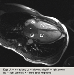
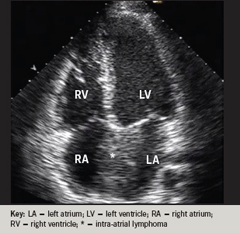
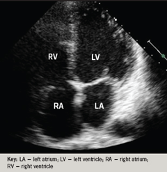
One year previously he had been treated with six cycles of chemotherapy for aplastic large cell lymphoma. He had a pyrexia (38.5oC), elevated C-reactive protein (14.6 mg/L) and low haemoglobin (10.8 g/dL). As part of a screen for infection, echocardiography was performed to exclude endocarditis (figure 2), but revealed a large intra-atrial mass. Cardiac magnetic resonance imaging (MRI) (figure 3) appearance was consistent with a lymphoma tumour. Fine-needle aspiration of an enlarged supra-clavicular node showed evidence of relapsed lymphoma. Further treatment with chemotherapy resulted in complete resolution (figure 4) of the intra-atrial mass, of his symptoms, and in restoration of sinus rhythm.

