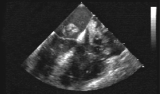
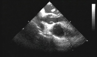
A 24-year-old male who had been bedridden for the last three weeks and was recovering from a traumatic injury to his leg presented to our echo lab for evaluation of abrupt onset dyspnoea preceded by presyncope. Clinically he was tachypnoiec, had a pulse rate of 142 beats per minute, blood pressure 100/70 mmHg, oxygen saturation at room air 97% and electrocardiogram (ECG) showing sinus tachycardia. Transthoracic echocardiography revealed dilated right heart chambers and a long multi-lobed vermicular mass in all four cardiac chambers. Trans-oesophageal echocardiography confirmed a large multi-lobed mass straddling the interatrial septum on both sides through a patent foramen ovale (figure 1). The highly mobile mass (consistent with thrombus) protruded like a worm into the aortic valve in systole through the left ventricular outflow tract (figure 2). Within three hours the patient had sudden bradycardia and cardiac arrest, probably from cerebral embolisation.
Discussion

The transit of a thrombus across a patent foramen ovale has been serendipitously documented on imaging studies in extremely rare instances.1-4 Nearly 40 cases of thrombus in transit diagnosed ante mortem using various imaging studies have been described in the literature and only one case documenting the serial passage of a thrombus across a patent foramen ovale in real time.1,3

The overall early mortality of these cases is as high as 30–40%. Management of these cases is challenging in view of concomitant pulmonary embolism. Therapeutic options in this case are few, thrombolytic therapy with anticoagulation or surgical removal of the thrombus, but mortality remains high.3-5
This case emphasises the role of echocardiography in such a clinically challenging setting and the need to use prophylactic heparin in every bedridden hospitalised patient so as to prevent such a catastrophic paradoxical embolism.
Conflict of interest
None declared.
References
- Thanigaraj S, Zajarias A, Valika A, Lasala J, Perez JE. Caught in the act: serial, real time images of a thrombus traversing from the right to left atrium across a patent foramen ovale. Eur J Echocardiogr 2006;7:179–81.
- Haddadin HA, Rahman H, Radparvar F. Massive pulmonary embolism with a mobile thrombus in the right atrium extending through the atrial septum to the left atrium into the left ventricle. Chest Meeting Abstracts2003;124(4):297S.
- Rousselle M, Ennezat P-V, Aubert J-M et al. Momentarily stuck in the foramen ovale. Eur J Echocardiogr 2007;8:223–6.
- Iwata A, Takazawa K, Tanaka N et al. Impending paradoxical embolism visualized by echocardiography combined with pulmonary embolism: a case report. J Cardiol 2000;36:123–7.
- The European Working Group on Echocardiography. The European Cooperative Study on the clinical significance of right heart thrombi. Eur Heart J 1989;10:1046–59.
