Left-sided dropped shoulder syndrome (DSS) can present with anterior chest pain that radiates to the left scapula and arm. Patients with atypical chest pain (ACP) of unknown cause (n=47) were investigated for left-sided DSS. Sixteen patients (34%) were diagnosed with left DSS. All the 47 patients were provided physiotherapy in two groups: the left DSS patients group (n=16) and a control group of 31 patients who did not show the criteria for the diagnosis of DSS.
Fourteen (87.5%) patients reported a satisfactory improvement of the ACP after physiotherapy. Satisfactory improvement has been judged by the reduction of the pain intensity, duration and frequency according to the patient’s report. Two (12.5%) patients showed no satisfactory improvement of the ACP. The control group showed no beneficial effect regarding their ACP after physiotherapy.
Physiotherapy aimed to strengthen the muscles that elevate the shoulder, could provide a treatment for atypical chest pain caused by left-sided DSS.
Introduction
Atypical or non-anginal chest pain (ACP) is any chest pain that has no cardiac cause. ACP is a common clinical condition encountered in everyday cardiology practice and constitutes about half of the cases of chest pain presented to emergency departments.1-5
The causes of this complaint are varied, and controversial aetiologies (musculoskeletal, neurological, psychological, mediastinal, pulmonary and gastrointestinal) have been suggested.3,4,6-9 ACP may result from cervical root compression, which can be confirmed by magnetic resonance imaging (MRI) findings.10
Scapular region pain is generally the initial symptom in cervical radiculopathy and can present alone before the arm or finger symptoms develop. The site of the pain is valuable for determining the localisation of the involved root.11
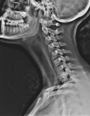
Cervical radiculopathy of C7, C8 and T1 roots could be caused by dropped shoulder syndrome (DSS) (figure 1).12 Left-sided DSS is presented with anterior chest pain that radiates to the left scapula, left upper arm and could be associated with numbness of the arm down to the hand.
Patients and methods
There were 112 patients with ACP who fulfilled the inclusion criteria included in this study between January 2013 to October 2014. The ethics committee of Welfare hospital has approved the research proposal and every patient has signed the consent form accepting the limited disclosure of his/her medical record according to the international standards of investigations on human subjects.
The inclusion criteria of the patients with ACP were:
- History of chest pain with at least one admission to an emergency department with normal cardiac enzymes and troponin T.
- Aged less than 40 years, of both sexes.
- Non-diabetic, normotensive, nonsmoker and not obese (central obesity: waist circumference ≥102 cm male, ≥88 cm female).
- Normal fasting blood sugar, complete blood count, renal function tests, lipid profile and thyroid function tests.
- Normal electrocardiogram (ECG) and stress ECG.
- Normal echocardiography.
The patients’ data were collected from their medical files. Diagnosis at discharge was: musculoskeletal (36 patients, 32%), gastrointestinal (13 patients, 12%), psychiatric (11 patients, 10%) and pulmonary (5 patients, 4%). The remaining 47 patients (42%) were labelled with ACP of unknown cause.
Those 47 patients were investigated for left DSS as a cause of their ACP. The ACP was central and/or left sided, of 1–6 years’ duration (average of 3.2 years). Pain lasted for more than 2–3 hours and came in varied frequencies. In 36 (76.6%) patients, the frequency was 1–3 times a week, while in the other 11 (23.4%) patients, pain occurred 1–2 times a month. In all the patients, pain was precipitated by heavy and/or prolonged manual work, and partially relieved by rest and over-the-counter (OTC) pain medications.
These 47 patients were sent for cervical plain X-ray, MRI, and electromyography/nerve conduction study (EMG/NC study). Single-blinded EMG/NC study of left (biceps, deltoid, triceps, flexor carpi ulnaris, abductor pollicis brevis, abductor digiti minimi, first dorsal interosseous, and the paraspinal) muscles and (musculocutaneous, axillary, median, ulnar, medial and lateral antebrachial) nerves were performed.

Sixteen patients (34%) were diagnosed with left DSS. The patients’ age was between 19 and 39 years. Three were males and 13 were females providing a male:female ratio of 1:4. Physiotherapy designed to elevate the shoulders was initiated for 12–16 weeks,12 and a fortnightly evaluation of the responses to the exercise were recorded.
All the 47 patients were included in the physiotherapy in two groups. The left DSS patients group and a control group of 31 patients who did not show the criteria for the diagnosis of DSS.
Results
Left DSS was found to be the cause of ACP in 16 patients (14.3%). Three major criteria were used for diagnosing left DSS: shoulder pain that radiates to the chest, neck, left scapular region, and the left upper limb down to the hand; lateral view of cervical X-ray showing eight or more vertebrae (the first, second, and, rarely, the third dorsal vertebrae) and a denervation EMG pattern of the C7, C8 and T1 innervated muscles.12
The other 31 patients were having ACP without left DSS. The MRI of one patient (0.9%) showed a prolapsed inter-vertebral disc at the level of C7–T1. The symptoms of the patients with left DSS are summarised in table 1. Clinical examination showed no evidence of vascular disorder. Motor power and tone examination were normal. Other clinical signs in left DSS patients are shown in table 1.
The cervical X-ray of DSS patients showed seven cervical vertebrae plus the first ± second thoracic vertebrae. No cervical rib could be detected on both sides. Neck muscle spasm was suggested by loss of normal lordosis in 12 patients (75.0%). The MRI and NC studies of all the patients with DSS were normal. Denervation EMG pattern was detected at left C7, C8 and T1 roots territories in 14 patients (87.5%), while two (12.5%) patients showed a denervation EMG pattern at left C8/T1 roots. All the patients showed a denervation EMG pattern of the lower segment of cervical paraspinal muscles (table 2).

Fourteen (87.5%) patients reported a satisfactory improvement of the ACP after physiotherapy. Satisfactory improvement has been judged by the reduction of the pain intensity, duration and frequency according to the patient’s report. Two (12.5%) patients showed no satisfactory improvement of the ACP. The control group showed no beneficial effect regarding their ACP after physiotherapy.
Discussion
Diagnosing the cause of a patient with ACP broadly as ‘neurogenic’ or ‘musculoskeletal’, without defining the real specific diagnosis, will lead, in most of the cases, to an empirical management approach. Most of these patients are treated by pain killers ± muscle relaxant, which is not always proved to be effective.
In this study, 14.3% of the ACP patients have been diagnosed with left DSS after fulfilling the criteria of the diagnosis.12 The neurogenic ACP caused by DSS has an explainable anatomical origin. In 2010, Mizutamari et al. demonstrated the anatomy behind the clinical presentations of pain in patients with C6, C7, C8/T1 radiculopathy.13
The vast majority of the patients (87.5%) who were diagnosed with left DSS reported a satisfactory improvement of their pain after physiotherapy. Physiotherapy aimed to strengthen the muscles that elevate the shoulder, which could also affect the natural history of the pain-producing mechanism/s in the cervical spine. This indicates that left DSS is a rather treatable cause and should be considered as one of the causes of ACP.
Conflict of interest
None declared.
Key messages
- Atypical chest pain in young adults could be caused by lower cervical radiculopathy due to left-sided dropped shoulder syndrome
- The diagnosis is based on history of chest pain with characteristic features of radiation, cervical plain X-ray and electromyography findings
- Physiotherapy aimed at strengthening the muscles that lift the shoulders showed a satisfactory improvement of atypical chest pain in most of the patients
References
1. Spalding L, Reay E, Kelly C. Cause and outcome of atypical chest pain in patients admitted to hospital. J R Soc Med 2003;96:122–5. http://dx.doi.org/10.1258/jrsm.96.3.122
2. Summers RL, Cooper GJ, Carlton FB, Andrews ME, Kolb JC. Prevalence of atypical chest pain descriptions in a population from the southern United States. Am J Med Sci 1999;318:142–5. http://dx.doi.org/10.1097/00000441-199909000-00008
3. Chambers J, Bass C. Atypical chest pain: looking beyond the heart. Q J Med 1998;91:239–44. http://dx.doi.org/10.1093/qjmed/91.3.239
4. Hung Cl, Hou CJY, Yeh HI, Chang WH. Atypical chest pain in the elderly: prevalence, possible mechanisms and prognosis. Int J Gerontology 2010;4:1–8. http://dx.doi.org/10.1016/s1873-9598(10)70015-6
5. Adamek RJ, Roth B, Zymanski CH, Hagemann B. Esophageal motility patterns in patients with and without coronary heart disease and healthy controls. Hepatogastroenterology 1999;46:1759–64.
6. Basha I, Mukerji V, Matt PL, Alpert M, Beitman B. Atypical angina in patients with coronary artery disease suggests panic disorder. Int J Psychiatry Med 1989;1914:341–6. http://dx.doi.org/10.2190/AK3T-V52N-7D5Y-GFF6
7. Arthur C. Treatment of morbidity with atypical chest pain. Can Fam Physcian 1987;33:1001–06. Available from: http://www.ncbi.nlm.nih.gov/pmc/articles/PMC2218465/
8. Beitman BD, Basha I, Flaker G, DeRosear L, Mukerji V, Trombka L, Katon W. Atypical or nonanginal chest pain, panic disorder or coronary artery disease? Arch Intern Med 1987;147:1548–52. http://dx.doi.org/10.1001/archinte.1987.00370090028005
9. Henderson RD, Douglas E. Atypical chest pain of cardiac and esophageal origin. Chest 1978;73:24–7. http://dx.doi.org/10.1378/chest.73.1.24
10. Stochkendahl MJ, Christensen HW. Chest pain in focal musculoskeletal disorders. Med Clin North Am 2010;94:259–73. http://dx.doi.org/10.1016/j.mcna.2010.01.007
11. Tanaka Y, Kokubun S, Sato T, Ozawa H. Cervical roots as origin of pain in the neck or scapular regions. Spine 2006;31:E568–E573. http://dx.doi.org/10.1097/01.brs.0000229261.02816.48
12. Abdul-Latif A. Dropped shoulder syndrome: a cause of lower cervical radiculopathy. J Clin Neurol 2011;7:85–89. http://dx.doi.org/10.3988/jcn.2011.7.2.85
13. Mizutamari M, Sei A, Tokiyoshi A, Fujimoto T, Taniwaki T, Togami W, Mizuta H. Corresponding scapular pain with the nerve root involved in cervical radiculopathy. J Orthop Surg (Hong Kong) 2010;18:356–60. Available from: http://www.josonline.org/pdf/v18i3p356.pdf
