Coronary angiography is commonly performed via the right femoral artery. Under local anaesthetic, the arterial lumen is initially cannulated with a wide-bore needle, then a long and soft J wire is inserted through the needle. The needle is then removed, and an arterial sheath is passed over the wire using a Seldinger technique.

Very rarely, the needle can displace following successful intraluminal cannulation. If the wire does not easily pass, it is important never to force it. Such an event occurred on this occasion.
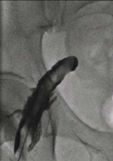
Following an unsuccessful attempt to pass the wire, contrast was injected through the needle. This demonstrated dissection of the common femoral artery, characterised by contrast holdup post-injection, due to contrast staying in one of the layers between arterial tunica intima and tunica adventitia (figure 1).
This was managed by removing the needle and externally compressing the artery with digital pressure. The patient made a full recovery, and had coronary angiography successfully performed via his left femoral artery.
Localised femoral artery dissection is an uncommon but well-recognised complication of coronary angiography, and usually resolves with external manual compression.
Conflict of interest
None declared.
