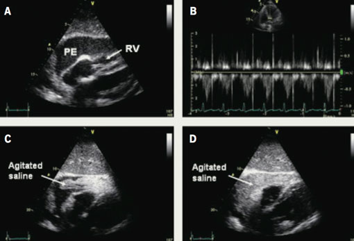
A 66-year-old male presented with increasing dyspnoea. He was an ex-smoker and had been diagnosed with stage IV undifferentiated large cell carcinoma of the left lung two months previously. Clinical examination revealed signs consistent with cardiac tamponade. Cardiac tamponade, a life-threatening condition, is a continuum of haemodynamic compromise, initiated by a collection of fluid in the pericardial space causing an increase in intra-pericardial pressure and cardiac compression. Transthoracic echocardiography confirmed the presence of a large global pericardial effusion with echocardiographic signs of cardiac tamponade (figure 1A and 1B).

The patient underwent therapeutic subxiphoid pericardiocentesis guided by contrast echocardiography. Pericardiocentesis is not without risk, and complications include laceration of cardiac chamber or coronary artery, aspiration of ventricular blood, arrhythmias, pneumothorax and puncture of peritoneum, oesophagus or aorta. Traditionally, many units still advocate pericardiocentesis drainage under fluoroscopy guidance given the dangers with blind drainage. However, transferring haemodynamically unstable patients to a cardiac catheter laboratory is not always safe and echocardiographic guidance allows pericardiocentesis to be performed safely at the bedside.
The echocardiogram was performed from the apical four-chamber view by an experienced operator. Pericardial needle aspiration revealed heavily blood-stained fluid. Agitated saline (9.5 ml normal saline, 0.5 ml air) was injected to exclude accidental puncture of a cardiac chamber with continuous echocardiographic monitoring (figure 1C and 1D).1 There was notable and immediate passage of contrast into the pericardial space. A pigtail catheter was then safely inserted and sutured in place.2 Over 1,000 ml of fluid was drained with immediate symptomatic improvement.
Subxiphoid pericardiocentesis guided by contrast echocardiography is a safe and effective technique to help differentiate accidental cardiac chamber puncture from haemopericardium in the management of cardiac tamponade.
Conflict of interest
None declared.
References
- Vayre F, Lardoux H, Pezzano M, Bourdarias JP, Dubourg O. Subxiphoid pericardiocentesis guided by contrast two-dimensional echocardiography in cardiac tamponade: experience of 110 consecutive patients. Eur J Echocardiogr 2000;1:66–71.
- Spodick DH. Acute cardiac tamponade. N Engl J Med 2003;349:684–90.
