The wait for a more convenient replacement for warfarin for use in atrial fibrillation (AF) patients is finally over in North America, where dabigatran is now available for stroke risk reduction in patients with non-valvular AF.
The wait for a more convenient replacement for warfarin for use in atrial fibrillation (AF) patients is finally over in North America, where dabigatran is now available for stroke risk reduction in patients with non-valvular AF.
This new anticoagulant, sold as Pradaxa® by Boehringer Ingelheim, was approved for the AF indication by both the US Food and Drug Administration (FDA) and the Canadian health authority at the end of October, and launched in both markets on November 3rd.The drug is priced at a wholesale-acquisition cost of $6.75 per day.
The approval is based on findings from the RE-LY (Randomised Evaluation of Long-Term Antiplatelet Anticoagulation Therapy) trial, in which dabigatran 150 mg twice daily significantly reduced the risk of stroke and systemic embolism compared with warfarin, the longtime standard of care, with a similar risk of bleeding.
In a surprise move, the FDA also approved a lower dose of 75 mg twice daily for patients with reduced kidney function. This dose was not tested in the RE-LY trial, but was arrived at by an FDA calculation that modelled the kinetics of the drug against renal function. The 110 mg dose that was tested in the RE-LY study was not approved.
Dabigatran should revolutionise treatment in that, unlike warfarin, it does not need monitoring and does not have a long list of contraindications and interactions, so should be suitable and easier to manage for a great many more patients.
Dabigatran is not yet available for stroke prevention in AF patients in the UK or the rest of Europe, where it is awaiting review by the European Medicines Agency. Currently in the UK dabigatran can only be prescribed for the primary prevention of venous thromboembolic events in adults who have undergone elective total hip or elective total knee replacement surgery.
Exercise can improve arterial function, aerobic capacity and induce left-ventricular remodelling in people over 65 but it does not reverse the cardiac stiffening effect of years of sedentary behaviour, a new study suggests.
Writing in Circulation (2010;122:1797-1805), the authors explain that the left-ventricular compliance of older people who have exercised intensely for most of their adult lives is indistinguishable from that of healthy younger people, suggesting physical activity plays a critical role in maintaining cardiovascular health. But it is not known whether exercise later in life can reverse this stiffening process. They therefore compared 12 months of regular endurance training in 12 previously sedentary people over 65 with that of 12 similar aged athletes.
They found that such exercise improved function but had no effect on the structure of the heart. They conclude that in persons over 65 it may be too late to reverse the stiffening effects of sedentary ageing.
People with diabetes over 40 years of age should be taking a statin to reduce their risk of stroke or coronary events, according to Dr John Betteridge (University College London).
Speaking at the European Association for the Study of Diabetes (EASD) 2010 Meeting, held in Stockholm in September, Dr Betteridge noted that the plaque burden is increased in diabetes and that low-density lipoprotein cholesterol in diabetics is more atherogenic.
In reviewing the evidence for statins in people with diabetes, he pointed out that CARDS (Collaborative Atorvastatin Diabetes Study) has shown a 37% reduction in cardiovascular events and a halving of the stroke rate over four years. Further encouraging data in this population came from the Cholesterol Treatment Triallists’ Collaboration.
But concerns about side-effects lead to many patients stopping taking statins inappropriately, he explained. Many of the side effects, particularly aches and pains, are often not related to the drug. Patients need to understand the importance of continuing to take this medication, he stressed.
A small study carried out by doctors in Texas, USA, on patients with heart failure suggests that giving a supplement of coenzyme Q10 can help heart failure patients.
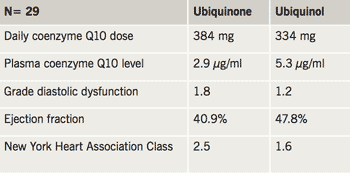
The authors of the study, presented at the 6th Conference of the International Coenzyme Q10 Association in Brussels, Belgium, said that giving the coenzyme in ubiquinol form improved plasma CoQ10 to higher levels than with ubiquinone. This improved level was associated with improvement in both diastolic and systolic myocardial function, along with improved New York Heart Association functional classification (table 1).
Other doctors from Melbourne, Australia, speaking at the conference said that they hoped an Integrative Cardiac Wellness Programme – in which coenzyme Q10, magnesium, alpha-lipoic acid, and fish oil were given three times daily to all cardiac surgery patients – may reduce troponin levels and lead to shorter hospital stays.
Table 1. Comparison of ubiquinone and ubiquinol in heart failure patients
Authors: Correspondence from the world of cardiology
The British Cardiovascular Society (BCS) has always had a significant role in education for cardiologists, largely through the BCS Annual Conference. The recognition of broader educational needs for trainees as well as trained cardiologists has led to a strategic change in educational activities within the BCS with a more comprehensive and structured approach to its delivery. In this column Dr Iain A Simpson, Vice President of Education and Research at the BCS, discusses educational activities within the BCS.
Responsibility for training of cardiologists in the UK does not lie with the BCS or its affiliated groups but rather with the General Medical Council, delivered through the Joint Royal College of Physicians Training Board, the Specialist Advisory Committee in Cardiology and through the individual postgraduate deaneries across the UK. The template for training in cardiology is the 2010 Cardiology Curriculum (available from: www.jrcptb.org.uk/specialties/ST3-SpR/Pages/Cardiology.aspx), which not only details the areas to be covered but also the assessment methods against which successful training is judged.
Simulator session at the 2010 Annual Conference
Although the BCS does not have a mandate for training, with its Affiliated Groups it has a membership uniquely positioned to coordinate, develop and deliver high-quality education to facilitate training requirements. In addition, through national training days and webcasting of key educational programmes it can ensure delivery of education is more uniform and accessible to all. There are already many excellent educational programmes being delivered on a regional basis and through the Affiliated Groups, so one of the aspirations of the BCS is to provide a detailed ‘map’ of educational activities, which are linked to the Cardiology Curriculum, on a regional and national basis. In addition to providing a resource of activities, the BCS has also agreed a number of quality indicators that will allow BCS Educational Endorsement of programmes, which trainees can use to ensure they are appropriate for the needs of their curriculum-based training. To achieve BCS Educational Endorsement, programmes should fulfil the following:
Be non-promotional educational programmes
Have an educational content that is evidence based and, where possible, includes reference sources
Include specific learning objectives
Incorporate formative assessment of learning objectives
Include feedback on educational course content, which is used to improve any subsequent programmes.
This does not mean that other educational programmes are not of good quality but rather that BCS endorsed programmes are specifically targeted at, and fulfil the needs of, cardiology training.
The template
The Cardiology Curriculum is the template for trainee education, but, as it covers the spectrum of clinical cardiology, it can also be used to structure educational programmes for trained cardiologists, and emphasises the need to look at education from the following perspectives:
Knowledge
Skills
Professionalism
Knowledge-based education lends itself well to be delivered by lectures, workshops, webcasting, etc. and, for trainees, this supplements the information available from the curriculum-based European Textbook of Cardiology. For the BCS this is also delivered through our Annual Conference and through links with Education in Heart. For trainees the development of two national training days has been supplemented by the highly successful Cardiology Review Course held each spring in conjunction with the Mayo Clinic. Using this as a template, a further Cardiology Review Course for trained cardiologists is planned to launch in the autumn of 2011.
Skills-based training is especially important in cardiology where procedural activities form so much of our clinical practice, yet skills training outside of the traditional ‘on-the-job’ experience is rare but gaining considerable interest as simulators for interventional cardiology, electrophysiology and trans-oesophageal echocardiography become increasingly sophisticated. For several years simulator training has occurred at the BCS Annual Conference, yet there are other aspects of skills training, such as basic surgical skills of suturing and handling of surgical instruments, that the BCS is supporting in conjunction with the Royal College of Surgeons and Heart Rhythm UK.
Professionalism is an area of education that is not specific to cardiology but spans the breadth of medicine. In the UK, Good Medical Practice Guidelines published by the General Medical Council (available from: www.gmc-uk.org/guidance/good_medical_practice.asp) are the template for professional standards. The areas of consent, communication, probity, clinical and fiscal management, medico-legal training, as well as commissioning of services, are something we have tried to embed within the BCS Annual Conference.
Summary
So the educational activities of the BCS are many and diverse. They continue to grow but in a structured manner, in full cooperation with the Affiliated Groups, with an aspiration to further improve the quality of education for trainees and trained cardiologists, and to hopefully facilitate the needs of impending revalidation, but ultimately for the benefit of our patients.
The outcome and complications of atrial fibrillation (AF) ablation in a UK patient cohort were investigated by offering symptomatic, drug-refractory patients ablation. Treatment goals were to disconnect all pulmonary veins electrically and improve symptoms using a state-of-the-art ablation method. Outcomes were defined as: ‘success’ (no symptoms or Holter AF); ‘partial success’ (substantially reduced AF symptoms); ‘clinical success’ (‘success’ and ‘partial success’); ‘failure’ (no symptom improvement).
A total of 100 consecutive patients (age: 49 years [range 37–76]; females: n=17; persistent AF: n=30; CHADS2 score >1: n=7) underwent a first ablation (between January 2004 and May 2007). Ultimately 167 procedures were performed until follow-up censure in May 2009. Complications occurred in 15 patients – acutely in 11, during follow-up in four. Cumulative ‘success’, ‘partial success’, ‘failure’ and ‘clinical success’ rates after 22 ± 14 months were 60%, 26%, 14% and 86%, respectively. ‘Clinical success’ rates for paroxysmal and persistent subgroups were 73% and 47% (first procedure) rising to 87% and 83% (all procedures). Numbers of patients needing anti-arrhythmic drugs reduced significantly (p<0.0001).
In conclusion, catheter ablation improves symptoms in 83–87% of patients, reduces objective AF burden and the need for anti-arrhythmic drug therapy. It should be recommended routinely to symptomatic patients with drug-refractory AF without demonstrable heart disease.
Introduction
Catheter-ablation is an established treatment for patients with uncontrolled symptoms of atrial fibrillation (AF).1 However, many cardiologists remain sceptical about its efficacy, and clinicians in general cannot relate ablation to the majority of AF patients.2-4 Although ablation techniques vary, there is general consensus that it is crucial to treat all pulmonary vein–left atrial junctions.5,6 Outcomes have usually been reported in terms of absolute success or failure after a short time period – not easily reconcilable with patient experiences subsequently.7 Recently, large amalgamated experiences have been published, which help explain some earlier inconsistencies, but these assume uniformity of patient selection, follow-up and ablation techniques.8
Unlike in other arrhythmias, AF presents ‘a moving target’ for ablation.9-11 As arrhythmia burden increases, ever more extensive ablation may be required to control it.12 In patients with persistent AF, ablation results remain tantalisingly unpredictable, even after extensive bi-atrial procedures.13-18 Similarly, patients with paroxysmal AF can be at different stages of atrial remodelling and do not respond uniformly to pulmonary vein isolation alone.19-21
The aim of this analysis was to report the AF ablation patient journey, based on a detailed evaluation of the course and outcome of 100 consecutive patients, treated in a uniform way by experienced operators at a single UK tertiary referral hospital. The object is to help clinicians better understand the nuances of AF ablation outcomes, largely concealed in larger series.
Methods
Patient selection
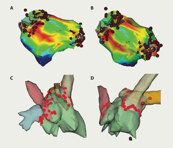
Figure 1. Example of CARTO (A and B) and NavX (C and D) computer maps of the left atrium showing lesion sets to isolate pulmonary veins
Patients with symptomatic AF without demonstrable heart disease, who were inadequately controlled by drug therapy, were considered for ablation. Structural heart disease and myocardial ischaemia were excluded as previously described.7 All were motivated to undergo ablation by intolerable symptoms or lack of acceptance of AF.
Ablation procedure
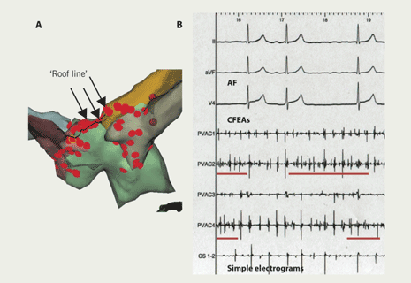
Ablation was performed in a uniform way. The left atrium was accessed by double-wiring of a single trans-septal puncture. An internal cardioversion/pacing catheter was positioned in the coronary sinus and a Lasso catheter (Biosense Webster Inc., CA, USA) used to guide ablation and confirm electrical isolation of veins. Ablation was performed using irrigation-tipped Webster 4 mm (Celsius ‘Thermocool’, Biosense Webster) or Navistar 4 mm catheters (Biosense Webster Inc., CA, USA). Patients were anticoagulated with unfractionated heparin, to maintain an activated clotting time longer than 250 seconds. Navigation and ablation were guided by either NavX-ESI (St Jude Medical Inc., St Paul, USA) or CARTO-XP (Biosense Webster Inc., CA, USA) mapping systems. Ablation lesions were sited around each pulmonary vein separately (figure 1). The acute procedural goal was to isolate all identified pulmonary veins (PVI). Ablation energy was power limited to 30 W, temperature to 50 ºC with irrigation flow rate of 17–30 ml/min. Additional linear lesions (figure 2A) were performed in patients with persistent AF or in any who continued in AF despite PVI. Focal sites at which complex fractionated electrograms were consistently recorded during AF – possible perpetuators of the arrhythmia – were also targeted for specific ablation at repeat procedures17,18,22 (figure 2B). Afterwards, patients with a history of long paroxysms or persistent AF continued their anti-arrhythmic drugs. All were on either warfarin or aspirin.
Figure 2. A NavX computer map of the left atrium (A), showing a ‘roof-line’ ablation linking right and left upper pulmonary vein ostia; and examples of focal complex fractionated electrogram ablation (CFEAs; underlined) and simple atrial electrograms during atrial fibrillation (AF) (B)
Follow-up
Patients were reviewed regularly out to 12 months following each procedure, their rhythm status determined on the basis of symptoms and a 12-lead electrocardiogram (ECG) at each visit. Only arrhythmias continuing beyond a two-month ‘blanking period’ are reported. All patients, free of AF symptoms, also had 48-hour to seven-day Holter ECG recordings. Anti-arrhythmic therapy was withdrawn three months after ablation. However, if symptoms returned later, therapy was restarted and the patient offered repeat ablation. Most patients who continued to experience symptoms despite anti-arrhythmic therapy opted for repeat ablation(s). Three outcome categories were defined: ‘success’ (i.e. no symptoms, ECG or Holter evidence of AF lasting >30 seconds; no class 1C or III anti-arrhythmic therapy); ‘partial success’ (i.e. AF symptoms and objective AF burden substantially reduced on or off previously ineffective, anti-arrhythmic therapy); ‘failure’ (i.e. no clinically relevant change in symptoms or AF burden). Follow-up was censured on 31st May 2009.
Patients’ own assessment of ablation outcome
An anonymised questionnaire was distributed by the hospital’s Clinical Governance Department in spring 2008 to assess:
symptom impact on quality of life pre-ablation
effect of ablation outcome on symptoms and quality of life
whether they would undergo ablation treatment again, or recommend it to others.
Results were correlated with objective arrhythmia outcomes to derive the fourth outcome category – ‘clinical success’ (i.e. the sum of ‘success’ and ‘partial success’ categories (clinician assessed) combined with patients’ own evaluation).
Statistical methods
All values are expressed as mean and one standard deviation. Comparisons between groups were made using Student’s t and Fishers’ exact testing. Subgroup comparisons were made using analysis of variance. Differences were considered significant at the 5% level.
Results
By 31st May 2009, over 1,000 AF ablation procedures had been performed at this hospital, using a variety of techniques.7 The 100 consecutive patients reported in detail here were those without demonstrable heart disease, who had a first procedure for AF in the period between June 2004 and June 2007 using a uniform, ‘state-of-the-art’ ablation method. Mean age was 55 ± 11 years (range 22–74), length of AF history was 66.5 months (range 3–240), 83 were male and 30 (30%) had persistent AF. In only seven was CHADS2 risk score greater than one (CHADS2 scores: 0 n=69; 1 n=24; 2 n=3; 3 n=3; 4 n=1). Scores were contributed to by the following: heart failure at presentation due to tachycardia myopathy (n=4), treated hypertension (n=25), age over 75 years (n=1), diabetes mellitus (n=5) and stroke (n=4). No patient had prior infarction, active myocardial ischaemia or idiopathic cardiomyopathy. AF symptoms had not been controlled by potent anti-arrhythmic drugs. Fifty-six had failed flecainide or propafenone and 13 had failed amiodarone or sotalol in high dose. Most patients were also taking beta-blocking agents, calcium channel blockers or digoxin prior to their first ablation.
Cumulative outcome results
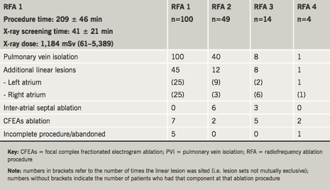
In total, 167 separate ablation procedures were performed in these 100 patients and procedure-related complications occurred in 15 (15%) (tables 1 and 2). Acute complications included cardiactamponade, requiring pericardiocentesis in five (3% of procedures), pericarditis with effusion treated conservatively in five (3% of procedures), significant discomfort at catheter access site in two (1%) and allergic iodine contrast reaction in one (0.6% of procedures).
Table 1. Extent of ablations performed by session
Late complications occurred in four (4%) patients – one (0.6% of procedures) each of – unexplained sudden death six months after ablation, minor stroke with normal brain imaging, pulmonary vein stenosis of more than 75% (treated conservatively), pulmonary embolus 10 days after ablation (managed conservatively). Left atrial flutter occurred in eight (8%) patients and was typically persistent and more symptomatic than AF. After a mean follow-up of 22 months (range 3–92 months) cumulative ‘success’, ‘partial success’ and ‘failure’, as defined, for the whole group were 60%, 26% and 14%, respectively. Outcomes for paroxysmal AF at time of first ablation were 61%, 26% and 13%, respectively, after 1.5 ± 0.7 procedures per patient and for persistent AF, 57%, 26% and 17%, respectively, after 2 ± 1 procedures (‘success’: p=0.66).
Patients’ own assessment of ablation outcome
Of the 100 patient questionnaires circulated 12 months after ablation, 80 were returned and analysed. Sixty-five (81%) described symptoms as “bad” or “terrible” prior to ablation. Of these, 58 (89%) felt “much better” afterwards – symptoms having improved to “none” or “not bad at all” (p<0.0001). Seventy-four (92%) described AF as interfering with their life at least “modestly” prior to ablation. Afterwards, 40 (58%) felt symptoms did not affect their quality of life “at all” (p<0.001), 29 (42%) rated interference as “only a little” and 11 (14%) as “moderately interfering” with their life. Seventy-four (92%) indicated that ablation had improved symptoms sufficiently for them to undergo the procedure, if faced with the same choice again, four were unsure and two said that they would not have opted for ablation. These results support the clinical impression that the outcome ‘partial (anti-arrhythmic) success’, as defined, also indicates patients who, although not free of AF, have benefited substantially from the procedure. Therefore, defining ‘clinical success’ as the sum of objective clinician-defined ‘success’ and ‘partial success’ indicates those who have either no AF or are sufficiently improved, in their own estimation, not to need further intervention. ‘Clinical success’ was achieved in 86% of the 100 patients (paroxysmal AF 87%; persistent AF 83%; p=0.75).
Does ‘failure’ of first AF ablation indicate a non-responder or the need for a repeat procedure?
As indicated by the following analysis, cumulative benefits accrue from repeat ablation procedures (table 2).
First ablation procedure
Although all identified pulmonary vein–left atrial junctions were treated, complete electrical isolation could only be confirmed in 77 (77%). Complications occurred in 10 (10%) patients. Overall ‘clinical success’ rate was 67%, but ‘success’ and ‘clinical success’ rates were significantly better for paroxysmal than for persistent AF (‘success’: 46% versus 16%, p=0.007; ‘clinical success’: 77% versus 43%, p=0.002).
Second ablation procedure
All patients continuing to experience AF symptoms were recommended repeat ablation and 49 had a second procedure 12.7 ± 8.1 months after the first. Pulmonary vein reconnections were found in some or all veins in 40 (82%) patients and complications occurred acutely in four (8%). There were no late complications. Overall ‘clinical success’ for the second procedure was 76%. Differences in ‘success’ and ‘clinical success’ rates between initially paroxysmal and persistent AF were no longer significant (‘success’: 47% versus 26%, p=0.23; ‘clinical success’: 80% versus 68%, p=0.49).
Third ablation procedure
Fourteen patients (14%) underwent a third AF ablation 26.7 ± 19.0 months after their first procedure. Pulmonary vein reconnections were found in eight (57%). One patient developed pericarditis, treated conservatively, and four developed atypical atrial flutter. Overall ‘clinical success’ for the third procedure was 71%. ‘Success’ and ‘clinical success’ rates were the same for paroxysmal and persistent AF patients (‘success’: 40% versus 22%, p=0.55; ‘clinical success’: 60% versus 78%, p=1.0).
Fourth ablation procedure
Four patients (4%) underwent a fourth ablation 29 ± 13 months after their first procedure. The indication was left atypical atrial flutter in three. Flutters terminated with addition of a left atrial ‘roof-line’ in one, with focal ablation around right lower pulmonary vein in another and with ablation at sites of complex fractionated electrograms in a third. AF arose from the right atrial appendage in the fourth patient. There were no complications. ‘Success’ rate, therefore, was 100%.
Patients changing from rhythm to rate-control management
Eight (8%) patients changed to rate-control management – four after the first and four after the second ablation procedure. They tended to be older (64 ± 9 versus 55 ± 11 years, p≤0.01). Six required complete AV-junction ablation for symptom control.
Anti-arrhythmic drug requirements before ablation and at most recent review
Prior to ablation 73 (73%) patients were taking Class 1C or III agents, alone or in combination therapy: flecainide 53 (53%), propafenone 3 (3%), disopyramide 4 (4%), high-dose sotalol 6 (6%) and amiodarone 7 (7%). At latest follow-up 22 ± 14.6 months later, only 20 (20%) still required these drugs: flecainide 14 (14%), propafenone 2 (2%) and amiodarone 4 (4%); and, in five, flecainide was only being used as a ‘pill-in-the-pocket’ regimen (p<0.0001).
Table 2. Summary of cumulative ablation procedures and their outcomes
Discussion
Although only a 100 consecutive patient cohort from a larger experience, this detailed analysis complements the perspective provided by larger, amalgamated AF-ablation series and specifically relates to a UK population.7,8 Its step-by-step analysis shows that outcomes are more nuanced than conveyed by earlier series, which simply reported recurrence or complete absence of AF at some arbitrary time point.23-25 The overall ‘clinical success’ rate of 86% confirms a role for ablation as a means of controlling medically refractory symptoms. The results, at mean follow-up of 22 ± 14.6 months, are consistent with those already reported previously by our group and by other authors from pioneering hospitals.7,13,26-29 On this basis, therefore, catheter ablation should be offered routinely to symptomatic patients with AF, inadequately controlled by potent anti-arrhythmic drug therapy.
The results also show that – in addition to patients who either have no AF (‘success’) or those whose arrhythmia is unchanged (‘failure’) – there is another group (‘partial success’) who benefit sufficiently to also consider their ablation successful.26 Ninety per cent of patients in this middle category reported ‘significant improvement’ after ablation – a figure similar to the 86% ‘clinical success’ rate identified by objective arrhythmia assessment. This ‘partial success’ group, inadequately discussed in most series, is readily recognisable to clinicians who, although not performing ablations themselves, routinely follow-up these patients.25
This report highlights the difficulty in achieving long-term electrical isolation of pulmonary veins with a ‘state-of-the-art’ ablation technique. Even when pulmonary vein isolation had been achieved acutely, almost all patients needing subsequent procedures had residual or re-established connections.30 If catheter ablation of AF is to be cost-effective and be made available to the majority of patients who might benefit, pulmonary vein isolation needs to be permanent, once achieved acutely. This cannot be guaranteed currently.19-21
Although ‘success’ following a first ablation was significantly better (p<0.007) for patients with paroxysmal (46%) as compared to persistent (16%) AF, eventual outcome was similar (60% versus 57%, p=0.66). Although at odds with most reports, this observation is supported by similar results from a recent, much larger series.8 It is probably explained by the fact that patients with paroxysmal or persistent AF behaviour are essentially the same, differing only in their stage of AF natural history.8,30
Complications occurred in 15 patients (9% of procedures).31 However, most were self-limiting or resolved with treatment. There were no instances of oesophageal injury, phrenic nerve palsy or pulmonary vein stenosis needing intervention.31,32 Atypical left atrial flutter – although not really a ‘complication’ – occurred in eight (8%) patients, was usually incessant, resistant to drugs and challenging to ablate.13 It was the main indication for third and fourth procedures.
This analysis reports medium-term outcome in a consecutive cohort of patients who have undergone catheter ablation, performed in a standard way, for AF at a UK tertiary referral hospital. The results are presented in a novel way with the aim of helping clinicians not involved in the delivery of this treatment, and patients, better understand what can be offered and expected. The overall conclusion, based on the outcomes achieved, is that catheter ablation should be offered routinely to all patients with symptomatic, medically refractory AF.
Conflict of interest
None declared.
Acknowledgement
Dr Bourke is part-funded by the Northumberland, Tyne & Wear Local Clinical Research Network (LCRN).
Key messages
Over 80% of patients with structurally normal hearts respond to catheter ablation of atrial fibrillation (AF), having less symptoms and less arrhythmia burden than they would have had with optimum anti-arrhythmic drug therapy
Catheter ablation allows withdrawal of anti-arrhythmic therapy in the majority of cases
Catheter ablation of AF carries a small risk of acute complications, but most are readily treatable and do not have long-term sequelae
Catheter ablation should be offered routinely to patients with symptomatic AF, inadequately controlled by trials of potent anti-arrhythmic drug therapy
References
Fuster V, Ryden LE, Cannom DS et al. ACC/AHA/ESC 2006 guidelines for the management of patients with atrial fibrillation. Europace 2006;8:651–745.
Testa L, Biondi-Zoccai GGL, Russo AD, Bellocci F, Andreotti F, Crea F. Rate-control versus rhythm-control in patients with atrial fibrillation: a meta analysis. Eur Heart J 2005;26:2000–06.
The AFFIRM Investigators.A comparison of rate control and rhythm control in patients with atrial fibrillation. N Engl J Med 2002;347:1825–33.
Naccarelli G, Gonzalez M. Atrial fibrillation and the expanding role of catheter ablation: do anti-arrhythmic drugs have a future? J Cardiovasc Pharmacol 2008;52:203–09.
Pappone C, Oreto G, Rosanio S et al. Atrial electro-anatomic remodelling after circumferential radiofrequency pulmonary vein ablation: efficacy of an anatomic approach in a large cohort of patients with atrial fibrillation. Circulation 2001;104:2539–44.
Gerstenfeld EP, Callans DJ, Dixit S et al. Incidence and location of focal atrial fibrillation triggers in patients undergoing repeat pulmonary vein isolation: implications for ablation strategies. J Cardiovasc Electrophysiol 2003;14:685–92.
Bourke JP, Dunuwille A, O’Donnell D, Jamieson S, Furniss SS. Pulmonary vein ablation for idiopathic atrial fibrillation: six month outcome of first procedure in 100 consecutive patients. Heart 2005;91:51–7.
Bhargave S, Di Biase L, Mohanty P et al. Impact and type of atrial fibrillation and repeat catheter ablation on long-term freedom from atrial fibrillation: results from a multi-centre study. Heart Rhythm 2009;6:1403–12.
Haïssaguerre M, Wright M, Hocini M, Jaïs P. The substrate maintaining persistent atrial fibrillation. Circulation 2008;1:2–5.
Calò L, Lamberti F, Loricchio ML et al. Left atrial ablation versus biatrial ablation for persistent and permanent atrial fibrillation: a prospective and randomized study. J Am Coll Cardiol 2006;47:2504–12.
Dixit S. Evolving strategies in catheter ablation of long-standing atrial fibrillation. Circulation 2008;1:324–6.
Allessie MA, Boyden PA, Camm AJ et al. Pathophysiology and prevention of atrial fibrillation. Circulation 2001;103:769–77.
Haissaguerre M, Hocini M, Sanders P et al. Catheter ablation of long-lasting persistent atrial fibrillation: clinical outcome and mechanisms of subsequent arrhythmias. J Cardiovasc Electrophysiol 2005;16:1138–47.
Vasavakul T, Khunnawat C, Ngarmukos T. A new approach to catheter ablation of atrial fibrillation: mapping of the electrophysiologic substrate. J Am Coll Cardiol 2004;43:2044–53.
Jais P, O’Neill M, Yoshihide T et al. Stepwise catheter ablation of chronic atrial fibrillation. J Cardiovasc Electrophysiol 2006;17(suppl):S28–S36.
Willems S, Hanno K, Rostock T et al. Substrate modification combined with pulmonary vein isolation improves outcome of catheter ablation in patients with persistent atrial fibrillation: a prospective randomised comparison. Eur Heart J 2006;27:2871–8.
Takahashi Y, O’Neill MD, Hocini M et al. Characterization of electrograms associated with termination of chronic atrial fibrillation by catheter ablation. J Am Coll Cardiol 2008;51:1003–10.
Oral H, Chugh A, Good E et al. Radiofrequency catheter ablation of chronic atrial fibrillation guided by complex electrograms. Circulation 2007;115:2606–12.
Lo L-W, Tai C-T, Lin Y-J et al. Progressive remodelling of the atrial substrate – a novel finding from consecutive voltage mapping in patients with recurrence of atrial fibrillation after catheter ablation. J Clin Electrophysiol 2007;18:258–65.
Oakes RS, Badger TJ, Kholmovski EG et al. Detection and quantification of left atrial structural remodelling with delayed-enhancement magnetic resonance imaging in patients with atrial fibrillation. Circulation 2009;119:1758–67.
Cheema A, Vasamreddy CR, Dalal D et al. Long-term single procedure efficacy of catheter ablation of atrial fibrillation. J Intervent Card Electrophysiol 2006;15:145–55.
Nademanee K, Oketane N. The role of complex fractionated atrial electrograms in atrial fibrillation ablation: moving to the beat of a different drum. J Am Coll Cardiol 2009;53:782–9.
Pappone C, Oreto G, Rossanio S et al. Atrial electro-anatomic remodelling after circumferential radiofrequency pulmonary vein ablation: effect of an anatomic approach in a large cohort of patients with atrial fibrillation. Circulation 2001;104:2539–44.
Mangrum JM, Mounsey JP, Kok LC, DiMarco JP, Haines DE. Intra-cardiac echocardiography-guided, anatomically based radiofrequency ablation of focal atrial fibrillation originating from the pulmonary veins. J Am Coll Cardiol 2002;39:1964–72.
Stabile G, Bertaglia E, Senatore G et al. Catheter ablation treatment in patients with drug-refractory atrial fibrillation: a prospective, multi-centre, randomised, controlled study. Eur Heart J 2006;27:216–21.
Bunch TJ, Munger TM, Friedman PA et al. Substrate and procedural predictors of outcomes after catheter ablation for atrial fibrillation in patients with hypertrophic cardiomyopathy. J Cardiovasc Electrophysiol 2008;19:1009–14.
Steven D, Rostock T, Lutomsky B et al. What is the real atrial fibrillation burden after catheter ablation of atrial fibrillation? A prospective rhythm analysis in pacemaker patients with continuous atrial monitoring. Eur Heart J 2008;29:1037–42.
Earley MJ, Abrams DJR, Staniforth A, Sporton SC, Schilling RJ. Catheter ablation of permanent atrial fibrillation: medium term results. Heart 2006;92:233–8.
Lo L-W, Tai C-T, Lin Y-J et al. Characteristics and outcome in patients receiving multiple (more than two) catheter ablation procedures for paroxysmal atrial fibrillation. J Cardiovasc Electrophysiol 2008;19:150–6.
Verma A, Kilicaslan F, Pisano E et al. Response of atrial fibrillation to pulmonary vein antrum isolation is directly related to resumption and delay of pulmonary vein conduction. Circulation 2005;112:627–35.
Spragg D, Dalal D, Cheema A et al. Complications of catheter ablation for atrial fibrillation: incidence and predictors. J Clin Electrophysiol 2008;19:627–31.
Cappato R, Calkins H, Chen S-A et al. Prevalence and causes of fatal outcome in catheter ablation of atrial fibrillation. J Am Coll Cardiol 2009;53:1798–803.
Sulphonylureas are well established in the treatment of type 2 diabetes mellitus. They are effective in improving glycaemic control and preventing microvascular complications. Side effects that can restrict use include hypoglycaemia and weight gain. Although there is no clear evidence for reduction of cardiovascular disease from randomised-controlled trials, follow-up data from the United Kingdom Prospective Diabetes Study (UKPDS) shows reduced cardiovascular risk. Concerns about sulphonylureas causing inhibition of ischaemic preconditioning are relevant in primary angioplasty, but there is a lack of clear evidence, with a need for randomised-controlled trials to investigate this further.
Cardiac auscultation is a critical part of the clinical examination. In this review we discuss the conventional approach to teaching and using the skill of cardiac auscultation. We then consider how recent technological advances may improve the teaching and implementation of this essential clinical skill.
The past: a historical perspective
The earliest description of the heart sounds comes from William Harvey’s De Motu Cordis in 1628, in which he likened the heart sounds to “two clacks of a water bellows to raise water”, but it was not until Laennec invented the stethoscope in 1816 that cardiac auscultation superseded percussion and direct auscultation. Laennec proposed the use of “a cylinder of wood, perforated in its centre longitudinally, by a bore three lines in diameter, and formed so as to come apart in the middle”; this he termed the cylinder or stethoscope. This was followed in 1819 by his landmark work De l’auscultation médiate ou traité du diagnostic des maladies des poumons et du coeur (On mediate auscultation or treatise on the diagnosis of the diseases of the lungs and heart), through which he achieved widespread recognition. The art of cardiac auscultation developed considerably during the nineteenth century and is now an essential component of the clinical examination.
Present: traditional teaching methods
Traditionally, cardiac auscultation has been taught best at the bedside during clinical undergraduate training and in preparation for postgraduate membership examinations. It is an essential component of the clinical examination, but like most clinical skills requires repetition1 and clinical experience to make an accurate diagnosis. Indeed, prior to the advent of echocardiography, physicians were totally reliant on their stethoscope and auscultatory skills to accurately diagnose and characterise cardiac murmurs.
The traditional clinical teacher will maintain that there is no substitute for clinical bedside teaching, while the modern educationalist will opt for multimedia applications, audio CDs and patient simulators.2,3 We would support the former, as evidenced by the decline in skills among medical graduates; cardiac auscultation, once the hallmark of an expert clinician, is rapidly becoming a lost art.4 The importance of cardiac auscultation cannot be underestimated and it remains an essential skill at the bedside, which, when performed well, can avoid the potential of ‘over-investigating’ patients and causing unnecessary anxiety. However, as many as three-quarters of American interns and two-thirds of cardiology trainees no longer receive formal teaching in cardiac auscultation.5 Of concern, several studies have reported an apparent lack of ability of interns to correctly diagnose a cardiac murmur, which we have reviewed recently.6 If this decline is related to lack of clinical exposure and deficiencies in current teaching methods, can this be rectified?
Modern approaches
Several studies have reported an improvement in diagnostic skills with the use of various educational aids and methods. A recent study of third-year medical students found that the use of a computer-based teaching application increased their ability and confidence in detecting cardiac murmurs and added heart sounds.7 Similarly, another study found that the use of interactive CD-ROMs was associated with an improvement in auscultation skills.8 A group based at University of California, Los Angeles compared the use of a virtual patient examination (VPE) with a conventional teaching model in third-year medical students and found that VPE-based teaching resulted in a higher level of competency and long-term retention of knowledge of cardiac auscultation.9 The use of web-based resources, in addition to clinical training, has also been proposed in two separate studies.10,11 The Mayo group reported an improvement in auscultation following attendance at a patient-centred teaching conference designed to improve such skills and taught by a master auscultator.12 These data suggest that a combined approach, which maintains clinical exposure, utilising clinical experience, but incorporates technological innovation to reinforce learning, may be the best way forward.
Patient simulators
Patient simulators first made their debut at the American Heart Association (AHA) scientific sessions in 1968. Nicknamed Harvey after Dr W Harvey Procter, this sophisticated mannequin is able to display a number of cardiovascular indices including blood pressure (by auscultation), jugular venous pulse waveforms and arterial pulses, precordial impulses and auscultatory findings in the four classic areas (synchronised with the pulse and varying with respiration).13 Harvey is capable of simulating a spectrum of cardiac disease by varying blood pressure, breathing, pulses, normal heart sounds, and murmurs. Harvey underwent rigorous testing as an educational model with pilot studies first reporting promising results in 1980.14 A National Heart, Lung and Blood Institute (NHLBI)-funded study in 1987 found that senior medical students who had trained with Harvey performed significantly better than their peers, who had only clinical training.15 Harvey has since undergone several modifications and remains an important learning resource for healthcare professionals and trainees. Several other simulators have since followed the advent of Harvey.
The future
With advances in technology, there has been considerable development of the traditional stethoscope and this has allowed us to overcome some of the previous acoustic limitations when using traditional stethoscopes. Electronic stethoscopes have the ability to amplify the heart sounds, filter sound frequency and eliminate background noise. Furthermore, a key to reinforcing teaching by the bedside, newer generation models are capable of storage and playback of heart sounds away from the patient through an external source or computer. The idea of combining such a stethoscope with computer software that could visualise the murmur and heart sounds as a means of facilitating undergraduate teaching was proposed several years ago.16
There are, however, very little published data comparing conventional and electronic stethoscopes. An early study in 1998 comparing some of the more primitive electronic stethoscopes with standard devices, concluded that the acoustic stethoscopes were preferable, however, they proposed that an ideal device would feature a combination of both.17 More recently, a Norwegian study randomised third-year medical students to either a traditional or an electronic stethoscope (Welch Allyn) and found no difference in terms of diagnostic accuracy when assessed by a cardiac auscultation test.18 A Danish study comparing a standard stethoscope with a ‘cardiology’ stethoscope also found no difference.19 Our own experience with electronic stethoscopes when teaching medical students has been a positive one based primarily on the ability to amplify sounds and reduce background noise. The ability to record the abnormal auscultatory findings and immediate facility to replay that sound has been particularly useful.
The most recent developments that may benefit day-to-day clinical practice include the European launch of the 3M Littmann 3200 electronic stethoscope, which has the ability to transmit heart sounds via Bluetooth technology. This technology has Food and Drug Administration (FDA) approval in the USA and works in conjunction with software (Zargis CardioscanTM), which is able to accurately interrogate and analyse heart sounds and murmurs. Using sophisticated algorithms, the software has been shown to predict whether a murmur is clinically significant based on AHA class I murmur criteria.20 We are currently conducting the first European clinical trial of this system to determine its accuracy in identifying innocent and pathological murmurs in real-world clinical practice in adult and paediatric populations. We believe this technology represents a major innovation in cardiac auscultation technology and such a device would be an invaluable screening tool and diagnostic aid in the settings of primary care and pre-op clinics, where often murmurs may be either missed or inappropriately investigated. Furthermore, the ability to digitally store these data may also allow the clinician to monitor and compare murmur intensity over time, and could prove useful in monitoring valvular disease, although this remains to be proven.
With regard to teaching and examining, 3M are about to introduce StethED, software that has been developed on Android technology but is now being transposed to the iPhone and Blackberry. Essentially, 3200s are linked via Bluetooth to the smartphone and the ‘teacher’ indicates which stethoscope is to act as the teacher, and which are the students. All individuals will use their stethoscope to auscultate and the sounds will be gathered. This will enable the examiner not only to ascertain whether the student has placed the stethoscope correctly on the praecordium and, hence, acquired the correct sounds, but also whether the student has interpreted the sounds correctly. Thus, the student can no longer ‘hide’ behind an inability to ‘hear’ the murmur and, for examination purposes, it will allow a standardised approach with a ‘listening examiner’ being able to confirm or refute the auscultatory findings exactly at the time of examination.
Conclusion
Many factors have conspired to limit adequate teaching and maintenance of cardiac auscultation skills. Indeed the requirements and expectations of junior doctors, with regard to auscultation, are much lower now than in previous generations. While technological advancements, such as echocardiography, may well have contributed to the demise of cardiac auscultation, technology in the form of integrated electronic auscultation may well revive its place in clinical medicine21.
Conflict of interest
The authors are currently performing an investigator-
initiated study funded by 3M to assess the clinical
utility of the 3200 stethoscope in conjunction with Zargis Cardioscan software.
Editors’ note
See also the previous editorial by Alam et al. in the January/February issue earlier this year (Br J Cardiol 2010;17:8–10).
Key messages
Cardiac auscultation is an essential part of the clinical examination
Cardiac auscultation skills have declined, possibly due to the introduction of newer technologies such as echocardiography
Technological innovations, such as electronic stethoscopes, multimedia applications and patient simulators, are now available to assist in the teaching of cardiac auscultation
A combined approach, which maintains clinical exposure, utilising clinical experience, but incorporates technological innovation to reinforce learning, may be the best way forward
References
Barrett MJ, Lacey CS, Sekara AE, Linden EA, Gracely EJ. Mastering cardiac murmurs: the power of repetition. Chest 2004;126:470–5.
Karnath B, Frye AW, Holden MD. Incorporating simulators in a standardized patient exam. Acad Med 2002;77:754–5.
Karnath B, Thornton W, Frye AW. Teaching and testing physical examination skills without the use of patients. Acad Med 2002;77:753.
Chizner MA. Cardiac auscultation: rediscovering the lost art. Curr Probl Cardiol 2008;33:326–408.
Mangione S, Nieman LZ, Gracely E, Kaye D. The teaching and practice of cardiac auscultation during internal medicine and cardiology training: a nationwide survey. Ann Intern Med 1993;119:47–54.
Alam U, Asghar O, Khan SQ, Hayat S, Malik RA. Cardiac auscultation: an essential clinical skill in decline. Br J Cardiol 2010;17:8–10.
Ostfeld RJ, Goldberg YH, Janis G, Bobra S, Polotsky H, Silbiger S. Cardiac auscultatory training among third year medical students during their medicine clerkship. Int J Cardiol 2009 Feb 3. [Epub ahead of print]
Stern DT, Mangrulkar RS, Gruppen LD, Lang AL, Grum CM, Judge RD. Using a multimedia tool to improve cardiac auscultation knowledge and skills. J Gen Intern Med 2001;16:763–9.
Vukanovic-Criley JM, Boker JR, Criley SR, Rajagopalan S, Criley JM. Using virtual patients to improve cardiac examination competency in medical students. Clin Cardiol 2008;31:334–9.
Criley JM, Keiner J, Boker JR, Criley SR, Warde CM. Innovative web-based multimedia curriculum improves cardiac examination competency of residents. J Hosp Med 2008;3:124–33.
Tuchinda C, Thompson WR. Cardiac auscultatory recording database: delivering heart sounds through the Internet. Proc AMIA Symp 2001:716–20.
March SK, Bedynek JL, Chizner MA. Teaching cardiac auscultation: effectiveness of a patient-centered teaching conference on improving cardiac auscultatory skills. Mayo Clin Proc 2005;80:1443–8.
Cooper JB, Taqueti VR. A brief history of the development of mannequin simulators for clinical education and training. Qual Saf Health Care 2004;13:i11–i18.
Gordon MS, Ewy GA, Felner JM et al. Teaching bedside cardiologic examination skills using ”Harvey,” the cardiology patient simulator. Med Clin North Am 1980;64:305–13.
Ewy GA, Felner JM, Juul D et al. Test of a cardiology patient simulator with students in fourth-year electives. J Med Educ 1987;62:738–43.
Woywodt A, Herrmann A, Kielstein JT et al. A novel multimedia tool to improve bedside teaching of cardiac auscultation. Postgrad Med J 2004;80:355–7.
Grenier MC, Gagnon K, Genest J Jr, Durand J, Durand LG. Clinical comparison of acoustic and electronic stethoscopes and design of a new electronic stethoscope. Am J Cardiol 1998;181:653–6.
Høyte H, Jensen T, Gjesdal K. Cardiac auscultation training of medical students: a comparison of electronic sensor-based and acoustic stethoscopes. Henning BMC Medical Education 2005;5:14.
Iversen K, Søgaard Teisner A, Dalsgaard M et al. Effect of teaching and type of stethoscope on cardiac auscultatory performance. Am Heart J 2006;152:85.e1–85.e7.
Watrous RL, Thompson WR, Ackerman SJ. The impact of computer-assisted auscultation on physician referrals of asymptomatic patients with heart murmurs. Clin Cardiol 2008;31:79–83.
Tavel ME. Cardiac auscultation: a glorious past — and it does have a future! Circulation 2006;113:1255–9.
Authors: David Turpie, Matthew Maycock, Chiala Crawford, Kathleen Aitken, Marwen Macdonald, Colin Farman, Maimie L P Thompson, Jamie Smith, Stephen J Cross, Stephen J Leslie
David Turpie
Cardiology Specialist Registrar
Matthew Maycock
Medical Student
Chiala Crawford
Staff Nurse
Kathleen Aitken
Staff Nurse
Marwen Macdonald
Echocardiographer
Colin Farman
Clinical Scientist
Maimie L P Thompson
Programme Manager –
18 Weeks RTT
Jamie Smith
Consultant Cardiologist
Stephen J Cross
Consultant Cardiologist
Stephen J Leslie
Consultant Cardiologist
Raigmore Hospital, Old Perth Road, Inverness, IV2 3UJ
Correspondence to: Professor S J Leslie,
Highland Heartbeat Centre, Cardiac Unit, Raigmore Hospital, Inverness, IV2 3UJ [email protected]
The number of patients with aortic stenosis (AS) in the UK is increasing. Patients with non-significant AS can be safely reviewed in technician-led clinics. The potential impact of this on healthcare services is unreported. The aim of this study was to describe the impact of establishing an AS surveillance clinic in a district general hospital setting and consider the potential impact of widespread implementation.
Criteria for an AS surveillance clinic were developed. Patients who were deemed suitable were identified from existing echocardiographic databases, discharge coding and review of the clinical notes. Patients with AS were identified (n=612). After a review of echocardiographic parameters, 117 patients were considered suitable for technician-led review. Of these, 47 patients (40%) were subsequently discharged from the cardiology clinic.
A small proportion of patients are reviewed in the general cardiology clinic for no other reason than asymptomatic mild AS (5% of follow-up appointments). Establishment of a national AS surveillance programme could result in a large number of patients (>9,000) being discharged from formal doctor-led cardiology clinic review in the UK. This would improve quality and consistency of follow-up monitoring for the patient and free up capacity to see new patients.
Introduction
Aortic stenosis (AS) is the most common form of valvular heart disease.1 The incidence is increasing due to an ageing population. Aortic sclerosis is present in up to 25% of adults over the age of 65 years with progression to severe AS within seven years in about 16%.2 Thus, surveillance of these patients is required. Recent guidelines suggest that patients with mild AS can be reviewed infrequently (up to every five years). Those with a higher degree of AS should be kept under more frequent review. Asymptomatic patients with mild AS make up a significant proportion of patients followed up in general out-patient cardiology clinics.3
Within the UK, demand for specialist cardiology services continues to increase. Service redesign, which allows a review of asymptomatic patients with AS without a clinic review by a doctor, could increase capacity to see new patients. This study describes the establishment of an AS surveillance clinic in a district general hospital.
Aim
To describe and discuss the potential benefits of widespread implementation of an AS surveillance clinic.
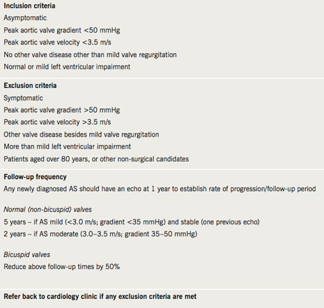
Table 1. Aortic stenosis (AS) surveillance clinic criteria for attendance at the clinic and for frequency of appointments
Methods
Setting
The study was performed in a Scottish district general hospital with a catchment population of around 200,000.
Identification of potential patients
Following local discussions, review of criteria from other centres and reference to national guidelines,5 specific criteria were developed for patients who would be suitable for follow-up in a technician-led AS surveillance clinic (table 1). Patients who were potentially suitable for inclusion were identified both in a prospective and retrospective manner. Prospective opportunistic identification of patients via cardiology clinic was undertaken by informing all cardiology staff of the presence of the clinic criteria. A retrospective review was undertaken of patients who were already known to have AS. They were identified from the echocardiography database or by discharge codes following opportunistic in-patient admission using International Classification for Disease-10 (ICD-10) code (I06.0, I06.2, I08.0, I08.2, I08.3, I08.8, I35, I42.1, I08.0, Q23.0, Q23.1, I06, I35.0, I35.1, I35.2, I35.8, I35.9, I39.1, Q23.0, Q23.8) in the year 2008 (01/01/2008 to 31/12/2008). Cases identified from these two sources were cross-referenced and a final cohort identified.
Data collection
Echocardiography reports were studied from all patients in the identified cohort. These had been produced from routine 2D echo (GE vivid 7) by British Society of Echocardiography (BSE) trained cardiac sonographers. Key data were extracted (maximum AV velocity, maximum AV pressure gradient, degree of aortic regurgitation, other valve pathology, i.e. mitral regurgitation or stenosis, pulmonary regurgitation or stenosis and tricuspid regurgitation or stenosis, left ventricular systolic function and valve calcification). The criteria for inclusion in the surveillance clinic (table 1) were then applied to the cohort to identify all patients suitable for follow-up based on echocardiography findings. The medical notes of this smaller group of patients were then examined to identify those patients who may need to be excluded from the surveillance clinic because of other elements requiring medical opinion such as other co-morbidities.
AS surveillance clinic
The final cohort of identified suitable patients were advised of their inclusion in the surveillance clinic, and allocated a follow-up appointment time based on the established criteria (table 1). The clinics have been established using a routine out-patient echo session and are run jointly by a BSE accredited echocardiographer and a trained cardiac nurse. On attendance, patients are asked about existing and new symptoms and a routine echo is performed (concentrating on obtaining aortic valve surveillance data – table 2). An electronic letter is generated following each clinic visit with paper copies sent to both the consultant in charge and the general practitioner with details of the echocardiogram result and the date of the next clinic appointment as determined by protocol. Where the patient symptoms or echo findings meet the exclusion criteria, patients are referred back to the cardiologist for follow-up in cardiology clinic.
Ethics
As this was a clinical case review, ethical approval was not required.
Results
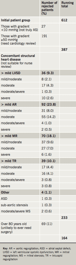
Table 2. Reasons and numbers for exclusion of patients from AS surveillance clinic follow-up
Of 723 patients initially identified, 113 (16%) were rejected (88 had a prosthetic aortic valve, five were miscoded as AS and data were missing on 23 patients). Thus, 612 patients with AS were identified. The agreed surveillance clinic referral criteria were then applied to this cohort. The numbers rejected for each element of the criteria (n=448), such as left ventricular systolic dysfunction (LVSD) or concomitant valve disease that was more than mild in severity, are detailed in table 2. Of the remaining 164 patients, clinical notes were available for 142 (88%).
Following review of the available clinical notes 37 (35%) of these patients were deemed not suitable for the clinic for the following reasons: dead (n=4), frail or significant co-morbidities (n=12), other significant valve disease (n=8), miscoded (n=10), other (n=3). This left 105 (74%) patients identified from the retrospective analysis as suitable for the AS surveillance clinic. An additional 12 patients (not identified by the above process) were prospectively identified in the cardiology clinic and, thus, in total, 117 patients were initially entered into the AS surveillance clinic. Of these, 47 (40%) patients were specifically attending the cardiology clinic for review of their asymptomatic AS. They were contacted by letter, enrolled in the AS surveillance clinic and their cardiology clinic review was cancelled.
Discussion
Patients with asymptomatic valve lesions, in particular AS, are reviewed in a variety of clinics, including general medicine and general cardiology. Echocardiography is arranged at the request of the physician/cardiologist. Thus, patients may have to attend both for echocardiography and clinic review. The often ad hoc request of echocardiography may result in the frequency of echocardiographic monitoring not being correct. This may result in a waste of valuable echocardiographic resource or increased risk to the patient. Even in centres that deliver a comprehensive ‘one-stop’ service, review by a doctor is not necessary in the majority of patients with asymptomatic AS.
This current study identified a cohort of patients (n=117) who did not need to be reviewed in a cardiology clinic. While the number of clinic appointments that were cancelled was relatively small, these are recurrent annual or two-yearly appointments and, therefore, the cumulative impact of this small change in clinical practice is significant, representing about 5% of review clinic appointments. Furthermore, due to the limited date range interrogated, we estimate that the above methodology identified approximately 50% of suitable patients. Given that the catchment population of our hospital represents approximately 4% of Scotland and 0.4% of the UK populations then extrapolation of these data suggests that in excess of 900 patients in Scotland could immediately be removed from cardiology clinic and >9,000 in the UK by employing our methodology. These would represent recurrent savings and are likely to underestimate the actual benefit that could be realised over time. Furthermore, as confidence grows with technician-led clinics it is likely that these clinics could be used to monitor other valvular lesions or patients with valve replacement. This could greatly increase the efficacy and clinical impact of this approach to valve disease management.
The presence of patients with various grades of AS is of value for the teaching of junior staff and medical students, and, clearly, the creation of aortic valve surveillance clinics would reduce the number of patients with murmurs in the general cardiology clinic. However, these patients will still attend hospital for echocardiography and nurse review and thus an aortic valve surveillance clinic could itself be a valuable teaching resource.
The object of this study was to identify patients who may benefit from a more systematic approach to the clinical care of their asymptomatic AS. The use of a structured specialist clinic should ensure that these patients receive evidence-based care and are not inadvertently ‘lost to follow-up’ (over 50% of our identified cohort were patients with AS who had not been scheduled for follow-up). Results from other centres show that this approach is sustainable and reduces the routine use of echocardiography5 and, thus, should result in increased capacity with no increased cost. Moreover, patients will likely have fewer journeys to hospital reducing costs for patients and healthcare providers. This may be particularly important for patients in remote areas. Notably, a reduction in carbon footprint is now a key target for the National Health Service (NHS).
Conclusions
The establishment of an AS surveillance clinic has resulted in a modest reduction in patients attending the cardiology out-patient department. This new system is more clinically efficient and is also likely to be cost-effective, as it negates the need for formal cardiology clinic review, saves consultant time, standardises care and potentially reduces the number of echocardiograms requested. Widespread national implementation of such clinics should be considered. Given current resource constraints, the ability to redesign services to optimise the contribution of consultant cardiologists has obvious benefits. Although impacts, in isolation, are modest, clinical leaders and managers need to encourage all departments to make improvements and refinements so that collective benefits for the organisation (NHS) and patients are realised •
Acknowledgement
The authors would like to thank Dr Gethin Ellis for discussions and suggestions regarding aortic valve surveillance clinic criterion.
Conflict of interest
None declared.
Key messages
The number of patients with aortic stenosis in the UK is increasing
Demand for specialist cardiology assessment is increasing
An AS surveillance clinic can result in a modest reduction in patients attending the cardiology out-patient department
Widespread national implementation of such clinics should be considered
References
Lindroos M, Kupari M, Heikkilä J, Tilvis R. Prevalence of aortic valve abnormalities in the elderly: an echocardiographic study of a random population sample. J Am Coll Cardiol 1993;21:1220–5.
Otto CM. Aortic stenosis: even mild disease is significant. Eur Heart J 2004;25:185–90.
Hughes ML, Leslie SJ, McInnes GK, McCormac K, Peden NR. Can we see more outpatients without more doctors? J Royal Soc Med 2003;96:333–7.
Bonow RO, Carabello BA, Kanu C et al. American College of Cardiology/American Heart Association Task Force on Practice Guidelines; Society of Cardiovascular Anesthesiologists; Society for Cardiovascular Angiography and Interventions; Society of Thoracic Surgeons. ACC/AHA 2006 guidelines for the management of patients with valvular heart disease: a report of the American College of Cardiology/American Heart Association Task Force on Practice Guidelines (writing committee to revise the 1998 Guidelines for the Management of Patients With Valvular Heart Disease): developed in collaboration with the Society of Cardiovascular Anesthesiologists: endorsed by the Society for Cardiovascular Angiography and Interventions and the Society of Thoracic Surgeons. Circulation 2006;114:e84–e231.
Taggu W, Topham A, Hart L et al. A cardiac sonographer led follow up clinic for heart valve disease. Int J Cardiol 2009;132:240–3.
Authors: Alistair C Lindsay, Scott W Murray, Robin P Choudhury
Alistair C Lindsay
Radcliffe Cardiovascular Fellow and Specialist Registrar in Cardiology
Robin P Choudhury
Wellcome Trust Senior Research Fellow in Clinical Science, Honorary Consultant Cardiologist and Clinical Director, Oxford Acute Vascular Imaging Centre
Division of Cardiovascular Medicine, Department of Cardiovascular Medicine, John Radcliffe Hospital, Oxford, OX3 9DU
Scott W Murray
Interventional Research Fellow and Specialist Registrar in Cardiology
Liverpool Heart and Chest Hospital, Thomas Drive, Broadgreen, Liverpool, L14 3PE
In recent years a large amount of research has focused on developing both invasive and non-invasive methods of assessing atherosclerosis. In this regard, magnetic resonance imaging (MRI) is safe, non-invasive, requires no ionising radiation, and is capable of giving high-resolution images of atherosclerotic plaque. As a result, MRI has been extensively applied to imaging of the vascular system – in particular, the carotid arteries – where it has been shown to have the ability to not only accurately quantify the extent of atherosclerotic plaque disease, but also to identify several compositional features suggestive of plaque vulnerability. Imaging of the relatively small coronary arteries has, until now, been limited by the problems of cardiac and respiratory motion, however, more recently, technological advancements have allowed more detailed plaque information to be acquired. This article will review the origins of MRI imaging of atherosclerotic disease, its current status, and its potential future applications.
Background: carotid/vascular MRI
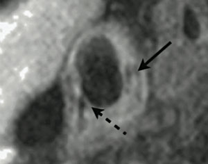
Figure 1. 3T magnetic resonance imaging (MRI) of atherosclerotic plaque in a right common carotid artery. The vessel wall is lined with complicated, lipid-rich plaque, which has a necrotic core (solid arrow). A thin fibrous cap can be seen in the bottom-left of the image (dashed arrow)
Magnetic resonance arteriography (MRA) has for many years been used as a non-invasive means of producing an arterial lumenogram, an image of flow down the arterial lumen, from which the presence of significant stenosis could often be detected, if needed, by comparison to the comparatively normal flow in the contralateral vessel. However, the first description of the use of magnetic resonance imaging (MRI) to describe direct imaging of atherosclerotic plaque itself was by Gold in 1993.1 The authors used ex vivo human aorta specimens containing atheroma to correlate MRI signal changes with the presence of specific histological features, including lipid-rich necrotic core, calcification, and fibrous plaque. Following this, the first in vivo description of MRI plaque imaging was performed in 1996 on six patients prior to carotid endarterectomy.2 Histological verification of these images with the carotid endarterectomy specimens clarified that MRI was capable of distinguishing a number of plaque features, including lipid cores, fibrous caps, calcification, haemorrhage and acute thrombosis, in addition to the normal media and adventitia of the vessel wall (figure 1). This discovery stimulated a large amount of subsequent research into carotid plaque MRI,3-6 ultimately leading to a modification of the American Heart Association (AHA) plaque classification system to allow MRI grading of carotid atherosclerotic plaque.7 More recent work has begun to move towards potential clinical uses of carotid MRI imaging. For example, Yuan et al. have described the positive association between identification of a ruptured fibrous cap on MRI, and a recent history of transient ischaemic attack (TIA) or stroke.3 In prospective studies, the presence of intraplaque haemorrhage on carotid MRI has been shown to predict the short-term combined risk of ipsilateral TIA and stroke,8 and, in addition, the chances of cerebral embolisation during surgery.8 Although larger long-term clinical studies are required, these studies indicate that carotid plaque MRI may have an important future role in the assessment and treatment of TIA and stroke.
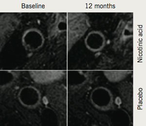
Figure 2. Effect of nicotinic acid on the carotid artery. Nicotinic acid is seen to prevent progression of carotid arterial wall thickening, while patients treated with placebo show clear progression of wall thickness
In addition to its potential clinical applications, the ability of MRI to provide highly accurate information on plaque burden and morphology has been used to examine the effect of both established and novel pharmacotherapies on the vasculature in the research and clinical trial domains. Corti et al. were the first to demonstrate a reduction in carotid and aortic atherosclerosis using serial MRI after 12 months of statin treatment.9 Subsequently, it was demonstrated that more intensive lipid lowering, to low-density lipoprotein (LDL)-cholesterol <100 mg/dL, was associated with a larger decrease in plaque size, also over 12 months.10 MRI has also been used to provide further mechanistic insights into how atherosclerotic plaque responds to cholesterol-altering medications. Lee et al. used MRI to demonstrate, in the carotid arteries and aorta, reduction in the plaque index (normalised vessel wall area) as early as three months after statin initiation. In the same patients, early changes in atherosclerosis (within three months) were significantly correlated with later change at 12 months.11 More recently, MRI has been used to investigate the effects of various treatment strategies, including the use of niacin to increase levels of high-density lipoprotein (HDL) (figure 2).12 As a result of these successes, MRI of atherosclerotic plaque is currently being used in phase III trials of novel therapeutic agents.
Coronary MRI
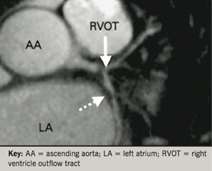
Figure 3. Magnetic resonance arteriography (MRA) of the left main stem, showing two separate plaques, which are seen as dark areas on the bright-blood signal (arrows). Adapted from Kim et al.13
MRI of the coronary arteries has progressed relatively more slowly. Not only are the coronary arteries small, but they are in constant motion and have similar characteristics to the surrounding myocardium and cardiac veins. Nonetheless, MRA of the coronary arteries, allowing the detection of stenotic plaque disease, has now been performed for over a decade, and was originally shown to be able to reliably detect patients with left main coronary or three-vessel disease (figure 3).13 Subsequently, in an attempt to improve acquisition times, whole heart magnetic resonance (MR) coronary angiography has been developed.14 This allows free-breathing images to be taken during a patient-specific time window of the cardiac cycle during which coronary artery motion is minimal. In a manner analogous to coronary computed tomography (CT), the upper and lower boundaries of the heart are defined in order to allow imaging over a single volume that covers both coronary arteries. As a result, images are simpler to acquire, and the scan can be performed in a much shorter time period. Using this technique, Sakuma et al. demonstrated that significant narrowing of coronary arterial segments with a diameter >2 mm could be detected with moderate sensitivity (82%) and high specificity (90%).15 However, despite technological advances such as this, the overall acquisition speed of 3D MRA remains considerably slower (>2 minutes) than that of multi-slice CT (<2 seconds). Nonetheless, it should be noted that MR coronary imaging does hold several advantages over CT plaque imaging: the ability to safely perform serial imaging, the lack of need for an injected contrast agent in image acquisition, and the ability to characterise heavily calcified areas of the coronary tree.16 Currently, however, clinical guidelines only recommend the use of coronary MRA for determining the proximal course of anomalous coronary arteries.
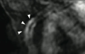
Figure 4. T1-weighted imaging of the right coronary artery wall. Arrows show bright signal from a recently symptomatic plaque in the right coronary artery. Adapted from Tanaka et al.22
More recent work has suggested a brighter future for MRI imaging of coronary atherosclerotic plaque. In addition to coronary MRA, black-blood imaging of the coronary artery wall is now possible, which allows detection of increased wall thickness in patients with angiographically documented coronary artery disease with high reproducibility.17,18 In a recent sub-study of the Multi-Ethnic Study of Atherosclerosis (MESA) trial, 179 asymptomatic patients with subclinical atherosclerosis underwent coronary wall MRI.19 Although no significant coronary artery narrowing was detected by MRA, direct wall imaging noted that as the arterial wall became thicker, the luminal diameter remained relatively constant; in contrast, the outer vessel wall was seen to expand with the thickened arterial wall. This phenomenon, termed “positive remodelling” and first described by Glagov,20 highlights the need to develop accurate methods of direct coronary plaque imaging using MRI, in a similar manner to those methods currently used for carotid plaque. Currently, non-contrast T1-weighted imaging of coronary plaque can identify high-intensity signal (HIP) (figure 4), which has recently been associated with positive coronary remodelling and low CT signal density.21 However, the prognostic significance of HIP lesions is currently unknown.22 Compared with carotid imaging, MRI of coronary atherosclerotic plaque is currently unable to identify individual plaque features with the same sensitivity and specificity.
Future directions
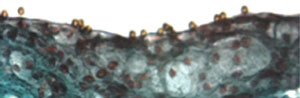
Figure 5. Microparticles of iron oxide (MPIO) bound to the aortic root of apolipoprotein E knockout mice. These particles have a paramagnetic effect, which leads to signal dropout when imaged using MRI
Ongoing improvements in imaging sequences, combined with higher field strength imaging, are likely to enhance the ability of MRI to provide high-resolution plaque images while simultaneously decreasing scan acquisition times. In addition, several novel contrast agents are under investigation that may permit MRI to simultaneously provide information on both vascular biology and morphology. For example, McAteer et al. constructed a dual-ligand microparticle of iron-oxide (4.5 µm diameter) to target endothelial P-selectin and vascular cell adhesion molecule (VCAM)-1 (figure 5).23 Binding of the microparticles to inflamed mouse endothelium was subsequently shown by high resolution ex vivo MRI (9.4 T). In human work, Tang et al. have demonstrated the use of ultrasmall particles of iron oxide (USPIO) to provide information on carotid plaque macrophage content using 1.5 T MRI.24
Conclusions
MRI is emerging as a leading non-invasive modality for the assessment of atherosclerotic plaque disease. While MRI of atherosclerotic plaque in the coronary arteries awaits further technical developments, imaging of plaque in the carotid arteries has already shown that MRI is capable of giving detailed information both on vessel wall thickness and plaque composition. With the ongoing development of novel contrast agents, MRI is likely to play a major role in the future development of imaging strategies to aid risk stratification and treatment in ischaemic vascular disease.
Conflict of interest
None declared.
Key messages
Magnetic resonance imaging (MRI) is one of the leading non-invasive plaque imaging modalities
Detailed images are available for carotid artery plaque and there is excellent promise for tissue characterisation
The absence of radiation exposure permits serial studies of plaque over time
There is the potential for a “one-stop shop” able to provide every facet of cardiovascular imaging in one scan
Routine coronary imaging will require further improvements in technology and resolution
References
Gold GE, Pauly JM, Glover GH et al. Characterization of atherosclerosis with a 1.5-T imaging system. J Magn Reson Imaging 1993;3:399–407.
Toussaint JF, LaMuraglia GM, Southern JF et al. Magnetic resonance images lipid, fibrous, calcified, hemorrhagic, and thrombotic components of human atherosclerosis in vivo. Circulation 1996;94:932–8.
Yuan C, Zhang S-X, Polissar NL et al. Identification of fibrous cap rupture with magnetic resonance imaging is highly associated with recent transient ischemic attack or stroke. Circulation 2002;105:181–5.
Yuan C, Mitsumori LM, Ferguson MS et al. In vivo accuracy of multispectral magnetic resonance imaging for identifying lipid-rich necrotic cores and intraplaque hemorrhage in advanced human carotid plaques. Circulation 2001;104:2051–6.
Shinnar M, Fallon J, Wehrli S et al. The diagnostic accuracy of ex vivo MRI for human atherosclerotic plaque characterization. Arterioscler Thromb Vasc Biol 1999;19:2756.
Hatsukami TS, Ross R, Polissar NL, Yuan C. Visualization of fibrous cap thickness and rupture in human atherosclerotic carotid plaque in vivo with high-resolution magnetic resonance imaging. Circulation 2000;102:959–64.
Cai J-M, Hatsukami TS, Ferguson MS et al. Classification of human carotid atherosclerotic lesions with in vivo multicontrast magnetic resonance imaging. Circulation 2002;106:1368–73.
Altaf N, MacSweeney ST, Gladman J, Auer DP. Carotid intraplaque hemorrhage predicts recurrent symptoms in patients with high-grade carotid stenosis. Stroke 2007;38:1633–5.
Corti R, Fayad Z, Fuster V et al. Effects of lipid-lowering by simvastatin on human atherosclerotic lesions: a longitudinal study by high-resolution, noninvasive magnetic resonance imaging. Circulation 2001;104:249–52.
Corti R, Fuster V, Fayad ZA et al. Lipid lowering by simvastatin induces regression of human atherosclerotic lesions: two years” follow-up by high-resolution noninvasive magnetic resonance imaging. Circulation 2002;106:2884.
Lee JMS, Wiesmann F, Shirodaria C et al. Early changes in arterial structure and function following statin initiation: quantification by magnetic resonance imaging. Atherosclerosis 2008;197:951–8.
. Lee JM, Robson MD, Yu LM et al. Effects of high-dose modified-release nicotinic acid on atherosclerosis and vascular function: a randomized, placebo-controlled, magnetic resonance imaging study. J Am Coll Cardiol 2009;54:1787–94.
. Kim W, Danias P, Stuber M et al. Coronary magnetic resonance angiography for the detection of coronary stenoses. N Engl J Med 2001;345:1863.
. Stehning C, Boernert P, Nehrke K. Advances in coronary MRA from vessel wall to whole heart imaging. Magn Reson Med Sci 2007;6:157–70.
Sakuma H, Ichikawa Y, Chino S et al. Detection of coronary artery stenosis with whole-heart coronary magnetic resonance angiography. J Am Coll Cardiol 2006;48:1946–50.
. Liu X, Zhao X, Huang J et al. Comparison of 3D free-breathing coronary MR angiography and 64-MDCT angiography for detection of coronary stenosis in patients with high calcium scores. AJR Am J Roentgenol 2007;189:1326–32.
Fayad ZA, Fuster V, Fallon JT et al. Noninvasive in vivo human coronary artery lumen and wall imaging using black-blood magnetic resonance imaging. Circulation 2000;102:506–10.
Botnar RM, Perez AS, Witte S et al. In vivo molecular imaging of acute and subacute thrombosis using a fibrin-binding magnetic resonance imaging contrast agent. Circulation 2004;109:2023–9.
Miao C, Chen S, Macedo R et al. Positive remodeling of the coronary arteries detected by magnetic resonance imaging in an asymptomatic population: MESA (multi-ethnic study of atherosclerosis). J Am Coll Cardiol 2009;53:1708–15.
Glagov S, Weisenberg E, Zarins C et al. Compensatory enlargement of human atherosclerotic coronary arteries. N Engl J Med 1987;316:1371–5.
Kawasaki T, Koga S, Koga N et al. Characterization of hyperintense plaque with noncontrast T(1)-weighted cardiac magnetic resonance coronary plaque imaging: comparison with multislice computed tomography and intravascular ultrasound. JACC Cardiovasc Imaging 2009;2:720–8.
Tanaka A, Kawasaki T, Noguchi T et al. Hyperintense plaque with noncontrast t1-weighted magnetic resonance coronary plaque imaging leading to acute coronary syndrome. Circulation 2009;120:2400–01.
McAteer MA, Schneider JE, Ali ZA et al. Magnetic resonance imaging of endothelial adhesion molecules in mouse atherosclerosis using dual-targeted microparticles of iron oxide. Arterioscler Thromb 2007;28:77–83.
Tang TY, Howarth SP, Miller SR et al. The ATHEROMA (atorvastatin therapy: effects on reduction of macrophage activity) study: evaluation using ultrasmall superparamagnetic iron oxide-enhanced magnetic resonance imaging in carotid disease. J Am Coll Cardiol 2009;53:2039–50.
You are not logged in
You need to be a member to print this page.
Find out more about our membership benefits