Hypertension, LVH, echo…and stuff

Br J Cardiol 2015;22:65–6 5 Comments
Click any image to enlarge
Authors:

In this regular series ‘ECGs for the fainthearted’ Dr Heather Wetherell will be interpreting ECGs in a non-threatening and simple way. In this issue, we apply the knowledge gained so far to a situation that may be encountered in general practice.

A 56-year-old woman attends for a well-woman blood pressure (BP) check. After three repeated measurements, a minute apart, her mean BP is 146/90 mmHg so the practice nurse decides to do an electrocardiogram (ECG), in accordance with the practice protocol. Figure 1 shows the ECG obtained.
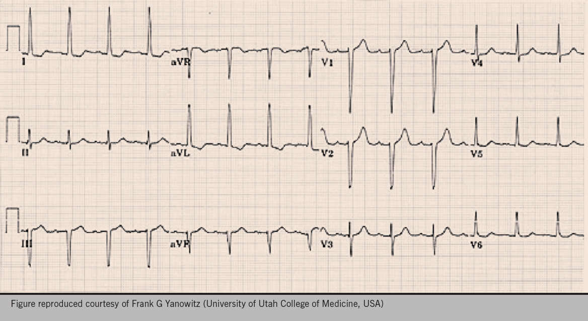
Figure 1. The electrocardiogram obtained during the well-woman check

What is your impression?

Does she need any further investigations?

Discussion

The main purpose of showing this ECG is to discuss the various voltage criteria for left ventricular hypertrophy (LVH).

Interestingly, there is no one universally agreed criteria for diagnosis of LVH by ECG.  A few of the more commonly accepted ones are listed below.

This ECG is compatible with LVH in that it meets the voltage criteria for LVH, in leads I and aVL.

As no other criteria for LVH are met, it is often better to say “This ECG meets the voltage criteria for LVH”, rather than diagnose hypertrophy. Don’t forget, LVH can only be truly diagnosed on scanned images, such as an echocardiogram (echo).

On crude eye-balling, the axis on this ECG is around -30 degrees (-30 degrees falls within normal axis parameters). So whilst we have possible borderline left axis deviation (LAD), it’s probably not significant without a predominantly negative deflection in lead II as well.

The ECG also shows T-wave inversion in lead I and aVL. When seen together, a leftward axis and T wave inversion, would further support the diagnosis of LVH and strain (see below).

In accordance with latest National Institute for Health and Care Excellence (NICE) Hypertension Guidelines,1 you may want to offer this woman ambulatory BP monitoring (ABPM), or home BP monitoring (HBPM), for confirmation of diagnosis.

Her cardiovascular disease risk and any target organ damage should also be assessed. Urinalysis should be checked for proteinuria, and bloods for underlying causes, or evidence of target organ damage (including U&E’s, and glucose).

Regardless of whether she turns out to have stage 1 or stage 2 hypertension, on the basis of her ABPM,1 her LVH will certainly sway you towards a lower threshold to treat.

The clinic BP described for this patient is surprisingly low for someone with LVH and strain. In this situation, ABPM may demonstrate higher pressures than seen in clinic – known as ‘masked hypertension’ (a condition less recognised and less discussed than ‘white coat hypertension’) − and that would reinforce need for antihypertensive treatment. More commonly, significant obesity can add to the effects of hypertension so an assessment of weight and lifestyle is also appropriate.

LVH

LVH is defined as an increase in the mass of the left ventricle, which can be secondary to an increase in wall thickness, an increase in cavity size, or both. LVH as a consequence of hypertension usually presents with an increase in wall thickness, with or without an increase in cavity size. Accurate assessment thus requires imaging with either echo or, even better, cardiac magnetic resonance. Various ‘voltage criteria’ seen on ECG, however, can be suggestive of LVH.

The left ventricle is represented by leads v5 and v6. 

So in LVH, we expect the ‘R’ waveforms to be enlarged/tall in these leads (conversely, we expect the ‘S’ waves to be deep in the right ventricular leads, that is V1 and V2).

Suggested voltage criteria for LVH include:

  • The sum of the S wave in v1 or v2, PLUS the R wave in v5 or 6 ≥35 mm

or

  • The sum of the deepest S wave + the tallest R wave >40 m
  • Any single, R or S, wave in leads v1−v6  ≥45 mm
  • The R wave in aVL  ≥11 mm
  • The R wave in lead I ≥12 mm
  • The R wave in aVF ≥20 mm

The predictive value of the voltage criteria is cumulative. i.e. the more voltage criteria met, the greater the likelihood of LVH.

An ECG diagnosis of LVH is also more secure when there are associated ST/T wave changes rather than voltage criteria alone, as in the example above.

In the ECG shown in figure 1, the precordial leads do not meet the voltage criteria for LVH (arguably borderline, at 35 mm).

Many clinicians are aware of the R and S wave criteria in the precordial (chest) leads, but this can be dependent on body habitus. The chest leads placed on a tall, thin man, with little adipose tissue, are in close proximity to the heart so naturally, the waveform amplitude in the chest leads will be large. The limb leads, being less affected by body habitus, are often more reliable in these individuals.

To echo or not to echo?

One of the most common questions I’m asked by GP colleagues when they see an ECG which is reported as ‘LVH’ is “do they need to request an echo?”.

Generally speaking, unless it is likely to change your management plan, an echo is not required to confirm a diagnosis of LVH. 

If a patient who is hypertensive is found to have LVH criteria on ECG, then an echo is only helpful if it will alter your threshold for treatment. If you plan to treat anyway, an echo is of dubious further value. (See 2011 NICE CG1271 for care management pathways).

As outlined above, voltage criteria for LVH in V1−V6 alone, in a tall thin person without other cause for concern, is not necessarily indicative of LVH and does not need an echo. Conversely, in an obese patient, the body fat between the ECG lead and the heart will result in a lower voltage in the precordial leads…and that may be the explanation in this case.

So remember − don’t just look at the chest leads! 

Likewise, with an ECG suggestive of LVH in a normotensive person, an echo would only be indicated if:

a) body habitus didn’t explain the amplitude, or

b) there were other signs/symptoms of concern (possible HOCM [hypertrophic myopathy]? or aortic stenosis?).

In such cases, there would often be other suspicious features on the ECG (such as T wave changes) or symptoms of possible cardiac origin. Sinister underlying causes are less likely on the basis of simple voltage criteria for LVH alone.

Strain pattern

When LVH is associated with other pathology, such as hypertension or aortic stenosis, a ‘strain pattern’ is often seen:

  • ST depression + flipped asymmetric T wave
  • ST elevation + upright asymmetric T wave
  • The strain pattern is greatest in the lead with the tallest/deepest QRS complex (seen in Lead I and aVL in the example above).

Simple isn’t it?

I hope this very simplified take on LVH helps clear up some of the conundrums around LVH, echo and hypertension – for GPs at least.

References

1. National Institute for Health and Care Excellence. Hypertension: clinical management of primary hypertension in adults. Clinical guideline 127. London: NICE, 2013. http://www.nice.org.uk/guidance/cg127

The interview

Br J Cardiol 2015;22:63–4 Leave a comment
Click any image to enlarge
Authors:

We continue our series in which Consultant Interventionist Dr Michael Norell takes a sideways look at life in the cath lab…and beyond. In this column, he advises on how to be the perfect candidate.

Dr Mike Norell
Dr Mike Norell

A senior trainee, who had taken an interest in an upcoming consultant post, sought advice from me as to how he might frame his application. In years gone by such a request would be expected only to receive an encouraging response along the lines of “I know the guys there; I’ll give them a ring”.

I doubt that such a supportive action − even if sincere − ever altered the final outcome of any consultant interview, and nowadays this type of communication runs contrary to good employment practice anyway. Over many years I have come to realise that the outcome of these events is down to two factors: you, and whether or not what you offer fits into the required role; essentially it’s “horses for courses”, as it were.

As I talked with our registrar about the philosophy involved, what might have been a cursory 30-second corridor chat ended up as an hour’s sermon and a later than usual trip home. I thought it might be useful to distil the elements of that conversation into this column so that readers in a similar position might − just might − benefit.

Top 10 tips

1. It’s not personal

So I present the 10 top tips for the consultant interview. These do not guarantee success; for the reasons above there may be factors outside your control that will determine the outcome. Nevertheless, you still have to do your utmost in order to give yourself the best chance; the rest is out of your hands. If unsuccessful, at least you will be satisfied that you could not have done more. Don’t take it personally; you were just not the right fit for the post.

2. To apply or not?

The job description will indicate the specific interests and activities of the unit and this knowledge might be enhanced by your own enquiries. You should craft your application to be relevant to the post on offer and indicate which of your attributes might be particularly valuable and suited to the department advertising the vacancy.

You should always apply for a job you want; never be put off by a locum in post or rumours that it is “sewn up”. I remember a world gone by of very senior registrars who were lined up for jobs they didn’t get.

The NHS application is not an easy document to complete. Still, you should deal with it diligently and avoid the perception that this is the same form you submit regardless of the nature of the post for which you are applying. You will still have the opportunity to present your well-constructed, comprehensive and more personalised CV when you visit − which brings us on to number three.

3. Should you visit before short listing?

I wonder how often I have been asked that question? My view is yes. Consultant posts used to be for the remainder of a doctor’s working life, although it is increasingly common to see ‘sideways movers’ nowadays, often after 10 years or so, as priorities, interests, aspirations and horizons inevitably alter (believe me).

Nevertheless, if you are genuinely interested in a position in which you might work for a good few years it seems appropriate, let alone sensible, to scope the job. It gives you the opportunity to meet key players and potential colleagues and thereby better orientate your approach to the job. It also serves as an opportunity to broaden your appreciation of the workings of a department other than your own, as well as non-clinical issues with which consultants must be familiar − particularly the dynamics of the Trust as a whole, not to mention the wider NHS.

Even if you end up withdrawing your application, the experience of visiting another unit and engaging with senior colleagues will be valuable when you are looking subsequently at a post that really whets your appetite.

4. Meet with senior managers

This is an important component of preparation enabling you to understand the threats to, and pressures upon, the Trust, as well as its aspirations and direction of travel. Much of the language and terminology used by managerial staff is not familiar, even to senior trainees. This type of meeting helps you to familiarise yourself with commonly-used terms so that you can begin to become fluent and are not surprised when they are used in an interview, for instance.

Meetings with the medical director, business manager and chief executive (CEO), should all be requested, although this often takes place after short listing. Meeting with these individuals should not be seen as box ticking and simply doing what you are supposed to. You should enter these discussions displaying genuine interest and with your own questions thought about beforehand and relevant to each interaction.

5. The interview

By the time you attend you should be utterly familiar with the post and how this appointment fits into the Trust’s strategic plan. On the panel will be clinicians, the CEO (or a deputy such as the medical director), a representative from the Royal College of Physicians (to ensure that you have the training background and credentials to be appointed) and a university representative who will focus on your research (if the post has academic involvement). It will be chaired sometimes by a lay individual but be aware that this list is not exhaustive; increasingly nursing and patient interests can also be represented at these interviews.

All the panel members will be introduced to you; take note of their designation and try not to look surprised!

6. Be honest…

The purpose of the interview is for the panel to get a complete picture of your experience, interests and aspirations, and see how well you would fit into the post on offer. Your job is to ensure that all this information comes across. You cannot pretend to be something you’re not, so don’t try to con anyone − for two reasons: firstly, it will be obvious and, secondly, why would you wish to take on a role for which you are not suited?

7. …objective

It is not a viva; there is usually no right or wrong answer to many – if not most – of the questions that will be posed. The important thing is to at least have a view and express it, acknowledging that there are usually a minimum of two sides to any issue, and therefore for you to be aware of other opinions. Provocative examples might be, “Do you think there is any utility in having MDT meetings?” or “Where have we gone wrong in the training of cardiologists?” Either way, pause and consider your reply; remember that there maybe someone on the panel who is evangelical about MDTs, or has spent most of their working life on training committees.

8. …and interesting

Some interviews are preceded by a slide presentation, the brief of which can itself be an attempt to discriminate between candidates. How applicants interpret a title such as “What do I do when not at work?” or present a talk on “What would I have done instead of cardiology?” is quite informative. It is a lot more interesting than the panel hearing five or six very similar versions of “How would I construct a cardiac department to deliver 21st century care?” or “What are the main issues threatening the sustainability and foundations of the NHS?”

9. Prepare…

You cannot revise for an interview but you can prepare for it. You can expect certain predictable questions that might be phrased differently but are still structured to get your familiarity with subjects such as clinical governance (“What do you understand by it?”), audit (“What is the most recent audit you have been involved with?”) and probity (“You notice that a colleague often smells of alcohol; what do you do?”).

Don’t panic or rush to answer; pause, take a breath and think. Often common sense will guide you and when all else fails, it is a safe bet to suggest that you would seek advice from a senior and more experienced colleague (unless he is the one with the alcohol problem!).

In answer to the question “What makes you special for this post?”, be prepared to highlight those sellable aspects that make you standout above the other candidates. 2,000 first operator cases, 20 first author publications and a passion for hard work, counts for nothing; the others will all have that. Mention that of course, but also emphasise elements that are unique to you and make you the right fit for the position. And be ready for the converse question and to identify any weaknesses or areas in which you might seek improvement; that will demonstrate insight and a willingness to learn, fit in and adapt.

10. …and practice

Go through typical interview questions with your supervisors and other colleagues beforehand so that you will learn to control any nerves and to come across as confident (but not forceful), considered (but not vague) and consultoid.

And for goodness sake smile occasionally (if appropriate). At the end of the process you should feel that you have given your all and, regardless of the outcome, the experience will make you stronger for your next encounter.

Oh! And good luck!

In brief

Br J Cardiol 2015;22:61–2 Leave a comment
Click any image to enlarge
Authors:

News in brief from the world of cardiology

Cholesterol lowering significantly reduces stroke in the elderly

Screen shot 2015-05-27 at 12.30.44 Use of cholesterol lowering drugs (statins and fibrates) is associated with a one third lower risk of stroke in older adults without previous disease, finds a study published in the BMJ.

A research team based in France set out to determine the association between use of lipid-lowering drugs in healthy older people and long-term risk of coronary heart disease and stroke. They tracked 7,484 men and women (average age 74 years) with no known history of vascular events, such as heart attacks and strokes, living in three French cities (Bordeaux, Dijon and Montpellier).

After an average follow-up time of nine years, the researchers found that use of statins or fibrates was associated with a one third lower risk of stroke compared with non-users. But no association was found between lipid-lowering drug use and coronary heart disease.

This is an observational study, so no definitive conclusions can be drawn about cause and effect. Nevertheless, if replicated, the study results suggest that lipid-lowering drugs might be considered for the prevention of stroke in older populations, say the researchers. Further details available at: http://www.bmj.com/content/350/bmj.h2335

…and cardiorespiratory fitness may delay onset of high cholesterol

Men who have higher levels of cardiorespiratory fitness may delay by up to 15 years increases in blood cholesterol levels that commonly occur with ageing, according to new research from the University of South Carolina.

“Age-related changes in cholesterol levels are usually unfavourable,” said study investigator, Dr Xuemei Sui (Arnold School of Public Health, University of South Carolina, Colulmbia, USA). “Our study sought to determine how cardiorespiratory fitness might modify the ageing trajectory for lipid and lipoproteins in healthy men.”

Dr Sui and colleagues used data from the Aerobics Center Longitudinal Study to assess levels of total cholesterol, low-density lipoprotein (LDL) cholesterol, high-density lipoprotein (HDL) cholesterol, non-high-density lipoprotein cholesterol and triglycerides in a total of 11,418 individuals who were observed during health examinations between 1970 and 2006 at the Cooper Clinic, Dallas, USA. Cardiorespiratory fitness was measured using a treadmill test. After cardiorespiratory fitness levels were standardised for age, subjects were placed into low, middle and high fitness categories, and cholesterol and triglycerides were analysed after an overnight fast.

Researchers found that total cholesterol, LDL cholesterol and triglycerides all increased up to a certain age and then decreased while the inverse was true for HDL cholesterol. Men with lower cardiorespiratory fitness had a higher risk of developing high cholesterol in their early 30s while men with high fitness did not see this development until their mid-40s. Additionally, men with low cardiorespiratory fitness reached abnormal HDL and non-HDL cholesterol levels around their early 20s and mid-30s, respectively, while those with higher fitness saw normal amounts for the entire lifespan.

The authors conclude that promoting cardiorespiratory fitness levels may help delay the development of dyslipidaemia. The study is published in the Journal of the American College of Cardiology (doi: 10.1016/j.jacc.2015.03.517).

NOAC news

Apixaban

The National Institute for Health and Care Excellence (NICE) has issued Final Appraisal Determination (FAD) recommending the NOAC (non-vitamin K oral anticoagulant) apixaban (Eliquis®) as an option for the treatment and prevention of recurrent deep vein thrombosis (DVT) and/or pulmonary embolism (PE) in adults.

The Appraisal Committee concluded that apixaban could be considered a clinically and cost-effective use of NHS resources and recommended the drug, within its marketing authorisation. The NICE FAD recommendation for apixaban, whose licensed dose is lower for the prevention of recurrent DVT and PE than for initial treatment of DVT and PE, provides clinicians with an additional oral single-drug option without the need for injectable therapy or routine monitoring of exposure.

The Scottish Medicines Consortium (SMC) has also announced that they have accepted apixaban for use within NHS Scotland.

Edoxaban

The European Committee for Medicinal Products for Human Use (CHMP) has recommended approval of the NOAC edoxaban (Lixiana®) for the prevention of stroke and systemic embolism (SE) in adult patients with non-valvular atrial fibrillation (NVAF) with one or more risk factors. The CHMP also recommended approval of edoxaban for the treatment of DVT and PE, and prevention of recurrent DVT and PE in adults.

Rivaroxaban

Results from the VENTURE-AF trial were presented recently at the Heart Rhythm Society’s 36th Annual Scientific Sessions, in Boston, USA, and published simultaneously in the European Heart Journal (doi: 10.1093/eurheartj/ehv177).

In this study, the NOAC rivaroxaban (Xarelto®) was investigated as an alternative to dose-adjusted vitamin K antagonists (VKAs) to reduce the risk of stroke and SE in patients with NVAF undergoing catheter ablation.

Patients with AF scheduled for catheter ablation were randomly assigned in a 1:1 ratio to rivaroxaban 20 mg orally once-daily or to dose-adjusted VKA (target INR 2.0–3.0). In patients treated with rivaroxaban, there were no thromboembolic events. In those patients receiving a VKA, two thromboembolic events occurred. There was one major bleeding event in the VKA treatment arm versus none in the rivaroxaban arm. No major bleeding events occurred in either group and the incidence of non-major bleeding events and procedure-attributable events was low for both treatment arms.

Increased MI risk in rheumatoid arthritis

Patients with rheumatoid arthritis are at increased risk of unsuspected myocardial infarction (MI), according to new research presented by Dr Adriana Puente (National Medical Centre, Mexico City, Mexico). Risk was increased even when patients had no symptoms and was independent of traditional cardiovascular risk factors such as smoking and diabetes.

Speaking at ICNC 12, a meeting organised by the Nuclear Cardiology and Cardiac CT section of the European Association of Cardiovascular Imaging, Dr Puente said: “Our study suggests that one quarter of patients with rheumatoid arthritis and no symptoms of heart disease could have a heart attack without prior warning”.

He concluded: “rheumatoid arthritis affects 1.6% of the general population. The condition nearly doubles the risk of a heart attack but most patients never knew they had heart disease and were never alerted about their cardiovascular risk”.

No excess in heart failure with sitagliptin

Initial positive findings have been reported from TECOS (Trial Evaluating Cardiovascular Outcomes with Sitagliptin) of the DPP-4 inhibitor sitagliptin (Januvia®). TECOS was an event-driven trial conducted in adults with type 2 diabetes and a history of cardiovascular (CV) disease. The study was designed to assess the cardiovascular safety of long-term treatment with sitagliptin as part of usual diabetes care compared with usual care without sitagliptin.

The primary end point was the composite of time to the first of any of the following confirmed events: CV-related death, non-fatal myocardial infarction, non-fatal stroke, or unstable angina requiring hospitalisation. The trial achieved its primary end point of non-inferiority for the composite CV end point. Among secondary endpoints, there was no increase in hospitalisation for heart failure in the sitagliptin group versus placebo.

The TECOS CV safety trial was led by an independent academic research collaboration between the University of Oxford Diabetes Trials Unit and the Duke University Clinical Research Institute. Complete results will be presented at the 75th Scientific Sessions of the American Diabetes Association.

News from the UK Stroke Forum

24911707_s copy

The association between obstructive sleep apnoea (OSA) and neurovascular disease was discussed by Dr David Hargroves (British Association of Stroke Physicians Education and Training Chair) and colleagues from East Kent during a training day at the recent UK Stroke Forum conference.

Dr Hargroves presented previously published data which shows that OSA is not uncommon: 24% of men and 9% of women in the general population may have OSA, 3−4% with clinically ‘obvious’ sequelae, and 60% of older and obese people may have OSA. Clinicians treating patients with neurological presentations should have a high index of suspicion, with clinical history taking being very important, as always. Patients with excessive daytime sleepiness with an Epworth sleepiness score of more than 12 but also evidence of nocturnal sympathetic over activity (sweating, thirst, dry mouth, nocturia etc…) without day-time somnolence should be investigated and have OSA excluded with a formal sleep study, he said.

More reports from the conference can be found online at www.bjcardio.co.uk. These include reports on the association between high blood pressure, stroke and the use of glyceryl trinitrate to the management of atrial fibrillation and the prevention of stroke.

BSH Training Day

A report from the 7th British Society for Heart Failure revalidation and training day is available online at www.bjcardio.co.uk. The day included thought-provoking case-based discussions with stimulating lectures on both the latest developments in advanced heart failure care and the visions of the future in a dynamic and progressive field.

The prevalence of heart failure is expected to increase over the coming years and the meeting highlighted the challenges that will need to be overcome in terms of both advancing medical therapies and service development.

Book review

Br J Cardiol 2015;22:67–8 Leave a comment
Click any image to enlarge
Authors:

Essential revision notes for cardiology KBA

Abedin coverAuthor: Khavandi A
Publisher: Oxford University Press, 2014
ISBN: 978-0-19-965490-1, Price: £59.99

The authors have been very successful at publishing the first knowledge-based assessment (KBA) revision aid – a well structured book which, over 32 chapters, comprehensively provides an excellent overview of the UK national curriculum and closely follows the European curriculum, making it an ideal preparation tool for the European Exam in Cardiology. The wealth of information in each chapter makes it relevant to both those in training but also consultants who may wish to use it as a quick reference book or to help with teaching. 

It has mostly been written by senior trainees and some new consultants, with input from specialists to deliver authoritative and assuring chapters, yet being sympathetic to the angle to which a trainee approaches cardiology. Chapters are bursting with diagrams, tables and electrocardiograms (ECGs), which make it light reading, and the bullet points allow concise information to be packed in, in a no-nonsense fashion. The text is easily digestible and the content up to date and accurate.  

This book bridges the gap between a large, highly detailed reference book, and a small pocket guide. It is an absolute must for those in training, and anyone interested in cardiology. 

Hannah ZR McConkey
Cardiology SpR
Oxford Deanery 

Inherited cardiac diseases

Editors: Elliott P, Lambiase PD, Kumar D
Publisher: Oxford University Press, 2011
ISBN: 978-0-19-955968-8 Price: £39.99

From the highly popular series of Oxford Specialist Handbooks in Cardiology, the first edition on the subject of inherited cardiac disease presents a concise and comprehensive overview of the conditions and issues encountered in the clinical practice of this subspecialty. Edited by leading experts in the field, with contributions from other consultant specialists and fellows, this book was written with the trainee in mind undertaking a rotation or specialising in this area. The insightful chapters on genetic counselling and point style management plans written for commonly encountered conditions in clinical practice also make this an indispensable pocket reference for specialists, nurses and counsellors. 

Arranged over 20 chapters, the first third of the book covers the fundamentals in genomics, common laboratory investigations, and pointers on genetic counselling, providing an appropriate introduction to this field of cardiovascular medicine. The following chapters on the inherited cardiac conditions each contain a wealth of information on pathophysiology, risk stratification and management, and are appropriately sub-headed to allow quick navigation and easy assimilation. The book finishes by including practical information on managing the cardiovascular manifestations of other inherited diseases, such as skeletal muscle disease, mitochondrial disease and metabolic disease, which are an important and easily neglected part of a trainee’s core knowledge.  

The authors have made an effort to include important clinical guidelines from Europe and North America, and kept the information current at the time of writing. Whilst new guidance and consensus documents have since been published (e.g in hypertrophic cardiomyopathy), this book’s contents and practical guidance still remain entirely relevant. Overall, the book is a worthy addition to the Oxford Specialist Handbooks series and is highly recommended to anyone with an interest in the inherited cardiac conditions. 

Kevin Leong
Cardiology Registrar

Fu Siong Ng
Clinical Lecturer in Cardiology

Imperial College London and Imperial College Healthcare NHS Trust, London

Oxford handbook of heart failure (second edition)

Authors: Gardner RS, McDonagh TA, Walker NL
Publisher: Oxford University Press, 2011
ISBN: 978-0-19-967415-2 Price: £39.99

‘Doing more with less’ is a concept few us can escape grappling with.  Remarkably, this handbook delivers. Despite being 10% shorter than the previous edition of this popular title within the Oxford Specialist Handbooks in Cardiology series, it continues to offer a comprehensive yet focused manual for all clinicians looking after patients with heart failure.

The structure of the book retains the logical approach of the first edition, dealing with pathophysiology, investigation and treatment, before moving on to a series of high-yield chapters targeting specific comorbidities. The authors have struck a good balance between describing management and facilitating more in-depth understanding. Further sections on procedures and the evidence base underlying specific interventions are equally valuable whether read in their entirety or dipped into. Chapters dealing with specific therapeutic agents cater for all, blending a useful summary of landmark randomised controlled trials (RCTs) with practical dosing suggestions.

Increased coverage of mechanical circulatory support (now meriting its own chapter) is particularly welcome and offers an accessible summary of the area for those of us not regularly involved with advanced heart failure. A second new chapter provides a valuable summary of the complexities of right ventricular dysfunction with a helpful division of management strategies by pathophysiology. Style, language and formatting follow the tried and tested Oxford Specialist Handbook template, with helpful cross-references to other areas of the book and candid ‘bomb sign’ annotation of controversial topics. Where uncertainty in the evidence base exists, for example in the heart failure with preserved ejection fraction (HF-PEF) chapter, the authors have transparently reminded readers at the start of the section.

As the prevalence of heart failure increases, more of us will encounter more patients with heart failure. The national audit for heart failure shows that specialist input has been associated with dramatically improved outcomes. Future therapeutic innovations are likely to be hard-won incremental advances, making it all the more important that we offer existing evidence-based practice to all patients who might benefit. The author’s preface to this edition aspires to improve outcomes for our patients and I believe this book is a useful tool to achieve this.

It is one thing to know “what to do” but another to know “why to do it,” and it is a real achievement to discuss both usefully within 500 pages. Writing a book that is accessible to all but maintains the interest of specialists is a tall order. The authors should be congratulated on a concise, balanced and well-pitched offering.

Graham Cole
Clinical Research Fellow
Imperial College London

The new NICE AF guideline and NOACs: safety first or safety last?

Br J Cardiol 2015;22:50–2doi:10.5837/bjc.2015.018 Leave a comment
Click any image to enlarge
Authors:

A non-fatal myocardial infarction (MI) is an inconvenience; a non-fatal stroke is a catastrophe. While this is a simplification, it draws attention to the fact that most patients with a non-ST-segment elevation MI (NSTEMI) do quite well. Conversely, most patients who suffer a non-fatal stroke suffer a massive change to their lives and their families’ lives, and experience a devastating change in their quality of life.

Continue reading The new NICE AF guideline and NOACs: safety first or safety last?

The new NICE AF guideline and NOACs: a response

Br J Cardiol 2015;22:53–5doi:10.5837/bjc.2015.019 Leave a comment
Click any image to enlarge
Authors:

We share Professor Brady et al.’s opinion1 that stroke prevention is the single greatest priority in the management of patients with atrial fibrillation (AF). It is reasonable to say that highlighting the inappropriately low levels of anticoagulant uptake as a major public health issue and seeking to improve anticoagulant uptake nationwide was uppermost in the minds of the Guideline Development Group (GDG) members.

We believe that the new guideline2 will be a major advance in stroke prevention in AF. We would suggest that Professor Brady and colleagues, in their focus on non-vitamin K oral anticoagulants (NOACs), have overlooked the importance of a number of crucial aspects of the guideline.

  • It represents a paradigm change in stroke management. The GDG were very keen to promote the concept that, whereas previously risk assessment was undertaken to define patients at high risk of stroke requiring anticoagulation, under the new guideline anticoagulation has become the norm for all but the lowest-risk patients.
  • It represents a considerable simplification in the approach to stroke prevention. The essential information on stroke prevention can be summarised in a single, relatively simple algorithm (figure 1).3
  • It has removed the confounding issue of aspirin. In many patients, particularly the elderly, aspirin has been a barrier to more effective stroke prevention.4
  • It promotes, for the first time, consideration of quality of anticoagulation among patients receiving vitamin K antagonists with specific recommendations for review of time in therapeutic range (TTR) and for consideration of alternative therapies, including NOACs, for patients with an inadequate TTR.
  • It promotes annual review of stroke and bleeding risk in all patients with AF.
  • It promotes equality of access to vitamin K antagonists and NOACs.
Figure 1. Simple algorithm for stroke prevention in people with non-valvular atrial fibrillation (AF)[3]

Professor Brady and colleagues have focused on the issue of NOACs. It is a principle of all National Institute for Health and Care Excellence (NICE) guidelines, and one understood by all GDG members, that they should not duplicate or seek to re-interpret existing NICE appraisals. In this case, a single technology appraisal on dabigatran5 had been published a matter of months before the commencement of work on the AF guideline and appraisals on rivaroxaban6 and apixaban7 were, at that time, ongoing. It was always the case that these individual technology appraisals would be incorporated directly into the new guideline without reassessment and without revision.

The brief of the GDG, therefore, was integration of the existing technology appraisals into the new guideline, with the aim of evidence-based, personalised decisions for stroke prevention. This is fundamentally different from didactic statements advocating an overly simplistic change in clinical practice at the level of the population. Professor Brady et al. have focused on the recent Ruff et al. meta analysis.8 As the accompanying editorial pointed out, the individual NOACs are not the same, nor are the trials homogeneous and “ultimately the drug could be fitted to the patient, or the patient to the drug, dependent on a focus on safety or efficacy, and on other patient factors…”.9

The GDG were cognisant of problems nationally in relation to the use of NOACs, and the fact that a number of commissioning groups had constructed local algorithms, at variance with the NICE technology appraisals, limiting their use. With this in mind, the GDG made the clear statement and recommendation: “Anticoagulation may be with apixaban, dabigatran etexilate, rivaroxaban or a vitamin K antagonist. Discuss the options for anticoagulation with the person and base the choice on their clinical features and preferences”.2

Equality of access to NOACs is made equally clear in the stroke prevention algorithm (figure 1). The GDG hoped that the promotion of equality of access would signal an end to local commissioning pathways, where these are at variance with existing NICE guidance in requiring patients to show poor control on a vitamin K antagonist before a NOAC can be considered.

NICE has been particularly concerned about the variable uptake of NOACs geographically following publication of the technology appraisals on dabigatran, rivaroxaban and apixaban, and the first task of the NICE Implementation Collaborative (NIC) was to encourage implementation of the existing appraisals. NICE supported the Academy of Royal Medical Colleges to produce a consensus statement to support the guideline,10 and produced its first patient decision aid, relating to anticoagulation and stroke prevention.11 The aid, published alongside the guidance, helps both patients and their doctors to assess the risk and benefits of commencing anticoagulation. The aid additionally considers the relative merits of a vitamin K antagonist or a NOAC, providing a checklist for patients to consider the relative merits of each, including such factors as monitoring, convenience, bleeding risk, drug interactions, side effects and reversibility. Patients are encouraged to assess the importance of individual factors to assist in reaching a decision as to the type of agent they would prefer.

There is, of course, a limit to the extent of the information that can be conveyed in this manner. In some cases, healthcare professionals may need to help the patient to understand the issues; in others, health professionals may wish to expand on the information in the decision aid, for example considering the more specific risks of intracranial haemorrhage or gastrointestinal bleeding, as discussed in the Ruff et al. meta-analysis.8 Obviously, there will also be patients where one or other agent is specifically indicated on clinical grounds, for example in cases of renal impairment. Nonetheless, within this constraint, the emphasis of the patient decision aid, and, indeed, the emphasis of the AF guideline as a whole, is on equality of access, shared decision-making and patient choice.

NICE, with others, is facilitating the many discussions required to review and revise complex existing local arrangements for stroke prevention in people with AF. Its Implementation Consultants, Regional Technical Advisers and Medicines and Prescribing Centre Associates are all active in such conversations, and it is encouraging to see a number of Academic Health Science Networks prioritising this topic for local implementation.

While there is still work to do, early results are encouraging. The NICE guideline was published in June 2014. Figure 2 shows the latest available data for primary care prescribing in England for the three available NOACs for all indications, including AF.

Figure 2. Trends in prescribing of new oral anticoagulants (NOACs) on NHS prescriptionsin England
Figure 2. Trends in prescribing of new oral anticoagulants (NOACs) on NHS prescriptions
in England

Conclusion

In conclusion, the GDG shared Professor Brady et al.’s concern that stroke prevention should be paramount in AF management. We would contend that the new guideline is a major step forward in stroke prevention and changes the paradigm for AF management. The new guideline emphasises the role of NOACs and promotes equality of access with vitamin K antagonists. Early data indicate implementation support is proving beneficial. Above all, the new guideline promotes patient education, patient empowerment and informed patient choice in anticoagulant selection.

Conflict of interest

MF is an ad hoc advisor to Oberoi Consulting. His practice partnership has received funding from Abbott, Bayer, Boehringer-Ingelheim, Bristol Myers Squibb, Dawn, INRStar, Medtronic, Oberoi Consulting, Pfizer, Roche, Sanofi-Aventis, and Servier. CC and NM:
none declared.

References

1. Brady AJB, Connolly DT, Docherty A. The new NICE AF guideline and NOACs: safety first or safety last? Br J Cardiol 2015;15:2015 http://dx.doi.org/10.5837/bjc.2015.018

2. National Institute for Health and Care Excellence. Atrial fibrillation: the management of atrial fibrillation. CG180. London: NICE, June 2014. Available from: http://www.nice.org.uk/guidance/cg180

3. National Institute for Health and Care Excellence. Atrial fibrillation. The management of atrial fibrillation. Clinical guideline. Methods, evidence and recommendations. London: NICE, 2014. Available from: http://www.nice.org.uk/guidance/cg180/resources/cg180-atrial-fibrillation-update-full-guideline3

4. Cowan C, Healicon R, Robson I et al. The use of anticoagulants in the management of atrial fibrillation among general practices in England. Heart 2013;99:1166–72. http://dx.doi.org/10.1136/heartjnl-2012-303472

5. National Institute for Health and Care Excellence. Dabigatran etexilate for the prevention of stroke and systemic embolism in atrial fibrillation. TA249. London: NICE, March 2012. Available from: http://www.nice.org.uk/guidance/ta249

6. National Institute for Health and Care Excellence. Rivaroxaban for the prevention of stroke and systemic embolism in people with atrial fibrillation. TA256. London: NICE, May 2012. Available from: http://www.nice.org.uk/guidance/ta256

7. National Institute for Health and Care Excellence. Apixaban for preventing stroke and systemic embolism in people with nonvalvular atrial fibrillation. TA275. London: NICE, February 2013. Available from: http://www.nice.org.uk/guidance/ta275

8. Ruff CT, Giugliano RP, Braunwald E et al. Comparison of the efficacy and safety of new oral anticoagulants with warfarin in patients with atrial fibrillation: a meta-analysis of randomised trials. Lancet 2014;383:955–62. http://dx.doi.org/10.1016/S0140-6736(13)62343-0

9. Larsen TB, Lip GYH. Warfarin or novel oral anticoagulants for atrial fibrillation. Comment. Lancet 2014;383:931–3. http://dx.doi.org/10.1016/S0140-6736(13)62376-4

10. NICE Implementation Collaborative. Consensus. Supporting local implementation of NICE guidance on use of novel (non-vitamin K antagonist) oral anticoagulants in non-valvular atrial fibrillation. London: NICE, June 2014. Available from: http://www.nice.org.uk/guidance/cg180/resources

11. National Institute for Health and Care Excellence. Patient Decision Aid. Atrial fibrillation: medicines to help reduce your risk of a stroke – what are the options? London: NICE, June 2014. Available from: http://www.nice.org.uk/guidance/cg180/resources

Introducing integrated care: potential impact on hospital cardiology clinic workload

Br J Cardiol 2015;22:75–7doi:10.5837/bjc.2015.020 Leave a comment
Click any image to enlarge
Authors:

Integration of healthcare services has been advocated to improve quality and cost-effectiveness. Different models of integrated care for cardiology have been suggested, but the cost-effectiveness of a consultant-run service has been questioned. We assessed the potential impact on secondary-care outpatient volumes of introducing a service run by GPs with a special interest, with support from consultant cardiologists. We retrospectively reviewed all cardiology outpatient attendances at the South London Healthcare NHS Trust for a period of three months in 2011. Using National Institute for Health and Care Excellence (NICE) guidelines and discussions between cardiologists and GPs, a novel outpatient referral triage protocol was drawn-up to decide the appropriate minimum level of care required for a range of cardiac conditions. Anonymised clinic letters were divided into new referrals and follow-ups, and were assessed to establish the diagnosis and clinical complexity. Implementing such an integrated community care service (ICC) would reduce new referrals to secondary care by 33%, and would enable transfer of 44% of patients currently followed up in secondary care to ICC. The study confirms that there is scope for significant transfer of care with the greatest gains in patients with valve disease, ischaemic heart disease and atrial fibrillation.

Introduction

Screen shot 2015-06-02 at 16.10.50The increasing burden on healthcare services, combined with effects of austerity, has placed the National Health Service (NHS) under pressure to achieve ever-greater efficiency, while improving the quality of service. An example of the increased demand is that the total number of hospital outpatient appointments in England has increased steadily by more than 3% per annum since 2011.1,2

The government, in the 2010 white paper, set out a redesigned pathway endorsing clinically led services based on more effective dialogue and communication between general practice and secondary care.3 Thus, integration of healthcare services has been advocated to improve patients’ care and cost-effectiveness.4 Integration may be defined as organisation of services within and between care sectors with the aim of enhancing quality of care.5

Different models of integrated care for cardiology have been suggested. However, the cost-effectiveness of a consultant-delivered service in the community has been questioned.6,7 Our aim was to assess the potential impact on secondary care outpatient volumes of a service run by GPs with specialist interest (GPwSIs) with support from consultant cardiologists.

Methods

We retrospectively reviewed clinic letters for all cardiology outpatient attendances, excluding rapid access chest pain clinics, for the two acute general hospitals of South London Healthcare NHS Trust (SLHT) for the period 1 August 2011 to 31 October 2011. Using National Institute for Health and Care Excellence (NICE) guidelines, and the results of discussions between consultant cardiologists and GPs, a novel outpatient referral triage protocol was drawn-up to decide the appropriate minimum level of care required for a range of cardiac conditions.

Anonymised patient letters were divided into new referrals and follow-ups, and were assessed by one person (AK) to establish the diagnosis and clinical complexity. New referrals were classified into groups according to the referring complaint, while follow-ups were classified according to their main diagnosis. This protocol gave three possible tiers of care for each condition/patient, namely secondary care, integrated community care (ICC) or GP-led care. In addition, for each outpatient visit we recorded whether the patient was discharged back to their GP or followed up.

GP-led care was considered suitable for low-risk conditions, i.e. those not requiring specialist investigations, or stable patients already on appropriate treatment. ICC, by a GPwSI, was defined as a service that would cover patients presenting with cardiac conditions not requiring urgent assessment, but still entailing a cardiology follow-up or further cardiac investigation. Secondary-care review would be reserved for patients with high-risk or complex conditions.

Results

There were 2,163 patient visits reviewed. There were 1,092 new referrals and 1,071 follow-ups.

New referrals

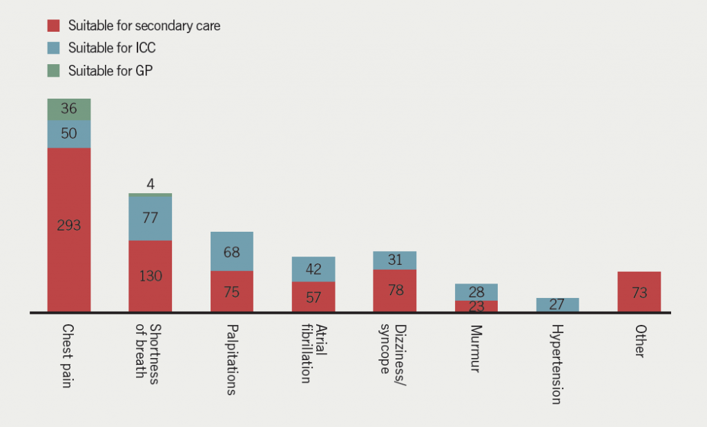
According to our referral triage protocol, 40 patients (4%) could have been treated by a GP, 323 patients (29%) by ICC and 729 patients (67%) would have required secondary care. Therefore, using an ICC model could reduce secondary-care referrals by 33%. The greatest percentage impact was in the murmur group (55% reduction), followed by the palpitations group (48% reduction), the atrial fibrillation (AF) group (42% reduction) and the shortness of breath (SOB) group (37% reduction) (figure 1).

Figure 1. Predicted impact of integrated care in the community (ICC) on new referrals to South London Hospital Trust (SLHT)

The outcome of the new referrals consultations were: 679 patients (62%) discharged back to their GP without secondary-care follow-up and 413 patients (38%) offered further appointments. The projected clinical outcome with the new protocol, suggests that 77 patients who were offered follow-up (7% of the initial total) could be followed up in an ICC setting, leaving only 31% of total new referrals being followed up in secondary care after the initial assessment.

Follow-ups

There were 1,071 patients seen as follow-ups in the cardiology service during this time period. Of these, 383 patients (36%) were discharged back to primary care and 688 (64%) were followed up. Predicted figures, assuming implementation of an ICC, project that the number of patients being followed up in secondary care would fall from the observed 688 to 384 (44% reduction). The greatest impact would be observed in the valve disease group (54% reduction). Figure 2 shows the predicted impact of implementing ICC for each diagnosis group.

Figure 2. Outcome of the followed-up patients when ICC implemented in SLHT

Discussion

Integration of healthcare services has been advocated to improve quality and cost-effectiveness. Integration may take different forms, but one model that has been suggested is for hospital consultants to carry out clinics in the community. Such an example of integrated care was reported by Knowsley PCT. Early reports of such service outcomes were positive, demonstrating a 10% reduction in unplanned hospital visits and a 12.6% decrease in short-stay admissions for cardiology-related events.8However, the value for money has been questioned,6 and our study examines the potential for improvements in efficiency by transferring services to primary care, supported by secondary care, rather than just putting consultants in a different setting. Our data demonstrate that 33% of new referrals to cardiology outpatient clinics, and 44% of patients currently seen as follow-ups in secondary care, could be seen in a GPwSI ICC service. The reduction in secondary care numbers is effected by providing the ICC and would not affect the usual primary care workload.

Targeted GPwSI clinics, with appropriate physiologist support, could provide a service for new patients with shortness of breath, murmurs and palpitations/AF, and for follow-up of valve disease, heart failure and AF, as well as chest pain and known coronary artery disease (CAD), taking significant numbers of patients out of secondary care. Clearly, good links would be required between primary and secondary care to enable patients to be fast-tracked to secondary or tertiary care once their clinical condition changed.

Integrated care is ultimately about achieving better care through better coordination,9 and current evidence suggests a potential to improve service quality and patient experience.10 However, there is a need for more solid evidence to decide how to develop this further, as longitudinal studies on the implementation of integrated care are scarce. Our study confirms that there is scope for significant transfer of care and indicates where the potential gains may be greatest.

Conflict of interest

None declared.

Key messages

  • Integrated cardiology care run by GPs with a special interest, with support from secondary care, would improve service quality
  • The study showed that the greatest gains in transferring care were in patients with valve disease, ischaemic heart disease and atrial fibrillation

Integrated care should be based on patient benefit: a GP comment

Screen shot 2015-05-13 at 17.27.21
Dr Terry McCormack (General Practitioner, North Yorkshire)

Getting the right patient to the right doctor has always been a major challenge for the NHS. Referral triage is becoming more common and is based on commissioners saving money for their local health economies. Providing an intermediate service is a half-way house in that the patient is directed to an alternative service rather than no service. Analysis of the impact on services and potential savings is to be welcomed, and Khwanda et al. provide such an analysis in this issue. The paper shows that the impact was mainly in taking pressure off the consultant service, rather than saving money. This was achieved particularly in valve disease, ischaemic heart disease and atrial fibrillation. Making an evaluation of the economic savings is complex and potentially difficult. Tariffs are simplistic and do not necessarily account for secondary costs, such as clinic support staff and investigation resources. Quality and patient outcomes are the most important measures. Is the service safe? What is the patient satisfaction? Who is responsible if things go wrong?

Only 4% were thought to be suitable to be treated by their GP, and this surprised me. I felt it would be more and I wonder if there were opportunities here for feedback to educate the GPs? It may reflect the bias of a paper with five cardiology authors and no primary care input.

Dr Terry McCormack
General Practitioner
Whitby, North Yorkshire

References

1. Health & Social Care Information Centre. Hospital Outpatient Activity 2012–13. London: HSCIC, 2013. Available from: http://www.hscic.gov.uk/catalogue/PUB13005/hosp-outp-acti-2012-13-summ-repo-rep.pdf

2. Health & Social Care Information Centre. Hospital Outpatient Activity 2011–12. London: HSCIC, 2012. Available from: http://www.hscic.gov.uk/catalogue/PUB09379

3. Department of Health. Equity and excellence: liberating the NHS. London: HMSO, July 2010; pp. 27–9. Available from: https://www.gov.uk/government/uploads/system/uploads/attachment_data/file/213823/dh_117794.pdf

4. Department of Health. High quality care for all. NHS next stage review final report. London: HMSO, 2008; pp. 65. Available from: https://www.gov.uk/government/uploads/system/uploads/attachment_data/file/228836/7432.pdf

5. Curry N, Ham C. Clinical and service integration. The route to improved outcomes. London: The King’s Fund, 2010: pp. 2. Available from: http://www.kingsfund.org.uk/publications/clinical-and-service-integration

6. Ham C, De Silva D. Integrating care and transforming community services: what works? Where next? Birmingham: Health Services Management Centre, 2009. Available from: http://www.birmingham.ac.uk/Documents/college-social-sciences/social-policy/HSMC/publications/PolicyPapers/Policy-paper-5.pdf

7. Sibbald B, McDonald R, Roland M. Shifting care from hospitals to the community: a review of the evidence on quality and efficiency. J Health Serv Res Policy 2007;12:110–17. http://dx.doi.org/10.1258/135581907780279611

8. Ham C, Smith J, Eastmure E. Commissioning integrated care in a liberated NHS. London: The Nuffield Trust, 2011; pp. 55. Available from: http://www.nuffieldtrust.org.uk/sites/files/nuffield/commissioning-integrated-care-in-a-liberated-nhs-report-sep11.pdf

9. Shaw S, Rosen R, Rumbold B. What is integrated care? London: The Nuffield Trust, 2011. Available from: http://www.nuffieldtrust.org.uk/sites/files/nuffield/publication/what_is_integrated_care_research_report_june11_0.pdf

10. Ramsey A, Fulop N. The evidence base for integrated care. London: NIHR King’s Patient Safety and Service Quality Research Centre, Kings College London, 2008. Available from: http://www.vision2020uk.org.uk/library.asp?libraryID=1293&section=

Treatment of VTE in primary care: building a new approach to patient management with rivaroxaban

Br J Cardiol 2015;22:78doi:10.5837/bjc.2015.021 Leave a comment
Click any image to enlarge
Authors:

Venous thromboembolism (VTE) is a common cardiovascular disorder associated with considerable morbidity and mortality. The standard treatment for VTE comprises parenteral heparin overlapping with, and followed by, a vitamin K antagonist, which, although effective, has several limitations. Currently, many patients commence treatment for VTE in hospital and are discharged after 5–10 days to ongoing care in the community. With the introduction of non-vitamin K oral anticoagulants (NOACs), there is now the possibility for the complete management of patients with uncomplicated VTE to be undertaken by primary care, reducing the burden on hospitals and improving the patient experience. The NOAC rivaroxaban, a direct factor Xa inhibitor, has been widely approved for the treatment of VTE. This article offers guidance to general practitioners on the practical use of rivaroxaban for the treatment of patients with VTE, along with a discussion of its potential benefits compared with standard therapy.

Introduction

Screen shot 2015-05-13 at 17.26.23Venous thromboembolism (VTE), comprising deep vein thrombosis (DVT) and pulmonary embolism (PE), causes considerable morbidity and mortality.1 VTE is associated with 370,000 deaths per year in the European Union (EU), an estimated 12% of annual deaths.1 The average incidence of VTE in Europe is approximately 160–180 per 100,000 person-years.1

Three large phase III trials with rivaroxaban, a direct factor Xa inhibitor approved for the treatment and prevention of VTE, have provided a strong safety and efficacy evidence base (table 1). The EINSTEIN-DVT study compared rivaroxaban (15 mg twice daily for 21 days, followed by 20 mg once daily) with the low molecular weight heparin (LMWH) enoxaparin in combination with, and followed by, a vitamin K antagonist (VKA) for up to 12 months for the treatment of DVT in patients without PE.2 The EINSTEIN-PE study compared rivaroxaban with enoxaparin/VKA for the treatment of PE in patients with or without DVT.3 EINSTEIN-EXT involved patients from EINSTEIN-DVT and EINSTEIN-PE who had already received 6–12 months of anticoagulation and were randomised to receive rivaroxaban (20 mg once daily) or placebo for a further six or 12 months.4 The primary efficacy end point for all three studies was symptomatic recurrent VTE (composite of DVT and non-fatal or fatal PE).2,3 In EINSTEIN-DVT and EINSTEIN-PE, rivaroxaban was non-inferior to enoxaparin/VKA, with a similar incidence of clinically relevant non-major bleeding.2,3 EINSTEIN-PE showed that rivaroxaban conferred a significant 51% relative-risk reduction in major bleeding (table 1), including reductions in intracranial haemorrhage and retroperitoneal bleeding.3 In EINSTEIN-EXT, rivaroxaban demonstrated superior efficacy compared with placebo, with no significant difference in the incidence of major bleeding.4

Table 1. Summary of safety and efficacy data from the EINSTEIN studies(2,3)

The NOACs, dabigatran and apixaban, are also approved for VTE treatment, based on the data from phase III studies, and a phase III trial of edoxaban, approved in Japan and the US, has demonstrated positive outcomes.5

Current dual-drug regimen versus rivaroxaban treatment

Screen shot 2015-05-22 at 12.22.41The standard dual-drug regimen of LMWH/VKA has a number of disadvantages compared with the direct OACs. For LMWH, the need for nurse administration, or patient education in self-administration, increases the cost of this regimen. Owing to a narrow therapeutic window and numerous drug and food interactions, VKAs require routine monitoring of the international normalised ratio (INR) and regular dose adjustment;6 conversely, the predictable pharmacokinetic and pharmacodynamic properties of NOACs, such as rivaroxaban, allow for a fixed-dosing regimen with no need for routine coagulation monitoring.6

Rivaroxaban has a rapid onset of action similar to LMWH, and can be used for VTE treatment from the outset, allowing for effective anticoagulation within four hours of suspected diagnosis in primary care, or alternatively, swiftly following initial treatment with LMWH, without the need for bridging.6 Patients in the EINSTEIN-PE study receiving rivaroxaban showed significantly higher treatment satisfaction scores (established using the Anti-Clot Treatment Scale, a 15-item patient-reported instrument assessing satisfaction with anticoagulant treatment).7 Adherence to therapy was also higher in the majority of patients, which should result in improved patient outcomes.7

In the EINSTEIN studies, patients who were hospitalised for the treatment of VTE had shorter lengths of stay when treated with rivaroxaban, providing significant savings, irrespective of the length of treatment.8 For example, in EINSTEIN-DVT the median length of stay was significantly lower in the rivaroxaban group compared with the enoxaparin/VKA group (5.0 days and 8.0 days, respectively; p<0.0001).8

Potential impact of rivaroxaban on primary care patient pathways

Current guidelines emphasise the desirability of community-based treatment for VTE after initial diagnosis. The simplicity of the rivaroxaban regimen makes it an attractive treatment choice for community pathways, allowing early discharge and early transfer of care to primary care settings. The lack of a need for routine coagulation monitoring has particular advantages in rural areas, increasing the number of patients who can be treated effectively by primary care teams.9

Practical implications of the introduction of rivaroxaban for VTE treatment in primary care

Despite the advantages that rivaroxaban therapy provides in treating patients with VTE, there are several factors that general practitioners must consider. The rivaroxaban dosing regimen is an initial 21-day, 15 mg twice-daily dose, followed by a 20 mg once-daily dose for the duration of treatment, and the importance of taking these doses with food must be emphasised.10

Although rivaroxaban may favour good patient adherence, a shorter half-life means that it is very important doses are not missed. If a dose is missed during the initial 21-day, 15 mg twice-daily treatment period, patients should take rivaroxaban immediately, and two 15 mg tablets may be taken together. Regular dosing should continue the next day. If a dose is missed during the 20 mg once-daily treatment phase, patients should be advised to take rivaroxaban immediately, and continue treatment as prescribed on subsequent days.10

Major bleeding is rare, with an incidence of 1% of rivaroxaban-treated patients in the pooled analyses of EINSTEIN-DVT and EINSTEIN-PE compared with 1.7% in the LMWH/VKA group;4 however, urgent reversal of the anticoagulant effect of rivaroxaban may be required in occasional circumstances in hospital settings. In primary care, the National Institute for Health and Care Excellence (NICE) recommends that rivaroxaban should not be stopped for minor surgical procedures or minor dental treatments.11

Patients taking rivaroxaban should be seen by their physician for regular check-ups to provide reassurance, assess treatment compliance and re-emphasise the importance of adherence to ensure optimal clinical outcomes.

VTE in challenging patient populations

Patients involved in clinical trials may not reflect the unselected patients seen in daily clinical practice, many of whom are frail or elderly. Patients with co-existent cognitive impairment may have a reduced ability to master complicated dosage regimens, increasing the risks of subtherapeutic treatment or overdose. The simple once-daily dosing regimen of rivaroxaban has the potential to improve adherence and outcomes for these patients. Elderly patients, not only have the highest incidence of thrombotic complications, but also the highest risk of bleeding.12 In the pooled analysis of EINSTEIN-DVT and EINSTEIN-PE, there was a significantly lower incidence of major bleeding in frail patients (1.3%) compared with LMWH/VKA (4.5%; p<0.001).4

Rivaroxaban is as effective as standard treatment in patients with mild or moderate renal impairment, with risk reductions for major bleeding of ~55% and ~78%, respectively.13 However, in patients with severe renal impairment, a reduction in rivaroxaban dose from 20 mg once daily to 15 mg once daily may be considered, particularly in patients at a high risk of bleeding, owing to increased rivaroxaban plasma levels further elevating bleeding risk.10 Use of rivaroxaban is not recommended for patients with a creatinine clearance <15 ml/min.10

Conclusion

The single-drug approach with rivaroxaban has the potential to introduce cost-effective, simplified pathways for the management of VTE in primary care from the point of diagnosis. Some clinical commissioning groups within England are now authorising a NOAC single-drug pathway for use in primary care to achieve significant cost savings. A simple regimen with no need for routine coagulation monitoring and few food or drug interactions is attractive to both patients and physicians. Improved patient satisfaction and adherence to treatment and reduced healthcare costs could also be achieved. With appropriate general practitioner and patient education, NOACs, such as rivaroxaban, have the potential to become the new standard of care.

Acknowledgements

The author would like to acknowledge Emma Marlow, who provided medical writing services with funding from Bayer HealthCare Pharmaceuticals and Janssen Scientific Affairs, LLC.

Conflict of interest

RH has been in receipt of funding from Bayer for lecturing and consultancy work.

Key messages

  • Venous thromboembolism (VTE) is a common cardiovascular disorder comprising deep vein thrombosis and pulmonary embolism
  • Standard therapy for the management of VTE is parenteral heparin overlapping with, and followed by, a vitamin K antagonist, which requires regular coagulation monitoring and may involve treatment as an inpatient
  • The NOAC rivaroxaban offers a single-drug approach that will allow general practitioners to implement new cost-effective pathways of care for patients with VTE that may improve patient satisfaction and adherence

References

1. Cohen AT, Agnelli G, Anderson FA et al. Venous thromboembolism (VTE) in Europe. The number of VTE events and associated morbidity and mortality. Thromb Haemost 2007;98:756–64. http://dx.doi.org/10.1160/TH07-03-0212

2. The EINSTEIN Investigators. Oral rivaroxaban for symptomatic venous thromboembolism. N Engl J Med 2010;363:2499–510. http://dx.doi.org/10.1056/NEJMoa1007903

3. The EINSTEIN–PE Investigators. Oral rivaroxaban for the treatment of symptomatic pulmonary embolism. N Engl J Med 2012;366:1287–97. http://dx.doi.org/10.1056/NEJMoa1113572

4. Prins MH, Lensing AWA, Bauersachs R et al. Oral rivaroxaban versus standard therapy for the treatment of symptomatic venous thromboembolism: a pooled analysis of the EINSTEIN-DVT and PE randomized studies. Thromb J 2013;11:21. http://dx.doi.org/10.1186/1477-9560-11-21

5. The Hokusai-VTE Investigators. Edoxaban versus warfarin for the treatment of symptomatic venous thromboembolism. N Engl J Med 2013;369:1406–15. http://dx.doi.org/10.1056/NEJMoa1306638

6. Eikelboom JW, Weitz JI. New anticoagulants. Circulation 2010;121:1523–32. http://dx.doi.org/10.1161/CIRCULATIONAHA.109.853119

7. Prins MH, Bamber L, Cano SJ et al. Patient-reported treatment satisfaction with oral rivaroxaban versus standard therapy in the treatment of pulmonary embolism: results from the EINSTEIN PE trial. Thromb Res 2014;135:281–8. http://dx.doi.org/10.1016/j.thromres.2014.11.008

8. van Bellen B, Bamber L, Correa de Carvalho F, Prins M, Wang M, Lensing AWA. Reduction in the length of stay with rivaroxaban as a single-drug regimen for the treatment of deep vein thrombosis and pulmonary embolism. Curr Med Res Opin 2014;30:829–37. http://dx.doi.org/10.1185/03007995.2013.879439

9. Hodge J, Janus E, Sundararajan V, Taylor S. Coordinated anticoagulation management in a rural setting. Aust Fam Physician 2008;37:280–3. Available from: http://www.racgp.org.au/afp/200804/23585

10. Bayer Pharma AG. Xarelto® (rivaroxaban) Summary of Product Characteristics. 2014. Available from: http://www.ema.europa.eu/docs/en_GB/document_library/EPAR_-_Product_Information/human/000944/WC500057108.pdf [accessed 6 January 2015].

11. National Institute for Health and Care Excellence. Clinical knowledge summary: rivaroxaban. London: NICE, 2014. Available from: http://cks.nice.org.uk/anticoagulation-oral#!scenario:2 [accessed 1 December 2015].

12. Beyer-Westendorf J, Ageno W. Benefit-risk profile of non-vitamin K antagonist oral anticoagulants in the management of venous thromboembolism. Thromb Haemost 2015;113:231–46. http://dx.doi.org/10.1160/TH14-06-0484

13. Bauersachs R, Lensing A, Pap A, Decousus H. No need for a rivaroxaban dose reduction in renally impaired patients with symptomatic venous thromboembolism. Abstract. J Thromb Haemost 2013;11(suppl 2):30–31. Abstract AS 20.2. http://dx.doi.org/10.1111/jth.12284

Implementation of point-of-care troponin T testing in clinical practice

Br J Cardiol 2015;22:79doi:10.5837/bjc.2015.022 Leave a comment
Click any image to enlarge
Authors:

Troponin testing is the cornerstone diagnostic test for evaluating acute coronary syndromes (ACS). Evidence suggests this test is increasingly being utilised in a less specific fashion. We sought to evaluate the appropriateness of bedside point-of-care (POC) troponin T (TnT) sampling in our hospital.

We examined the case records for 109 consecutive patients who underwent admission troponin testing. We reviewed the clinical presentation, baseline electrocardiogram (ECG) and final diagnoses.

Only 55% of patients presented with actual cardiac chest pain. A troponin-positive result was found in 19.2% of patients (n=21); however, only half of these had a final diagnosis of ACS. The troponin assay was negative in 80.8% of patients (n=88); under one-quarter of these (n=16) underwent further ischaemia assessment. Almost one-third of patients had neither chest pain nor ECG changes (n=35), but still underwent troponin testing. None had a final diagnosis of coronary artery disease.

Troponin assays are requested for patients with a relatively low suspicion of an ACS. A failure to undertake further ischaemia assessment suggests a lack of initial conviction of a cardiac diagnosis. True ACS was diagnosed in less than half of troponin-positive cases. These data support a need for more selective usage.

Introduction

Current evidence suggests there has been a marked proliferation of troponin testing within medical units as the troponin assay has become the cornerstone biomarker in the diagnosis of an acute myocardial infarction (AMI).1,2 Both troponin T (TnT) and troponin I (TnI) are cardio-specific structural subunits and highly sensitive and specific markers of myocardial injury.3,4 Newer generation high-sensitivity troponin (hs-Tn) assays can detect increasingly lower troponin concentrations within an earlier time window of up to three hours.5 Early implementation of first-generation assays were accompanied with poor patient selection; availability of point-of-care (POC) ‘bedside’ sampling may exacerbate this.6

The increasing sensitivity of troponin assays is inevitably associated with an erosion of test sensitivity; more patients may be detected with non-acute coronary syndrome (ACS) related myocardial injury from medical pathology (pulmonary thromboembolism [PTE], sepsis) or chronic cardiac disease (congestive cardiac failure [CCF], cardiac dysrhythmias).7,8

Additionally, the 2012 European Society of Cardiology (ESC)/American College of Cardiology Foundation (ACCF)/American Heart Association (AHA)/World Heart Federation (WHF) task force suggest cardiac biomarker changes alone are insufficient for a final diagnosis of AMI.2 They must be accompanied by evidence of ischaemia (clinical or electrocardiographic) and cardiac imaging suggestive of myocardial damage. Current practice also recommends that a further ischaemia assessment be undertaken in troponin-negative patients to evaluate for unstable angina.

As POC and hs-Tn testing become more popularised, appropriate patient test selection will be essential to reduce false-positive results and inappropriate treatment.

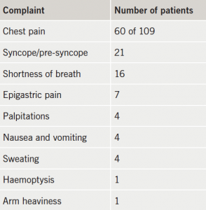
Table 1. Presenting complaint prior to troponin testing

Methods

The medical records of 109 consecutive patients, who had a POC troponin requested from either the emergency department or the medical admissions unit, were prospectively reviewed over a one-month period at a district general hospital. The samples were analysed using a POC TnT assay (Roche Cardiac T-Quantitative test). The local hospital policy recommends troponin sampling on admission and again at 12 hours post-event. Tests are reported as being either negative (<50 ng/ml), intermediate (50–100 ng/ml) or positive (>100 ng/ml). All intermediate and positive results are confirmed by formal laboratory assessment. Both POC and laboratory assays are standardised against the fifth generation Roche Elecsys hs-TnT.

Figure 1. Final diagnosis in troponinpositive patients
Figure 1. Final diagnosis in troponin positive patients

We collected data on three principal areas: the clinical presentation, subsequent relevant investigations and the final diagnosis on discharge. Data were collected on the presenting complaint, electrocardiographic (ECG) changes and coronary risk factors. We noted all subsequent cardiac tests requested (exercise treadmill tests, dobutamine stress echo, computed tomography [CT] coronary angiography, conventional angiography) and non-routine medical tests (D-dimer, CT pulmonary angiography and oesophagogastroduodenoscopy [OGD]). Each patient was followed through until completion of their clinical journey, and the final discharge diagnosis was noted.

Results

Of the 109 patients in the audit, 46.8% (n=51) were male, and 53.2% (n=58) were female. The mean age of the patients was 67.5 years. Only 55% (n=60) patients tested had a history of cardiac chest pain; while many presented with a combination of non-cardiac symptoms (table 1).

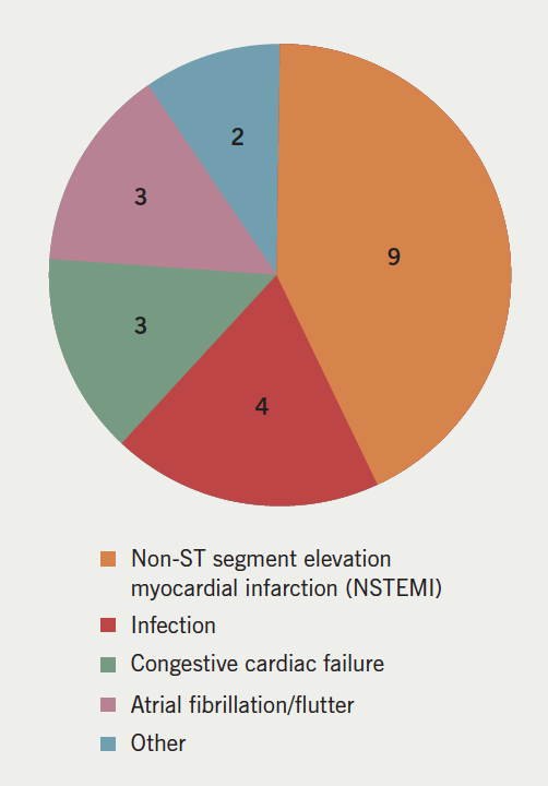
A positive TnT result was found in 19.2% of patients (n=21). Of these, nine were diagnosed with an ACS or coronary artery disease (CAD). In the majority of instances, the troponin rise was attributed to other causes including tachyarrhythmias (n=6), congestive cardiac failure (n=3), and sepsis (n=2) (figure 1).

Figure 1. Final diagnosis in troponin positive patients
Figure 2. Final diagnosis in troponin negative patients

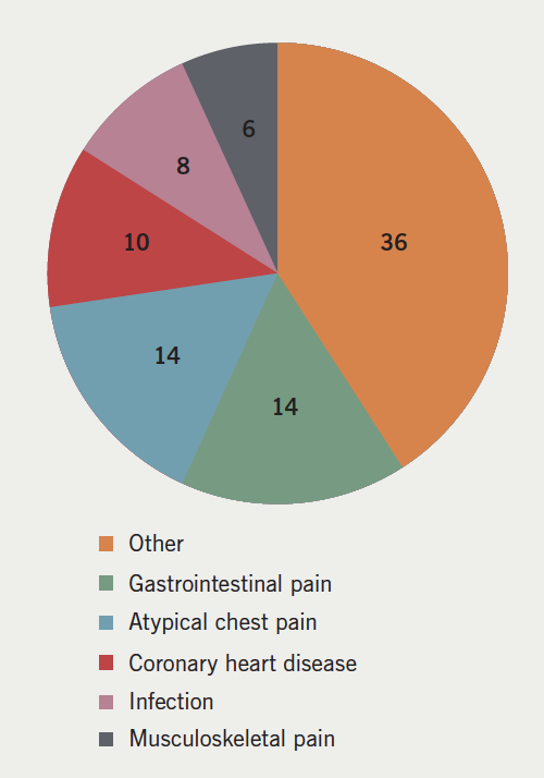
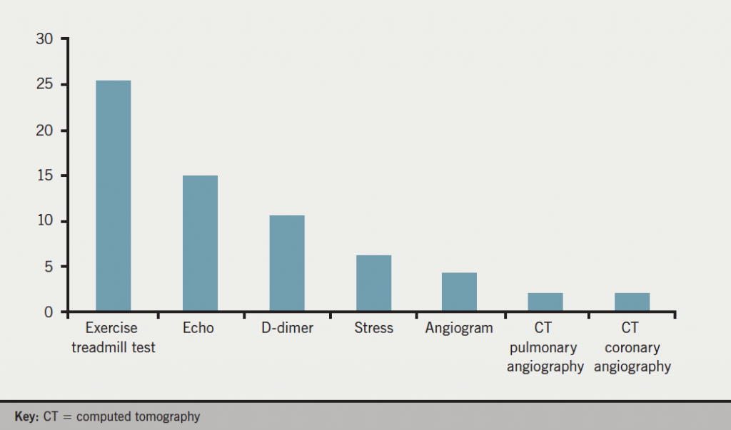
A negative troponin was found in 80.8% (n=88) of patients. A majority of these patients were discharged with no further ischaemia assessment (54%; n=40). A small number were diagnosed with stable angina, but only two referred for coronary revascularisation (final diagnoses shown in figure 2). The exercise tolerance test was the most common investigation following a negative TnT result (most common investigations shown in figure 3).

We identified 35 patients (32.1%), with neither chest pain nor ECG changes (therefore ‘low-risk’), who had a TnT requested, regardless. In all these cases, the TnT assay was negative and none were given a final diagnosis of ACS or coronary heart disease (CHD).

From the study, 93 patients (85%) had a troponin checked at baseline, and in around half of cases (46.8%; n=51) a 12-hour troponin was required to complete the ACS screen. All patients with positive or intermediate bedside troponin results had a laboratory sample obtained to confirm the findings: 100% of patients had a completed TnT screen within the 12-hour assessment period.

Figure 3. Additional tests performed in chest pain and troponin T negative patients (% of patients)

Discussion

Higher-sensitivity troponin testing is a powerful tool for the assessment of myocardial infarction. The real world implementation of newer POC ‘bedside’ testing has not been extensively documented.

POC testing was frequently performed in patients with a non-ACS clinical presentation. The majority had no history of cardiac chest pain; instead, non-specific cardiovascular complaints (pre-syncope, palpitations, etc.) triggered a troponin check. Almost one-third of patients had neither chest pain nor ECG changes suggestive of ischaemia. In these cases, there was, unsurprisingly, no final diagnosis of CAD.

In suspected ACS, where the troponin samples are negative, some further form of functional assessment should be performed to evaluate inducible ischaemia.9 In this analysis, a negative TnT test was found in 80.8% of patients (n=88), but an ischaemia assessment was omitted in over half (n=48). This could imply a lack of clinical conviction in suspecting a genuine cardiac event (and, hence, reflect poor patient selection for POC testing). It may also suggest that a negative test is being incorrectly utilised to underwrite safe discharge without follow-up.

A higher detection rate of true AMI could have been anticipated if our assay was used appropriately. From our study, 19.2% of patients were troponin positive; this is higher than the troponin positive rate of 9% from a recent multi-centre analysis by Groarke et al.1 However, in our cohort, the majority of these rises were of non-ACS aetiology (12/21 cases). Elevated cardiac TnT with CCF, PTE and sepsis is associated with increased morbidity, however (with the possible exception of PTE), haphazard troponin sampling is not recommended, as the influence on clinical management is not conclusively established.

In many cases, POC requests are initiated by less experienced ‘front-door’ staff to accelerate patient throughput. However, use of the bedside assay still allows timely and complete sampling at appropriate time intervals.

As patient comorbidity increases and higher-sensitivity assays are more readily available, adequate patient selection must be further emphasised. Indiscriminate testing will yield more non-coronary troponin rises, which can cloud clinical judgement, misguide therapy and incur additional medical expenses from further investigations and prolonged inpatient stay.

Better clinical selection could be engendered by protocolising sample requests, continuous screening or requiring senior authorisation prior to testing. However, these strategies slow turnover time and are impractical for a bedside assay. The role of the clinician is essential to adjudicate testing for more equivocal cases. Our data suggest further education regarding thoughtful patient selection and result interpretation will be essential for effective implementation of the POC, and eventually the hs-Tn assay.

Conflict of interest

None declared.

Sources of support

No external funding.

Key messages

  • Point-of-care (POC) troponin T (TnT) testing can be implemented effectively in acute coronary syndromes (ACS)
  • TnT assays are requested for patients with a relatively low suspicion of ACS
  • A lack of initial conviction is further evidenced by a failure to undertake further ischaemia assessment
  • Higher-sensitivity assays yield a greater number of non-coronary troponin rises; a final diagnosis of genuine ACS was made in less than half of troponin-positive cases suggesting non-optimal pre-test selection
  • Effective use of the POC and higher-sensitivity TnT assays will require more selective usage and consideration of the clinical context

References

1. Groarke JD, Browne L, Margey R et al. A multicentre analysis of troponin use in clinical practice. Ir J Med Sci 2013;182:185–90. http://dx.doi.org/10.1007/s11845-012-0853-2

2. Thygesen K, Alpert JS, Jaffe AS et al. Third universal definition of myocardial infarction. Circulation 2012;126:2020–35. http://dx.doi.org/10.1161/CIR.0b013e31826e1058

3. Saenger AK. A tale of two biomarkers: the use of troponin and CK-MB in contemporary practice. Clin Lab Sci 2010;23:134–40.

4. Saenger AK, Jaffe AS. The use of biomarkers for the evaluation and treatment of patients with acute coronary syndromes. Med Clin North Am 2007;91:657–81. http://dx.doi.org/10.1016/j.mcna.2007.04.001

5. Apple FS, Collinson PO. Analytical characteristics of high-sensitivity cardiac troponin assays. Clin Chem 2012;58:54–61. http://dx.doi.org/10.1373/clinchem.2011.165795

6. Rajappan K, Murphy E, Amber V et al. Usage of troponin in the real world: a lesson for the introduction of biochemical assays. QJM 2005;98:337–42. http://dx.doi.org/10.1093/qjmed/hci052

7. Gupta S, de Lemos JA. Use and misuse of cardiac troponins in clinical practice. Prog Cardiovasc Dis 2007;50:151–65. http://dx.doi.org/10.1016/j.pcad.2007.01.002

8. Mahajan VS, Jarolim P. How to interpret elevated cardiac troponin levels. Circulation 2011;124:2350–4. http://dx.doi.org/10.1161/CIRCULATIONAHA.111.023697

9. Bertrand ME, Simoons ML, Fox KA. Management of acute coronary syndromes in patients presenting without persistent ST-segment elevation. Eur Heart J 2002;23:1809–40. http://dx.doi.org/10.1053/euhj.2002.3385

Correspondence: coenzyme Q10 supplementation for the treatment and prevention of heart failure

Br J Cardiol 2015;22:68 Leave a comment
Click any image to enlarge
Authors:

Correspondence from the world of cardiology

Dear Sirs,

We wish to comment on two recent clinical trial studies relating to the use of supplementary coenzyme Q10 (CoQ10) for the treatment and prevention of cardiovascular disease.

In the first randomised controlled trial, the Q-Symbio study by Mortensen et al,1 oral supplementation with CoQ10 (300 mg/day for two years, as an adjuvant to conventional medication) in 420 patients with chronic heart failure (New York Heart Association class III or IV) reduced the risk of cardiac related mortality by 47% compared to placebo.

In the second randomised controlled trial, the KiSel-10 study by Alehagen et al,2 oral supplementation with CoQ10 (200 mg/day) and selenium (200 µg/day) for five years in 440 normal elderly Swedish individuals reduced the risk of cardiovascular related mortality by 54%.

The Q-Symbio and KiSel-10 clinical trials represent landmark studies in the treatment and prevention of cardiovascular disease. However, what is not immediately apparent from reading the above papers is the key importance of the type of supplemental CoQ10 used and also the blood CoQ10 level subsequently achieved. The type of CoQ10 used in the Q-Symbio and KiSel-10 studies, Bio-Quinone Q10 100 mg (licensed in the EU as Myoqinon®), was chosen because of its documented bioavailability; specifically the ability of a defined dosage to raise blood CoQ10 levels above the 3 µg/ml threshold required for clinical efficacy in cardiovascular disease.3,4

In addition to the study by Mortensen et al,1 three relevant smaller-scale randomised controlled clinical trials were identified (as listed on Medline) in which changes in blood CoQ10 levels were documented together with changes in clinical status, following CoQ10 supplementation in patients with chronic heart failure. One of these studies5 reported significantly improved functional capacity, left ventricular contractility and endothelial function in NYHA class II/III patients; supplementation with CoQ10 (300 mg/day for two months) correspondingly raised mean blood CoQ10 levels to 3.6 µg/ml. In the remaining studies,6,7 supplementation with CoQ10 (100–200 mg/day for three to six months) in NYHA class II/III patients did not result in significant clinical benefit; corresponding mean blood CoQ10 levels following supplementation in these studies were 2.0 µg/ml and 2.2 µg/ml respectively.

These data illustrate the importance of dosage and bioavailability of supplemental CoQ10, and the importance of raising blood CoQ10 levels above a therapeutic threshold of 3 µg/ml, for the effective management of chronic heart failure.

Conflict of interest

DM is medical adviser to Pharma Nord (UK) Ltd.

David Mantle
Medical Adviser
Pharma Nord (UK) Ltd, Morpeth, Northumberland NE61 2DB
([email protected])

References

1. Mortensen SA, Rosenfeldt F, Kumar A, et al. The effect of coenzyme Q10 on morbidity and mortality in chronic heart failure. J Am Coll Cardiol HF 2014;2:641–9. http://dx.doi.org/10.1016/j.jchf.2014.06.008

2. Alehagen U, Johansson P, Bjornstedt M, et al. Cardiovascular mortality and N-terminal proBNP reduced after combined selenium and CoQ10 supplementation. Int J Cardiol 2013;167:1860–6. http://dx.doi.org/10.1016/j.ijcard.2012.04.156

3. Weis M, Mortensen SA, Rassing MR, et al. Bioavailability of four oral coenzyme Q10 formulations in healthy volunteers. Mol Aspects Med 1994:15:273–80.

4. Langsjoen PH, Langsjoen AM. Overview of the use of CoQ10 in cardiovascular disease. Biofactors 1999;9:273–84. http://dx.doi.org/10.1002/biof.5520090224

5. Belardinelli R, Mucaj A, Lacalaprice F, et al. CoQ10 and exercise training in chronic heart failure. Eur Heart J 2006;27:2675–81. http://dx.doi.org/10.1093/eurheartj/ehl158

6. Watson PS, Scalia GM, Galbraith A, et al. Lack of effect of CoQ10 on left ventricular function in patients with congestive heart failure. J Am Coll Cardiol 1999;33:1549–52. http://dx.doi.org/10.1016/S0735-1097(99)00064-9

7. Khatta M, Alexander BS, Krichten CM, et al. The effect of CoQ10 in patients with congestive heart failure. Ann Intern Med 2000;132:636–40. http://dx.doi.org/10.7326/0003-4819-132-8-200004180-00006