News from the European Atherosclerosis Society Congress

Br J Cardiol 2015;22:59–60 Leave a comment
Click any image to enlarge
Authors:

Glasgow was the host of the recent 83rd Annual Congress of the European Atherosclerosis Society (EAS), held from 22nd–25th March 2015 and attended by more than 1,500 delegates from across 77 countries. We report its highlights.

FH initiative

366242-buchanan-street-glasgow copyHeadlining the Congress was the launch of the EAS Familial Hypercholesterolaemia Studies Collaboration (FHSC), a consortium of major FH registries across Europe, Asia-Pacific, Africa and South America, led by Professor Kausik Ray (Imperial College, London). As shown by the previous EAS Consensus Panel statement, FH is one of the most common inherited conditions, yet it is underdiagnosed and undertreated in almost all countries.1 The FHSC will provide information on key aspects relating to FH care which will be critical in leveraging public policy to improve detection and management. Linking patient and clinician empowerment underpins the mission of the FHSC: in recognition of this common goal, EAS Glasgow brought together representatives of FH Patient Advocacy groups from around the world, including HEART UK − The Cholesterol Charity (http://heartuk.org.uk/), to gain insights into how barriers to optimum FH care can be overcome.

eas logoWith an estimated prevalence of one in 200–250 people, heterozygous FH accounts for most of the burden of FH care. Recent recognition of the genetic and phenotypic heterogeneity of homozygous FH2 has highlighted the need for further information into this severe FH presentation. This is the aim of a substudy of the EAS-FHSC Initiative − the HoADH International Clinical Collaboration (HICC) − jointly led by Professor Derick Raal (University of the Witwatersrand, Johannesburg, South Africa) and Dr G Kees Hovingh (Academic Medical Center, Amsterdam, the Netherlands). Professor Raal discusses why the FHSC, including homozygous autosomal dominant hypercholesterolaemia (HoADH), are critical for FH care worldwide in our podcast.

Hot off the press

The Clinical Latebreaker session gave a tantalising insight into a prospective EAS Consensus Paper on paediatric FH expected shortly. In his presentation, Dr Albert Wiegman (Academic Medical Center, Amsterdam, the Netherlands) made the case for targeting children and adolescents with FH. Childhood provides the ideal opportunity for screening for FH on the basis of an elevated low-density lipoprotein cholesterol (LDL-C) value, given the lack of dietary and hormonal influences. Dr Wiegman presented data showing that if the child is not overweight and has normal thyroid function, an elevated LDL-C (>95th percentile) suggests that FH is highly likely. This EAS Consensus Paper will have important impact for all stakeholders in FH care.

Statins back in the news

Statins were again in the news at EAS Glasgow. Dr David Preiss (Institute of Cardiovascular and Medical Sciences, University of Glasgow) presented findings from a new collaborative meta-analysis of 17 statin trials including 132,568 subjects (with and without coronary disease), followed for an average of 4.3 years. Statin treatment led to a significant 10% reduction in first hospital admission for heart failure (relative risk 0.90, 95% CI 0.84 to 0.97). The study was published simultaneously in the European Heart Journal.3 Dr Preiss commented that with evidence of greater benefit from statins in the long-term, this analysis may underestimate the true benefit of statins. Already heart failure accounts for almost 2% of the UK NHS budget: with an ageing population, it has been estimated that this may increase by at least 50% over the next 25 years.4,5

There was also much interest in the recently published EAS Consensus Panel statement on statin associated muscle symptoms (SAMS),6 which featured in two key Educational Symposia. One session entitled ‘Statin Intolerance, an impactful and yet unresolved clinical challenge’ highlighted the clinical impact of SAMS, which is responsible for up to 40% of referrals to lipid clinics, according to Professor Erik Stroes (Academic Medical Center, Amsterdam, the Netherlands) co-chair of this session. Importantly, in most cases muscle pain is not associated with creatine kinase (CK) elevation. In support, a general practice study in Spain (n=3,845) showed that 78% of patients with muscle pain on statins did not have CK elevation.7 Strategies for identifying and managing SAMS were discussed; taking sufficient time with patients to discuss the need for statin treatment, investigating the symptoms with statin de-challenge and re-challenge, and considering a low dose of an alternative statin, were all key to management.

The other Educational Session ‘Latest developments in difficult-to-treat patients with hypercholesterolemia’ highlighted the limitations of current treatments for addressing the unmet needs of patients at high cardiovascular risk, including those with FH or SAMS. Novel agents, specifically PCSK9 monoclonal antibody therapy, clearly offer potential. Professor Stroes overviewed evidence from recent studies, including GAUSS-28 and ODYSSEY Alternative,9 in patients who were intolerant to at least two statins (including one at lowest dose in ODYSSEY Alternative). Both studies showed additional substantial LDL-C lowering with the PCSK9 inhibitor (either evolocumab or alirocumab, respectively), with no indication of increase in myalgia, CK elevation or muscle pain. Moreover, interim long-term safety data from ODYSSEY Alternative showed that only 0.7% of patients treated with alirocumab discontinued treatment due to muscle symptoms.9

Novel therapies: PCSK9 inhibitors

Much of the interest in recent meetings has been on PCSK9 inhibitors, and Glasgow was no exception. A new analysis of 4,166 patients in six phase III trials from the ODYSSEY programme, presented during the Clinical Latebreaker session, showed that the efficacy of PCSK9 inhibition was not blunted by background high-intensity statin therapy, or other lipid-lowering treatment.10 These findings provide important reassurance to clinicians, in the light of evidence that statins upregulate PCSK9 expression.11

A pooled analysis of 4,564 patients in eight phase 3 trials included in the ODYSSEY programme showed that treatment with the PCSK9 inhibitor alirocumab significantly improved LDL-C goal attainment; 75−79% on alirocumab attained LDL-C goal at week 24, compared with 52% on ezetimibe and 6−8% on statin alone.12 A key question, therefore, is what proportion of patients at high cardiovascular risk who are unable to attain LDL-C goal with current treatments would potentially qualify for PCSK9 inhibitor therapy? Evidence from DYSIS II (Dyslipidemia International Study II) provides some insight. Among 461 patients (76% male, mean age 64.1 years) with a recent acute coronary syndrome in Germany, only 22% attained the European Society of Cardiology/EAS guideline recommended LDL-C goal of 1.7 mmol/L (70 mg/dL).13 DYSIS II Belgium also showed that nearly half (46%) of patients with diabetes and coronary heart disease did not attain LDL-C goal.14 Furthermore, an analysis from LTAP-2 (the Lipid Treatment Assessment Project -2) including 9,955 patients (40% with atherosclerotic vascular disease) showed that 11.5% of patients were potential candidate for PCSK9 inhibitor treatment. However, if the LDL-C threshold was increased to >2.6 mmol/L (100 mg/dL), 21% of patients with cardiovascular disease would qualify.15

Reassertion of lifestyle

Lifestyle was also very much in the news at EAS Glasgow. In a special lecture ‘Investing in your arteries: importance of a healthy lifestyle for lifetime cardiovascular risk reduction’, Professor John Deanfield (British Heart Foundation Vandervell Professor of Cardiology, University College Hospital, Londonand lead author of the Joint British Societies Guidelines) argued for a renewed emphasis on ‘primordial prevention’, targeting cardiovascular risk factors such as smoking, cholesterol, blood pressure and body weight from an early age to optimise clinical benefit. He cited evidence from the Framingham Offspring Cohort which showed that long-term exposure (by 11−20 years) to even moderately elevated cholesterol was associated with about a four-fold higher risk of coronary events compared with individuals with no exposure before age 55 years.16 Professor Deanfield argued that public education should be a priority so that individuals can take responsibility for their cardiovascular health and sustain lifestyle change; this call for a primordial approach to cardiovascular disease prevention was echoed in a simultaneous publication in the European Heart Journal.17

In the EAS-International Chair on Cardiometabolic Risk (ICCR) Joint Session: ‘Changing lifestyle patterns: the challenge for cardiovascular prevention’, Professor Naveed Sattar (Institute of Cardiovascular and Medical Sciences, University of Glasgow) took a global view of the need for primordial prevention. Increasing urbanisation in developing economies has led to adoption of Westernised diets and increasing exposure to poor diet choices and physical inactivity, resulting in increasing obesity, diabetes and cardiovascular disease. The impact has been greatest in low to middle income countries: in Bangladesh, for example, diabetes prevalence in urban areas has increased seven-fold over the period 1986–2006. Smoking is a key challenge: in China, now with the world’s highest prevalence of smokers, one million people die each year due to smoking-related causes, and over the next 15 years this will triple.18 Professor Sattar emphasisesd the importance of focusing on lifestyle, and discusses the practicalities of sustaining change in our podcast.

A pertinent issue to this debate is the need for micronutrient supplementation, as discussed by Professor Ian Young (Centre for Public Health, Queen’s University Belfast, Northern Ireland). While experimental and epidemiological data, as well as studies in individuals with intermediate traits such as metabolic syndrome are supportive of benefit, clinical trials have less consistent results. Given that PREDIMED (Primary Prevention of Cardiovascular Disease with a Mediterranean Diet) 19 showed that inclusion of nuts or extra virgin olive oil in a Mediterranean diet reduced cardiovascular events (primarily stroke) by 30% in a high-risk primary prevention population, eating a diet rich in micronutrients rather than taking micronutrient supplements should be the preferred approach for preventing cardiovascular disease.

Novel biomarkers

The case for biomarkers for improving risk prediction especially in individuals at low to intermediate cardiovascular risk was a hot topic. Professor Kausik Ray emphasised the need to take account of the numbers needed to screen for treatment decisions and impact on clinical benefit.

Lipoprotein(a) (Lp(a)) is already established as a cardiovascular risk factor, independent of LDL-C or other lipids, mediated by pro-atherogenic, pro-thrombotic and antifibrinolytic effects which enhance atherothrombosis.20 Professor Børge Nordestgaard (University of Copenhagen & Copenhagen University Hospital, Denmark) discusses the evidence for Lp(a) and how to manage this in the clinic in our podcast.

New findings from an analysis of 98,097 subjects from the Copenhagen City Heart Study and the Copenhagen General Population Study link Lp(a) with risk for heart failure.21 Individuals with Lp(a) levels in the top 10% (68-289 mg/dL) had a 60−80% increase in heart failure (p<0.001 for trend). The effect of elevated Lp(a) was likely causal; the risk of heart failure was 18% (hazard ratio 1.18, 95% CI 1.04−1.34) for individuals carrying genetic variants known to be associated with elevated Lp(a), consistent with observational data (hazard ratio 1.22, 95% CI 1.11−1.35).

With the withdrawal of niacin/laropiprant in Europe, there are no therapeutic options that specifically target elevated Lp(a). Ongoing studies are evaluating the potential of an antisense oligonucleotide to apolipoprotein(a) contained on the Lp(a) molecule. Initial studies show 40−80% reduction in Lp(a) with doses of 100−300 mg; a phase 2 study in patients with elevated Lp(a) (>50 mg/dL) is ongoing.22

There was also news to strengthen the case for elevated non-fasting remnant cholesterol (defined as total cholesterol – HDL-C – LDL-C). This analysis included about 90,000 Danish individuals free of cardiovascular disease at baseline, of whom 4,435 developed ischaemic heart disease, 1,722 developed myocardial infarction, and 8,121 died over a 22-year follow-up period. Whereas both LDL-C and Lp(a) were associated equally with risk of ischaemic heart disease and myocardial infarction, only non-fasting remnant cholesterol concentrations were associated stepwise with increased risk of all-cause mortality. Subjects with the highest non-fasting remnant cholesterol levels (≥1.50 mmol/L) had a 60% increased risk of all-cause mortality.23

Accumulating evidence for these markers strengthens the case for consideration of their inclusion in future dyslipidaemia guidelines. Clearly, the lipid field continues to be ‘hot news’ for 2015 and beyond.

References

1. Nordestgaard BG, Chapman MJ, Humphries SE et al.; European Atherosclerosis Society Consensus Panel. Familial hypercholesterolaemia is underdiagnosed and undertreated in the general population: guidance for clinicians to prevent coronary heart disease: consensus statement of the European Atherosclerosis Society. Eur Heart J 2013;34:3478−90a. http://dx.doi.org/10.1093/eurheartj/eht273

2. Cuchel M, Bruckert E, Ginsberg HN et al.; European Atherosclerosis Society Consensus Panel on Familial Hypercholesterolaemia. Homozygous familial hypercholesterolaemia: new insights and guidance for clinicians to improve detection and clinical management. A position paper from the Consensus Panel on Familial Hypercholesterolaemia of the European Atherosclerosis Society. Eur Heart J 2014;35:2146−57. http://dx.doi.org/10.1093/eurheartj/ehu274

3. Preiss D, Campbell RT, Murray HM et al. The effect of statin therapy on heart failure events: a collaborative meta-analysis of unpublished data from major randomized trials. Eur Heart J 2015 Mar 23. pii: ehv072. [Epub ahead of print]. http://dx.doi.org/10.1093/eurheartj/ehv072

4. National Institute for Health and Clinical Excellence. New NICE guidance will improve diagnosis and treatment of chronic heart failure. London: NICE, 2010. Link: http://www.nice.org.uk/newsroom/pressreleases/chronicheartfailureguidance.jsp

5. Sutherland K. Bridging the quality gap: heart failure. The Health Foundation, 2010. Link: http://www.health.org.uk/public/cms/75/76/313/583/Bridging%20the%20quality%20gap%20Heart%20Failure.pdf?realName=cXqFcz.pdf

6. Stroes ES, Thompson PD, Corsini A et al.; European Atherosclerosis Society Consensus Panel. Statin-associated muscle symptoms: impact on statin therapy-European Atherosclerosis Society Consensus Panel Statement on Assessment, Aetiology and Management. Eur Heart J 2015 Feb 18. pii: ehv043. [Epub ahead of print].

7. Pedro-Botet J, Millán Núñez-Cortés J, Flores JA, Rius J. Muscle symptoms related with statin therapy in general practice. 83rd Congress of the EAS, Glasgow, 22nd−25th March 2015. Abstract EAS-0232.

8. Stroes E, Colquhoun D, Sullivan D et al. Anti-PCSK9 antibody effectively lowers cholesterol in patients with statin intolerance: the GAUSS-2 randomized, placebo-controlled phase 3 clinical trial of evolocumab. J Am Coll Cardiol 2014;63:2541−8. http://dx.doi.org/10.1016/j.jacc.2014.03.019

9. Moriarty PM, Thompson PD, Cannon CP et al. ODYSSEY ALTERNATIVE: efficacy and safety of alirocumab versus ezetimibe, in patients with statin intolerance as defined by a placebo run-in and statin rechallenge arm. http://my.americanheart.org/idc/groups/ahamah-public/@wcm/@sop/@scon/documents/downloadable/ucm_469684.pdf

10. Krempf M, Bergeron J, Elassal J et al. Efficacy of alirocumab according to background statin intensity and other lipid-lowering therapy in heterozygous familial hypercholesterolemia or high CV risk populations: Phase 3 sub-group analyses. 83rd Congress of the EAS, Glasgow,22nd−25th March 2015. Abstract EAS-0493.

11. Mayne J, Dewpura T, Raymond A et al. Plasma PCSK9 levels are significantly modified by statins and fibrates in humans. Lipids Health Dis 2008;7:22. http://dx.doi.org/10.1186/1476-511X-7-22

12. Farnier M, Gaudet D, Valcheva V et al. Efficacy of alirocumab in heterozygous familial hypercholesterolemia or high CV risk populations: pooled analyses of eight phase 3 trials. 83rd Congress of the EAS, Glasgow, 22nd−25th March 2015. Abstract EAS-0563.

13. Gitt A, Ashton V, Horack M et al. Low LDL-C target achievement among treated ACS patients in Germany: The Dyslipidemia International Study (DYSIS) IIACS results. 83rd Congress of the EAS, Glasgow, 22nd−25th March 2015. Abstract EAS-0397.

14. Hermans M, Cools F, Gevaert S et al. Lipid abnormalities remain high among diabetic patients with stable CHD: results of the Dyslipidemia International Study (DYSIS) II Belgium. 83rd Congress of the EAS, Glasgow, 22nd−25th March 2015. Abstract EAS-0405.

15. Waters D, Santos R, Abreu P et al. Who might benefit from PCSK9 inhibitor treatment? An analysis from the Lipid Treatment Assessment Project-2 (LTAP-2). 83rd Congress of the EAS, Glasgow, 22nd−25th March 2015. Abstract EAS-0744.

16. Navar-Boggan AM, Peterson ED, D’Agostino RB Sr et al. Hyperlipidemia in early adulthood increases long-term risk of coronary heart disease. Circulation 2015;131: 451−8. http://dx.doi.org/10.1161/CIRCULATIONAHA.114.012477

17. Ayer J, Charakida M, Deanfield JE, Celermajer DS. Lifetime risk: childhood obesity and cardiovascular risk. Eur Heart J 2015 Mar 24. pii: ehv089. [Epub ahead of print]. http://dx.doi.org/10.1093/eurheartj/ehv089

18. World Health Organization. Smoking in China. http://www.wpro.who.int/china/mediacentre/factsheets/tobacco/en/

19. Estruch R, Ros E, Salas-Salvadó J et al. Primary prevention of cardiovascular disease with a Mediterranean diet. N Engl J Med 2013;368:1279−90. http://dx.doi.org/10.1056/NEJMoa1200303

20. Nordestgaard BG, Chapman MJ, Ray K et al. Lipoprotein(a) as a cardiovascular risk factor: current status. Eur Heart J 2010;31:2844−53. http://dx.doi.org/10.1093/eurheartj/ehq386

21. Nordestgaard BG, Kamstrup PR. Elevated lipoprotein(a) levels and increased risk of heart failure. 83rd Congress of the EAS, Glasgow, 22nd−25th March 2015. Abstract EAS-0432.

22. Tsimikas S et al., data in press.

23. Nordestgaard BG, Freiberg JJ, Varbo A. Extreme nonfasting remnant cholesterol vs extreme LDL cholesterol as contributors to cardiovascular disease and all-cause mortality in 90,000 individuals from the general population. 83rd Congress of the EAS, Glasgow, 22nd−25th March 2015. Abstract EAS-0450.

Pulmonary hypertension in UK clinical practice: an update

Br J Cardiol 2015;22(suppl 1):S2–S15doi:10.5837/bjc.2015.s01 Leave a comment
Click any image to enlarge
Authors:
Sponsorship Statement: This supplement is based on the proceedings of a roundtable meeting held at the Royal Society, London, on 17th December 2014. The meeting was convened by the British Journal of Cardiology (BJC), who approached Actelion Pharmaceuticals UK Ltd for support. Actelion had no input into the content of the meeting or in the selection of the participants but provided an unrestricted grant to cover the meeting costs, honoraria and travel expenses, the services of a medical writer and the development and distribution of this supplement by the BJC. The faculty all reviewed and approved this supplement before publication. Actelion also reviewed the supplement for product-related technical accuracy.

Insights from the world of cardiology

Continue reading Pulmonary hypertension in UK clinical practice: an update

Disclaimer: Medinews Cardiology Limited advises healthcare professionals to consult up-to-date Prescribing Information and the full Summary of Product Characteristics available from the manufacturers before prescribing any product. Medinews Cardiology Limited cannot accept responsibility for any errors in prescribing which may occur.

The effectiveness of a mobile ECG device in identifying AF: sensitivity, specificity and predictive value

Br J Cardiol 2015;22:70–2doi:10.5837/bjc.2015.013 Leave a comment
Click any image to enlarge
Authors:
First published online April 15, 2015

Early identification of atrial fibrillation (AF), especially when asymptomatic, is increasingly important when there are interventions that can reduce the risk of stroke. One mobile ECG device that has the potential for doing just that is the AliveCor® device, which is non-invasive and easy to use. We aimed to assess its utility in primary care by establishing its sensitivity and specificity, and consider the predictive value for identifying AF in a general practice population.

We used the device on a population known to have AF in order to calculate the sensitivity, and on a population who did not have AF at the time of recording, to establish specificity. Using the known prevalence of AF in a UK population, we were able to calculate the predictive values for identification of AF. All AliveCor® traces we compared with a gold-standard 12-lead electrocardiogram (ECG).

The device has a high sensitivity and specificity in the hands of experienced clinicians. In particular, the sensitivity was consistently high, which would ensure a high true-positive rate of identification. Furthermore, the negative predictive value in populations with a prevalence of AF as in the UK is sufficiently high to be useful.

In conclusion, the AliveCor® device should be considered as an option for early identification of patients with unknown AF. It has a high negative-predictive value and is sufficiently sensitive to be useful in a general practice population, but does not rule out the need for a definitive ECG in suspected cases.

Introduction

People with atrial fibrillation (AF) are five times more likely to have a stroke.1 AF is an increasing problem as our population gets older.2 It is, therefore, important to be able to identify this condition as early as possible, when intervention with anticoagulation can prevent stroke, as is recommended by the National Institute for Health and Care Excellence (NICE), in most cases.3 Several studies have attempted to identify the most effective way of screening for, or case-finding, AF.4-7

Stock

The gold standard for diagnosis of AF is a 12-lead electrocardiogram (ECG). However, the 12-lead ECG is an impractical diagnostic tool for assessing those without symptoms, or without an apparently irregular pulse. Devices are increasingly being manufactured that could identify pre-symptomatic AF, such as WatchBP Home A device.8

One of the new devices that may be useful for case-finding asymptomatic people is the AliveCor® ‘App’ that can be used with a digital telephone (www.AliveCor.com). The AliveCor® device provides a single-lead ECG, which, if no ‘P-waves’ are identified or the rhythm is irregular, would trigger a 12-lead diagnostic ECG to be done. For it to be useful, it has to be able to demonstrate a sufficiently high sensitivity to identify cases, and a high negative-predictive value to be able to exclude cases in the primary care population. This means having a high specificity even in a relatively high prevalence population.

This study aims to establish the sensitivity and specificity of this device, and estimate the predictive values in an average general practice population.

The study was conducted independently of the manufacturer, who had no input into the purchase of the device, conception, design analysis or interpretation of the study.

Methods

To assess the sensitivity of the AliveCor® device, patients with known AF, diagnosed by 12-lead ECG, were recruited. To establish the specificity of the device, it was necessary to recruit patients without AF and confirm by 12-lead ECG.

The device was used to assess the rhythm of patients attending their regular AF clinic at the North West Heart Centre located at University Hospital South Manchester in Wythenshawe. Upon attending their outpatient appointment, the patients had a routine 12-lead ECG performed. At the same time, the patient was asked to use the AliveCor® device and a 30-second reading was taken using the application on the phone. A 12-lead ECG reading was recorded and printed at the same time. After the patient had concluded their ECG, they were allocated a number and their patient details removed from the 12-lead ECG for the purposes of blinding the readings for future interpretation. The AliveCor® reading was saved to a SD memory card once the reading had been converted into PDF format. Patient consent was established for conducting this study.

Alongside the known AF cohort of patients, were also many patients who were attending for reasons unrelated to AF. Those individuals, on whom the consultant wanted a 12-lead ECG performed in any case, were also asked to take a reading using the AliveCor® device. These patients underwent the same procedure as those with AF, and again were assigned a number, anonymised and their AliveCor® readings saved to memory card after conversion to PDF format.

Once the data had been collected, the 12-lead ECG recordings were then interpreted by a cardiac physiologist, used to reporting ECGs (KP) at the North West Heart Centre, and a general practitioner with a special interest in cardiology (IB). The recordings were designated as either being in AF or not. Once these had been interpreted, the AliveCor® readings were also assigned as either AF or not.

The designation of the 12-lead ECG was compared with the AliveCor® designation. The 12-lead ECG is regarded as the gold-standard diagnostic test for the purposes of this study. After the data regarding the readings had been analysed, it was then possible to calculate the sensitivity and specificity of the device. This was calculated by whether or not the AliveCor® reading corresponded to the 12-lead ECG designation. From this information it is possible to also calculate the positive- and negative-predictive values of the device depending on the prevalence of AF in any given population.

Results

In total, there were 99 patients who participated in the collection of ECG readings. Due to artefacts being present in some of the ECG recordings or the recordings were illegible, four patients were removed from the cohort. Therefore, the number of patients for analysis totalled 95. Of these 95 patients, 29 had AF using the 12-lead ECG as the method of interpretation, with 66 being in sinus rhythm.

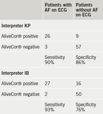
Table 1. Respective atrial fibrillation (AF) designation of 12-lead electrocardiogram (ECG) and AliveCor
Table 1. Respective atrial fibrillation (AF) designation of 12-lead electrocardiogram (ECG) and AliveCor®

Of the 29 patients identified as being in AF via the 12-lead ECG recordings, using the AliveCor®, KP correctly identified 26 alongside a negative test for the remaining three individuals who were in AF according to the 12-lead ECG. With AliveCor®, IB identified nine with AF who did not have AF on the diagnostic 12-lead ECG, but identified 27 out of the 29 who did have AF on 12-lead ECG (table 1). Thus, in the case of KP the sensitivity of the AliveCor® device can be calculated as 90%, and the specificity as 86%. For IB the sensitivity was 93% and the specificity was 76%.

Discussion

The AliveCor® device is a simple and easy-to-use device. It is also lightweight and easily portable. The device can be used by professionals in primary care with minimal training. The diagnosis of AF is made by the absence of ‘P-waves’ and an irregularly spaced ‘QRS complex’. Even after potential identification with the AliveCor® device, there needs to be a diagnostic ECG.

The prevalence of AF on general practice registers in Central Manchester is 0.7%. This is lower than the national average of 1.5%, according to 2013 returns. However, much of the apparent under-ascertainment can be explained by the age distribution. The population of Central Manchester consists of 7.5% aged over 65 years compared with 17% over 65 years in England, according to 2011 census data. The prevalence of AF in over 65-year-olds in Central Manchester is likely to be similar to that throughout England.9

The high negative-predictive values suggest that this test is a good ‘rule-out’ test for AF in a population with a prevalence of AF, such as we have in England. A ‘positive’ AliveCor® test should still be combined with a 12-lead ECG to confirm the diagnosis of AF, but the test is more sensitive than assessing pulse regularity alone (77–84%).7

As the prevalence of AF increases with age, so too will the predictive values change. In one study,10 in the age range 75–79 years, the prevalence of AF was found to be 9%. So the positive-predictive value in this cohort is 39%, and the negative-predictive value is 99%. The prevalence of AF in the same study in those over 85 years was 18%. Therefore, the positive-predictive value in this cohort is about 59%, and the negative-predictive value in this cohort is about 98%. This prevalence is similar to a UK population, where the prevalence of AF in over 75-year-olds was 12%.6 In this population, the positive-predictive value will be about 47%, and the negative-predictive value will be 98%.

However, when case finding in a high-risk population, it is also important to have a low false-negative rate, in other words a high sensitivity. We do not want to miss cases. This may be sacrificed for a low false-positive rate, or specificity, since the final diagnosis will be made by a 12-lead ECG. For both analysers, an experienced electrophysiologist and a GP with a special interest in cardiology, the sensitivity is about 90%. In addition, in the population likely to be tested, the negative-predictive value is almost 100%.

Interpretation of the AliveCor® reading depends on the skill and experience of the clinician. So for less experienced clinicians, the sensitivity and specificity may be lower. This will lead to more false-positive and negative tests, and will result in more 12-lead ECGs being done than in experienced hands. This can be mitigated by a simple training programme to identify AF on the AliveCor® strip, or by commissioning a reporting service. The priority is to retain a high sensitivity, so that true positives are identified, which may be at the expense of a lower specificity.

The AliveCor® app can be used with a digital mobile telephone. It has a high sensitivity and negative-predictive value, which makes it potentially useful in case-finding AF, especially in the over 75-year-old population. It also has other uses within a cardiological assessment of patients. It should be considered as an option for use in early detection of AF.

Conflict of interest

None declared. The study was conceived, designed and conducted without any contribution from the manufacturer. The device was bought by IB.

Key messages

  • AliveCor® is a simple and non-invasive way of identifying atrial fibrillation (AF)
  • It has a high sensitivity in experienced hands and so will identify potential cases
  • It has a high negative-predictive value in a general practice population and so is a good ‘rule-out’ test
  • Suspected cases should have a gold-standard 12-lead electrocardiogram (ECG) as a diagnostic test

References

1. Wolf PA, Abbott RD, Kannel WB. Atrial fibrillation as an independent risk factor for stroke: the Framingham study. Stroke 1991;22:983–8. http://dx.doi.org/10.1161/01.STR.22.8.983

2. Rothwell PM, Coull AJ, Giles MF et al. Change in stroke incidence, mortality, case-fatality, severity, and risk factors in Oxfordshire, UK from 1981 to 2004 (Oxford Vascular Study). Lancet 2004;363:1925–33. http://dx.doi.org/10.1016/S0140-6736(04)16405-2

3. National Institute for Health and Care Excellence. Atrial fibrillation: the management of atrial fibrillation. CG180. London: NICE, July 2014. Available from: http://www.nice.org.uk/guidance/cg180

4. Morgan S, Mant D. Randomised trial of two approaches to screening for atrial fibrillation in UK general practice. Br J Gen Pract 2002;52:373–80. Available from: http://bjgp.org/content/52/478/373

5. Sudlow M, Rodgers H, Kenny RA et al. Identification of patients with atrial fibrillation in general practice: a study of screening methods. BMJ 1998;317:327–8. http://dx.doi.org/10.1136/bmj.317.7154.327

6. Hobbs FD, Fitzmaurice DA, Mant J et al. A randomised controlled trial and cost-effectiveness study of systematic screening (targeted and total population screening) versus routine practice for the detection of atrial fibrillation in people aged 65 and over. The SAFE study. Health Technol Assess 2005;9:iii–iv, ix–x, 1–74. http://dx.doi.org/10.3310/hta9400

7. Somerville S, Somerville J, Croft P, Lewis M. Atrial fibrillation: a comparison of methods to identify cases in general practice. Br J Gen Pract 2000;50:727–9. Available from: http://bjgp.org/content/50/458/727

8. National Institute for Health and Care Excellence. WatchBP Home A for opportunistically detecting atrial fibrillation during diagnosis and monitoring of hypertension. MTG13. London: NICE, January 2013. Available from: http://www.nice.org.uk/guidance/mtg13

9. Langenburg M, Hellemons BSP, van Ree JW et al. Atrial fibrillation in elderly patients: prevalence and comorbidity in general practice. BMJ 1996;313:1534. http://dx.doi.org/10.1136/bmj.313.7071.1534

10. Heeringa J, van der Kuip DA, Hofman A et al. Prevalence, incidence and lifetime risk of atrial fibrillation: the Rotterdam study. Eur Heart J 2006;27:949–53. http://dx.doi.org/10.1093/eurheartj/ehi825

NICE guidelines for acute heart failure: long on pills, short on pumps

Br J Cardiol 2015;22:(2)doi:10.5837/bjc.2015.014 Leave a comment
Click any image to enlarge
Authors:
First published online April 15, 2015

The heart beats 120,000 times a day pumping 7,000 litres. Ischaemia or inflammation decimate this workload causing end organ dysfunction. New National Institute for Health and Care Excellence (NICE) guidelines for acute heart failure (AHF) acknowledge very high early mortality. Of 67,000 acute admissions, 11% die in hospital, 50% are readmitted and 33% are dead within 12 months.1,2 When cardiogenic shock ensues, prognosis is poor.3-5 Most patients are elderly but several thousand deaths occur in those under 65 years. Many are salvageable using advanced resuscitation techniques. When the prospectively randomised-controlled trial (RCT) IABP-SHOCK II trial (Intra-Aortic Balloon Pump in Cardiogenic Shock II) revealed the intra-aortic balloon pump (IABP) as ineffective, a New England Journal of Medicine (2012) editorial stated “we must move forward understanding that a condition with 40% mortality at 30 days remains unacceptable”.3,4 So do the guidelines help with this? 

Westaby
Professor Stephen Westaby

NICE’s medical therapy is excellent, until the end game, but drugs alone have limitations.5 Inotropes worsen ischaemia, while vasopressors elevate afterload. Injured myocardium needs rest to promote recovery, not a flogging.6 Consider two real patients. A 21-year-old female with ion-channelopathy is admitted to a tertiary care centre. After 75 DC shocks and cardiac massage she is in shock. She needs extracorporeal membrane oxygenation (ECMO) circulatory support but cannot be transferred.6 She dies. A 56-year-old male with ischaemic cardiomyopathy suffers acute on chronic heart failure. Renal impairment and pulmonary hypertension preclude transplantation. Though an ideal candidate for a long-term left ventricular assist device (LVAD) he dies.6 In the US, this mode of avoidable death is designated ‘failure to rescue’ through inadequate care.7

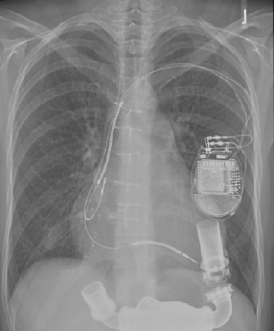
Figure 1. Electric-powered implantable rotary blood pump inserted into the apex of the failing left ventricle. Would the UK taxpayer prefer to see this £120,000 device discarded within weeks (bridge to transplant) or used for years (lifetime therapy)?

Rotary blood pumps are appropriately simple for surgeons. They are less complicated than cardiac resynchronisation or implantable defibrillators. Unlike the IABP they restore flow to physiological levels and unload the injured ventricle. Supported by laboratory research in a non-transplant centre, we first achieved LVAD ‘bridge to recovery’ for acute myocarditis in 1996.8 This young patient is still doing well. With the World’s first permanent implant of a rotary LVAD (2000) we achieved 7.5 years of good quality life in end stage dilated cardiomyopathy (15% of overall lifetime).8,9 After many successes, charitable funding expired. Now we have nothing to offer the sickest patients.

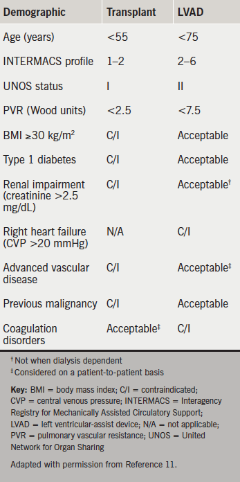
NICE guidelines state “people with potentially reversible severe AHF or potential transplant candidates should be discussed with a centre which provides mechanical circulatory support”.1 This is problematic. Five designated centres perform 100–120 transplants per year with funding for a small number of LVADs.10 They cannot triage all deteriorating patients. Many are too sick to transfer. The National Specialist Commissioning Group fund long-term LVADs only for bridge to transplantation (figure 1). Consequently, devices costing £120,000 are discarded in weeks or months when a donor becomes available. Designed for ‘lifetime’ use, implantable LVAD survival approaches that of transplantation with excellent symptomatic relief (table 1).1-13 The longest exceeds eight years. The oldest patients are nonagenarians.

Figure 1. Electric-powered implantable rotary blood pump inserted into the apex of the failing left ventricle. Would the UK taxpayer prefer to see this £120,000 device discarded within weeks (bridge to transplant) or used for years (lifetime therapy)?
Table 1. Preferred patient characteristics with regards to suitability for cardiac transplant or rotary blood pumps

Unhelpful categorisation

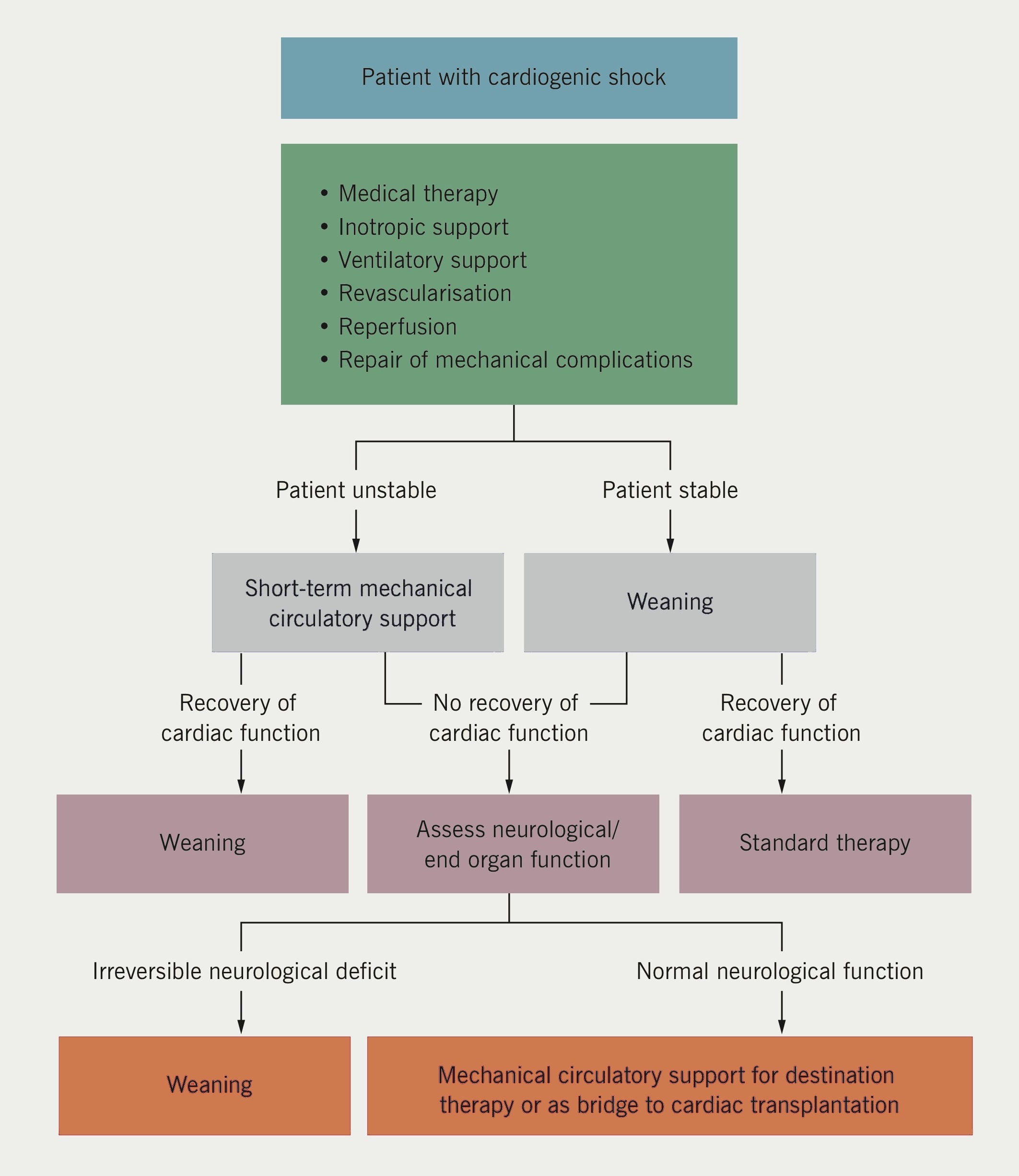
An expert Society of Thoracic Surgeons (US) and Food and Drugs Administration ‘Think Tank’ on circulatory support (2012) deemed categorisation into ‘bridge to transplant’, ‘bridge to recovery’ or ‘lifetime’ use as unhelpful.14 Heart failure is unpredictable.15 The objectives are to relieve symptoms and extend life with the techniques available.16 European Society of Cardiology guidelines specify the use of LVADs and ECMO in AHF and during myocardial revascularisation (figure 2).17,18 The American College of Cardiology and American Heart Association also provide circulatory support algorithms.19 NICE Interventional Procedures Panel (2006) published supportive guidance for temporary LVAD use, but funding was not forthcoming for non-transplant centres.20 Had inexpensive LVADs and ECMO been provided to tertiary centres, several thousand lives could have been saved.21

This time, NICE pronounced on LVAD use based solely upon the review question “for people with AHF is IABP counter pulsation more clinically/cost effective compared to LVADs, medical care alone or each other?”1 Three RCTs showed no advantage of IABP above drugs for shock.3 Three RCTs showed no advantage for two very short-term (2–3 days) percutaneous LVADs over IABP.6 Not included were effective longer-term (weeks or months) extracorporeal LVADs and ECMO, which for ethical reasons cannot be randomised in dying patients. Between 50% and 75% of shock patients survive with a pump.6 Finally, one RCT showed better survival with LVAD over medical management. Deemed ‘low quality evidence’ by NICE, this was the pivotal Randomised Evaluation of Mechanical Assistance in the Treatment of Congestive Heart failure (REMATCH) trial widely considered the most remarkable endeavour in the history of RCTs.22,23 REMATCH launched LVADs as a transplant alternative.11-13

Figure 2. European Society of Cardiology (ESC)/European Association for Cardio- Thoracic Surgery (EACTS) guidelines on myocardial revascularisation. Treatment of patients with cardiogenic shock(18)

NICE did not acknowledge the inappropriateness of RCTs in dying patients, therefore, the role of circulatory support remains poorly defined. Even the research recommendations make no mention of LVADs.1 The General Medical Council’s guidelines for end-of-life care are relevant in AHF.24 They state “you should not withhold treatment if doing so would involve significant risk to the patient and the only justification is resource constraints”. Duty of candour would suggest that the chances for survival with circulatory support should be discussed with the patient or their relatives, whether or not the devices are immediately available in their hospital. The onus is on the system to provide the means for survival irrespective of geography. Given the impressive survival rates, ECMO and LVADs must not be limited to transplant centres, as European guidelines indicate.25 Every cardiac surgery centre must have temporary circulatory support technology available. In turn, at least 1,000 systolic heart failure patients annually could benefit from an implantable long-term LVAD. This does not need a Medical Innovations Bill. Lifetime LVADs have been funded by US Medicare and Medicaid since 2003. One thousand times £150,000 (LVAD plus hospital costs) is £150 million, and this is the problem. A week after guideline publication, the BBC’s prime time One Show (8 October 2014) raised public expectations by featuring LVADs as the miracle cure for severe heart failure. But the long and short of it is that palliative care and early death are much cheaper.

Conflict of interest

SW was the ‘co-opted’ surgical representative on the NICE Acute Heart Failure Guidelines panel and was invited to attend two meetings. He was overseas representative on the Society of Thoracic Surgeons/Food and Drugs Administration ‘Think Tank’ on long-term mechanical circulatory support. He is co-founder and Medical Director of Calon Cardio-technology.

References

1. National Institute for Health and Care Excellence. Acute heart failure: diagnosing and managing acute heart failure in adults. CG187. London: NICE, 2014. Available from: http://www.nice_org.uk/guidance/cg187

2. Cleland J, Dargie H, Hardman S, McDonagh T, Mitchell P. National heart failure audit. April 2012–March 2013. London: National Institute for Cardiovascular Outcomes Research (NICOR), 2013. Available from: http://www.ucl.ac.uk/nicor/audits/heartfailure/documents/annualreports/hfannual12-13.pdf

3. Thiele H, Zeymer U, Neumann EJ. Intra-aortic balloon support for myocardial infarction with cardiogenic shock. N Engl J Med 2012;367:1287–96. http://dx.doi.org/10.1056/NEJMoa1208410

4. O’Connor CM, Rogers JG. Evidence for overturning the guidelines in cardiogenic shock. N Engl J Med 2012;367:1349–50. http://dx.doi.org/10.1056/NEJMe1209601

5. Westaby S, Kharbanda R, Banning AP. Cardiogenic shock in ACS. Part 1: prediction, presentation and medical therapy. Nat Rev Cardiol 2012;9:158–71. http://dx.doi.org/10.1038/nrcardio.2011.194

6. Westaby S, Anastasiadis K, Weiselthaler GM. Cardiogenic shock in ACS. Part 2: role of mechanical circulatory support. Nat Rev Cardiol 2012;9:195–208. http://dx.doi.org/10.1038/nrcardio.2011.205

7. Ahmed EO, Butler R, Novick RJ. Failure to rescue as a measure of quality of care in a cardiac surgery recovery unit: a five year study. Ann Thorac Surg 2014;97:147–52. http://dx.doi.org/10.1016/j.athoracsur.2013.07.097

8. Westaby S, Katsumata T, Piggot D et al. Mechanical bridge to recovery in fulminant myocarditis. Ann Thorac Surg 2000;70:278–82. http://dx.doi.org/10.1016/S0003-4975(00)01450-8

9. Westaby S, Frazier OH, Banning A. Six years of continuous mechanical circulatory support. N Engl J Med 2006;355:325–7. http://dx.doi.org/10.1056/NEJMc060715

10. MacGowan GA, Parry G, Schueler S, Hassan A. The decline in heart transplantation in the UK. BMJ 2011;342:d2483. http://dx.doi.org/10.1136/bmj.d2483

11. Westaby S. Cardiac transplant or rotary blood pump: contemporary evidence. J Thorac Cardiovasc Surg 2013;145:24–31. http://dx.doi.org/10.1016/j.jtcvs.2012.08.048

12. Kirklin JK, Naftel DC, Pagani FD et al. Long-term mechanical circulatory support (destination therapy): on track to compete with heart transplantation. J Thorac Cardiovasc Surg 2012;144:584–603. http://dx.doi.org/10.1016/j.jtcvs.2012.05.044

13. Shumway SJ. Heart transplantation versus long term mechanical assist devices: clinical equipoise? Eur J Cardiothorac Surg 2013;44:195–7. http://dx.doi.org/10.1093/ejcts/ezt210

14. Acker MA, Pagani FD, Gattis Stough W et al. Statement regarding the pre and post market assessment of durable, implantable ventricular assist devices in the United States. Ann Thorac Surg 2012;94:2147–58. http://dx.doi.org/10.1016/j.athoracsur.2012.09.040

15. Goodlin SJ. Palliative care in congestive heart failure. J Am Coll Cardiol 2009;54:386–96. http://dx.doi.org/10.1016/j.jacc.2009.02.078

16. Westaby S, Deng M. Continuous flow blood pumps: the new gold standard for advanced heart failure. Eur J Cardiothorac Surg 2013;44:4–8. http://dx.doi.org/10.1093/ejcts/ezt248

17. McMurray JJV, Adamopoulos S, Anker SD et al. ESC guidelines for the diagnosis and treatment of acute and chronic heart failure 2012. Eur Heart J 2012;33:1787–847. http://dx.doi.org/10.1093/eurheartj/ehs104

18. Windecker S, Kolh P, Alfonso F et al. 2014 ESC/EACTS guidelines on myocardial revascularization: the Task Force on Myocardial Revascularization of the European Society of Cardiology (ESC) and the European Association for Cardio-Thoracic Surgery (EACTS). Developed with the special contribution of the European Association of Percutaneous Cardiovascular Interventions (EAPCI). Eur Heart J 2014;35:2541–619. http://dx.doi.org/10.1093/eurheartj/ehu278

19. Yancy CW, Jessup M, Bozkurt B et al. 2013 ACCF/AHA guideline for the management of heart failure: a report of the American College of Cardiology Foundation/American Heart Association Task Force on Practice Guidelines. J Am Coll Cardiol 2013;62:147–239. http://dx.doi.org/10.1016/j.jacc.2013.05.019

20. National Institute for Health and Care Excellence. Interventional Procedure Guidance 177. Short-term circulatory support with the left ventricular assist devices as a bridge to cardiac transplantation or recovery. London: NICE, June 2006. Available from: http://www.nice.org.uk/guidance/ipg177

21. Westaby S, Taggart D. Inappropriate restrictions on life saving technology. Heart 2012;98:1117–19. http://dx.doi.org/10.1136/heartjnl-2011-301365

22. Rose EA, Gelijns AC, Moskowitz AJ et al. Long term mechanical left ventricular assistance for end stage heart failure. N Engl J Med 2001;345:1435–43. http://dx.doi.org/10.1056/NEJMoa012175

23. Leitz K, Miller LW. Will left ventricular assist device therapy replace heart transplantation in the foreseeable future? Curr Op Cardiol 2005;20:132–7.

24. General Medical Council. Treatment and care towards the end of life: good practice in decision making. Manchester: GMC, 20 May 2010. Available from: http://www.gmc-uk.org/guidance/ethical_guidance/end_of_life_care.asp

25. Westaby S. Lifetime circulatory support must not be limited to transplant centres. Heart Fail Clin 2007;3:369–75. http://dx.doi.org/10.1016/j.hfc.2007.05.008

Patients with a mechanical mitral valve are potential candidates for TAVI

Br J Cardiol 2015;22:(2)doi:10.5837/bjc.2015.015 Leave a comment
Click any image to enlarge
Authors:
First published online April 15, 2015

We present a review of transcatheter aortic valve implantation (TAVI) in the presence of a mechanical mitral valve. We conclude that in patients with a prior mechanical prosthesis, TAVI is feasible and can be carried out without complication. Based on proof of feasibility, evidence to date would suggest that patients with mechanical prostheses be actively considered for TAVI going forward. 

Introduction

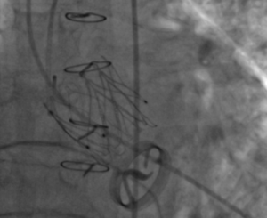
Figure 1. View at time of implantation demonstrating a newly deployed JenaValveTM in a patient with a Medtronic-Hall disc valve in the mitral position

Transcatheter aortic valve implantation (TAVI) has become standard of care for patients with severe aortic stenosis at prohibitive operative risk for surgical aortic valve replacement (SAVR).1 The first randomised-controlled trial of TAVI stipulated the presence of a mitral valve prosthesis as an exclusion criterion for enrolment in the trial.2 The main reason was concern that dysfunction of the mitral valve prosthesis might arise during TAVI valve deployment.3 Further concerns were that the presence of a rigid mitral prosthesis would inhibit complete valve deployment or would result in the valve slipping; a ‘watermelon seed’ effect.4 Despite this, a number of authors have published successful cases of TAVI in the presence of mechanical valve prostheses.

Methods

In order to determine the performance of TAVI in the presence of a mechanical mitral prosthesis we performed a systematic review of all published cases. Specifically, we sought to evaluate the clinical efficacy of TAVI in patients who had previously undergone mechanical mitral valve replacement. The review was undertaken in accordance with Preferred Reporting Items for Systematic Reviews and Meta-Analyses (PRISMA) guidelines. The primary outcome of interest was mortality at 30 days. Secondary outcomes included dysfunction of the mechanical prosthesis, TAVI valve embolisation, endocarditis, under-expansion of the TAVI valve and paravalvular leak, procedural success, and conversion to SAVR. Outcomes are reported in accordance with the Valve Academic Research Consortium (VARC) 2 consensus document.5

An online search of PubMed was performed on 30 August 2014. Search terms used included permutations of percutaneous, transcutaneous, transcatheter, transarterial, transfemoral, transapical, TAVI, transcatheter aortic valve replacement (TAVR), mechanical mitral prosthesis. Additional studies were identified using references cited within appropriate articles. Data were extracted by two independent authors (KOS, EH) and discrepancies were resolved by discussion.

Results

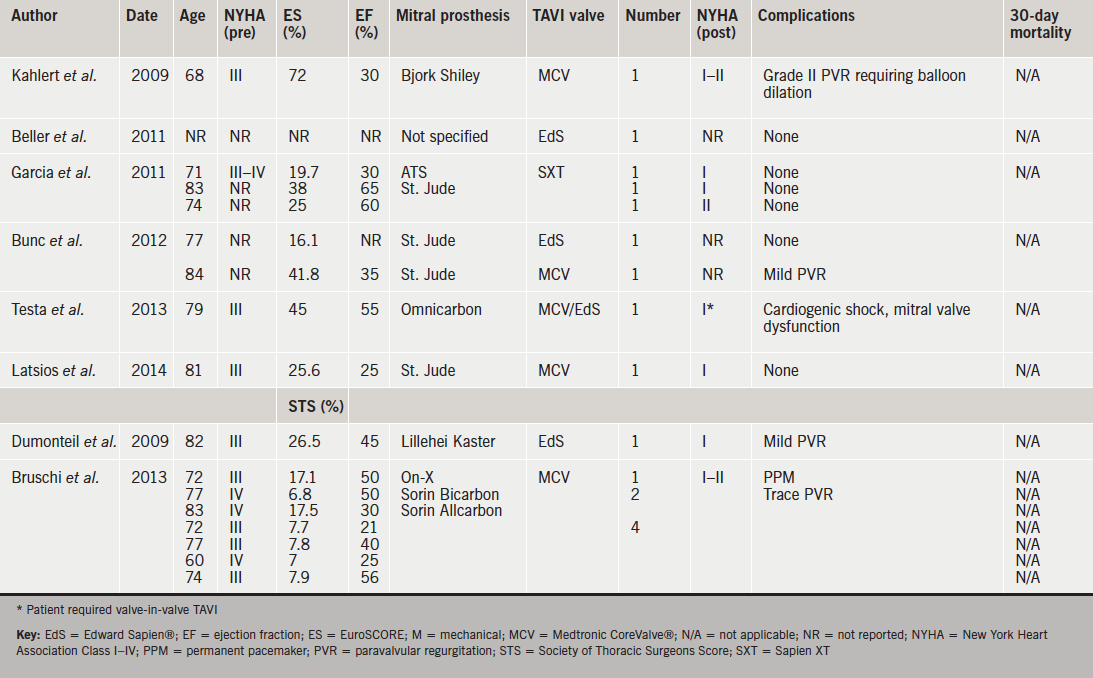
We identified 397 studies of which 16 were selected for full review. Of these, 11 were isolated case reports, and five were case series reporting two or more cases. A total of 35 cases were identified. Fifteen cases describe Medtronic CoreValve® implantation, 15 Edward Sapien®, three Sapien XT, one Medtronic Evolut™, and one JenaValve™ (figure 1. One case describes CoreValve® implantation with an interval subsequent implantation of an Edward Sapien®. Of these, eight authors report transfemoral cases (49%, n=17), outlined in table 1. Ten authors describe a transapical approach (40%, n=14), outlined in table 2. One author outlines cases performed via a transaortic approach (11%, n=4).

Table 1. Transfemoral transcatheter aortic valve implantation (TAVI)
Table 1. Transfemoral transcatheter aortic valve implantation (TAVI) (click to enlarge)
Table 1. Transfemoral transcatheter aortic valve implantation (TAVI)
Table 2. Transapical TAVI (click to enlarge)

The 30-day mortality rate post-TAVI was 5.7% (n=2). One patient was a 37-year-old woman with severe familial cholesterolaemia who had undergone a portocaval shunt in childhood and implantation of an apico-descending conduit four years prior, due to severe aortic calcification and a porcelain aorta. She underwent a subsequent mechanical mitral valve replacement. Her ejection fraction was 10%, EuroSCORE 85% and Society of Thoracic Surgeons predicted risk of mortality (STS PROM) was 75%. In this case, the valve was successfully deployed via the apex of the heart but severe left ventricular failure necessitated extracorporeal membrane oxygenation and the patient developed septicaemia and died on day 5.6 Long-term follow-up of reported cases was limited. However, one mortality eight months post-procedure was reported in a patient with aortic valve endocarditis requiring re-operation who died of multi-organ failure post-procedure.6

Late transcatheter heart valve embolisation has been identified in two reported cases.7 The first, a 75-year-old female with a prior mechanical prosthesis who, subsequent to transapical TAVI, presented with acute cardiac failure and an embolised Edward Sapien® prosthesis 21 days following implantation with a tilted valve and severe aortic regurgitation. The valve was retrieved at SAVR, however, she suffered a stroke and died seven days following surgery, so is, therefore, considered a 30-day mortality.8 The second was an 82-year-old female who re-presented 14 days following transapical TAVI with recurrent cardiac failure symptoms. Again, in this case, the TAVI prosthesis, an Edward Sapien® valve was tilted, resulting in severe aortic regurgitation. She underwent SAVR and aortic root replacement and was discharged successfully on day 14.9

Procedural success is reported in 97% of cases. There was no conversion to SAVR reported at the time of initial implantation. Testa et al. report implantation of a CoreValve® prosthesis in a patient with an Omnicarbon single-leaflet mechanical mitral prosthesis via the transfemoral approach.10 Authors describe the occurrence of cardiogenic shock, secondary to impairment of the mechanical prosthesis, due to it being situated too inferiorly. Suspecting the aetiology of the complication straightaway, they responded rapidly by first attempting to snare the CoreValve® and pull it back, then releasing it from the annulus and moving it into the ascending aorta. The patient recovered, and two months later underwent a transfemoral Edward Sapien® implantation by crossing the CoreValve®, which remained in the aortic position. She was discharged with significant symptomatic improvement on day 6.10

Procedural complications were reported in 6% (n=2). In one case of transfemoral CoreValve® implantation in a patient with a Bjork Shiley mitral prosthesis, grade two paravalvular regurgitation following valve deployment warranted balloon dilation.11 The second case describes transient mechanical prosthesis dysfunction resulting from the transapical delivery system touching a leaflet of the mechanical prosthesis. Upon immediate retraction the issue resolved and the subsequent implantation was uneventful.6 Interestingly, despite frequently cited concerns regarding the ‘watermelon seed’ effect, there have been no reported cases of this occurring to date. Furthermore, there were no reported cases of valve deformation. Post-procedural complications are reported in 11.4% (n=4) of cases. Specifically, one instance of pleural effusion following transapical TAVI, which required drainage. Also one patient in whom a permanent pacemaker was required.12,13 Paravalvular regurgitation, described as trace-mild, was reported in three cases.12,14,15

Discussion

Despite initial concerns, feasibility of transapical, transaortic and transfemoral TAVI in the presence of a mechanical prosthesis has been demonstrated. Experience to date, however, is limited and this patient cohort represents a relatively small proportion of the overall group eligible for TAVI. Considering our systematic review predominantly identified case reports, it is difficult to draw any firm conclusions regarding complication rates and 30-day mortality compared with larger patient cohorts. Furthermore, there is a significant risk of publication bias with authors more likely to publish successful cases of implantation than incidences of failure. This creates a likelihood of underestimation of the potential dangers involved. Nonetheless, the complications reported in cases published are well accepted; pacemaker insertion, endocarditis and mild paravalvular leak.2 What makes this group unique is the propensity for mechanical valve dysfunction, either intra- or post-procedurally. While two authors report this, it is likely that there is under-reporting of its incidence.6,10 It is difficult to recommend an optimum vascular approach based on the limited experience to date. Some would argue that the transaortic route offers the benefits of the control obtained via transapical TAVI without the additional risk of delivering the delivery sheath ‘past’ the mitral prosthesis in the left ventricle.16 This belief is unsubstantiated by evidence to date and is, therefore, speculative at present. In the absence of solid evidence, it is still reasonable that the least invasive approach should be adopted. Furthermore, it is possible that prosthesis choice is an important factor in this setting, one case report describes the use of the JenaValve, whereby a potential advantage of the prosthesis is the fact that there is a limit to the lower extent of valve deployment in the left ventricle due to the leaflet clipping mechanism built into the valve design.13 Again, while logical, there is insufficient evidence to date to conclusively recommend this.

One concern is that of late valve embolisation, as identified in two reported cases. While other issues likely to predispose to late embolisation exist, such as valve under sizing, under expansion of an appropriately sized valve due to aortic root calcification, asymmetric aortic root calcification, basal septal bulging and low valve implantation, the presence of a mechanical valve is likely to precipitate superior displacement of the TAVI prosthesis and remains an associated risk.7 Indeed, one of the reported cases at SAVR noted non-circularity of the aortic annulus in the region of the non-coronary sinus, the specific area in which the TAVI prosthesis had slipped into the ventricle.9

This study has a number of limitations. Case reports comprise the majority of the reported patients; therefore, there is likely bias towards under-reporting of adverse events and complications. This represents a small subpopulation of TAVI patients and is likely to be a scenario encountered quite rarely by each reporting author. Nonetheless, the review we have carried out does have value to those clinicians being faced with this challenge.

Conclusion

We demonstrate that TAVI, in patients with a prior mechanical prosthesis, is not only feasible but can be carried out without complication. Meticulous procedural planning is a necessity. Based on proof of feasibility, evidence to date would suggest that patients with mechanical prostheses be actively considered for TAVI going forward. Further studies are required to identify the potential benefits of certain prostheses in this setting.

Conflict of interest

None declared.

Key messages

  • Presence of a mechanical mitral valve was an exclusion criterion of the PARTNER (Placement of Aortic Transcatheter Valves) trial, however, to date 35 cases have been described in the literature
  • TAVI deployment in the presence of a mechanical mitral prosthesis is feasible, but should be approached with caution; incidences of mitral valve ‘locking’ have been reported
  • Strategies for success include pre-procedural study of the existing prosthesis, close examination of the aorto-mitral area, and shape of balloon at valvuloplasty, careful prosthesis selection and slow valve deployment

References

1. Holmes DR Jr, Mack MJ, Kaul S et al. 2012 ACCF/AATS/SCAI/STS expert consensus document on transcatheter aortic valve replacement. J Am Coll Cardiol 2012;59:1200–54. http://dx.doi.org/10.1016/j.jacc.2012.01.001

2. Leon MB, Smith CR, Mack M et al. Transcatheter aortic-valve implantation for aortic stenosis in patients who cannot undergo surgery. N Engl J Med 2010;363:1597–607. http://dx.doi.org/10.1056/NEJMoa1008232

3. Chao VT, Chiam PT, Tan SY. Transcatheter aortic valve implantation with preexisting mechanical mitral prosthesis – use of CT angiography. J Invasive Cardiol 2010;22:339–40. Available from: http://www.invasivecardiology.com/articles/Transcatheter-Aortic-Valve-Implantation-with-Preexisting-Mechanical-Mitral-Prosthesis-—-Use

4. Garcia E, Albarran A, Heredia-Mantrana J et al. [Transcatheter aortic valve implantation in patients with a mechanical mitral valve]. Rev Esp Cardiol 2011;64:1052–5. http://dx.doi.org/10.1016/j.recesp.2011.02.022

5. Kappetein AP, Head SJ, Genereux P et al. Updated standardized endpoint definitions for transcatheter aortic valve implantation: the Valve Academic Research Consortium-2 consensus document. J Thorac Cardiovasc Surgery 2013;145:6–23. http://dx.doi.org/10.1016/j.jtcvs.2012.09.002

6. Drews T, Pasic M, Buz S et al. Transapical aortic valve implantation after previous mitral valve surgery. J Thorac Cardiovasc Surg 2011;142:84–8. http://dx.doi.org/10.1016/j.jtcvs.2010.08.034

7. Mylotte D, Andalib A, Theriault-Lauzier P et al. Transcatheter heart valve failure: a systematic review. Eur Heart J 2014;published online. http://dx.doi.org/10.1093/eurheartj/ehu388

8. Maroto LC, Rodriguez JE, Cobiella J, Silva J. Delayed dislocation of a transapically implanted aortic bioprosthesis. Eur J Cardiothorac Surg 2009;36:935–7. http://dx.doi.org/10.1016/j.ejcts.2009.03.072

9. Baumbach H, Hill S, Hansen M, Franke UF. Severe aortic insufficiency after transapical aortic valve implantation. Ann Thorac Surg 2011;92:728–9. http://dx.doi.org/10.1016/j.athoracsur.2011.02.047

10. Testa L, Gelpi G, Bedogni F. Transcatheter aortic valve implantation in a patient with mechanical mitral prosthesis: a lesson learned from an intraventricular clash. Catheter Cardiovasc Interv 2013;82:E621–E625. http://dx.doi.org/10.1002/ccd.24948

11. Kahlert P, Eggebrecht H, Thielmann M et al. Transfemoral aortic valve implantation in a patient with prior mechanical mitral valve replacement. Herz 2009;34:645–7. http://dx.doi.org/10.1007/s00059-009-3295-5

12. Bruschi G, De Marco F, Barosi A et al. Self-expandable transcatheter aortic valve implantation for aortic stenosis after mitral valve surgery. Interact Cardiovasc Thorac Surg 2013;17:90–5. http://dx.doi.org/10.1093/icvts/ivt086

13. O’Sullivan KE, Casserly I, Hurley J. Transapical JenaValve in a patient with mechanical mitral valve prosthesis. Catheter Cardiovasc Interv 2014;published online. http://dx.doi.org/10.1002/ccd.25415

14. Dumonteil N, Marcheix B, Berthoumieu P et al. Transfemoral aortic valve implantation with pre-existent mechanical mitral prosthesis: evidence of feasibility. JACC Cardiovasc Interv 2009;2:897–8. http://dx.doi.org/10.1016/j.jcin.2009.05.023

15. Bunc M, Ambrozic J, Music S et al. Successful Tavi in patients with previous mitral valve replacement. Int J Clin Med 2012;3:603–06. http://dx.doi.org/10.4236/ijcm.2012.37109

16. Bruschi G, De Marco F, Oreglia J et al. Percutaneous implantation of CoreValve aortic prostheses in patients with a mechanical mitral valve. Ann Thorac Surg 2009;88:e50–e52. http://dx.doi.org/10.1016/j.athoracsur.2009.07.028

Cardiac orientation: is there a correlation between the anatomical and the electrical axis of the heart?

Br J Cardiol 2015;22:(2)doi:10.5837/bjc.2015.016 Leave a comment
Click any image to enlarge
Authors:
First published online April 15, 2015

Data have suggested that in vivo cardiac orientation has the greatest effect on the cardiac electric field, and, thus, surface electrical activity. We sought to determine the correlation between in vivo cardiac orientation using cardiac computed tomography (CT) and the electrical cardiac axis in the frontal plane determined by surface electrocardiogram (ECG). 

Patients aged between 30 and 60 years old with a normal body mass index (BMI), who underwent CT coronary angiography between July 2010 and December 2012 were included. Patients with diabetes, hypertension, arrhythmias, structural heart disease or thoracic deformities were excluded. In vivo cardiac orientation was determined along the long axis and correlated with the electrical cardiac axis on surface ECG.

There were 59 patients identified, with 47% male, mean age of 49.9 years and a mean BMI of 22.39 kg/m2. The mean cardiac axis on CT was 38.1 ± 7.8°, while the mean electrical cardiac axis on ECG was 51.8 ± 26.6°. Bi-variate analysis found no correlation between the two readings (Pearson r value 0.12, p=0.37).

We conclude, there is no simple relationship between the anatomical cardiac axis and the ECG determined electrical axis of the heart. The electrical axis of the heart, however, showed more variability, reflecting possible underlying conduction disturbances.

Introduction

Due to the asymmetry of the heart, it has long been described in what is known as the ‘Valentine’ position, in which the heart is oriented vertically downwards. It defines the heart as a solitary organ and provides no reference point for its location within the chest. This description has since been found to be inaccurate, as we know the heart is positioned in a direction extending from the right shoulder to the left hypochondrium. The in vivo orientation of the heart takes into account its surrounding bony structures and is the best definition of true anatomical heart position.1,2

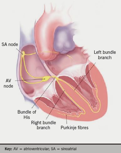
Figure 1. Pathway of cardiac electrical activation

In 1951, Fowler and Braunstein noted a significant association between electrocardiographic and anatomical positions of the heart about the antero-posterior and longitudinal axes, but notably not along the transverse axis. This study used X-ray and angiocardiogram to assess anatomical cardiac position.3 As seen in figure 1, cardiac electrical activation is generated from the sinus node located high in the right atrium and spreads throughout the atria to the atrioventricular node in the infero-posterior region of the interatrial septum. It then enters the base of the ventricle at the bundle of His and follows the left and right bundle branches along the interventricular septum. The pathway of activation described, in approximation, travels along the long axis of the heart. Therefore, an association between the electrocardiographic axis and the anatomical position along the long axis seems feasible.4,5 The electrical cardiac axis on electrocardiogram (ECG), however, represents the mean direction of the electrical action potential during ventricular depolarisation.

A small canine study in 2005 by Arteeva et al. supported Fowler’s findings, and concluded that the orientation of the heart within the thorax affected the formation of the cardiac electric field on the body surface much more than the torso geometry.6 The same year, Engblom et al. conducted a study using cardiac magnetic resonance imaging (CMR) that suggested there was no simple relationship between the electrical and anatomical axes of the heart.7 Therefore, it remains largely unclear whether the electrical cardiac axis and the anatomical cardiac axis are entirely separate entities or whether they are, in fact, related.

We sought to identify in vivo cardiac orientation along the long axis using computed tomography (CT) to calculate an individual’s true anatomical axis. We then subsequently assessed its correlation with their electrical cardiac axis determined on ECG in the
frontal plane.

Methods

This was a retrospective study that included patients who underwent CT coronary angiography (CTCA) at three private radiology centres across Sydney between July 2010 and December 2012.

A Siemens SOMATOM Definition Flash dual-source 128-slice and a General Electric Lightspeed VCT 64-slice CT scanner were both used across the three radiology centres. Six CTs were performed using the 64-slice CT scanner while the remaining were all performed using the 128-slice CT scanner.

Participants

There were 59 patients identified as appropriate for the study. Patients aged between 30 and 60 years old with a normal body mass index (BMI) were included. Normal BMI was defined as between 18.5 kg/m2 and 24.99 kg/m2 according to the World Health Organization (WHO) guidelines.8 Patients with diabetes, hypertension, arrhythmias, structural heart disease or thoracic deformities were excluded. Diabetes and hypertension were defined as any individual on one or more diabetic or anti-hypertensive medications, respectively. Arrhythmias referred to tachyarrhythmias only, and patients were excluded regardless of whether they were paroxysmal or chronic in nature. Patients who had previously had ablative procedures for these tachyarrhythmias were also excluded. Those with congenital or acquired structural heart disease, or thoracic abnormalities were also excluded.

CT analysis

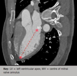
Figure 2. Multi-planar reconstruction (MPR) view of the cardiac long axis

The CT scanning technique used was at the discretion of the radiographers and cardiologists present at the time of the scan. This often adhered to the scanning protocols available at each of the individual radiology centres.

In vivo cardiac orientation was determined along the long axis using Osirix image analysis. Multi-planar reconstruction (MPR) views were used to mark a point at the left ventricular apex and the centre of the mitral annulus. The line connecting these points was marked as the cardiac long axis, as seen in figure 2.

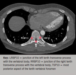
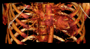
A point at the most distal aspect of the sternum was marked as the xiphisternum. 3D volume-rendered images and axial views were used in conjunction to mark points along the vertebral column. Points were marked along the most posterior aspect of the vertebral foramen, as well as at both junctions of the ribs with the vertebral body at each vertebral level (figures 3 and 4).

Figure 3. Vertebral points

The angle of the long axis was measured and subtracted from the bony landmarks to give in vivo cardiac orientation along the long axis.

ECG analysis

A standard 12-lead ECG performed in the frontal plane was obtained for each patient. The majority of patients had ECGs performed within 12 months of the CTCA. Three patients did not have recent ECGs and their most recent ECG was approximately five years prior to their CTCA.

The electrical cardiac axis was determined using the formula below. This formula uses leads I and aVF. The correlation factor in the formula counteracts the inaccuracy created by the difference in the electrical strength of aVF, which is a bipolar lead and lead I which is a unipolar lead.9,10

Equation

Statistical analysis

Figure 4. Volume-rendered image of thechest with multiple bony and cardiacstructures marked
Figure 4. Volume-rendered image of the
chest with multiple bony and cardiac
structures marked

Bi-variate analysis was performed using SPSS, version 21. The results are all presented as mean ± standard deviation (SD), followed by the range of values in brackets. All axes, both anatomical and electrical are displayed in degrees. The anatomical axis was calculated on Osirix image analysis in radians and subsequently converted to degrees using the formula below:

Degrees = Radians × 180/π

Results

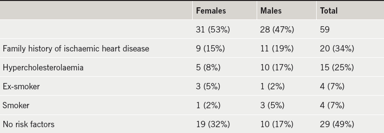
There were 59 patients suitable to be included in the study with similar numbers of men and women, as shown in table 1. Their cardiac profile is also demonstrated in this table. There were no cardiac risk factors in 49% of the patients. The average age was 49.9 ± 7.4 (31–60) years. The average BMI was 22.39 ± 1.75 (18.67–24.88) kg/m2. A double-flash scan was performed in 23 (39%) of the patients, as shown in table 2.

Table 1. Patient demographics
Table 2. Computed tomography (CT) scan technique

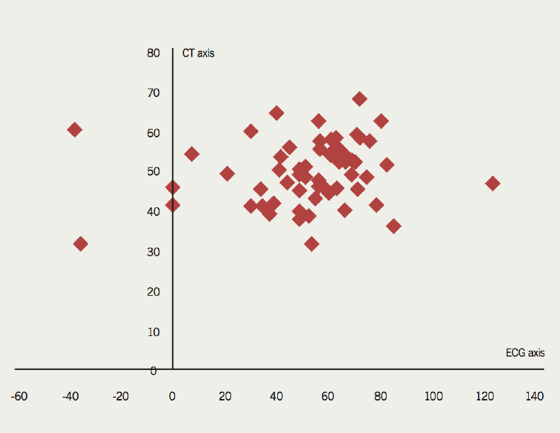
The scatter plot in figure 5 shows the distribution of in vivo cardiac axis on CT and the electrical cardiac axis on ECG. It demonstrates that there is no clear relationship between these two axes.

Figure 5. Plot of electrocardiogram (ECG) axis versus computed tomography (CT) axis
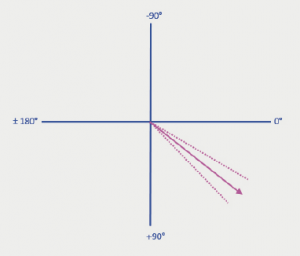
Figure 6. Mean cardiac axis on CT,38.1 ± 7.8° (broken lines demonstratestandard deviation)
Figure 6. Mean cardiac axis on CT, 38.1 ± 7.8° (broken lines demonstrate standard deviation)

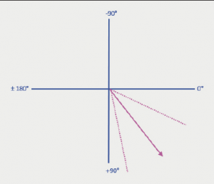
Bi-variate analysis was performed on the data. A Pearson correlation coefficient (r) of 0.12 (p=0.37) and Spearman correlation coefficient (ρ) of 0.16 (p=0.23) confirms that there is no correlation between the in vivo cardiac axis determined on CT and the electrical cardiac axis determined on ECG. The average in vivo cardiac axis on CT was 38.1 ± 7.8° (20.9–56.2°) (figure 6). The average electrical cardiac axis on ECG was 51.8 ± 26.6° (–37.6–125.8°) (figure 7). The calculated in vivo cardiac axis showed significantly less variation compared with the electrical cardiac axis calculated from the ECG.

The average difference between the two readings was 24.0 ± 18.0° (0.91–89.79°). Three patients who had the greatest difference between their in vivo cardiac axis and their electrical cardiac axis are detailed in table 3.

Figure 7. Mean cardiac axis on ECG,51.8 ± 26.6° (broken lines demonstratestandard deviation)
Figure 7. Mean cardiac axis on ECG, 51.8 ± 26.6° (broken lines demonstrate standard deviation)

Two of these patients had left-axis deviation on their ECG and one patient had right-axis deviation on their ECG. The left-axis deviation measured in the two patients was –35.1° and –37.6°, which corresponded to a CT anatomical axis of 20.9° and 50.5°, respectively. The right-axis deviation measured in patient 2 was 125.8°, which corresponded to a CT anatomical axis of 36.0°. These three patients all had a normal QRS width ranging from 60–100 ms. Of note, both patients with left-axis deviation had abnormalities in their transition zone. One had a delayed transition zone, while the other had evidence of R-wave regression. Patient 2 with the right-axis deviation had no electrocardiographic abnormalities of their transition zone. The remaining patients all had an electrical cardiac axis within the normal range (–30° to 90°).

Table 3. Patients with abnormal electrical cardiac axis

Discussion

Our study suggests that there is no correlation between in vivo cardiac orientation and the ECG-determined electrical cardiac axis. The in vivo cardiac orientation refers to the true anatomical cardiac axis, which accounts for its surrounding structures. The electrical cardiac axis, however, represents the mean direction of the electrical action potential during ventricular depolarisation. It has previously been thought that the anatomical position of the heart would be reflected in the electrical cardiac axis. Our study, however, suggests that there is no simple relationship between these two axes in healthy, young to middle-aged individuals. Although, it certainly seems plausible that changes in the anatomical cardiac axis would result in changes in the electrical cardiac axis, there does not appear to be a simple measure of how exactly this occurs.

The mean in vivo cardiac axis calculated on CT was 38.1°. There was little variability around this measurement, suggesting this is a good approximation of the in vivo cardiac axis in young to middle-aged healthy individuals. The mean electrical axis calculated on ECG was 51.8°. There was, however, a great deal more variability in the calculated electrical cardiac axis. This is suspected to be due to variations in the pathway of cardiac activation due to subclinical conduction disturbances.

Intra-individual variability of ECGs has been described in detail by Schijvenaars et al. Limb-lead interchanges are quite common and also, unfortunately, quite difficult to recognise on ECG. He, nevertheless, concludes that intra-individual variability seems to contribute less to ECG readings when compared with inter-individual variables such as age, obesity and sex.11-16 In our series, we used young to middle-aged individuals between the ages of 30 and 60 years old. Our study used patients with a normal BMI based on the WHO guidelines. The WHO guidelines for the definition of a normal BMI are the same for different ages and sex, although some debate has been raised as to whether these values need to be modified for Asian and Pacific populations. This was, however, studied by the committee and it was concluded that the guidelines should remain unchanged as international classification for all ethnic groups.5 There was also an approximate equal distribution of men and women in our study, which should potentially avoid any significant biases based on gender.

Thus, assuming any potential inaccuracies of ECG recordings are negligible, the variability noted in the electrical cardiac axis may, in fact, be due to subclinical conduction disturbances. These conduction disturbances can delay intra-cardiac conduction and alter the course of the cardiac action potential. This, in turn, may cause a change in the electrical cardiac axis. However, our study shows that these changes in the electrical cardiac axis are not necessarily reflected in changes in the true anatomical cardiac axis.

Additionally, Foster et al. used CMR to determine the effects of respiration and cardiac cycle changes on the left ventricular long axis. Although his study had relatively small numbers, it concluded that these variables did not contribute significantly to the long axis orientation.16,17

Variations in the transitional zone on ECG were noted in those with a left electrical cardiac axis. A left-axis deviation was seen with a delayed transition zone in one patient, and with R-wave regression in another. A delayed transition zone was identified as a shift of the transitional zone to the left, beyond lead V4. The shift of the transitional zone to the left precordial leads is generally referred to as a clockwise rotation of the heart, which is a widely accepted definition.3,18-21 Tahara et al. used cardiac CT to describe that a shift in the transitional zone is associated with an anatomical rotation about the long axis in approximately two-thirds of people.22 His study, however, did not explore the relationship between the transition zone and the electrical cardiac axis.

Two of the major limitations of this study were the small sample size and the retrospective study design. The small sample size was predominantly due to the narrow inclusion and exclusion criteria used. This is attributable to the fact that it is uncommon for relatively well individuals without significant cardiac problems or risk factors to have a CTCA performed.

Conclusion

Our study suggests that there is no correlation between the in vivo anatomical cardiac axis and the electrical cardiac axis in healthy young to middle-aged individuals. The electrical cardiac axis was, however, found to be subject to a greater degree of variability, and this is thought to be due to subclinical conduction disturbances. The study also raises the possibility of an association between a shift in the transitional zone on ECG and the electrical cardiac axis. Further studies with larger numbers are warranted to support these findings.

Acknowledgements

This abstract has been presented at CSANZ New Zealand, 2013 and at SCCT Montreal, 2013.

Funding

This research received no grant from any funding agency in the public, commercial or not-for-profit sectors.

Conflict of interest

None declared.

Key messages

  • In vivo cardiac orientation is a more accurate definition of the anatomical position of the heart
  • There is no simple relationship between the anatomical and electrical cardiac axis of the heart in young to middle-aged healthy individuals
  • There was more variability in the electrical cardiac axis, possibly due to underlying conduction abnormalities
  • There may be a relationship between the transition zone and the cardiac electrical axis

References

1. Partridge JB, Anderson RH. Left ventricular anatomy: its nomenclature, segmentation, and planes of imaging. Clin Anat 2009;22:77–84. http://dx.doi.org/10.1002/ca.20646

2. Anderson RH, Loukas, M. The importance of attitudinally appropriate description of cardiac anatomy. Clin Anat 2009;22:47–51. http://dx.doi.org/10.1002/ca.20741

3. Fowler NO, Braunstein JR. Anatomic and electrocardiographic position of the heart. Circulation 1951;3:906–10. http://dx.doi.org/10.1161/01.CIR.3.6.906

4. Boineau JP, Spach MS. The relationship between the electrocardiogram and the electrical activity of the heart. J Electrocardiol 1968;1:117–24. http://dx.doi.org/10.1016/S0022-0736(68)80014-7

5. Pullan AJ, Buist ML, Sands GB, Cheng LK, Smith NP. Cardiac electrical activity – from heart to body surface and back again. J Electrocardiol 2003;36(suppl):63–7. http://dx.doi.org/10.1016/j.jelectrocard.2003.09.016

6. Arteeva NV, Roshcevskaya M, Vityazev VA, Shmakov DN, Roshchevskii MP. Effect of torso shape and heart location in the chest on formation of cardiac electric potentials on body surface in dogs. Bull Exp Biol Med 2005;140:165–7. http://dx.doi.org/10.1007/s10517-005-0435-7

7. Engblom H, Foster JE, Martin TN et al. The relationship between electrical axis by 12-lead electrocardiogram and anatomical axis of the heart by cardiac magnetic resonance in healthy subjects. Am Heart J 2005;150:507–12. http://dx.doi.org/10.1016/j.ahj.2004.10.041

8. World Health Organization. Global database on body mass index. Available from: http://www.who.int/bmi/index.jsp [accessed June 2009].

9. Hoseini SS, Moeeny A, Shoar S et al. Designing nomogram for determining the heart’s QRS axis. J Clin Basic Cardiol 2011;14:12–15. Available from: http://www.kup.at/kup/pdf/10452.pdf

10. Hancock EW, Deal BJ, Mirvis DM, Okin P, Kligfield P, Gettes LS. AHA/ACCF/HRS recommendations for the standardization and interpretation of the electrocardiogram. Part V: electrocardiogram changes associated with cardiac chamber hypertrophy: a scientific statement from the American Heart Association Electrocardiography and Arrhythmias Committee, Council on Clinical Cardiology; the American College of Cardiology Foundation; and the Heart Rhythm Society. Endorsed by the International Society for Computerized Electrocardiology. J Am Coll Cardiol 2009;53:992–1002. http://dx.doi.org/10.1016/j.jacc.2008.12.015

11. Schijvenaars BJA, Gerard van Herpen P, Kors JA. Intraindividual variability in electrocardiograms. J Electrocardiol 2008;41:190–6. http://dx.doi.org/10.1016/j.jelectrocard.2008.01.012

12. Hoekema R, Uijen GJ, van Erning L, van Oosterom A. Interindividual variability of multilead electrocardiographic recordings – influence of heart position. J Electrocardiol 1999;32:137–48. http://dx.doi.org/10.1016/S0022-0736(99)90092-4

13. van Oosterom A, Hoekema R, Uijen GJ. Geometrical factors affecting the interindividual variability of the ECG and the VCG. J Electrocardiol 2000;33(suppl 1):219–27. http://dx.doi.org/10.1054/jelc.2000.20356

14. Wenger W, Kligfield P. Variability of precordial electrode placement during routine electrocardiography. J Electrocardiol 1996;29:179–84. http://dx.doi.org/10.1016/S0022-0736(96)80080-X

15. Peberdy MA, Ornato JP. Recognition of electrocardiographic lead misplacements. Am J Emerg Med 1993;11:403–05. http://dx.doi.org/10.1016/0735-6757(93)90177-D

16. Foster JE, Engblom H, Thomas NM et al. Determination of left ventricular long-axis orientation using MRI: changes during the respiratory and cardiac cycles in normal and diseased subjects. Clin Physiol Funct Imaging 2005;25:286–92. http://dx.doi.org/10.1111/j.1475-097X.2005.00624.x

17. Hoffman EA, Ritman EL. Shape and dimensions of cardiac chambers: Importance of CT section thickness and orientation. Radiology 1985;155:739–44. http://dx.doi.org/10.1148/radiology.155.3.400137

18. Goldberger E. Unipolar lead electrocardiography and vectorcardiography. Philadelphia: Lea & Febiger, 1953.

19. Goldberger E. Effect of clockwise rotation of the heart on the electrocardiogram. Am J Med 1949;7:756–9. http://dx.doi.org/10.1016/0002-9343(49)90414-3

20. Goldman MJ. Principles of clinical electrocardiography. 11th edition. Los Altos: Lange Medical Publications, 1982.

21. Cooksey JD, Dunn M, Masae E. Clinical vectorcardiography and electrocardiography. 2nd edition. Chicago: Year Book Medical Publishers, 1977.

22. Tahara Y, Mizuna H, Ono A, Ishikawa K. Evaluation of the electrocardiographic transitional zone by cardiac computed tomography. J Electrocardiol 1991;24:239–45. http://dx.doi.org/10.1016/0022-0736(91)90029-L

Coronary and bypass graft angiography using a single catheter via the left trans-radial artery

Br J Cardiol 2015;22:(2)doi:10.5837/bjc.2015.017 Leave a comment
Click any image to enlarge
Authors:
First published online April 15, 2015

Using the left trans-radial artery access route for coronary and bypass angiography has been frowned upon by the majority of operators due to several catheter changes during the procedure, patient discomfort because of spasm, positioning of the patients left arm and longer radiation exposure times, to name a few reasons. This short scientific article demonstrates one of a series of 22 cases where a single catheter was successfully used via this access route.

Introduction

The traditional approach to coronary catheterisation in patients with a history of coronary artery bypass grafting (CABG) with left internal mammary artery (LIMA) graft is via the femoral approach. However, trans-radial catheterisation still remains a distinct option for such patients. Operators tend to be cautious about using this approach due to the nature of having to exchange diagnostic catheters several times during the procedure, risk of spasm, alternative set-up for the procedure, arm positioning, etc. A retrospective audit of 22 single-operator coronary graft angiography cases over a six-month period demonstrates the feasibility and option to use the left trans-radial approach (LTRA) to perform a coronary and graft study with the use of a single diagnostic catheter – the TIGER (TIG) catheter (Terumo Medical Corporation).

Most operators, who opt to use the LTRA, routinely practice with the standard femoral diagnostic catheters to access the native coronary arteries and grafts.1 A disadvantage of using LTRA for graft angiography is because several catheter exchanges are necessary to engage both native coronary arteries and the bypass grafts.

My standard approach for performing coronary and graft angiography in patients is via the left radial artery (LRA) in those with a LIMA graft and via the right radial artery in those without. My personal preference is to use the TIG catheter as the exclusive standard catheter of choice for all coronary angiography via the radial artery. The TIG is designed to enable coronary angiography of the left coronary artery (LCA) and right coronary artery (RCA) via the right radial artery without the need for any catheter exchange.2 Over a six-month period, I performed a series of 22 cases using the TIG catheter via the left radial to successfully perform both coronary and graft angiography. This case description illustrates the last in the short series of successful procedures.

Case description

A 68-year-old man with a history of a three-vessel bypass in 2007. The bypass grafts were: saphenous vein graft (SVG) to obtuse marginal (OM), SVG to RCA and LIMA to the left anterior descending artery (LAD).

The patient was referred for coronary angiography because he was experiencing a central crushing chest pain radiating to his left arm, relieved by glyceryl trinitrate (GTN) spray. Exercise tolerance had reduced in recent weeks with increasing shortness of breath. Other significant medical history to note included hypertension, diabetes mellitus and peripheral vascular disease, and is an ex-smoker.

I performed a left radial Allen’s test before the procedure. Ulnar arch patency using pulse oximetry was confirmed with a Barbeau score of A.3 A 6f Terumo Glidesheath (Terumo Medical Corporation) was inserted using the standard Seldinger technique. My standard radial angiography protocol is to administer 200 µg of nitrate, and 5000 IU heparin intra-arterially through the radial sheath before the catheter is inserted. The TIG catheter was fed up through the left arm with a 0.35”/150 cm Emerald (Cordis, Johnson & Johnson) guidewire and into the left subclavian artery.

The catheter was used to first intubate the LCA. Figure 1A shows severe distal left main stem disease, a blocked proximal LAD and a severe lesion in the circumflex artery just proximal to a blocked OM artery. After disengagement, the TIG was rotated to the RCA (figure 1B) and showed it was blocked proximally. The TIG was disengaged and pulled back to engage the SVG to the obtuse marginal (OM) (figure 1C) showing that there was a mild ostial narrowing in an otherwise patent graft. The catheter was then disengaged and manipulated to intubate the SVG to posterior descending artery (PDA) (figure 1D). The SVG to PDA was patent with a good run off. The catheter was disengaged for a final time before being withdrawn slowly into the subclavian artery where the LIMA graft was successfully engaged to show a widely patent graft with a severe lesion just beyond the anastomosis site (figure 1E). The guidewire was re-inserted to remove the TIG catheter over the wire. Haemostasis was achieved using a TR band (Terumo Medical Corporation).

Figure 1. Coronary angiograms for native and grafts that demonstrate good engagement and co-axial alignment of the TIG catheter with native coronaries, saphenous vein grafts (SVGs) and left internal mammary artery (LIMA) via left trans-radial approach (LTRA) approach

Discussion

Figure 2. Standard 5.2 French diagnostic catheters used during native coronary and graft angiography. From left to right: TIG, Judkins right 4.0 (modified) catheter, Judkins left 4.0 catheter, right coronary bypass catheter, left bypass catheter and internal mammary (IM)

Operators who use the left trans-radial approach for coronary and graft angiography achieve comparable outcomes to those operators using the trans-femoral approach.4 However, this has been through the use of several different diagnostic catheters – Judkins, Amplatz, internal mammary or multi-purpose catheters (figure 2).

Several years ago, using the left trans-radial technique, initially meant another learning curve in what has now proven to be an effective and more patient friendly access route. Learning to handle and manipulate a single catheter through this particular access site will mean another exciting step in trans-radial education.

I found there was a learning curve of 15 cases before I had successfully adapted my practice. The structural association between the coronary arteries and aortic arch in the left radial approach varies from that of the right radial approach and even the femoral approach.5 This implies that re-education of certain manipulation techniques of the TIG catheter have to be developed to ensure a positive outcome, as well as patient satisfaction.

The TIG was originally designed to be used exclusively as a right radial catheter (Terumo Medical Corporation, 2008). It is my standard work-horse diagnostic catheter for right trans-radial procedures, and it is now my first choice for left trans-radial approaches as well.

Table 1. Mean comparison of screening time and radiation dose for left transradial cases using a single catheter versus a multi-catheter approach via the trans-femoral route

We found that there was no spasm present in any of the cases – this is attributed to the lack of catheter exchanges. Radiation and exposure times can be prolonged due to the catheter exchanges or spasm that may occur. We compared the mean doses and screening times for these cases against the same number (n=22) of retrospectively performed trans-femoral graft angiography cases. The results indicated that by using this single-catheter approach via the left trans-radial approach, the procedure dose and screening times were reduced by 25.4% and 21.3%, respectively (table 1).

Although challenging at first, due to its manipulation, the TIG was found to intubate the LIMA when it was the last artery to review. We have deduced that this may be due to the catheter having ‘softened’ during the procedure and, therefore, being more malleable to co-axially align itself to the angulation of the vessel.

Conclusion

When considering using the left radial artery for coronary and graft angiography, the TIG catheter can be safely used as a single catheter of choice to successfully engage all grafts, as well as the native coronary arteries, thereby, enhancing the patient experience, promoting less spasm, reducing radiation doses and exposure times.

Conflict of interest

None declared.

References

1. Suh WM, Kern M. Coronary and bypass graft angiography via the right radial approach using a single catheter. J Invasive Cardiol 2012;24:295–7. Available from: http://www.invasivecardiology.com/articles/coronary-and-bypass-graft-angiography-right-radial-approach-using-single-catheter

2. Terumo Medical Corporation (2008). Website: http://www.terumomedical.com

3. Barbeau GR, Arsenault F, Dugas L, Simard S, Lariviere MM. Evaluation of the ulno-palmar arterial arches with pulse oximetry and plethysmography: comparison with the Allen’s test in 1010 patients. Am Heart J 2004;147:489–93. http://dx.doi.org/10.1016/j.ahj.2003.10.038

4. Sanmartin M, Cuevas D, Moxica J et al. Trans-radial cardiac catheterization in patients with coronary bypass grafts: feasibility analysis and comparison with trans-femoral approach. Catheter Cardiovasc Interv 2006;67:580–4. http://dx.doi.org/10.1002/ccd.20633

5. Saito S. Right or left side? Catheter Cardiovasc Interv 2003;58:305. http://dx.doi.org/10.1002/ccd.10462

News from ACC.15

Br J Cardiol 2015;22:(2) Leave a comment
Click any image to enlarge
Authors:
First published online April 15, 2015

As well as the high-tech interventional advances including radial access percutaneous coronary intervention (PCI), new antibodies against cholesterol, and coronary angiography for heart disease diagnosis, maybe the most important studies presented at this year’s American College of Cardiology (ACC) focused on the considerably low-tech interventions of avoiding sitting for too long, and losing weight to reduce important cardiovascular risk factors. We report from the 64th Annual Scientific Session held in San Diego, California, USA from 14th–16th March 2015.

Screen shot 2015-04-14 at 16.36.19Too much sitting increases coronary artery calcification

Sitting for many hours per day is associated with increased coronary artery calcification, a marker of subclinical heart disease, a new study suggests.

The study found no association between coronary artery calcification (CAC) and the amount of exercise a person gets, suggesting that too much sitting might have a greater impact than exercise on this particular measure of heart health.

The results suggest that exercise may not entirely counteract the negative effects of a mostly sedentary lifestyle on coronary artery calcium.

Presenting the study at the ACC meeting, Dr Jacquelyn Kulinski (Medical College of Wisconsin, USA), said:  “It’s clear that exercise is important to reduce your cardiovascular risk. But this study suggests that reducing how much you sit every day may represent a more novel, companion strategy (in addition to exercise) to help reduce your cardiovascular risk.”

Other studies published recently have linked excess sitting with an increased risk for cardiovascular disease, diabetes, cancer and early death. This study suggests that the mechanism behind these observations may be coronary calcium.

For the study, Dr Kulinski and colleagues analysed data from 2,031 participants in the Dallas Heart Study who had undergone a computed tomography (CT) scan to measure CAC. All participants wore a watch accelerometer for at least four days to measure body movements, which were classed as sedentary, light activity, or moderate to vigorous physical activity. On average, participants were sedentary for 5.1 hours a day, but this ranged from 1.1 to 11.6 hours a day.

Results showed that each hour of sedentary time per day on average was associated with a 14 % increase in CAC burden. The association was independent of exercise activity and other traditional heart disease risk factors.

“I think the study offers a promising message. Reducing the amount of time you sit by even an hour or two a day could have a significant and positive impact on your future cardiovascular health,” Dr Kulinski said.

“The lesson here is that it’s really important to try to move as much as possible in your daily life; for example, take a walk during lunch, pace while talking on the phone, take the stairs instead of the elevator and use a pedometer to track your daily steps,” Dr Kulinski said. “And if you do have a very sedentary job, don’t go home at night and sit in front of the TV for hours on end.”

PEGASUS: ticagrelor shows benefits post-MI but bleeding is concern

The antiplatelet agent, ticagrelor, was associated with a reduction in myocardial infarction (MI), stroke, or cardiovascular death versus placebo in secondary prevention patients, but this was accompanied by a higher risk of clinically significant bleeding and transfusion, as well as a higher rate of dyspnoea in the PEGASUS-TIMI 54  (Prevention of Cardiovascular Events in Patients with Prior Heart Attack Using Ticagrelor Compared to Placebo on a Background of Aspirin-Thrombolysis in Myocardial Infarction) trial.

Presenting the results, Dr Marc Sabatine (Brigham and Women’s Hospital, Boston, USA) said he would “consider such therapy as long as it was well tolerated”.

But writing in an editorial (doi: 10.1056/NEJMe1502137) accompanying publication of the PEGASUS-TIMI 54 paper (doi: 10.1056/NEJMoa1500857) in the New England Journal of Medicine Dr John F Keaney Jr (Massachusetts Medical School, Worcester, USA) points out the “fragile balance between efficacy and adverse events” and asks whether dual platelet inhibition with high-potency agents is approaching the point of diminishing returns.

Dr Sabatine explained that dual antiplatelet therapy is recommended for the first year after an acute coronary syndrome (ACS) event; post-hoc analysis from the CHARISMA (Clopidogrel and Aspirin versus Aspirin Alone for the Prevention of Atherothrombotic Events) trial suggested that prolonged therapy might be beneficial for patients with a prior MI.

The PEGASUS-TIMI 54 study involved 21,162 patients who had had an MI a mean of 1.7 years previously. They also had at least one additional risk factor: age 65 years or older, diabetes, a second prior MI more than a year ago, multi-vessel coronary artery disease, or chronic kidney disease.

Patients were randomised to high dose (90 mg twice daily), low dose (60 mg twice daily) ticagrelor or placebo, with all patients continuing to take aspirin.

Table 1. Major results from the PEGASUS-TIMI 54 study

Results (see table 1) showed that after a median follow-up of 33 months, both doses of ticagrelor significantly lowered the risk of the primary composite efficacy outcome – cardiovascular death/MI/stroke. But this was at the cost of a two- to three-fold increase in major bleeding and risk of dyspnoea.

The researchers point out that the lower dose of ticagrelor may offer a more attractive risk/benefit profile. They estimate that for every 10,000 patients who began treatment, taking 90 mg ticagrelor twice daily would prevent 40 primary end points and lead to 42 TIMI major bleeding events per year, whereas taking 60 mg ticagrelor twice daily would prevent 40 primary end points and lead to 31 TIMI major bleeding events per year.

While fatal bleeding and non-fatal intracranial haemorrhage occurred in less than 1% of patients in each group, they note that the study excluded patients with recent bleeding, prior stroke, or on anticoagulants, so “the safety profile of long-term ticagrelor should not be generalised to people at high risk of bleeding”.

MATRIX: radial access should be first choice for PCI in ACS

Radial access percutaneous coronary intervention (PCI) was associated with a large significant reduction in both major bleeding and all-cause mortality compared with femoral artery access in the MATRIX study in acute coronary syndrome (ACS) patients.

Lead investigator, Dr Marco Valgimigli (Erasmus Medical Center, Rotterdam, the Netherlands) said the results “are so compelling that radial access should be the first approach for interventionalists treating ACS patients undergoing invasive management”.

The MATRIX (Minimising Adverse Haemorrhagic Events by Transradial Access Site and Systemic Implementation of angioX) study follows on from the RIVAL (Radial versus Femoral Access for Coronary Angiography and Intervention in Patients with Acute Coronary Syndromes) trial which did not show a reduced rate of cardiovascular events or major bleeds with the radial approach but did find a large reduction in the risk of large vascular-access complications.

Dr Sanjit Jolly (McMaster University, Hamilton, Canada), who led the RIVAL study, said he thought MATRIX “will be the study to change guidelines”.

The MATRIX study randomised 8,404 ACS patients (48% ST-elevation myocardial infarction) to PCI with transradial versus transfemoral access.

Table 1. Clinical end points in the MATRIX study

Results (table 1) showed a reduction in the co-primary end point of net clinical events (death, myocardidal infarction, stroke, and major bleeding), which was driven by a 33% relative reduction in the rate of major bleeding and a 28% reduction in all-cause mortality.

The other co-primary endpoint − major adverse cardiac events (MACE) − was reduced by 15% but this did not meet pre-specified significance requirements of a level of 2.5%

The MATRIX study was published online in The Lancet (doi: 10.1016/S0140-6736(15)60292-6) on 16th March 2015 to coincide with its presentation at the ACC meeting.

MATRIX part 2: mixed results with bivalirudin

A second part of the MATRIX trial compared bivalirudin with unfractionated heparin as the antithrombotic regimen used during PCI, and found no significant difference with regard to the co-primary endpoints.

However, bivalirudin was associated with a significant reduction in cardiovascular death and all-cause mortality, as well as a significant reduction in any bleeding measures including non–access-site bleeding, bleeding requiring a blood transfusion, and fatal bleeding.

Trying to explain how the two primary composite end points could have not been reduced when there were large relative reductions in death and bleeding, lead investigator Dr Marco Valgimigli pointed out that death and bleeding, contributed relatively little to the composite end points, which were dominated by myocardial infarction, which was higher than expected.

In response, Dr Jolly pointed out that “mortality is the most important part of the primary outcome”. He added that the mortality benefit is hypothesis-generating at this point, but a meta-analysis of the various bivalirudin studies may confirm the finding.

These results from the MATRIX trial with bivalirudin appear to conflict with those from the HEAT-PPCI (How Effective Are Antithrombotic Therapies in Primary PCI) study, reported last year, which showed a significantly lower rate of MACE in heparin-treated patients compared to those given bivalirudin and no differences in bleeding complications.

LEGACY: long-term weight loss decreases AF symptoms

A meaningful reduction in the burden of atrial fibrillation (AF) can be achieved just by losing weight, a new study suggests.

Presenting the data, Dr Rajeev Pathak (University of Adelaide, Australia) noted that obesity and AF often coexist, and previous studies have shown that weight loss can reduce AF symptoms in the short term and improve outcomes of ablation. But this is the first study to track the long-term effects of weight loss and the degree of weight fluctuation on AF burden

The study included 355 adults with AF and a body-mass index (BMI) of at least 27 kg/m2 who were enrolled in a long-term weight loss programme. The programme included three in-person visits per month, detailed dietary guidance, low-intensity exercise, support counseling and maintenance of a daily diet and physical activity diary.

Participants returned to the clinic annually for a health examination, including an echocardiogram and AF monitoring which involved completing questionnaires and wearing a Holter monitor for seven days.

Table 1. Percentage of patients free of atrial fibrillation symptoms without surgery or medication

After an average of four years, there was a significant inverse relationship between the amount of weight lost and AF symptoms (see table 1).

Sustained weight management and a linear weight loss trajectory were also associated with greater freedom from AF. Patients who lost and then regained weight, causing a fluctuation of more than 5% between annual visits, were twice as likely to have recurrent rhythm problems than those who did not experience such fluctuations.

Weight loss was also associated with significant beneficial structural changes in the heart and other markers of heart health including blood pressure, cholesterol and blood sugar levels. In an analysis that took all of these factors into account, patients who lost at least 10% of their body weight were six times more likely to achieve freedom from AF than patients who lost less than 3% of their weight or gained weight.

This study was published online in the Journal of the American College of Cardiology on 15th March 2015 (doi: 10.1016/j.jacc.2015.03.002).

Folic acid lowers stroke risk in Chinese study

Daily supplementation with folic acid reduced stroke risk by more than 20% in a Chinese hypertensive population in the CSPPT (China Stroke Primary Prevention Trial).

Table 1. Major results in the China Stroke Primary Prevention Trial

The trial included 20,702 adults with hypertension but without a history of stroke or myocardial infarction (MI) who received enalapril 10 mg plus folic acid 0.8 mg daily or enalapril alone. Results (see table 1) showed that after an average of 4.5 years of follow-up, those taking folic acid had a 21% reduction in the risk of having a first stroke. The folic acid group also showed significant risk reduction in first ischaemic stroke and a composite of cardiovascular events, but not in haemorrhagic stroke or MI.

Presenting the study, Dr Yong Huo (Peking University First Hospital, Beijing, China), explained that in contrast to the West, mortality from stroke is rising in China. He also pointed out that folate deficiency is very common in China, occurring in 20−60% of individuals.

However he added: “We believe these findings are universal. They”re applicable not only to the Chinese population but also populations throughout the world”.

The study was published in JAMA (doi: 10.1001/jama.2015.2274)to coincide with its presentation at the ACC. In an accompanying editorial (doi: 10.1001/jama.2015.1961), Drs Meir Stampfer and Walter Willett (Brigham and Women’s Hospital and Harvard Medical School, Boston, USA) say the trial has “important implications for stroke prevention worldwide”.

“This study seems to support fortification programs where feasible, and supplementation should be considered where fortification will take more time to implement,” they conclude.

PCSK9 Inhibitors: more encouraging data

The latest data with the new PCSK9 inhibitor cholesterol-lowering drugs continues to show reductions in low-density lipoprotein (LDL) greater than ever achieved before and preliminary outcome results suggest a halving of major cardiovascular events.

These latest results come from long-term follow up of the OSLER-1 and OSLER-2 trials with evolocumab (Repatha®, Amgen) and the ODYSSEY Long Term trial with alirocumab (Sanofi/Regeneron).

“And all the data we have point to the fact that the reduction in clinical events is tied to the reduction in LDL cholesterol,” he added.

OSLER

The OSLER (Open Label Study of Long Term Evaluation Against LDL-C) programme included a total of almost 4,500 patients who had been included in previous phase 2 or 3 studies with evolocumab. They had been assigned for approximately one year to either standard therapy alone (n=1,489) or with an injection of evolocumab (n=2,976) taken once every two weeks at a dose of 140 mg or once monthly at 420 mg.

The primary end point was treatment-related adverse events which occurred in 69.2% of the evolocumab group and 64.8% of the standard-therapy group. Serious adverse events occurred in 7.5% of each group. Neurocognitive adverse events were reported in 0.9% of the evolocumab patients versus 0.3% of those in the standard therapy group.

LDL cholesterol was lowered by 61% in the evolocumab group (from a median of 120 mg/dL [3.10 mmol/L] to 48 mg/dL [1.24 mmol/L]), and adverse cardiovascular events occurred in 2.18% of the standard-therapy group versus 0.95% of the evolocumab group (p=0.003).

Lead investigator, Dr Marc Sabatine (Brigham and Women’s Hospital, Boston, USA) said: “Ongoing are several dedicated cardiovascular-outcomes trials. But in the interim, we”re starting to accumulate enough patients and enough follow-up that we can now get some sense of the effect of these drugs on cardiovascular outcomes.”

ODYSSEY

The ODYSSEY Long-term trial included 2,341 patients considered to be at high risk for cardiovascular events, with LDL cholesterol levels of at least 70 mg/dL (1.81 mmol/L) and receiving a maximum tolerated dose of statins. They were randomised to 150 mg alirocumab (every two weeks) or placebo. LDL cholesterol was lowered by 61% for the alirocumab group to 48 mg/dL (1.24 mmol/L) versus 0.8% (to 119 mg/dL [3.08 mmol/L]) for the placebo group. After 78 weeks of treatment, major adverse cardiovascular events had occurred in 1.7% of the alirocumab groups versus 3.3% of those receiving placebo.

Treatment-related adverse events that were more prevalent in the alirocumab group versus the placebo group included injection-site reactions (5.9% vs. 4.2%), myalgia (5.4% vs. 2.9%), and neurocognitive issues (1.2% vs. 0.5%).

Both the OSLER and ODYSSEY trials were published in the New England Journal of Medicine (doi: 10.1056/NEJMoa1500858 and 10.1056/NEJMoa1501031) to coincide with their presentation at the ACC.

In an accompanying editorial (doi: 10.1056/NEJMe1502192), Drs Neil J Stone and Donald M Lloyd-Jones (Bluhm Cardiovascular Institute, Northwestern University, Chicago, USA) caution that endorsing these drugs for widespread use now, before formal outcome studies have been completed, would be “premature”. They add that: “Much work remains to be done, but PCSK9 inhibitors appear on track to become important arrows in our quiver for targeting reduction of cardiovascular events among higher-risk patients when statins are not enough.”

When to use CTA to diagnose heart disease

Two new studies presented at the ACC meeting have shed more light on the role of anatomic testing with coronary computed-tomography angiography (CTA) as a diagnostic tool for coronary artery disease.

PROMISE (Prospective Multi-center Imaging Study for the Evaluation of Chest Pain), CTA did not improve clinical outcomes compared with functional testing but did increase overall radiation exposure.

However in the SCOT-HEART (CT Coronary Angiography in Patients with Suspected Angina due to Coronary Heart Disease) trial, CTA clarified the diagnosis in one in four patients, led to investigations being changed in one in six patients, treatment being changed in one in four patients and was associated with a 38% reduction in subsequent myocardial infarction (MI) rate, although this was not statistically significant.

In trying to explain the seemingly different results, SCOT-HEART author, Dr David Newby (Centre for Cardiovascular Science, University of Edinburgh) pointed out to the BJC that the studies had very different designs and inclusion criteria. The main difference was that PROMISE assigned patients to CTA or a stress test. In SCOT-HEART patients had standard of care (which included a stress test in 85% of cases) and they were then randomised to CTA or no CTA.

Also PROMISE was a very diverse and heterogenous population, and much lower risk than SCOT-HEART, Dr Newby noted. “In PROMISE, 25% of patients did not have chest pain, only 11% had coronary artery disease (CAD) and very few had coronary heart disease events. In contrast, the SCOT-HEART population all had chest pain; 38% had non-obstructive CAD and 25% had obstructive CAD and they had an event rate that was two to three times higher than in PROMISE.”

Dr Newby concluded that “CTA should be performed where there is uncertainty about the diagnosis, investigation or treatment of the patient presenting with chest pain”.

PROMISE

The PROMISE trial involved 10,003 symptomatic patients, who were randomised to CTA or functional testing with exercise electrocardiography, nuclear stress testing, or stress echocardiography.  After a median follow-up of two years, the primary end point − a composite of death, MI, hospitalisation for unstable angina, or major procedural complications − occurred in 3.3% of those who received CTA versus 3.0% of those given functional testing.

Lead author, Dr Pamela Douglas (Duke Clinical Research Institute, Durham, USA) noted that the low event rate in the study made it very unlikely that any difference between the two strategies would be seen.

Commenting on the PROMISE results, chair of the ACC.15 meeting, Dr Athena Poppas (Rhode Island Hospital, USA), said the data suggest that “functional testing is still the best first step for patients in whom coronary disease is suspected”.

The PROMISE trial was published in the New England Journal of Medicine (doi: 10.1056/NEJMoa1415516) to coincide with its presentation at the meeting.

SCOT-HEART

The SCOT-HEART trial included 4,146 patients, who had been referred for assessment of suspected angina due to coronary heart disease. Of these patients, 47% had a baseline clinic diagnosis of coronary heart disease and 36% had angina due to coronary heart disease. At six weeks, CTA reclassified the diagnosis of coronary heart disease in 27% of patients and the diagnosis of angina due to coronary heart disease in 23% patients. This changed planned investigations in 15% of patients and treatments in 23%, but did not affect six-week symptom severity or subsequent admittances to hospital for chest pain. After 1.7 years, CTA was associated with a 38% reduction in fatal and non-fatal myocardial infarction (26 vs. 42, HR 0.62, 95% CI 0.38–1.01; p=0.0527), but this was not significant.

In an editorial (doi: 10.1016/ S0140-6736(15)60463-9) accompanying publication of the SCOT-HEART trial (doi: 10.1016/S0140-6736(15)60291-4) in The Lancet, PROMISE author Dr Pamela Douglas urged caution in the interpretation of the reduced MI rate as it was one of 22 secondary end points.

News from the UK Stroke Forum

Br J Cardiol 2015;22:(2) Leave a comment
Click any image to enlarge
Authors:
Sponsorship Statement: This extended report from the UK Stroke Forum has been made possible through sponsorship from Bayer HealthCare. BJC selected the content of the report. Bayer HealthCare sponsored the writing of it and also checked the report for accuracy before publication (job code L.GB.03.2015.10359). Prescribing information for rivaroxaban (Xarelto®▼) can be accessed here

The UK’s premier stroke conference − the ninth UK Stroke Forum conference − took place on the 2nd−4th of December 2014 at the Harrogate International Conference Centre. Over 1,300 delegates from multidisciplinary backgrounds attended numerous plenary and concurrent sessions covering the entirety of the stroke pathway. Drs Praphull Shukla and David Hargroves report highlights relevant to the cardiovascular community.

Obstructive sleep apnoea and neurovascular disease

UKSF_LogoThe association between obstructive sleep apnoea (OSA) and neurovascular disease was discussed by Dr David Hargroves (British Association of Stroke Physicians Education and Training Chair) and colleagues from East Kent during the training day at the conference. Dr Hargroves presented previously published data which shows that OSA is not uncommon: 24% of men and 9% of women in the general population may have OSA, 3−4% with clinically ‘obvious’ sequelae, and 60% of older and obese people may have OSA.1 Clinicians treating patients with neurological presentations should have a high index of suspicion, with clinical history taking being very important, as always. Patients with excessive daytime sleepiness with an Epworth sleepiness score of more than 12 but also evidence of nocturnal sympathetic over activity (sweating, thirst, dry mouth, nocturia etc.) without daytime somnolence should be investigated and have OSA excluded with a formal sleep study.

24911707_s copyA mechanism by which OSA may be related to stroke was hypothesised as being the result of a reduction in cerebral blood flow due to negative intrathoracic pressure. This, in turn, induces cerebral ischaemia and reduces middle cerebral artery (MCA) velocity, increasing sympathetic tone, which in return, increases oxidative stress and contributes to endothelium dysfunction. Patients with OSA have an increased incidence of hypertension, coronary artery disease, and patent foramen ovale.

Patients with OSA are well known to have an increased incidence of cardiovascular disease. In an observational study of 1,022 patients referred to the Yale Sleep service, 68% were found to have an apnoea-hypopnoea index (AHI) of more than 5 (the OSA group), who were compared to those patients found to have an AHI of less than 5 (the control group).2

Dr David Hargroves (British Association of Stroke Physicians Education and Training Chair)

After three years of follow-up, data revealed that the risk of stroke, hypertension and oxygen desaturation in the OSA group was significantly higher. Those in the OSA cohort had a mean AHI of 35 compared to a mean AHI of 2 in the control group. Adjusted hazard ratio for the risk of stroke or death from any cause was again significantly higher (2.24) in the OSA group. Increased severity of OSA was directly proportional to the incidence of stroke or death from any cause. Untreated OSA has previously been reported as being the cause of the highest incidence of stroke.

The take home message given was that all patients with neurovascular disease should have OSA actively excluded with appropriate history taking and, if required, exclusion with overnight polysommography. The audience (60% stroke physicians/neurologists) had the opportunity to vote via electronic pads and demonstrated that following the talk, 70% would now actively seek to exclude OSA as a diagnosis in their neurovascular patients.

Blood pressure control and use of GTN

High blood pressure is associated with poor outcome after stroke. Whether blood pressure should be lowered early after stroke, and whether to continue or temporarily withdraw existing antihypertensive drugs, is not known. Professor Philip Bath’s group from the University of Nottingham presented a Cochrane literature review of the current available evidence.3  Dr Kailash Krishnan (Division of Clinical Neuroscience, University of Nottingham) said that he and his colleagues had identified 100 trials of which 66 were excluded; this was due to unavailability of blood pressure data in nine studies and confounding factors in another 57 trials. Some 26 trials with a total number of 17,011 patients were included in the review.

The review objective was to assess clinical effectiveness of altering blood pressure in acute stroke. The primary outcome was death or dependency and death or disability. The secondary outcome was blood pressure after randomisation, on treatment, and heart rate (HR), death (less than one month or more than one month), late disability or dependency, early neurological deterioration and length of stay. The review did not demonstrate that lowering blood pressure improves outcome.

Despite this one agent has shown some promise: nitric oxide (NO) is a candidate treatment for acute stroke due to its multimodal activity, including vasodilator, blood pressure lowering and neuroprotection. Glyceryl trinitrate (GTN), a NO donor, has been studied in multiple trials in patients with ultra-acute, hyperacute, acute and subacute stroke. Randomised controlled trials of GTN versus control were identified through electronic searches of databases. Five trials (four phase II studies, one phase III study) were identified, involving 4,197 patients with acute or subacute stroke (2,113 treated with GTN, and 2,084 not treated with GTN). Patients enrolled within six hours of the stroke numbered 312 (7%). The review found GTN significantly improved the functional outcome of patients when administered within six hours but not beyond.

In the ENOS (Efficacy of Nitric Oxide in Stroke) trial, the trialists assessed outcomes after stroke in patients given drugs to lower their blood pressure. It was a multicentre, partial-factorial trial, which randomly assigned patients admitted to hospital with an acute ischaemic or haemorrhagic stroke and raised systolic blood pressure (systolic 140–220 mmHg) to seven days of transdermal GTN (5 mg per day), started within 48 hours of stroke onset, or to no GTN (control group). A subset of patients who were taking antihypertensive drugs before their stroke were also randomly assigned to continue or stop taking these drugs.

The primary outcome was function, assessed with the modified Rankin Scale at 90 days by observers masked to treatment assignment. Between 20th July 2001 and 14th October 2013, 4,011 patients were enrolled. Mean blood pressure was 167 (SD 19) mmHg/90 (SD 13) mmHg at baseline (median 26 hours [16−37] after stroke onset). Blood pressure was significantly reduced on day 1 in 2,000 patients allocated to GTN compared with 2,011 controls (difference -7.0 [95% CI -8.5 to -5.6] mmHg/-3.5 [-4.4 to -2.6] mmHg; both p<0.0001). This reduction was still seen on day 7 in 1,053 patients allocated to continue antihypertensive drugs compared with 1,044 patients randomised to stop them (difference -9.5 [95% CI -11.8 to -7.2] mmHg/-5.0 [-6.4 to -3.7] mm Hg; both p<0.0001).

Functional outcome at day 90, however, did not differ in either arm of the study, although immediate continuation of pre-stroke antihypertensive drugs was associated with more disability and reduced cognition. Dr Karishnana concluded that in patients with acute stroke and high blood pressure, transdermal glyceryl trinitrate lowered blood pressure and had acceptable safety, despite a correlation between continuation of antihypertensive drugs and pneumonia in patients with dysphagia, but did not improve functional outcome. There is no evidence to support continuing prestroke antihypertensive drugs in patients in the first few days after acute stroke therefore.

Dr Krishnan pragmatically advised withholding a patient’s previously prescribed antihypertensive until the patient was able to swallow this or a suitable oral route existed (e.g. NG tube in situ).

Princess Margaret Memorial Lecture

Professor Peter Rothwell (University of Oxford) spoke on the urgency of improving prevention of stroke in an ageing and older population. He eloquently distinguished between life expectancy and life span. The latter is relatively fixed, at just over 100, but the former is amenable to aggressive intervention, particularly in older age. He went on to confirm that many older patients are still under investigated in the UK. Through the implementation of relatively simple and already known preventative measures, delaying stroke onset by five years would see a 50% reduction in stroke incidence by 2030. He cited evidence of the 70−90% reduction in stroke if aspirin is used immediately in suspected transient neurological deficit (TIA)/minor stroke. He was enthusiastic for a similar campaign to the FAST campaign for stroke to occur for patients with TIA.

Atrial fibrillation and stroke

Atrial fibrillation (AF) is one of the most common cardiac arrhythmias and its prevalence increases with age. As life expectancy worldwide increases so will the prevalence of AF and its adverse sequelae from associated ischaemic stroke. Dr David Hargroves (East Kent Hospitals) presented data from England Hospital Emergency Admissions data (HES) for the year 2013−14. AF was associated with stroke in at least 29% of patients either during or prior to their admission. This compares to previous estimates of 20−21%. The mortality rate, length of stay and readmission rate was significantly higher from AF versus non-AF associated stroke patients.

There is still a significant problem with a failure to appropriately treat patients at high risk of cardioembolic events with anticoagulation. The Sentinel Stroke National Audit Programme (January−March 2013) showed that in patients suffering a stroke with AF, 36% were treated with an oral anticoagulant and 38% were treated with an antiplatelet; 26% received neither.

This is despite evidence from both the BAFTA (Birmingham Atrial Fibrillation Treatment of the Aged) and AVERROES (Apixaban versus Aspirin to Prevent Stroke) studies, which compared the efficacy and safety of warfarin or apixaban, respectively, with aspirin in patients aged 75 or older, and found similar, non-statistically different risks from haemorrhagic risk with both.4,5

Recent updated guidance from the National Institute for Health and Care Excellence guidance (CG180)6 recommends a personalised package of care in managing AF and preventing stroke. It also advises use of SPARCtool (www.sparctool.com) for risk assessment and prescription of a suitable oral anticoagulant. NICE recommends offering first-line anticoagulation to all patients with CHA2DS2-VASc score of >2, and consideration in men with score of >1. It recommends use of novel oral anticoagulants (NOACs) or vitamin K antagonists. There is no place for single antiplatelet use as a mode of stroke prevention.

A recent meta-analysis7 of randomised trials compared the efficacy and safety of the following NOACs with warfarin:

  • dabigatran (RE-LY study)
  • rivaroxaban (ROCKET-AF study)
  • apixaban (ARISTOTLE study)
  • edoxaban (ENGAGE AF-TIMI 48 study)

The meta-analysis found NOACs to be superior in stroke prevention with a lower risk of cerebral haemorrhage (p<0.0001) but there was a statistically increased risk of gastrointestinal bleeding with the NOACs (p = 0.043). Trials have also shown that NOACs are non-inferior to warfarin for safety and efficacy.

Dr David Hargroves
Consultant Physician and Clinical Lead for Stroke Medicine

Dr Praphull Shukla,
Registrar in Geriatric Medicine

East Kent Hospitals University Foundation Trust

References

1. Young T, Palta M, Dempsey J, Skatrud J, Weber S, Badr S. The occurrence of sleep-disordered breathing among middle-aged adults. N Engl J Med 1993;29:1230–5. http://dx.doi.org/10.1056/NEJM199304293281704

2. Yaggi HK, Concato J, Kerman WN, Lichtman JH, Brass LM, Mohsenin V. Obstructive sleep apnea as a risk factor for stroke and death. N Engl J Med 2005;353:2034−41. http://dx.doi.org/10.1056/NEJMoa043104

3. Leonardi-Bee J, Bath PMW, Phillips SJ, et al. Efficacy of nitric oxide, with or without continuing antihypertensive treatment, for management of high blood pressure in acute stroke (ENOS): a partial-factorial randomised controlled trial. Lancet 2015;385:617–28. http://dx.doi.org/10.1016/S0140-6736(14)61121-1

4. Mant J, Hobbs FDR, Fletcher K et al. Warfarin versus aspirin for stroke prevention in an elderly community population with atrial fibrillation (the Birmingham Atrial Fibrillation Treatment of the Aged Study, BAFTA): a randomised controlled trial. Lancet 2007;370:493−503. http://dx.doi.org/10.1016/S0140-6736(07)61233-1

5. Diener HC, Elkelboom J, Connnolly SJ et al. Apixaban versus aspirin in patients with atrial fibrillation and previous stroke or transient ischaemic attack: a predefined subgroup analysis from AVERROES, a randomised trial. Lancet Neurol 2012;11:225–31. http://dx.doi.org/ 10.1016/S1474-4422(12)70017-0

6. National Institute for Health and Care Excellence. Atrial fibrillation: the management of AF (cG180). London: NICE, June 2014. http://www.nice.org.uk/guidance/cg180

7. Ruff CT, Giugliano RP, Braunwald E, et al. Comparison of the efficacy and safety of new oral anticoagulants with warfarin in patients with atrial fibrillation: a meta-analysis of randomised trials. Lancet 2014:383:955–62. http://dx.doi.org/10.1016/S0140-6736(13)62343-0

Disclaimer: Medinews Cardiology Limited advises healthcare professionals to consult up-to-date Prescribing Information and the full Summary of Product Characteristics available from the manufacturers before prescribing any product. Medinews Cardiology Limited cannot accept responsibility for any errors in prescribing which may occur.

Where there’s life, there’s hope: debating the Saatchi Bill

Br J Cardiol 2015;22:(1) 1 Comment
Click any image to enlarge
Authors:

Opinion from the world of cardiology

Editors’ introduction: The Medical Innovation Bill proposed by Lord Maurice Saatchi, which would have allowed doctors to treat terminally ill patients with new and experimental treatments instead of having to stick to standard procedures, has given rise to heated debate across the medical community. Recently vetoed by the Liberal Democrats, who now promise a full examination of the issue, it is even more important for healthcare professionals to voice their opinions. We present one view below on the Bill’s ambitions and implications, which we hope will stimulate debate. Have your say now in the comments section at the end of the article.

We believe we live in a world of choice. We believe that we choose our futures. We believe that the power of choice makes us invincible. Yet, we seem to have forgotten what makes us truly human: one day, our brief, precious lives will end. Society today still cowers at the spectre of death − but why? It is because the thought of being deprived of choice has become such an alien concept to us. We cannot choose the way in which we leave this world, so we fear death. But what if, when the time came, you were given the choice to try and cheat death? Even if it were risky, wouldn’t you grasp for one more chance at life? The Saatchi Bill, which would allow doctors to treat terminally ill patients with new and innovative treatments instead of having to stick to failed standard procedures, supports the most fundamental human instinct: the will to survive.

Superficially, we have become indifferent to the endless parade of distressing photographs and reports about the Ebola outbreak in Africa. Beyond that initial insensitivity, we start to feel pity for helpless victims of the virus. Temporary medical facilities in crisis zones are rapidly overwhelmed by its ruthless nature. Therefore it is astonishing to learn that the Ebola sufferers in Liberia have better access to innovative treatments than the people of Britain. For example, in Britain today, the death toll of cancer is 100 times higher than that of Ebola globally, and access to such groundbreaking treatments is non-existent, even for those desperate patients whose needs have far extended the capacities of current medicine. 150,000 Britons will die of cancer this year. How can we stand and watch our loved ones lose the fight, knowing that we could arm them with a cure?

It is understandable why some fear this potentially radical leap forward in medical standards. Firstly, patients fear that the Bill will strip them of their protection against bad practice. This is false. The Bill will not result in patients being treated like laboratory mice, nor will it invite reckless experimentation: instead, it will marshal in bold, but responsible, innovation. New treatments administered under the Saatchi Bill would not only require the consent of the patient (or the patient’s representative) but also the doctor would have to be supported by at least one other qualified doctor, carry out a full risk assessment, and obtain any other consents required by law. Freedom of choice, respect for the patient and preservation of dignity are thoroughly preserved. Secondly, critics state the Bill is simply unnecessary because of our steady success in fighting cancer and other diseases throughout the decades. However, this is only half the story. There is not nearly enough research funding for less common diseases because there are not enough patients to run the trials and the potential profit for the drug companies is small so does not provide enough of an incentive. Cancer research, for example, is focused around the most substantial killers; breast, lung, prostate and bowel. Nevertheless, one in six of us will die from a rarer cancer. By giving doctors the opportunity to explore new avenues of medical practice, everyone will be equally accounted for.

Polio, tetanus, rabies, yellow fever…we have conquered so many afflictions with the weapons of modern medicine. But how did we achieve it? The process has to begin with a first trial. Although the treatment may not be perfect at the outset, doctors are given the opportunity to learn and improve. Then, one day, after trials and perseverance, success comes about. The story is true for every disease; cancer, multiple sclerosis, motor-neurone disease — there will never be a cure for any of these debilitating conditions unless “real doctors with real patients in real hospitals can be given the opportunity to innovate”. When people are dying, they want their suffering to mean something. By monotonously trotting out our tired, futile procedures, we are devaluing their deaths. They become another line in the tally carved on death’s door.

It is not too late to change our ways. We have come so far and, together, we have pushed the boundaries of medicine to extraordinary limits. As Stephen Hawking said: “Where there is life, there is hope”. Therefore I exhort you to join us in facing the forces that fight the passage of the Saatchi Bill, for the sake of the hope which can save lives today, the lives of those we love and the lives of so many generations to come.

India Fox
Student
Aswan Heart Centre, Aswan, Egypt

What do you think about the Saatchi Bill?  Leave your comments below.