Aetiology by valve position
Aortic stenosis and regurgitation
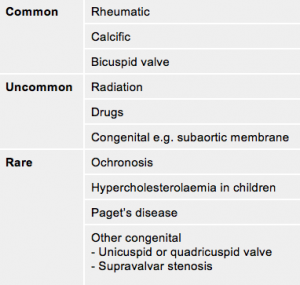
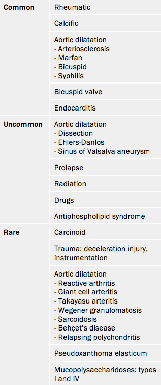
In industrially underdeveloped regions, rheumatic disease remains the most common cause. In the industrially-developed regions and in the elderly throughout the world, aortic valve disease is predominantly a result of calcific disease (see tables 4 and 5), which will be further explored in module 2. Aortic valve sclerosis is defined by valve thickening with a peak transaortic velocity on echocardiography of <2.5 m/s. Around 16% of patients with sclerosis progress to stenosis within seven years.
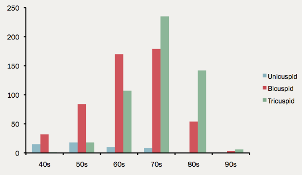
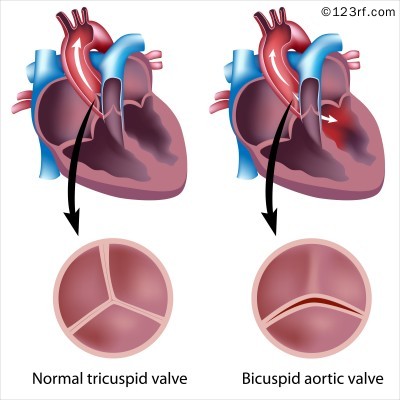
The most common congenital anomaly is bicuspid aortic valve and this is found in 0.5–0.8% of large population studies12 although it is reported in up to two thirds all valves excised during valve replacement for aortic stenosis (see figures 7 and 8)13 and up to 2.0% of the population based on autopsy series.
The risk of a bicuspid valve or aortopathy is about 10% in first-degree relatives of probands. The ratio of male to female is approximately 2:1.
The valve is ‘anatomically’ or truly bicuspid in a third of cases or ‘functionally’ bicuspid in two thirds as a result of incomplete separation of two cusps in utero. The most common pattern, in 80% of cases, is failure of right-left separation which is more likely to be associated with aortic dilatation. Failure of separation of right and non-coronary cusps is more likely to be associated with mitral prolapse.
During a 20 year follow-up, 24% of patients with a bicuspid aortic valve developed severe stenosis or regurgitation requiring surgery.14 Events are far more common in those with even mild valve thickening at baseline, with surgical rates of 75% at 12 years in the presence of thickening compared with only 8% without thickening.
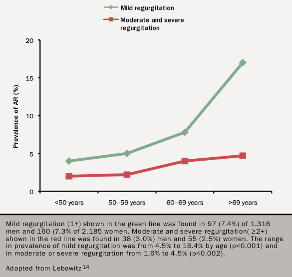
The frequency of aortic regurgitation increases with age (figure 9).15 Aortic regurgitation of any degree occurred in 29% and severe regurgitation in 13% in the Helsinki Ageing Study.11 Functional aortic regurgitation results from dilatation of the aortic root usually as a result of arteriosclerosis or medial necrosis. There may often be associated organic regurgitation as a result of a bicuspid aortic valve or arteriosclerosis.
The risk factors for aortic dilatation are:
- age,
- weakness of the aortic wall,
- the arteriosclerotic risk factors: hypertension; dyslipidaemia; smoking; and diabetes.
Weakness of the aortic wall as a result of medial necrosis occurs in Marfan Syndrome (see figure 10) and Ehlers-Danlos Syndrome Type IV.
Bicuspid aortic valve should be regarded as a general thoracic aortopathy and is associated with significant dilatation of the aorta (>40 mm) in about 20% of cases.14 Approximately one half affects the root, and the rest the ascending aorta.
