Biological valves
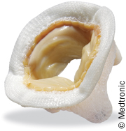
The most frequently implanted biological replacement valve is the stented xenograft. The stent is a plastic or wire structure covered in fabric with the cusps of the valve placed usually inside (although occasionally outside) and a sewing ring attached outside (see figure 7).

The valve cusps usually consist of pericardium or a porcine aortic valve. The porcine aortic valve may be used in its entirety as in the Carpentier-Edwards valve, but there is a muscle bar at the base of the right coronary cusp which reduces the orifice available for flow. This cusp may therefore be excised and replaced by a single cusp from another pig or, more frequently, each cusp may be taken from up to three different pigs as in the Medtronic Mosaic® (see figure 8), or the St Jude Epic™ (see figure 9) or Carbomedics Synergy valve. Pericardium is cut using a template and sewn inside the stent posts or occasionally, as in the Carbomedics Mitroflow™, to the outside. Usually the pericardium is bovine, but sometimes porcine and, occasionally equine or kangaroo!6

A stentless heterograft valve usually consists of a preparation of porcine aorta. The aorta may be relatively long (Freestyle®, Medtronic) or may be sculpted to fit under the coronary arteries (Toronto SPV®, St. Jude Medical).
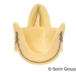
Some stentless valves are tricomposite and one is made out of bovine pericardium (Sorin, Freedom) (see figure 10). Most designs are technically hard to implant and require long bypass times which may be a disadvantage in a high-risk patient. The potential advantages of better hemodynamic function, durability and complication rates than for stented valves have not been borne out. For these reasons stentless valves are not commonly implanted but may still be useful for example in patients with endocarditis.
A homograft is a human aortic valve or, less commonly, pulmonary valve which is usually cryopreserved and left unstented.
A homograft has good durability if harvested early after death and does not need anticoagulation and so may be used as an alternative to a mechanical valve in the young. A homograft may also be used for choice in the presence of endocarditis since it allows wide clearance of infection with replacement of the aortic root and valve and the possibility of using the attached flap of donor mitral leaflet to repair perforations in the base of the recipient’s anterior mitral leaflet.
The disadvantage is that durability may be disappointing when the valve is harvested relatively late after death and particularly when a pulmonary homograft is placed in the aortic position. Re-operation in the presence of a calcified homograft root may be difficult.


