

We present a case of carcinoid heart disease as a consequence of a metastatic neuroendocrine tumour. The importance of recognising carcinoid syndrome as a cause of tricuspid and pulmonary valve pathology, as well as potential treatment, are discussed.
Introduction
Carcinoid heart disease (CHD) represents a rare yet important aetiology of intrinsic tricuspid and pulmonary valve pathology, associated with significant morbidity and mortality due to right heart failure. With timely medical therapy and, when indicated, surgical intervention, substantial improvements in both quality of life and long-term survival can be achieved. Notably, clinical manifestations indicative of cardiac involvement often remain subtle until CHD reaches an advanced stage.
The cardiac manifestations stem from the paraneoplastic effects elicited by vasoactive substances, notably 5-hydroxytryptamine (5-HT or serotonin), histamine, tachykinins, and prostaglandins, released by malignant cells.1 This process occurs independently of any direct metastatic infiltration into cardiac tissues. Normally, these vasoactive mediators are inactivated within the liver, lungs, and brain. In the setting of hepatic metastases, however, this detoxification pathway is bypassed, allowing bioactive substances to reach the right heart chambers and trigger valvular damage.
Early recognition and management of carcinoid syndrome (CS) are, therefore, critical in preventing CHD. Once CS is identified, prompt initiation of treatment with a somatostatin analogue is recommended,2 together with regular screening for CHD using transthoracic echocardiography.
Patient presentation
A 69-year-old woman with an unremarkable past medical history presented to the cardiology clinic with a six-month history of progressive exertional dyspnoea and intermittent ankle swelling. She reported no chest pain, orthopnoea, or paroxysmal nocturnal dyspnoea, but described occasional palpitations. Systemic enquiry revealed a concurrent six-month history of diarrhoea, which had been investigated without identifying any organic cause. There was no family history of cardiac conditions or cardiomyopathy. Her only regular medication was lansoprazole. She had no history of ischaemic heart disease, pulmonary embolism, or chronic lung disease. She was a non-smoker, with no alcohol excess history, and remained independent in her activities of daily living.
Physical examination revealed a blood pressure of 142/80 mmHg with a resting heart rate of 90 bpm and body mass index (BMI) of 27 kg/m². Auscultation revealed a systolic murmur at the right mid-sternal border. Lung auscultation was unremarkable, and no peripheral oedema was noted during the consultation.
Initial work-up
Blood tests, including full blood count, renal function, thyroid function and lipid profile, were all within normal limits, apart from a slightly raised N-terminal pro-B-type natriuretic protein (NT-proBNP) level of 828 ng/L.
Ambulatory Holter monitoring demonstrated sinus rhythm with ventricular and supraventricular ectopics (<1%), an average heart rate of 77 bpm with a maximum of 98 bpm and a minimum of 61 bpm.
Transthoracic echocardiogram showed impaired left ventricular (LV) function with biplane ejection fraction (EF) of 40%. The right ventricle was dilated but preserved in function, and the right atrium was also dilated. Severe tricuspid regurgitation was noted, with a maximum pressure gradient across the tricuspid valve of 22 mmHg. In addition, moderate pulmonary regurgitation was present (figure 1).
Diagnosis and management
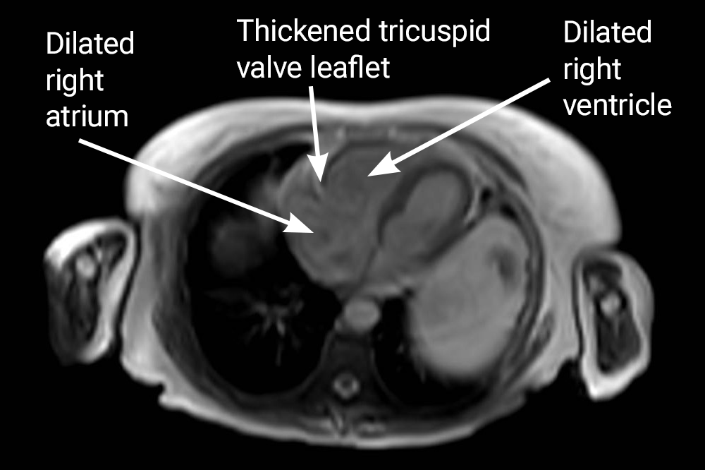
Cardiac magnetic resonance imaging (MRI) was requested to further investigate the dilatation of the right atrium (RA) and right ventricle (RV), as well as tricuspid regurgitation. Results revealed a dilated RV and RA, along with thickened and retracted tricuspid and pulmonary valves, indicating a morphology suggestive of carcinoid valve disease (figure 1). Additionally, a large liver lesion was observed.
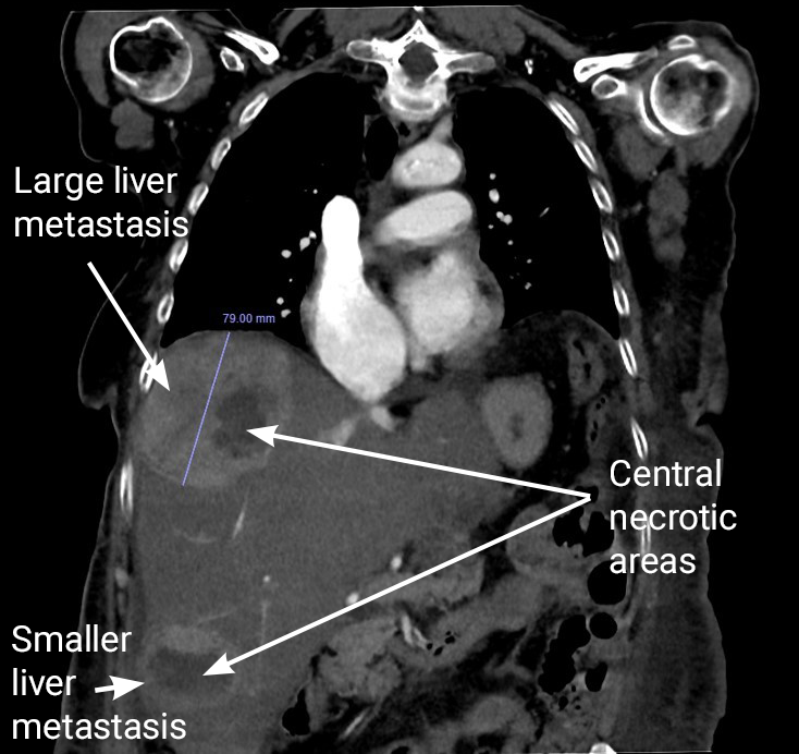
The patient was subsequently admitted to the hospital for further evaluation. Computed tomography (CT) abdominal imaging revealed disseminated intrahepatic metastatic disease, with multiple tumours displaying areas of necrosis (figure 2). Additionally, there was evidence of diffuse anasarca, ascites, and bilateral hydronephrosis, likely secondary to retroperitoneal fibrosis.
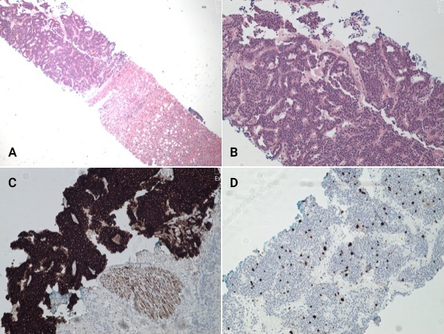
A liver biopsy confirmed the presence of a metastatic neuroendocrine tumour, demonstrating positive staining and a Ki67 index of 5% (figure 3). During admission, the patient developed acute kidney injury, alongside bilateral oedematous legs extending above the knees.
Following the diagnosis of a neuroendocrine tumour, the patient’s management included initiation of diuretic therapy and lantreotide infusions, with input from the oncology team. Bilateral nephrostomies were performed to relieve the obstructive bilateral hydronephrosis.
The patient was evaluated and discussed at the joint surgical and cardiology (JSC) meeting for consideration of tricuspid valve intervention. However, due to the prohibitive surgical risk, a decision was made to pursue conservative management. Despite initiation of targeted therapy and supportive measures, the patient’s condition deteriorated rapidly, and she unfortunately died.
Discussion
CHD can be diagnosed in up to 50% of patients with CS, and it may manifest as the initial presentation of the disease.3 Clinicians should have a high index of suspicion for CHD in patients exhibiting tricuspid regurgitation (TR) without pulmonary hypertension or cor pulmonale. Retroperitoneal fibrosis is a recognised complication that can lead to acute kidney injury (AKI) due to ureteric obstruction, as observed in our patient.
CHD is associated with poor clinical outcomes, with a three-year survival rate of 31%, which is half that of patients without CHD.4 The diagnosis of CHD can be facilitated through serum studies, CT scans, echocardiography, and, as in this case, cardiac MRI, which can expedite diagnosis and treatment.
While surgery was not felt to be feasible in our patient, there is evidence that valve intervention can improve outcomes for patients.5 Surgical intervention to the tricuspid or pulmonary valve has been shown to improve life-expectancy for patients with CHD and should not be overlooked as a treatment option. A discussion with a surgical centre is essential to identify whether patients are suitable for surgery to improve their outcomes.
Optimal management of patients with CHD necessitates a multi-disciplinary team experienced in dealing with these complex cases. This team should include cardiologists, oncologists and cardiothoracic surgeons, and is essential for providing informed decisions regarding the optimal management of patients with CHD.
Conflicts of interest
None declared.
Funding
None.
Patient consent
Informed consent was obtained from the patient’s family prior to publication.
Editors’ note
See also the editorial by Simwa and Cowburn from this issue at https://doi.org/10.5837/bjc.2026.053.
References
1. Kulke MH, Mayer RJ. Carcinoid tumors. N Engl J Med 1999;340:858–68. https://doi.org/10.1056/NEJM199903183401107
2. Basson MD, Ahlman H, Wangberg B, Modlin IM. Biology and management of the midgut carcinoid. Am J Surg 1993;165:288–97. https://doi.org/10.1016/S0002-9610(05)80529-X
3. Khan MA, Herzog CA, St Peter JV et al. The prevalence of cardiac valvular insufficiency assessed by transthoracic echocardiography in obese patients treated with appetite-suppressant drugs. N Engl J Med 1998;339:713–18. https://doi.org/10.1056/NEJM199809103391101
4. Strickman NE, Rossi PA, Massumkhani GA, Hall RJ. Carcinoid heart disease: a clinical pathologic, and therapeutic update. Curr Probl Cardiol 1982;6:1–42. https://doi.org/10.1016/0146-2806(82)90013-5
5. O’Malley TJ, Jimenez DC, Saxena A et al. Outcomes of surgical treatment for carcinoid heart disease: a systematic review and meta-analysis. Surgery 2021;170:390–6. https://doi.org/10.1016/j.surg.2021.02.054


