Criteria for referral to designated centres
When the general practitioner suspects that the patient may have PH, the patient should be referred as an out-patient for assessment by a cardiologist or chest physician. The cause of the patient’s symptoms will be sought, using investigations such as ECG, chest X-ray, spirometry and echocardiography.
Adults with confirmed or suspected pulmonary arterial hypertension (PAH), PH due to chronic thrombotic or embolic disease, pulmonary hypertension which appears to be out of proportion to lung disease or heart disease, another cause of PH, or where the cause is unclear, should be referred to a designated centre (see box), where cardiac catheterisation is carried out.1 Referral should not be delayed since some patients can deteriorate rapidly. Children in Great Britain should be referred to the UK Children’s Service if they have confirmed or suspected PAH.
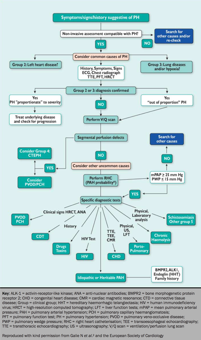
A diagnostic algorithm to guide clinicians when faced with a patient with signs, symptoms or history suggestive of PH is shown in figure 1.2

Designated pulmonary hypertension centres in the UK and Ireland
Cambridge
- Pulmonary Vascular Diseases Unit, Papworth Hospital NHS Trust, Papworth Everard, Cambridge CB23 8RE
Tel: 01480 830 541
www.papworth-hospital.org.uk
Dublin
- Mater Misericordiae University Hospital, Eccles Street, Dublin 7
Tel: 00 353 1803 44 20
www.mater.ie
Glasgow
- Golden Jubilee National Hospital, Glasgow. Scottish Pulmonary Vascular Unit, Beardmore Street, Clydebank, Western Dunbartonshire, Glasgow G81 4HX
Tel: 0141 951 5497
www.spvu.co.uk
London
- Pulmonary Hypertension Service, Hammersmith Hospital, Du Cane Road, London W12 0HS
Tel: 0208 383 2330 0203 313
www.pulmonary-hypertension.org.uk - Royal Brompton Pulmonary Hypertension and Adult Congenital Heart Centre, Sydney Street, London SW3 6NP
Tel: 0207 351 8362
www.rbht.nhs.uk - Royal Free Hospital, Pond Street, London NW3 2QG
Tel: 0207 794 0500 extension 8648
www.royalfree.nhs.uk - UK Pulmonary Hypertension Service for Children, Great Ormond Street Hospital, London WC1N 1EH
Tel: 0207 405 9200 extensions 1005, 1007, 8495
www.ich.ucl.ac.uk
Newcastle
- Northern Pulmonary Vascular Unit, Regional Cardiothoracic Centre, Freeman Hospital, Newcastle upon Tyne, NE7 7DN
Tel: 0191 223 1968
www.newcastle-hospitals.org.uk
Sheffield
- Pulmonary Vascular Unit, Royal Hallamshire Hospital, Glossop Road, Sheffield S10 2JF
Tel: 0114 271 3940
www.phsheffield.org.uk
Investigations for pulmonary hypertension
Whenever PH is suspected, patients must have a thorough diagnostic work-up: this is important in order to establish a precise diagnosis and to recommend the most appropriate treatment (see figure 1). Initial investigations comprise ECG, chest radiograph, lung function tests, echocardiogram and routine blood tests.
The ECG
The ECG can provide evidence to support the diagnosis of pulmonary hypertension by showing right ventricular hypertrophy or strain, right atrial enlargement and right axis deviation of the QRS complex. A normal ECG does not exclude significant pulmonary hypertension. In idiopathic PAH, 80-90% of patients are likely to have an abnormal ECG on presentation.
In severe PH, atrial arrhythmias are often poorly tolerated. A finding of atrial fibrillation suggests underlying heart disease, pulmonary embolism or cor pulmonale as seen in chronic lung disease. Atrial arrhythmias should be suspected in patients with syncope in the absence of severe pulmonary hypertension. Atrial arrhythmias should also be suspected in any patient with sudden or rapid deterioration in symptoms of pulmonary hypertension in whom a diagnosis of PH is already established. Atrial flutter (figure 2),3 in particular, is common in this setting.
Learning point:
- The ECG may provide supportive evidence for a diagnosis of PH but the findings are non-specific

