Assessing the health-related quality of life in patients hospitalised for acute heart failure

Br J Cardiol 2013;20:72–6doi:10.5837/bjc.2013.013 Leave a comment
Click any image to enlarge
Authors:
First published online April 23rd 2013

Acute heart failure (AHF) is a common cause of hospitalisation, presenting substantial economic and humanistic burden for healthcare systems and patients. This study was designed to capture proxy UK health-related quality of life (HRQoL) data for hospitalised patients with AHF. 

Proxy assessments of HRQoL for patients were obtained from 50 experienced UK cardiac nurses (formal caregivers) and from 50 UK individuals who acted as caregivers for patients who had experienced an AHF event leading to hospitalisation (informal caregivers). Data were collected retrospectively for four time points (days 1, 3, 5 and 7 post-hospital admission for AHF event) using the EQ-5D. Results show a disparity in reported HRQoL at day 1 values between caregiver types (mean single utility index 0.20 vs. 0.68, respectively, p<0.001). By day 7, formal caregivers rated typical patients’ HRQoL as being comparable to informal caregivers’ assessments (0.82 vs. 0.73, respectively, p=0.145). 

In conclusion, collection of utility data in severe acute conditions is challenging. This study captures values through the use of proxy assessment. Data suggest that AHF hospitalisation is associated with a significant HRQoL burden and that there exists a need for development of new treatments aimed at improving hospitalisation outcomes.

Introduction

splAcute heart failure (AHF) has been defined by the European Society of Cardiology (ESC) as the rapid onset of, or change in, symptoms and signs of heart failure, and is a life-threatening condition that requires immediate medical attention.1 These symptoms and signs include shortness of breath at rest or during exertion, fatigue, pulmonary or peripheral fluid retention, a cough, and evidence of an abnormality of the structure or function of the heart at rest.2-4 This change in cardiac function results in an urgent need for therapy, and AHF is among the most common causes of hospitalisation.5 AHF can, therefore, be seen to represent a huge burden on healthcare resources,6 with the need for hospitalisation in order to stabilise the condition of patients being the single largest contributor to the costs of managing AHF.7 The situation is further worsened in that readmission rates are high following discharge; approaching 50% within six months.8

As well as the substantial economic burden on healthcare services, a diagnosis of AHF is associated with a significant health-related quality of life (HRQoL) burden for patients. When making decisions about allocation of healthcare resources, many decision makers consider the impact of the intervention on both costs and health outcomes.9 Health outcomes are commonly aggregated into quality-adjusted life years (QALYs), which is a metric that combines both survival and quality of life. AHF represents a significant challenge to quality of life for patients, due to a combination of the debilitating effects of the condition and the necessity for hospitalisation. Despite the incidence of the condition and the extent of burden experienced there is a paucity of utility data available for AHF, which could be used in economic analyses.

Most HRQoL assessment relies on self-report from patients, but for individuals who are severely or acutely ill this is often not possible for a variety of practical and ethical reasons. Capturing HRQoL information accurately from individuals who are unable or unwilling to complete appropriate instruments, therefore, poses a challenge. One potential approach to overcoming this issue is through the use of proxy assessment. Proxy measures have been successfully used previously in a number of different patient populations including rehabilitation patients10 and individuals with developmental disabilities.11 These studies have demonstrated agreement between self-reported assessments and those of proxies, but other findings suggest that proxy assessment may be subject to bias and measurement error.12

The purpose of the current study was to capture quality of life data for patients in the days immediately following AHF hospitalisation. Given the practical and ethical challenges posed by collecting data directly from acutely ill patients, a proxy assessment approach was employed in which the caregivers of AHF patients were surveyed. In an attempt to address potential sources of bias, proxy assessments were made both by relatives of patients that had experienced recent AHF and by healthcare professionals who regularly manage patients with the condition.

Materials and methods

Participants and methods

Fifty nurses with a minimum of two years’ experience, who regularly treat cardiac patients (formal caregivers), and 50 close relatives of different patients who had been hospitalised as a result of AHF within the last six months (informal caregivers), were recruited in the UK via a specialist agency. Prior to any data collection taking place, ethical approval was sought for the study protocol from an institutional review board. Potential participants were forwarded a URL for the study website where they were presented with additional information pertaining to the study. Basic sociodemographic data were obtained before proceeding with the assessment exercise.

Participants were asked to provide assessments of the HRQoL of either a ‘typical’ AHF patient (formal caregivers) or the affected relative who was under their care (informal caregivers) at each of four time points (time elapsed since hospital admission = one day, three days, five days, seven days). All assessments were made using the EuroQol five dimension (EQ-5D) instrument.

EQ-5D

The EQ-5D is a widely used generic HRQoL instrument and is the preferred measure of health status for bodies such as the National Institute for Health and Clinical Excellence (NICE).9 It comprises of five dimensions (mobility, self-care, usual activities, pain/discomfort and anxiety/depression) each with three levels detailing the extent of problems experienced. Subsequent to these evaluations, the EQ-5D contains a 100 mm visual analogue scale (VAS) addressing the individual’s reported current overall health. The EQ-5D has been used previously to assess critically ill individuals in intensive care settings with a variety of different conditions.13,14 Additionally, it has previously demonstrated sensitivity to the HRQoL impact of a number of different heart conditions (including coronary artery bypass and aortic valve surgery, coronary heart disease, and acute coronary syndrome).15-18 Although the instrument was originally designed to be self-completed, many studies have used proxy administration where patient completion may be inappropriate or even impossible.19-21 Calculation of the EQ-5D single index utility value involves the application of preference weights collected from large scale data collection efforts with the general public. Application of the UK weights typically leads to a range of scores from 0 (representing states of health equivalent to being dead), up to a maximum of 1 (representing a state with no health problems). Negative values are possible for states of extremely poor health and represent states rated worse than being dead. The VAS component of the EQ-5D produces scores from 0 (worst imaginable health) to 100 (best imaginable health).

Statistical analysis

Demographic data were summarised using frequency counts and percentages, and independent t-tests were performed to assess differences between caregiver groups at each time point.

Results

Study participants

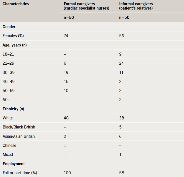
The characteristics of the two caregiver groups are presented in table 1.

Table 1. Participant characteristics for the two caregiver groups

Proxy EQ-5D assessments

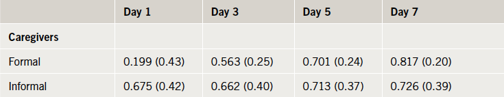
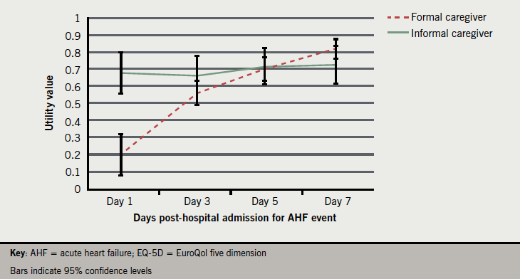
The mean EQ-5D single index utility values calculated for the four time points post-AHF event are reported in table 2. Formal caregiver assessments of HRQoL are significantly lower than those of informal caregivers at day 1 (t[98]=–5.570, p<0.001). Later assessments do not differ significantly between groups and demonstrate a steady improvement in perceived HRQoL over the course of the seven-day period post-AHF event. Figure 1 presents the utility values plotted over time.

Table 2. EuroQol five dimension (EQ-5D) utility values for the four time points following acute heart failure event, mean (standard deviation)
Table 2. EuroQol five dimension (EQ-5D) utility values for the four time points following
acute heart failure event, mean (standard deviation)
Figure 1. EQ-5D utility values by caregiver group across time points

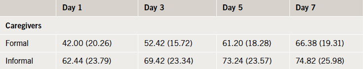
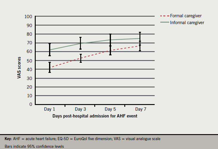
The EQ-5D VAS scores for the four time points post-AHF event are presented in table 3. The disparity in HRQoL assessment at day 1 between the two groups is less marked than seen with the utility values. Formal caregivers assessments of HRQoL based on their experience do, however, remain lower than informal caregivers across all time points and differ significantly at day 1 (t[98]=–4.63, p<0.01), day 3 (t[98]=–4.27, p<0.01) and day 5 (t[98]=–2.85, p<0.01). Visual analogue scale scores for day 7 approximate those for an existing survey of the UK general population (mean 82.5, standard deviation [SD] 17).22 Unlike the trend witnessed with the utility values, the rate of improvement in HRQoL for the two groups is broadly similar (albeit with the formal caregivers suggesting greater overall improvement due to lower baseline values). Figure 2 presents VAS scores plotted over time.

Table 3. EQ-5D visual analogue scale (VAS) values for the four time points following AHF event, mean (standard deviation)
Table 3. EQ-5D visual analogue scale (VAS) values for the four time points following AHF
event, mean (standard deviation)

Discussion

This study aimed to assess HRQoL for patients who have been hospitalised as a result of AHF. Health utilities were captured via proxy administration of the EQ-5D instrument with both formal and informal caregivers. The findings suggest that AHF hospitalisation is associated with very poor quality of life, at least in the short term. Over the course of the seven days following hospitalisation, significant improvement occurs as evidenced by the increase in reported utility values. The results also demonstrate some disparity exists as to the average extent of problems experienced by patients as judged by the different caregiver groups. The utility values for the formal and informal caregivers deviate greatly at day 1 after the AHF event but, as expected, both suggest that patients experience substantial problems related to ill health and hospitalisation.

Figure 2. EQ-5D VAS scores by caregiver group across time points

Previous studies have found the EQ-5D instrument can be valid when administered by proxy.23-24 The decision to use two different sources of proxy values in the study was an attempt to avoid introducing systematic bias into the results. It is likely that both the formal and informal caregiver group assessments are influenced by a number of factors unique to their relationship with patients. Formal caregiver assessments of a ‘typical’ AHF patient suggest that HRQoL is more detrimentally affected by event-related hospitalisation. This may be a product of a more ‘functional capacity’ based approach to evaluating health. Given the extremely debilitating immediate impact of an AHF event this could lead to reports of greatly diminished HRQoL. In contrast, informal caregiver assessments of relatives HRQoL suggest that the deterioration is somewhat less pronounced. It is likely that these particular assessments are informed by a more intimate knowledge of the pre-event baseline health status of patients and reflect a ‘relative change’ based evaluation approach. Although the assessment exercises conducted by the raters are essentially different in terms of their focus, it is difficult to argue that either assessment is objectively superior when looking solely at data collected using the EQ-5D. As the instrument was designed specifically for use by members of the general public with no formal medical training this should not disadvantage informal caregivers in making valid assessments. Future work looking at the level of agreement between retrospective patient assessments of HRQoL and those made by proxies may give a clearer indication of which values correspond more closely. Of course, such retrospective assessments are themselves subject to confounds in the form of recall biases.

The study has some important limitations that should be considered when interpreting the results. No assessment was captured directly from any patient, which introduces concerns over the validity of the values obtained. However, there are clearly a number of logistical and ethical issues, such as the difficulty in obtaining informed consent from severely ill patients, which make prospective data collection challenging. Retrospective data collection presents one possible alternative method for obtaining suitable HRQoL data. Such an approach could be nonetheless affected by a selection bias with only those who survived (and perhaps, therefore, those with the least severe events or best outcomes) being capable of providing data. It is possible that this selection bias may have influenced the current study, with the caregivers who participated being relatives of patients who are recovering or have recovered from the AHF event (although we do not have sufficient data to verify this assumption). Informal caregivers may have been assessing a less severely affected group of patients than formal caregivers, and this could, therefore, at least partly explain differences in the HRQoL ratings from the two groups. The retrospective assessment approach taken in this study may also have led to recall bias. Given the nature of their occupation, formal caregivers are more likely to have had recent and frequent experience of patients in the immediate post-AHF state. In practical terms, this problem is difficult to avoid when employing such a methodology. We have attempted to limit the potential for this to occur by including an inclusion criteria that the patients under the informal caregivers care had to have experienced the AHF event a maximum of six months previously (with the majority doing so within a three-month period). Although this shorter period of time should facilitate more accurate recall, the relatively small window of time between each study time point (day 1, 3, 5 and 7 post-AHF event) may serve to blur distinctions in the patient’s quality of life at these intervals. This may be reflected in the fact that the range of utility values for the informal carer group is narrower than that of the formal caregivers. All of these potential sources of bias were recognised when the study was designed, which led us to make the decision to include data collection from two independent groups. Despite these limitations we believe that the study has provided some insight into the HRQoL effects of AHF hospitalisation as viewed by different caregiver groups. Interestingly, a recent draft of the NICE guidelines for health technology assessment has proposed the use of caregiver derived utility values in preference to those provided by healthcare professionals when collecting HRQoL data.25

The study supports previous research suggesting that there is a marked decline in HRQoL associated with hospitalisation as a result of experiencing an AHF event. Recent research has suggested that there is significant variation in prescribing practices, which may have led to suboptimal outcomes for many patients.26-27 Improved pharmacological management of AHF to reduce the incidence of hospitalisation and length of stay, could confer substantial benefits for patients and healthcare systems.

Acknowledgement

The authors thank Georgia Tarnesby, employed by Novartis Pharma, for reviewing and providing comments on an earlier draft of the manuscript.

Funding

The research was funded by Novartis Pharma AG, Postfach, CH-4002 Basel, Switzerland.

Conflict of interest

SHO and PL are employed by Novartis Pharma AG. Oxford Outcomes were paid a fixed fee to design and conduct this study by Novartis Pharma AG.

Key messages

  • Acute heart failure (AHF) is a common cause of hospitalisation, presenting a substantial burden for healthcare systems, as well as patients. This study was designed to capture proxy UK health-related quality of life (HRQoL) data for hospitalised patients with AHF
  • This study demonstrates the feasibility of capturing HRQoL data for acutely unwell patients, perhaps unable or unwilling to self-report such issues
  • Results show a disparity in reported HRQoL at day 1 values between caregiver types; however, by day 7, formal caregivers rated typical patients’ HRQoL as being comparable to informal caregivers ratings
  • Findings suggest AHF hospitalisation is associated with notable HRQoL burden and that improved pharmacological management could lead to significant benefits for both patients and healthcare systems

References

  1. ESC (European Society of Cardiology) Task Force. ESC guidelines for the diagnosis and treatment of acute and chronic heart failure 2012. Eur Heart J 2012;33:1787–847. http://dx.doi.org/10.1093/eurheartj/ehs104
  2. Dickstein K, Cohen-Solal A, Filippatos G et al. ESC guidelines for the diagnosis and treatment of acute and chronic heart failure 2008. The Task Force for the Diagnosis and Treatment of Acute and Chronic Heart Failure 2008 of the European Society of Cardiology. Eur J Heart Fail 2008;10:933–89. http://dx.doi.org/10.1016/j.ejheart.2008.08.005
  3. Goldberg R, Spencer F, Szklo-Coxe M et al. Symptom presentation in patients hospitalized with acute heart failure. Clin Cardiol 2010;33:e73–e80. http://dx.doi.org/10.1002/clc.20627
  4. Gheorghiade M, Filippatos G, De Luca L, Burnett J. Congestion in acute heart failure syndromes: an essential target of evaluation and treatment. Am J Med 2006;119(suppl 1):S3–S10. http://dx.doi.org/10.1016/j.amjmed.2006.09.011
  5. Lloyd-Jones D, Adams RJ, Brown TM et al. Heart disease and stroke statistics – 2010 update: a report from the American Heart Association. Circulation 2010;121:e45–e215. http://dx.doi.org/10.1161/​CIRCULATIONAHA.109.192667
  6. Ross EA, Bellamy FB, Hawig S, Kazory A. Ultrafiltration for acute decompensated heart failure: cost, reimbursement and financial impact. Clin Cardiol 2011;34:273–7. http://dx.doi.org/10.1002/clc.20913
  7. O’Connell JB. The economic burden of heart failure. Clin Cardiol 2000;23(suppl 3):III6–III10. http://dx.doi.org/10.1002/clc.4960231503
  8. Krumholz H, Chen Y, Wang Y et al. Predictors of readmission among elderly survivors of admission with heart failure. Am Heart J 2000;139:72–7. http://dx.doi.org/10.1016/S0002-8703(00)90311-9
  9. National Institute for Health and Clinical Excellence (NICE). Guide to the methods of technology appraisal. London: NICE, June 2008. Available from: http://www.nice.org.uk/niceMedia/pdf/TAP_Methods.pdf
  10. McPhail S, Beller E, Haines T. Two perspectives of proxy reporting of health-related quality of life using the Euroqol-5D, An investigation of agreement. Med Care 2008;46:1140–8. http://dx.doi.org/10.1097/MLR.0b013e31817d69a6
  11. Schmidt S, Power M, Green A et al. Self and proxy rating of quality of life in adults with intellectual disabilities: results from the DISQOL study. Res Dev Disabil 2010;31:1015–26. http://dx.doi.org/10.1016/j.ridd.2010.04.013
  12. Tamim H, McCusker J, Dendukuri N. Proxy reporting of quality of life using the EQ-5D. Med Care 2002;40:1186–95. http://dx.doi.org/10.1097/00005650-200212000-00006
  13. Badia X, Diaz-Prieto A, Rue M, Patrick DL. Measuring health and health state preference among critically ill patients. Intensive Care Med 1996;22:1379–84. http://dx.doi.org/10.1007/BF01709554
  14. Cristina G, Armando TP, Altamiro CP. Quality of life after intensive care – evaluation with EQ-5D questionnaire. Intensive Care Med 2002;28:898–907. http://dx.doi.org/10.1007/s00134-002-1345-z
  15. Ellis JJ, Eagle KA, Kline-Rogers EM, Erickson SR. Validation of the EQ-5D in patients with a history of acute coronary syndrome. Curr Med Res Opin 2005;21:1209–16. http://dx.doi.org/10.1185/030079905X56349
  16. Kahyaoglu Süt H, Unsar S. Is EQ-5D a valid quality of life instrument in patients with acute coronary syndrome? Anadolu Kardiyol Derg 2011;11:156–62. http://dx.doi.org/10.5152/akd.2011.037
  17. Markou AL, de Jager MJ, Noyez L. The impact of coronary artery disease on the quality of life of patients undergoing aortic valve replacement. Interact Cardiovasc Thorac Surg 2011;13:128–32. http://dx.doi.org/10.1510/icvts.2011.269209
  18. Xie J, We EQ, Zheng ZJ, Sullivan PW, Zhan L, Labarthe DR. Patient-reported health status in coronary heart disease in the United States: age, sex, racial and ethnic differences. Circulation 2008;118:491–7. http://dx.doi.org/10.1161/CIRCULATIONAHA.107.752006
  19. Hung MC, Yan YH, Fan PS et al. Measurement of quality of life using EQ-5D in patients on prolonged mechanical ventilation: comparison of patients, family caregivers, and nurses. Qual Life Res 2010;19:721–7. http://dx.doi.org/10.1007/s11136-010-9629-1
  20. Matza LS, Secnik K, Mannix S, Sallee FR. Parent-proxy EQ-5D ratings of children with attention-deficit hyperactivity disorder in the US and the UK. Pharmacoeconomics 2005;23:777–90. http://dx.doi.org/10.2165/00019053-200523080-00004
  21. Wolfs CA, Dirksen CD, Kessels A, Willems DC, Verhey FR, Severens JL. Performance of the EQ-5D and the EQ-5D+C in elderly patients with cognitive impairments. Health Qual Life Outcomes 2007;14:33. http://dx.doi.org/10.1186/1477-7525-5-33
  22. Kind P, Dolan P, Gudex C et al. Variations in population health status: results from a United Kingdom national questionnaire survey. BMJ 1998;316:736–41. http://dx.doi.org/10.1136/bmj.316.7133.736
  23. Klaassen RJ, Barr RD, Hughes J, Rogers P, Anderson R, Grundy P. Nurses provide valuable proxy assessment of health-related quality of life of children with Hodgkin disease. Cancer 2010;116:1602–07. http://dx.doi.org/10.1002/cncr.24888
  24. Pickard S, Jeffrey A, Johnson A, Feeny DH, Shuaib A, Carriere KC. Agreement between patient and proxy assessments of health-related quality of life after stroke using the EQ-5D and Health Utilities Index. Stroke 2004;35:607–12. http://dx.doi.org/10.1161/01.STR.0000110984.91157.BD
  25. National Institute for Health and Clinical Excellence (NICE). Guide to the methods of technology appraisal, third edition draft for consultation. London: NICE, 2012. Available from: http://www.nice.org.uk/media/CB1/43GuideToMethodsOfTechnologyAppraisal2012.pdf
  26. Fonarow GC, Yancy CW, Albert NM et al. Heart failure care in the outpatient cardiology practice setting. Circ Heart Fail 2008;1:98–106. http://dx.doi.org/10.1161/CIRCHEARTFAILURE.108.772228
  27. Albert NM, Yancy CW, Liang Li et al. Use of aldosterone antagonists in heart failure. JAMA 2009;302:1658–65. http://dx.doi.org/10.1001/jama.2009.1493

A pilot study to investigate the safety of exercise training and testing in cardiac rehabilitation patients

Br J Cardiol 2013;20:78doi:10.5837/bjc.2013.012 Leave a comment
Click any image to enlarge
Authors:
First published online April 23rd 2013

We conducted a pilot study to evaluate the safety of the shuttle walking test (SWT) and exercise training for cardiac patients in community-based cardiac rehabilitation settings. Overall, 33 cardiac patients were tested (19 males and 14 females, 67 ± 8 years). Eleven cardiac patients (testing group) and 22 cardiac patients (training group) underwent ambulatory electrocardiogram (ECG) monitoring during the SWT and exercise training during a long-term cardiac rehabilitation programme. Frequency of ECG events was reported for the two groups. Chi-square test was performed to determine associations between the incidence of cardiovascular events and poor functional capacity (SWT <450 m). 

The findings showed only minor events provoked during the SWT or exercise training, and no event-related hospitalisation, syncope episodes or fatality. The most important cardiac event was silent myocardial ischaemia (testing group: 27.3%; training group: 18%). Poor functional capacity was not associated with the risk of a cardiac event during exercise (testing group: χ2=0, p=0.99, phi=0.24; training group: χ2=2.1, p=0.15, phi=–0.42). 

In conclusion, supervised exercise testing and training are accompanied only by minor cardiovascular events and they can be carried out safely in community-based cardiac rehabilitation settings.

Introduction

SPExercise is well recognised as a tool for assessment, prevention and management of cardiovascular disease.1 Cardiac patients are encouraged to attend cardiac rehabilitation programmes including elements of supervised exercise. Such programmes can reduce mortality and morbidity rates by up to 27%.2,3

Despite the benefits derived from participation in exercise-based cardiac rehabilitation, exercise itself may act as a trigger for myocardial ischaemia or cardiac arrest in patients with established coronary heart disease.4 During rehabilitation, cardiovascular event rates range from 12.3 to 37.4 per million patient hours of exercise.4-8 Fatal cardiac events are more rarely reported, but range from 8.6 per million patient hours of exercise in 19784 to zero in the present day.5-8 Event rates during exercise are also lower in more recent studies of supervised cardiac rehabilitation exercise programmes,5-8 events during exercise sessions do still occur, however.

The American Heart Association9,10 emphasise the importance of pre-training cardiovascular risk assessment, including medical history, physical examination and ambulatory electrocardiogram (ECG) monitoring during exercise testing. These actions are recommended to minimise exercise-induced cardiovascular events by identifying patients at greatest risk.

Cardiovascular events during exercise testing using traditional laboratory exercise protocols (treadmill, cycle ergometer) are also rare,4,8,11 and appear difficult to predict.5,8 Laboratory-based exercise protocols are often replaced in daily clinical practice by functional capacity tests, such as the incremental shuttle walking test (SWT). This test is reliable,12 relatively simple to perform and inexpensive.13 Only one study has evaluated the safety of this test in a cardiac population.14 The authors found a very low number of cardiovascular events during exertion, but questioned whether this was the result of low individual effort during exertion or successful treatment of coronary arteries during surgical intervention. The small and selective subsample (19 post coronary artery bypass graft surgery patients) studied, increases the necessity for new studies in this field.

The Scottish Intercollegiate Guidelines Network (SIGN) recommends the use of exercise testing15 and the British Association for Cardiac Rehabilitation (BACR) recommends the use of exercise training,16 to detect exercise-induced cardiovascular events. Thus, the aim of this study was to verify to what extent a recommended exercise testing protocol (SWT) and an exercise training session are safe for a mixed cohort of cardiac rehabilitation patients.

Materials and methods

Study participants

Overall, 33 cardiac patients were tested (19 males and 14 females, 67 ± 8 years). Eleven patients were assessed while undertaking a sub-maximal SWT (testing group) and 22 patients were assessed during cardiac rehabilitation exercise training (training group).

All patients were clinically stable and had been enrolled in a community-based, phase IV cardiac rehabilitation programme for at least 10 weeks. Patients with severe locomotor limitations were excluded from this study.

All procedures were approved by the University ethical committee and conformed to the declaration of Helsinki guidelines for research with human subjects.17 Patients were verbally recruited by instructors and informed written consent was obtained from all patients before enrolment.

Protocol and measurements

Anthropometric and cardiovascular assessment

Patients were assessed once, each received a primary health assessment (pre-exercise health questionnaire, medical, pharmacological history, resting heart rate, resting blood pressure) and anthropometric assessment (stature, body mass, waist circumference) prior to testing. Body mass index (BMI) was calculated (kg/m2). Table 1 summarises the participants’ clinical characteristics and baseline measurements.

Table 1
Table 1. Descriptive characteristics and baseline measurements of patients

All patients were monitored with an ambulatory ECG event monitor (C.NET5000, version 1.2, Cardionetics Ltd., United Kingdom), either during the SWT (testing group) or during the cardiac rehabilitation class (training group). A single-lead ECG was recorded using an electrode configuration analogous to lead standard V5 in a traditional 12-lead clinical ECG monitoring. The recording device was placed into a pouch fitted to the patient’s waist using a particular type of belt.14

Major cardiovascular events were defined as one of the following: myocardial infarction requiring hospitalisation, ventricular fibrillation, ventricular tachycardia requiring treatment, atrial arrhythmias requiring treatment, asystole, stroke and death. Minor cardiovascular events were defined as: isolated ventricular arrhythmias or atrial arrhythmias not requiring intervention, chest pain and bradycardia not requiring intervention.11

Exercise testing

Patients performed a SWT13 by walking back and forth between two cones set 0.5 m from either end of a 10 m course. Initial walking speed, indicated by an audible signal, was 0.5 m/s and increased by 0.13 m/s each minute. During the test, heart rate was also recorded with a heart rate monitor (Polar Electro Sports Tester S810I, Heart Rate Monitor, Kempele, Finland) and ratings of perceived exertion (RPE) were measured using the Borg (6–20) scale.18

The SWT was terminated when the patient (a) felt too breathless or fatigued to continue at the required speed, (b) failed to complete the shuttle within the allowed time, (c) reached 85% of the predicted maximal heart rate: 210 – (0.65×age), (d) reached RPE ≥15 (Borg 1998), or (e) completed all the levels.

Exercise training programme

The programme offers the opportunity of life-long supervised exercise for cardiac patients. The programme consisted of two supervised sessions per week, made up of 60 minutes of circuit-based exercise classes. Supervisory staff included a physiotherapist specialised in cardiac rehabilitation, also trained in immediate life support methods. Exercise sessions comprised: a 15-minute warm-up, a 35-minute main conditioning component and a 10-minute cool-down period. The conditioning component of the session included cardiovascular and strength exercises at an intensity of 60–80% of the age-predicted maximal heart rate, or at 12 to 15 on the RPE scale. During this component patients had access to a variety of exercise equipment, such as arm ergometers, rowing ergometers, progressive resistance equipment, balance equipment, steppers and free weights.

Data analysis

A cardiologist verified the presence or absence of each cardiovascular event. Frequency of the cardiovascular events was reported for both groups. Patients were divided into event-free versus cardiac-event subgroups. Differences in functional capacity between the two subgroups were examined using a two-sample t-test. A chi-square test was performed to determine associations between incidences of cardiovascular events with poor functional capacity (SWT <450 m).19

All statistical analyses were performed using SPSS version 16.0 (SPSS nc., Chicago, IL, USA). The statistical significance was set at p<0.05.

Results

Event rates

No major event related to hospitalisation was detected in both testing and training groups. The most clinically important event was silent myocardial ischaemia, which occurred in 27.3% of the testing group patients, and in 18% of the training group patients. There was no significant difference in functional capacity between the event-free subgroup and the subgroup with cardiovascular events in both testing and training group (p>0.05).

Frequency of cardiovascular events during exercise testing

Eight cardiovascular events were detected automatically by the ECG monitoring system during exercise testing. Seven (82%) were verified by the cardiologist. According to the cardiologist’s review of the ECG recordings, of the eleven patients, 5 (45.5%) had atrial ectopic beats, four had isolated ventricular ectopic beats (36.4%), three (27.3%) had ischaemic ST segment depression, two (18.2%) had an atrial fibrillation event, one (9.1%) had a bradycardia event (under 50 beats per minute), while four (36.4%) of the patients were free from cardiovascular events (table 2).

Table 2
Table 2. ECG event monitoring during the shuttle walking test performance (n=11)

Frequency of cardiovascular events during exercise training

Among 18 cardiovascular events automatically detected during exercise training, 16 (73%) were verified by the cardiologist. According to the cardiologist’s review of the ECG recordings; 15 patients (68%) had isolated ventricular ectopic beats, four (18%) had atrial ectopic beats, four (18%) had ischaemic ST segment depression, one (4.6%) had atrial fibrillation. Six (27.3%) of the patients were free from cardiovascular events (table 3).

Table 3
Table 3. ECG event monitoring during cardiac rehabilitation exercise training

In all, 76.5% of thecardiovascular eventswere detected around the time of the main conditioning component of the exercise training, 11.8% during the warm up and 23.5% during the cool down.

Association between frequency of cardiovascular events and patient functional capacity

A chi-square test showed no significant association between poor functional capacity (SWT <450 m) and risk for cardiovascular events in either testing group or training group (testing group: χ2=0, p=0.99, phi=0.24; training group: χ2=2.1, p=0.15, phi=–0.42). Among patients with poor functional capacity, cardiovascular events were found in half of those (n=1) who were assessed during exercise testing with the SWT, and in all of those (n=7) who were assessed during exercise training.

Discussion

The present study provides information regarding the safety of exercise training during a community-based, phase IV cardiac rehabilitation programme and a standard functional exercise test.

Risk of cardiovascular events during exercise testing and training

According to cardiologist-verified ECG data only minor, non-fatal cardiovascular events were observed during both the SWT (with an event rate of 0.64 per patient per test) and in exercise training (with an event rate of 0.73 per patient per session).No major cardiovascular events were detected during exercise testing or training. These findings agree with those of Hollenberg et al.20 who found no complex arrhythmias or symptomatic ischaemia during exercise in participants without suspected cardiac disease.

Contrary to previous studies,4,5,8 we showed that exercise training is associated with more events than exercise testing. This may be explained by use of the SWT, whereas previous studies have used treadmill tests, which may provoke greater stresses on the cardiovascular system than the incremental SWT.21-23The most serious (major) event, silent ischaemia, was observed more often during the SWT than during exercise training. This may indicate that the SWT induces fewer, yet, potentially more serious cardiovascular events than exercise training. This is likely due to differences in work rate and duration of exercise. For example, during the SWT the patients eventually achieve exercise at a higher intensity than during circuit-based exercise training, resulting in greater physiological stresses and, thus, major events. The nature of the observed cardiovascular events is discussed below.

Myocardial ischaemia

The most clinically important event was silent (asymptomatic-painless) myocardial ischaemia indicated by ST segment depression. This event is likely associated with untreated coronary artery stenosis8 or, more likely, exercise-induced transient oxygen deprivation.6 Silent exercise-induced ST segment depression should not be regarded as a severe cardiac event. Its survival rate is similar to silent non-ST ischaemic event, and significantly greater than symptomatic ST changes, in cardiac patients.24

A significant association was found between silent myocardial ischaemia and sudden cardiac death in men without a history of coronary heart disease with a high number of risk factors.25 However, this result cannot be used in the present study due to different criteria for subject selection. This highly significant relationship between silent ischaemia and cardiac death might be associated with the large number of false-positive diagnostic exercise tests,25 as the presence of false-positive or negative exercise tests are higher in asymptomatic individuals (Baye’s rule).26,27 Thus, the presence of silent myocardial ischaemia does not necessarily signify a severe cardiovascular condition.

Cardiac arrhythmias

There was a relatively high prevalence of ventricular and atrial arrhythmias, during exercise but these were not related to any major cardiac event. Particularly, non-complex ventricular ectopic activity, atrial ectopics and atrial fibrillation were detected in 70% of patients. Atrial ectopic beats and fibrillation commonly occur in 15–40% of patients after coronary artery bypass graft surgery,28-30 10–11% of patients after a percutaneous coronary intervention,31,32 5–10% after a myocardial infarction,33 37–50% after a valve replacement25,34,35 and 11–24% after a cardiac transplantation.36,37 The above data were collected at rest, our study showed similar frequencies, as atrial arrhythmias were detected in 45% of patients during the SWT and 23% of patients during exercise training. The high percentage of atrial arrhythmias may be explained by higher participation rates of post-vascularisation patients and the fact that they were stress-induced arrhythmias.38

Ventricular ectopic beats were observed in 36.4% of patients during the SWT and in 68.2% during exercise training. The prognostic value of exercise-induced ventricular ectopic beats remains unclear.39,40 There is some evidence that they can be independent predictors of cardiac mortality in patients without a pre-existing diagnosis of coronary heart disease.41 These are common cardiovascular events during exercise for cardiac patients.40 Exercise can increase catecholamines by more than 10-fold, decrease serum pH and increase serum potassium by twofold.41 These changes can all predispose patients to arrhythmias, which are common and usually well tolerated during exercise.

Exercise-induced arrhythmias can also occur during the recovery period after exercise, when catecholamine levels usually continue to increase.42,43 No major cardiovascular events were reported post-exercise in our study. A small number of minor cardiovascular events were recorded during the cool-down phase. Most events (76.5%) were detected during the main component of the session; however, none of them were identified as a major event. The relative safety of the exercise training experienced by the present study group is possibly the result of a well-supervised exercise programme, which was risk stratified and gave special consideration to warm-up and cool-down elements.44

If any of these physiological changes occur during rest, there is an increased risk of cardiac arrest, particularly for patients with diagnosed coronary heart disease.42,45 The current data collection protocol did not include a resting ECG for comparison with the exercise data. No episodes of transient ventricular fibrillation were observed, however, supporting the case that the isolated ventricular ectopic beats were physiologically induced by exercise and not a contraindication for exercise.39

The presence of ventricular ectopic beats can also relate to elevatedlevelsof exercise stress. During treadmill exercise testing
the reproducibility of exercise-induced ventricular ectopic beats is low and a second exercise stress test was recommended.46This finding was not explained by the authors as a learning effect, but as a decrease in myocardial oxygen consumption and a product of the pressure-rate, which has not been substantiated. In the present study, only one trial of the SWT was used, since a previous study showed no learning effect.12 Moreover, the target of this study was to investigate events during exercise in a realistic cardiac rehabilitation setting.

Is it possible to predict the risk of cardiovascular events considering the functional capacity level?

Low exercise capacity (less than 5 metabolic equivalents [METs] or 450 m in SWT) is used in risk stratification for entry into cardiac rehabilitation programmes, exercise prescription and prognosis of mortality.19,47 Patients who walked <450 m in the SWT had no greater risk of cardiovascular events during exercise than patients who walked >450 m.

The lack of significant association between event rate and functional capacity may be due to the small sample and the lack of major events observed. The cut-off value of 450 m may also be inappropriate for this population. It was originally used to categorise cardiac patients as high and low performers, but was developed for use in heart failure patients. This group differ from the present cohort as they did not regularly exercise,19,47 due to severe cardiorespiratory problems, exercise intolerance and fatigue.48

Study limitations

The lack of resting ECG data means we cannot confirm whether the isolated ventricular ectopic beats recorded here are exercise-induced or related to other cardiovascular pathologies. The presence of isolated ventricular ectopic beats is not a contraindication to exercise, whether there was evidence that these arrhythmias are exercise-induced or not, the recommended management of a patient with such events would be similar.

The present sample size was relatively small, principally because ECG monitoring is not frequently used in the routine evaluation of low-risk, asymptomatic, cardiac rehabilitation patients. We had, therefore, to introduce this to our clinic and the sample actually represents all non-paced patients who were attending at the time of the study. This sample represents well a typical participation in a long-term cardiac rehabilitation programme.

Conclusions

During exercise testing or training, complex arrhythmias or symptomatic ST segment depression is not induced in cardiac patients enrolled regularly for more than two months in supervised cardiac rehabilitation maintenance programmes. Moreover, the overall minor cardiovascular event incidence of 0.7 per patient exercise session seems to be a safe rate in long-term cardiac rehabilitation settings. The minor cardiovascular events, such as arrhythmias and silent ST depression, are not to be ignored, but are a reason to suggest additional cardiac assessment and risk modifications.

This study provides important safety information for cardiac rehabilitation. It is suggested, first, that the routine evaluation of low-risk cardiac rehabilitation patients with a functional capacity higher than 5 METs (or >450 m performance in SWT), does not require ECG monitoring during exercise and they can safely exercise in community-based cardiac rehabilitation settings. Second, exercise prescription is safe for long-term cardiac rehabilitation patients, if the contraindications to testing and training are followed.

Further investigations should determine whether the presence of isolated ventricular arrhythmias is related to exercise; they should then examine the prognostic value of these minor events over a longer-term follow-up period.

Funding

No funding received for this work.

Conflict of interest

None declared.

Key messages

  • Poor functional capacity is not shown to be associated with the risk of a cardiac event during exercise
  • Supervised exercise testing and training are accompanied only by minor cardiovascular events and they can be carried out safely in community-based cardiac rehabilitation settings
  • Although, exercise can sometimes trigger symptoms in cardiac patients, exercise prescription is safe for long-term supervised cardiac rehabilitation patients, if the contraindications to testing and training are followed
  • When clinicians evaluate low-risk cardiac rehabilitation patients, there is no need for ECG monitoring during exercise in community-based cardiac rehabilitation settings
  • Patients can safely exercise in a community-based cardiac rehabilitation setting

References

  1. Saha M, Redwood SR, Marber MS. Exercise training with ischaemia: is warming up the key? Eur Heart J 2007;28:1543–4. http://dx.doi.org/10.1093/eurheartj/ehm187
  2. O’Connor GT, Buring JE, Yusuf S et al. An overview of randomized trials of rehabilitation with exercise after myocardial infarction. Circulation 1989;80:234–44. http://dx.doi.org/10.1161/01.CIR.80.2.234
  3. Jolliffe JA, Rees K, Taylor RS et al. Exercise-based rehabilitation for coronary heart disease. Cochrane Database Syst Rev 2000;(4):CD001800. http://dx.doi.org/10.1002/14651858.CD001800
  4. Haskell WL. Cardiovascular complications during exercise training of cardiac patients. Circulation 1978;57:920–4. http://dx.doi.org/10.1161/01.CIR.57.5.920
  5. Van Camp SP, Peterson RA. Cardiovascular complications of outpatient cardiac rehabilitation programs. JAMA 1986;256:1160–3. http://dx.doi.org/10.1001/jama.1986.03380090100025
  6. Franklin BA, Bonzheim K, Gordon S, Timmis GC. Safety of medically supervised outpatient cardiac rehabilitation exercise therapy: a 16-year follow-up. Chest 1998;114:902–06. http://dx.doi.org/10.1378/chest.114.3.902
  7. Scheinowitz M, Harpaz D. Safety of cardiac rehabilitation in a medically supervised, community-based program. Cardiology 2005;103:113–17. http://dx.doi.org/10.1159/000083433
  8. Pavy B, Iliou MC, Meurin P et al. Safety of exercise training for cardiac patients: results of the French registry of complications during cardiac rehabilitation. Arch Intern Med 2006;166:2329–34. http://dx.doi.org/10.1001/archinte.166.21.2329
  9. American College of Sports Medicine. ACSM’s guidelines for exercise testing and prescription. 6th edition. Philadelphia: Lippincott Williams & Wilkins, 2000.
  10. Fletcher GF, Balady GJ, Amsterdam EA et al. Exercise standards for testing and training: a statement for healthcare professionals from the American Heart Association. Circulation 2001;104:1694–740. http://dx.doi.org/10.1161/hc3901.095960
  11. Gibbons L, Blair SN, Kohl HW, Cooper K. The safety of maximal exercise testing. Circulation 1989;80:846–52. http://dx.doi.org/10.1161/01.CIR.80.4.846
  12. Pepera G, McAllister J, Sandercock G. Long-term reliability of the incremental shuttle walking test in clinically stable cardiovascular disease patients. Physiotherapy 2010;96:222–7. http://dx.doi.org/10.1016/j.physio.2009.11.010
  13. Singh SJ, Morgan MD, Scott S et al. Development of a shuttle walking test of disability in patients with chronic airways obstruction. Thorax 1992;47:1019–24. http://dx.doi.org/10.1136/thx.47.12.1019
  14. Tobin D, Thow MK. The 10 m shuttle walk test with Holter monitoring: an objective outcome measure for cardiac rehabilitation. Coronary Health Care 1999;3:3–17. http://dx.doi.org/10.1016/S1362-3265(99)80028-5
  15. Scottish Intercollegiate Guidelines Network (SIGN). Cardiac rehabilitation. A national clinical guideline. Edinburgh: SIGN, 2002. Available from: http://www.sign.ac.uk/pdf/sign57.pdf [accessed 10/08/2011].
  16. British Association for Cardiac Rehabilitation. Exercise instructor. Training module. 4th edition. Leeds: Human Kinetics Europe, 2006.
  17. World Medical Association Inc. Declaration of Helsinki. Ethical principles for medical research involving human subjects. J Indian Med Assoc 2009;107:403–05.
  18. Borg G. Borg’s perceived exertion and pain scales. Champaign: Human Kinetics, 1998.
  19. Lewis ME, Newall C, Townend JN et al. Incremental shuttle walk test in the assessment of patients for heart transplantation. Heart 2001;86:183–7. http://dx.doi.org/10.1136/heart.86.2.183
  20. Hollenberg M, Ngo LH, Turner D, Tager IB. Treadmill exercise testing in an epidemiologic study of elderly subjects. J Gerontol A Biol Sci Med Sci 1998;53:B259–B267. http://dx.doi.org/10.1093/gerona/53A.4.B259
  21. Singh SJ, Morgan MD, Hardman AE et al. Comparison of oxygen uptake during a conventional treadmill test and the shuttle walking test in chronic airflow limitation. Eur Respir J 1994;7:2016–20.
  22. Zwierska I, Nawaz S, Walker RD et al. Treadmill versus shuttle walk tests of walking ability in intermittent claudication. Med Sci Sports Exerc 2004;36:1835–40. http://dx.doi.org/10.1249/01.MSS.0000145471.73711.66
  23. Fowler SJ, Singh SJ, Revill S. Reproducibility and validity of the incremental shuttle walking test in patients following coronary artery bypass surgery. Physiotherapy 2005;91:22–7. http://dx.doi.org/10.1016/j.physio.2004.08.009
  24. Mark DB, Hlatky MA, Califf RM et al. Painless exercise ST deviation on the treadmill: long-term prognosis. J Am Coll Cardiol 1989;14:885–92. http://dx.doi.org/10.1016/0735-1097(89)90459-2
  25. Laukkanen JA, Makikallio TH, Rauramaa R, Kurl S. Asymptomatic ST-segment depression during exercise testing and the risk of sudden cardiac death in middle-aged men: a population-based follow-up study. Eur Heart J 2009;30:558–65. http://dx.doi.org/10.1093/eurheartj/ehn584
  26. Miranda CP, Lehmann KG, Lachterman B et al. Comparison of silent and symptomatic ischemia during exercise testing in men. Ann Intern Med 1991;114:649–56.
  27. Lakka TA, Venalainen JM, Rauramaa R et al. Relation of leisure-time physical activity and cardiorespiratory fitness to the risk of acute myocardial infarction. N Engl J Med 1994;330:1549–54. http://dx.doi.org/10.1056/NEJM199406023302201
  28. Mathew JP, Fontes ML, Tudor IC et al. A multicenter risk index for atrial fibrillation after cardiac surgery. JAMA 2004;291:1720–9. http://dx.doi.org/10.1001/jama.291.14.1720
  29. Villareal RP, Hariharan R, Liu BC et al. Postoperative atrial fibrillation and mortality after coronary artery bypass surgery. J Am Coll Cardiol 2004;43:742–8. http://dx.doi.org/10.1016/j.jacc.2003.11.023
  30. Dogan SM, Buyukates M, Kandemir O et al. Predictors of atrial fibrillation after coronary artery bypass surgery. Coron Artery Dis 2007;18:327–31. http://dx.doi.org/10.1097/MCA.0b013e3281689a2c
  31. Celik T, Iyisoy A, Kursaklioglu H et al. Effects of primary percutaneous coronary intervention on P wave dispersion. Ann Noninvasive Electrocardiol 2005;10:342–7. http://dx.doi.org/10.1111/j.1542-474X.2005.00647.x
  32. Gorenek B, Parspur A, Timuralp B et al. Atrial fibrillation after percutaneous coronary intervention: predictive importance of clinical, angiographic features and P-wave dispersion. Cardiology 2007;107:203–08. http://dx.doi.org/10.1159/000095418
  33. Bhatia GS, Lip GY. Atrial fibrillation post-myocardial infarction: frequency, consequences, and management. Curr Heart Fail Rep 2004;1:149–55. http://dx.doi.org/10.1007/s11897-004-0002-y
  34. Asher CR, Miller DP, Grimm RA et al. Analysis of risk factors for development of atrial fibrillation early after cardiac valvular surgery. Am J Cardiol 1998;82:892–5. http://dx.doi.org/10.1016/S0002-9149(98)00498-6
  35. Banach M, Goch A, Misztal M et al. Predictors of paroxysmal atrial fibrillation in patients undergoing aortic valve replacement. J Thorac Cardiovasc Surg 2007;134:1569–76. http://dx.doi.org/10.1016/j.jtcvs.2007.08.032
  36. Creswell LL, Schuessler RB, Rosenbloom M, Cox JL. Hazards of postoperative atrial arrhythmias. Ann Thorac Surg 1993;56:539–49. http://dx.doi.org/10.1016/0003-4975(93)90894-N
  37. Pavri BB, O’Nunain SS, Newell JB et al. Prevalence and prognostic significance of atrial arrhythmias after orthotopic cardiac transplantation. J Am Coll Cardiol 1995;25:1673–80. http://dx.doi.org/10.1016/0735-1097(95)00047-8
  38. Bunch TJ, Chandrasekaran K, Gersh BJ et al. The prognostic significance of exercise-induced atrial arrhythmias. J Am Coll Cardiol 2004;43:1236–40. http://dx.doi.org/10.1016/j.jacc.2003.10.054
  39. Gibbons RJ, Balady GJ, Bricker JT et al. ACC/AHA 2002 guideline update for exercise testing: summary article: a report of the American College of Cardiology/American Heart Association Task Force on Practice Guidelines (Committee to Update the 1997 Exercise Testing Guidelines). Circulation 2002;106:1883–92. http://dx.doi.org/10.1161/01.CIR.0000034670.06526.15
  40. Dewey FE, Kapoor JR, Williams RS et al. Ventricular arrhythmias during clinical treadmill testing and prognosis. Arch Intern Med 2008;168:225–34. http://dx.doi.org/10.1001/archinte.168.2.225
  41. Beckerman J, Wu T, Jones S, Froelicher VF. Exercise test-induced arrhythmias. Prog Cardiovasc Dis 2005;47:285–305. http://dx.doi.org/10.1016/j.pcad.2005.02.011
  42. Dimsdale JE, Hartley LH, Guiney T et al. Postexercise peril. Plasma catecholamines and exercise. JAMA 1984;251:630–2. http://dx.doi.org/10.1001/jama.1984.03340290044018
  43. Fleg JL, Tzankoff SP, Lakatta EG. Age-related augmentation of plasma catecholamines during dynamic exercise in healthy males. J Appl Physiol 1985;59:1033–9.
  44. Balady GJ, Williams MA, Ades PA et al. Core components of cardiac rehabilitation/secondary prevention programs: 2007 update: a scientific statement from the American Heart Association Exercise, Cardiac Rehabilitation, and Prevention Committee, the Council on Clinical Cardiology; the Councils on Cardiovascular Nursing, Epidemiology and Prevention, and Nutrition, Physical Activity, and Metabolism; and the American Association of Cardiovascular and Pulmonary Rehabilitation. Circulation 2007;115:2675–82. http://dx.doi.org/10.1161/CIRCULATIONAHA.106.180945
  45. Futterman LG, Lemberg L. The clinical significance of exercise-induced ventricular arrhythmias. Am J Crit Care 2006;15:431–5.
  46. Sheps DS, Ernst JC, Briese FR et al. Decreased frequency of exercise-induced ventricular ectopic activity in the second of two consecutive treadmill tests. Circulation 1977;55:892–5. http://dx.doi.org/10.1161/01.CIR.55.6.892
  47. Morales FJ, Montemayor T, Martinez A. Shuttle versus six-minute walk test in the prediction of outcome in chronic heart failure. Int J Cardiol 2000;76:101–05. http://dx.doi.org/10.1016/S0167-5273(00)00393-4
  48. Troosters T, Gosselink R, Decramer M. Chronic obstructive pulmonary disease and chronic heart failure: two muscle diseases? J Cardiopulm Rehabil 2004;24:137–45. http://dx.doi.org/10.1097/00008483-200405000-00001

The SERVE-HF study: investigating the impact of central sleep apnoea on heart failure

Br J Cardiol 2013;20:50–1doi:10.5837/bjc.2013.011 Leave a comment
Click any image to enlarge
Authors:
First published online April 23rd 2013

When reference is made to sleep-disordered breathing (SDB), obstructive sleep apnoea (OSA) often springs to mind. Indeed, much research has been centred on identifying individuals at risk of OSA, determining the most effective form of therapy and unearthing the manner by which OSA increases cardiovascular disease (CVD) risk. As a result, factors such as central adiposity, neck circumference and age have been identified as OSA risk factors, and continuous positive airway pressure (CPAP) has become a well-recognised treatment for OSA. Studies also indicate that OSA may increase CVD risk via mechanisms involving tissue hypoxia and increased sympathetic nervous system activity, and that CPAP therapy counteracts these mechanisms.1 The case for the OSA–CVD link has been further strengthened by additional research showing that CPAP can reduce elevated blood pressure and reduce the risk of cardiovascular events, such as heart attack and stroke.2,3

An overlooked form of SDB

Professor Martin Cowie

Another form of sleep apnoea – central sleep apnoea with Cheyne Stokes respiration (CSA-CSR) – has received less in-depth investigation than OSA. Nonetheless, it carries significant importance, particularly in heart failure patients. Moderate-to-severe forms have been reported to occur in up to 50% of chronic heart failure patients.4-7 Unlike OSA patients, whose loud night-time snores are punctuated with dramatic apnoeic episodes, CSA-CSR patients exhibit a different style of breathing. In this patient group, night-time breathing follows a waxing and waning pattern in which successive breaths grow larger and then diminish in size until breathing stops. The cycle is then repeated again and again. A 2007 study by Javaheri et al. demonstrated that CSR increases the risk of death among heart failure patients.8

It has also been hypothesised that CSA-CSR can be treated with CPAP ventilation, thus improving survival and morbidity outcomes for heart failure patients.9 However, only one large-scale study – the Canadian Continuous Positive Airway Pressure for Patients with Central Sleep Apnoea and Heart Failure (CANPAP) – has attempted to evaluate this to date. Unfortunately, the CANPAP study had to be terminated early – after only six months’ follow-up among 200 patients – and the results obtained indicated no survival benefit associated with the use of CPAP therapy in this patient group.10

Positive steps forward

A group of physicians, including myself, have since come together to conduct a large international, randomised study designed to assess the effect of adaptive servo ventilation (ASV) on all-cause mortality and hospitalisation rates among patients with chronic heart failure and CSA-CSR. The ventilatory device used in this study – Resmed’s AutoSet CS™2 – not only delivers additional ventilation but also adjusts the amount of ventilatory support provided depending on a patient’s breathing pattern and depth. In short, it stabilises the breathing of heart failure patients exhibiting CSA-CSR.

The study, known as the SERVE-HF (Treatment of Predominant Central Sleep Apnoea by Adaptive Servo Ventilation in Patients with Heart Failure) study, began recruitment in late 2007 and has just recruited its 1,100th patient. The aim is to enrol around 1,300 patients who will be followed-up for at least two years. It is being conducted in over 70 centres in Europe and Australia.

SERVE-HF really is a landmark study. It is the world’s largest study for any aspect of SDB in CVD to date. SERVE-HF has been designed as a cardiac outcome study, with the primary end points of morbidity and mortality. The SERVE-HF study also includes a substudy in which echocardiogram, magnetic resonance imaging (MRI) scanning, biomarker analysis and the assessment of different patient-centred outcomes are being performed. The aim is not only to identify if ASV helps heart failure patients live longer, but also to examine its effects on quality of life, sleep quality and heart function – factors that often deviate from the norm in heart failure patients.

Potential for great change in clinical practice

This study has led to an exciting collaboration between a huge number of cardiologists and sleep physicians in many countries across Europe and in Australia, and it is likely to be a landmark study in terms of its design and results. If its findings show that ASV improves heart failure outcomes, heart failure management will inevitably undergo a huge change. At present, few cardiologists actively seek to get involved in diagnosing and managing SDB. This may be because SDB is an area traditionally believed to fall under the remit of sleep or respiratory physicians. A large randomised study like SERVE-HF is, therefore, needed to provide conclusive data about the importance and impact of treating SDB on heart failure outcomes.

Simply being involved in this study has increased my awareness of SDB and its prevalence among heart failure patients. All patients in my clinics are now actively screened for SDB using Resmed’s ApneaLink TM – a SDB detection tool that monitors air flow and oximetry. My colleagues and I have found that a sizeable percentage of these patients do have SDB – either obstructive or central.

The greatest challenge to SDB management today is under-diagnosis and under-treatment. The SERVE-HF study hopes to address this issue. By identifying if SDB offers a new strategy for improving the burden of heart failure for patients and physicians alike, this study is set to potentially revolutionise the specialties of cardiology, respiratory, and sleep medicine. With a study completion date set for 2015, SERVE-HF is a study to watch closely.

Conflict of interest

MC is co-principal investigator of the SERVE-HF study referred to in this article.

References

  1. Bradley TD, Floras JS. Sleep apnoea and heart failure. Part I: obstructive sleep apnoea. Circulation 2003;107:1671–8. http://dx.doi.org/10.1161/01.CIR.0000061757.12581.15
  2. Marin JM, Carrizo SJ, Vicente E, Agusti AG. Long-term cardiovascular outcomes in men with obstructive sleep apnoea-hypopnoea with or without treatment with continuous positive airway pressure: an observational study. Lancet 2005;365:1046–53. http://dx.doi.org/10.1016/S0140-6736(05)71141-7
  3. Doherty LS, Kiely JL, Swan V, McNicholas WT. Long-term effects of nasal continuous positive airway pressure therapy on cardiovascular outcomes in sleep apnoea syndrome. Chest 2005;127:2076–84. http://dx.doi.org/10.1378/chest.127.6.2076
  4. Javaheri S. Sleep disorders in systolic heart failure: a prospective study of 100 male patients. The final report. Int J Cardiol 2006;106:21–8. http://dx.doi.org/10.1016/j.ijcard.2004.12.068
  5. Oldenburg O, Lamp B, Faber L, Teschler H, Horstkotte D, Topfer V. Sleep-disordered breathing in patients with symptomatic heart failure: a contemporary study of prevalence in and characteristics of 700 patients. Eur J Heart Fail 2007;9:251–7. http://dx.doi.org/10.1016/j.ejheart.2006.08.003
  6. Schulz R, Blau A, Borgel J et al.; Working Group Kreislauf und Schlaf of the German Sleep Society (DGSM). Sleep apnoea in heart failure. Eur Respir J 2007;29:1201–05. http://dx.doi.org/10.1183/09031936.00037106
  7. Vazir A, Hastings PC, Dayer M et al. A high prevalence of sleep disordered breathing in men with mild symptomatic chronic heart failure due to left ventricular systolic dysfunction. Eur J Heart Fail 2007;9:243–50. http://dx.doi.org/10.1016/j.ejheart.2006.08.001
  8. Javaheri S, Shukla R, Zeigler H, Wexler L. Central sleep apnoea, right ventricular dysfunction, and low diastolic blood pressure are predictors of mortality in systolic heart failure. J Am Coll Cardiol 2007;49:2028–34. http://dx.doi.org/10.1016/j.jacc.2007.01.084
  9. Teschler H, Dohring J, Wang YM, Berthon-Jones M. Adaptive pressure support servo-ventilation: a novel treatment for Cheyne-Stokes respiration in heart failure. Am J Respir Crit Care Med 2001;164:614–19.
  10. Bradley TD, Logan AG, Kimoff RJ et al.; CANPAP Investigators. Continuous positive airway pressure for central sleep apnoea and heart failure. N Engl J Med 2005;353:2025–33. http://dx.doi.org/10.1056/NEJMoa051001

Is training shaping up?

Br J Cardiol 2013;20:48–9doi:10.5837/bjc.2013.010 Leave a comment
Click any image to enlarge
Authors:
First published online April 23rd 2013

The Cardiology Curriculum1 describes the specialty of cardiology as a subspecialisation for physicians who were predominantly concerned with the care of patients with cardiovascular disorders. It goes on to state that care of such patients embraces a wide range of clinical activities and cardiologists need a broad view of the cardiovascular needs of individual patients and the communities in which they live, including an understanding of any prevailing health inequalities. This requires knowledge of not only the diagnostic and therapeutic modalities available, but also an appreciation of the importance of the epidemiology and potential for prevention of cardiovascular disease. Although cardiology is generally stereotyped as a highly practical skill-based medical specialty, with invasive and interventional skills as high-profile components of the workload, competence in other areas of practice such as cardiovascular clinical pharmacology and non-invasive imaging are equally important.

Multiple skills required

istockCardiologists need the ability to work as leaders of, or within, teams and systems involving other healthcare professionals in order to effectively provide optimal patient care. Cardiologists generally work as hospital-based specialists and need to integrate their work with, not only community-based primary care colleagues, but also other hospital-based physicians, as well as working closely with cardiothoracic surgeons and anaesthetists and the imaging specialties, e.g. radiology and nuclear medicine. Cardiologists may work some of their time as part of acute medical admissions teams looking after emergency medical admissions admitted to acute medicine units. Further subspecialisation within cardiology has become commonplace, with individuals focusing the development of their expertise in areas such as cardiac imaging, coronary intervention, heart rhythm disorders, adult congenital heart disease or heart failure.

Providing this range of cardiological care is a tall order for any one individual, but this last decade has seen major changes in the delivery of cardiology services, and there has been a move away from the often single-handed general cardiologist model to a more team-based, subspecialised approach. During this period, and to accommodate these changes, the Cardiology Curriculum has effectively been rewritten. Two versions1 have been created since 2007 under guidance from the Postgraduate Medical Education Board (PMETB) and later the General Medical Council (GMC), once the former organisation had been subsumed by the latter. The curricula are based around a new model (one year shorter than the previous specialist registrar system), which splits specialty training into an initial three-year period of core general cardiology, which includes general medicine (GIM), followed by a two-year period of modular subspecialty training. Training is now intended to be more transparent, reproducible and competency based, and an assessment strategy is available to monitor effectiveness.

In the same time frame, the working week for trainee doctors has reduced significantly from approximately 72 hours to 48 hours, when in August 2009, we saw the full implementation of the European Working Time Directive (EWTD) into UK legislation.2

The current cardiology training programme has to negotiate tensions between the requirement for high procedure numbers, a demonstrably high standard of competence and rapid completion of different modules of training, all within the constraint of limited weekly hours. The difficulty of achieving this balance is compounded by a sometimes mainly service-based general medicine on-call requirement.

Impact of changes

There are little data collected nationally to provide information on the impact of all these changes on trainees. In addition, although the NHS Plan,3 published by the Department of Health in 2000, directed a significant increase in health spending and wide-ranging changes to health service delivery, consultant posts may not have increased to match the increase in training numbers, and there is a perception that many qualified trainees will struggle to find consultant appointments. Since 2004, the British Junior Cardiac Association (BJCA) has conducted five cardiology trainee surveys to address these issues and inform debate. The results of the most recent survey were published in this journal.4 There have been changing subspecialty choices of cardiology trainees; changing experience of training and changing views on their prospects of a consultant appointment.

With one-third of the trainee base responding, we see a predominantly male population, but a steady increase in female trainees now reaching 21%, although Joint Royal Colleges of Physicians Training Board (JRCPTB) figures5 would suggest an increase to 25% by 2016.

An interesting observation in this survey sample is that 51% of trainees intend to dual accredit in cardiology and general medicine, whereas certification figures for 20125 show that only 19 trainees out of 95 (20%) dual certified, with most of them being on the pre-2007 curriculum (which is a six-year programme). This is consistent with the survey observations that many trainees (now on the five-year curriculum) are finding difficulty in achieving cardiology competencies, e.g. echo and angiography skills, within this time period because of an onerous service commitment to GIM, as a consequence of which, many trainees opt for single certification. The cardiology Specialty Advisory Committee (SAC) is very much aware of this concern and has made an approach in the Shape of Training Consultation6 to extend the programme back to six years. We believe that an extension in the training time will also facilitate increased exposure to clinical procedures in the craft subspecialties of percutaneous coronary intervention (PCI) and electrophysiology, which is again consistent with the BJCA survey findings of a decreased experience between 2009 and 2012.

Shifts in training choice

There has certainly been a shift in subspecialty training choices over the last five years with a marked decrease in applications for training in interventional cardiology, but this is mirrored by an increased demand for training in the three imaging modalities of cardiac computed tomography (CT), magnetic resonance (MR) and echocardiography. Adult congenital, electrophysiology, heart failure and academia, however, have remained relatively constant over the years. Onerous and demanding rotas in primary PCI may be one of the explanations for a decrease in PCI popularity, and this issue may need to be addressed in time by the relevant bodies so that the already excellent service provided for patients is maintained within an acceptable work–life balance for the interventionalists and their teams.

An alternative explanation, however, may be more general, as reflected in the survey findings of a perceived dearth of consultant posts in future years, with a view that many of the intervention posts are already ‘filled’. Our recent workforce assessment in a joint project with the Centre for Workforce Intelligence (CfWI), Royal College of Physicians (RCP) and British Cardiovascular Society (BCS) should dispel such apprehensions with latest figures suggesting a balance between new post creation (5%) and retirements (3%) against new certificate of completion of training (CCT) holders. Indeed, there are no immediate plans to cut training numbers in cardiology. A survey of cardiology Training Programme Directors (TPDs) in 2011 showed that of the 89 CCT holders that year, 66% were appointed to UK consultant posts, 15% became UK locum consultants and 6% obtained overseas consultant appointments; 13% went abroad to do post-CCT fellowships in electrophysiology and PCI.

The present Cardiology Curriculum appears well adapted to train tomorrows’ cardiologists into a workforce capable of not only accommodating the changes in the delivery of cardiac care that have occurred over the last decade, but also in allowing flexibility to change with the times ahead. The results of this well-constructed survey of the trainees, however, would suggest that significant service pressures within a restricted working week is impeding delivery and that an extension in training time may be required. Such surveys prove invaluable to those who write the curricula and who are ultimately responsible for delivering the programme, and the SAC and BCS warmly encourages such interactions.

Conflict of interest

None declared.

Further resource

Shape of training review available at
www.shapeoftraining.co.uk

Editors’ note

Results from the BJCA survey were published in the last issue Br J Cardiol 2013;20:22–4 with an accompanying editorial by Niall Keenan Br J Cardiol 2013;20:8–9.

References

  1. Joint Royal Colleges of Physicians’ Training Board (JRCPTB) website. Available from: http://www.jrcptb.org.uk/trainingandcert/ST3-SpR/Pages/Cardiology.aspx [accessed 13/02/13].
  2. Directive 2003/88/EC of the European Parliament and of the Council of 4 November 2003, concerning certain aspects of the organisation of working time. The European Parliament and the Council of the European Union. Available from: http://eur-lex.europa.eu/LexUriServ/LexUriServ.do?uri=CELEX:32003L0088:EN:NOT [accessed 13/02/13].
  3. Department of Health. The NHS plan: a plan for investment, a plan for reform. Crown Copyright 2000. Available from: http://www.dh.gov.uk/en/Publicationsandstatistics/Publications/PublicationsPolicyAndGuidance/DH_4002960 [accessed 13/02/13].
  4. Holdsworth D. Cardiology training in the UK – an observational study based on the 2012 BJCA survey. Br J Cardiol 2013;20:22–4.
  5. Joint Royal Colleges of Physicians’ Training Board (JRCPTB) database. Available from: http://www.jrcptb.org.uk/trainingandcert/ST3-SpR/Pages/Cardiology.aspx [accessed 13/02/13].
  6. Shape of training review of postgraduate medical education and training. General Medical Council 2013. Available from: http://www.shapeoftraining.co.uk [accessed 13/02/13].

The BJC – leading opinion for 20 years

Br J Cardiol 2013;20:5doi:10.5837/bjc.2013.003 Leave a comment
Click any image to enlarge

Twenty years ago, when we launched the British Journal of Cardiology (BJC) our intention was to produce a peer-reviewed journal, which linked cardiologists and general practitioners (GPs) with an interest in cardiovascular medicine. We have not waivered and, indeed, have grown to be a unique publication widely read across both primary and secondary care, leading opinion for 20 years.

bjc20

Much has happened in cardiovascular medicine since the early 1990s. Deaths from coronary heart disease have more than halved during this period. We have seen the introduction of drug-eluting coronary stents, implantable cardioverter defibrillators (ICDs), cardiac resynchronisation therapy (CRT), ventricular assist devices and a host of new pharmacological treatments, many of which are disease modifying. The more recent national implementation of primary percutaneous coronary intervention (PPCI) will continue to help improve outcomes.

Groundbreaking innovations such as the National Service Frameworks (NSF) have contributed to the dramatic decline in UK rates of coronary heart disease in this period and improved survival following acute myocardial infarction. The National Institute for Health and Clinical Excellence (NICE), set up in 1999, has also helped to reduce variation in the availability and quality of NHS treatments and care across the country. Similarly in Scotland, the Scottish Intercollegiate Guideline Network (SIGN) is supporting healthcare providers to deliver high quality, evidence-based, safe, effective and person-centred care.

We are now entering another new era of pharmacogenomics and workers are currently exploring the use of human embryonic stem cells (hESCs), skeletal myoblasts and adult bone marrow stem cells to limit infarct size, and to regenerate myocardium in infarcted hearts, attenuate heart remodelling and to contribute to left ventricle (LV) systolic force development.

Similarly, we are entering the era of revalidation for doctors, a process which also involves patients and carers in order to consistently raise standards of competencies in provision of healthcare nationally.

The BJC is moving with the times and we now offer a variety of publication platforms through our original print journal to our website, podcasts, newsletters, supplements, handbooks and reprints. Always our aim is to update our readers with practical and digestible information. Most recently, we have developed a series of ‘state of the art’ e-learning programmes on topics including chronic stable angina, pulmonary hypertension, anticoagulation and heart valve disease. These will be available to healthcare professionals without charge. More materials will be available online to accommodate the needs of busy people who prefer to keep updated and to gain their continuing professional development in flexible ways.

We have enjoyed providing high quality, peer reviewed articles, reviews and other educational materials over the past 20 years. We thank all our readers, contributors, reviewers, editorial board, associate organisations, sponsors and advertisers, with whom we are proud to be associated. We look forward to reporting and publishing cardiology’s key developments in the years to come.

The Editors

Percutaneous coronary intervention in old age – effective or intrusive?

Br J Cardiol 2013;20:6–7doi:10.5837/bjc.2013.004 Leave a comment
Click any image to enlarge
Authors:

Cardiovascular disease is one of the leading causes of morbidity and mortality among the elderly,1,2 and interventional cardiologists are well aware that they are treating an increasing number of very elderly patients. It is clearly good news that life-expectancy is increasing and that more patients remain alive and active well into their eighties and nineties. While there is no obvious pathophysiological rationale for elderly patients to have a different therapeutic response to cardiovascular treatments there are important issues to consider. 

istockFirst, there is a much higher incidence of comorbidities in the elderly, which increases the potential for complications and may limit the scope for symptomatic improvement. For example, there may be little point in treating exertional angina when the patient is more limited by an arthritic knee. Second, care needs to be exercised when considering the benefits of prognostic interventions in a group that statistically have a relatively short remaining lifespan. These concerns emphasise the importance of studies specifically examining the response of the elderly to cardiovascular treatments – historically an area that has been overlooked. We need data if we are to determine whether cardiac interventions in the old are effective or intrusive.

It is, therefore, very welcome that in this edition of the British Journal of Cardiology, Rana et al.3 publish a paper investigating short-term survival at 30 days and long-term survival at four years, in 294 patients over the age of 85 years undergoing percutaneous coronary intervention (PCI) for acute coronary syndrome (ACS) (see pages 27–31). They show that one-year mortality rates were 17.7% in this group of patients. They also found that there were no significant differences in rates of mortality between patients having PCI electively or for the treatment of ACS. In addition, in elderly patients who underwent revascularisation during the index admission, there was a larger absolute reduction in mortality at six months compared with patients <70 years of age (7% vs. 1.8%, respectively). The authors conclude that PCI appears to be safe in this elderly group of patients.3

How does this information from the group in Bournemouth compare with previous studies? In current practice, up to 25% of all PCIs are performed in patients over the age of 75 and 12% are performed in those aged over 80 years, and this number has increased steeply over the past decade.4,5 Although clinical trials have generally excluded elderly patients, a number of observational studies have investigated outcomes after PCI in elderly patients. A large observational study carried out in New York between 2000 and 2001 investigated outcomes after PCI in 82,140 consecutive cases. This was a multi-centre study that reported in-hospital mortality and major adverse cardiac events (MACE) following both elective and emergency procedures in <60, 60–80 and >80 years of age. They concluded that in-hospital mortality and MACE were highest in the >80 age group at 11.5% and 13.1%, respectively.6 In a European registry of 47,407 consecutive patients from 2005 to 2008, there were over 8,000 patients aged >75 years.5 They separately analysed ACS patients and elective patients undergoing PCI. This study concluded that patients aged 75 and over were at higher risk of in-hospital death and age was an independent risk factor, even after adjustment for baseline characteristics and severity of coronary artery disease.

Increased risks of PCI in the elderly

We know from experience that elderly patients have an increased risk of procedure-related complications, which are associated with higher mortality rates. A recent study in the USA, between 2001 and 2006, observed a decreasing rate of in-hospital mortality among patients aged >80 years.4 However, mortality was still five-times higher in this group than those aged <80 years. Mortality in the elderly group represented almost 30% of all deaths related to PCI. Similar differences in mortality in the elderly versus young after PCI were seen in a European study.5

Nearly all studies have shown increased risk of access-related complications and associated bleeding events in elderly patients.5 Studies have also demonstrated a higher risk for in-hospital stroke in elderly patients.7,8 This increased risk of vascular complications in the elderly is likely to be due to a higher prevalence of native vascular disease and, therefore, a greater risk associated with medical therapy after PCI.8,9

In addition, elderly patients have an increased risk of contrast-induced nephropathy (CIN) following PCI. Since elderly patients generally have more complex lesions, requiring greater amounts of contrast during procedures, the risk of CIN increases in this cohort. We already know from studies that CIN has been associated with early and late mortality.10

Quality of life in the elderly after PCI

An American study measured quality of life using the Short Form Health Survey (SF36) at six months following PCI in 55 patients aged over 80 years compared with younger patients.11 They concluded that benefits of PCI were at least equal to younger patients as compared with the elderly.11 In addition, the TIME study (Trial of Invasive Versus Medical Therapy in Older Patients) showed significant improvements in quality of life after revascularisation.12

Lowering risk of PCI in the elderly

Over the last 10 years, a number of therapeutic developments have reduced in-hospital complications following PCI. Some of these include the growth of drug-eluting stents and increased use of transradial access. In 2011, 58.5% of all PCIs in the UK were undertaken using transradial access. This is a dramatic rise from only 10.2% in 2004.13 The benefits of radial over femoral approach are reduced access-site complications, earlier ambulation of patients and increased patient satisfaction.14 In the elderly, early ambulation is particularly important since this group of patients have an increased risk of venous thromboembolism and hospital-acquired infections.

Recently, one of the largest randomised trials investigating femoral versus radial access for PCI (the RIVAL study – Radial Versus Femoral Access for Coronary Intervention) compared a total of 7,021 patients undergoing PCI after ACS.15 In this study, at 30 days, there were no significant differences in rates of death, myocardial infarction, stroke or non-coronary-artery-bypass-graft-related major bleeding. However, the rates of local vascular complications (including pseudoaneurysm requiring closure and large haematoma) were significantly higher in the femoral group compared with the radial group. The study concluded that both radial and femoral approaches are safe and effective for PCI.15 However, the lower rates of local vascular complications may be a reason to use the radial approach for PCI. Since elderly patients have significantly higher baseline risk of arterial access complication,16 the reduced rates of lower local vascular complications may be beneficial in this group.

Conclusions

With the increase in the elderly population, the proportion of these patients undergoing PCI has increased considerably. The elderly are, unsurprisingly, at higher risk for mortality and morbidity following PCI, and this is despite adjusting for baseline characteristics. However, the elderly appear to derive significant benefits in terms of quality of life. PCI is clearly effective in old age but a careful and holistic approach to patient selection is essential to get the best outcomes.

Conflict of interest

None declared.

Editors’ note

See also the article by Rana et al. 

References

  1. Alexander KP, Roe MT, Chen AY et al.; for the CRUSADE Investigators. Evolution in cardiovascular care for elderly patients with non-ST segment elevation acute coronary syndromes: results from the CRUSADE National Quality Improvement Initiative. J Am Coll Cardiol 2005;46:1479–87. http://dx.doi.org/10.1016/j.jacc.2005.05.084
  2. Mehta RH, Rathore SS, Radford MJ et al. Acute myocardial infarction in the elderly: differences by age. J Am Coll Cardiol 2001;38:736–41. http://dx.doi.org/10.1016/S0735-1097(01)01432-2
  3. Rana O, Ryan M, O’Kane P et al. Percutaneous coronary intervention in the very elderly (>85 years old): trends and outcomes. Br J Cardiol 2013;20:27–31
  4. Singh M, Peterson ED, Roe MT et al. Trends in the association between age and in-hospital mortality after percutaneous coronary intervention: National Cardiovascular Data Registry experience. Circ Cardiovasc Interv 2009;2:20–6. http://dx.doi.org/10.1161/CINTERVENTIONS.108.82617
  5. Bauer T, Mollmann H, Weidinger F et al. Predictors of hospital mortality in the elderly undergoing percutaneous coronary intervention for acute coronary syndromes and stable angina. Int J Cardiol 2011;151:164–9. http://dx.doi.org/10.1016/j.ijcard.2010.05.006
  6. Feldman DN, Gade CL, Slotwinder AJ et al. Comparison of outcomes of percutaneous coronary interventions in patients of three age groups (<60, 60–80, and >80 years). Am J Cardiol 2006;98:1334–9. http://dx.doi.org/10.1016/j.amjcard.2006.06.026
  7. Aggarwal A, Dai D, Rumsfeld JS et al. Incidence and predictors of stroke associated with percutaneous coronary intervention. Am J Cardiol 2009;104:349–53. http://dx.doi.org/10.1016/j.amjcard.2009.03.046
  8. Weintraub WS, Veledar E, Thompson T et al. Percutaneous coronary intervention outcomes in octogenarians during the stent era. Am J Cardiol 2001;88:1407–10. http://dx.doi.org/10.1016/S0002-9149(01)02120-8 
  9. Kinnaid TD, Stabile E, Mintz GS et al. Incidence, predictors and prognostic implications of bleeding and blood transfusion following percutaneous coronary interventions. J Am Coll Cardiol 2003;92:930–5. http://dx.doi.org/10.1016/S0002-9149(03)00972-X
  10. McCullough PA, Wolyn R, Rocher LL et al. Acute renal failure after coronary intervention: incidence, risk factors, and relationship to mortality. Am J Med 1997;103:368–75. http://dx.doi.org/10.1016/S0002-9343(97)00150-2
  11. Kaehler J, Koester R, Hamm CW et al. Quality of life following percutaneous coronary interventions in octogenarians. Dtsch Med Wochenscher 2005;130:639–43. http://dx.doi.org/10.1055/s-2005-865073
  12. Pfisterer M, Buser P, Osswald S et al. Outcome of elderly patients with chronic symptomatic coronary artery disease with an invasive vs optimized medical treatment strategy: one-year results of the randomized TIME trial. JAMA 2003;289:1117–23. http://dx.doi.org/10.1001/jama.289.9.1117
  13. BCIS. British Cardiac Intervention Society audit returns of interventional procedures 2011. Available from: http://www.bcis.org.uk/
  14. Louvard Y, Lefevre T, Allain A et al. Coronary angiography through the radial or the femoral approach: the CARAFE Study. Catheter Cardiovasc Interv 2001;52:181–7. http://dx.doi.org/10.1002/1522-726X(200102)52:2<181::AID-CCD1044>3.0.CO;2-G
  15. Jolly SS, Yusuf S, Cairns J et al. Radial versus femoral access for coronary angiography and intervention in patients with acute coronary syndromes (RIVAL): a randomised, parallel group, multicentre trial. Lancet 2011;377:1409–20. http://dx.doi.org/10.1016/S0140-6736(11)60404-2
  16. Piper WD, Malenka DJ, Ryan TJ et al. Predicting vascular complications in percutaneous coronary interventions. Am Heart J 2003;145:1022–9. http://dx.doi.org/10.1016/S0002-8703(03)00079-6 

Underuse of beta blockers in patients with heart failure

Br J Cardiol 2013;20:11–13doi:10.5837/bjc.2013.005 Leave a comment
Click any image to enlarge
Authors:

Angiotensin-converting enzyme (ACE) inhibitors/angiotensin receptor blockers (ARBs) and beta blockers improve outcomes in patients with chronic heart failure secondary to left ventricular systolic dysfunction. 

1
Our letter from Nepal below shows underuse of beta blockers is a widespread problem

As outlined in the recent European Society of Cardiology (ESC) guidelines for the treatment of heart failure,1 the pivotal trials with beta blockers were conducted in patients with continuing symptoms and a persistently low ejection fraction (EF), despite treatment with an ACE inhibitor and, in most cases, a diuretic. Despite this, “there is consensus that these treatments are complementary and that a beta blocker and an ACE inhibitor should both be started as soon as possible after diagnosis of heart failure with reduced ejection fraction (HF-REF)”.1 This is, in part, because ACE inhibitors have a modest effect on left ventricular remodelling, whereas beta blockers often lead to a substantial improvement in EF. Furthermore, beta blockers are anti-ischaemic, are probably more effective in reducing the risk of sudden cardiac death, and lead to a striking and early reduction in overall mortality.

Surprisingly, for whatever reasons, these drugs are not always prescribed. The EuroHeart Failure Study showed that heart failure drugs, particularly beta blockers, were underused, and when they were prescribed, this was in inappropriately low doses.2 It has also been shown, within UK primary care practice, that even when commenced beta blockers are commonly used at low dose and over half of patients have stopped taking them by three years.3

The study from Chitwan, Nepal, reported overleaf shows that this problem is not unique to Europe. The retrospective analysis suggests that only 22–32% of heart failure patients were prescribed beta blockers. Why? It may be a throwback to the past, where beta blockers were deemed to be contraindicated in patients with left ventricular dysfunction, the author suggests.

This view may still prevail among some prescribers in the UK. But, in an era where the National Institute for Health and Clinical Excellence (NICE)4 recommends that ACE inhibitors (or ARBs licensed for heart failure, if ACE inhibitors are not tolerated) and beta blockers are first-line drug treatment for heart failure due to left ventricular systolic dysfunction, it is crucial that we move forward to ensure that our patients receive optimal care and dispel this myth. The Quality Standards from NICE published in 20115 form a basis from which heart failure services can be developed to ensure that all potential patients receive best possible treatments (including education) and have access to the multi-disciplinary heart failure team •

Conflict of interest

PK has received speaker fees and/or advisory board reimbursement from A Menarini, Boehringer Ingelheim, Novartis, Pfizer, Servier and Vifor. HP: none declared.

References

  1. McMurray JJV, Adamopoulos S, Anker S et al. ESC guidelines for the diagnosis and treatment of chronic and acute heart failure 2012. Eur Heart J 2012;33:1787–847. http://dx.doi.org/10.1093/eurheartj/ehs104
  2. Komajdaa M, Follath F, Swedberg K et al.; The Study Group of Diagnosis of the Working Group on Heart Failure of the European Society of Cardiology. The EuroHeart Failure Survey programme – a survey on the quality of care among patients with heart failure in Europe. Part 2: treatment. Eur Heart J 2006;27:2725–36. http://dx.doi.org/10.1016/S0195-668X(02)00700-5
  3. Kalra PR, Morley C, Barnes S et al. Discontinuation of beta-blockers in cardiovascular disease: UK primary care cohort study. Int J Cardiol 2012; e-publication. http://dx.doi.org/10.1016/j.ijcard.2012.06.116
  4. National Institute for Health and Clinical Excellence. Chronic Heart Failure. Management of chronic heart failure in primary and secondary care. London: NICE, 2010.
  5. National Institute for Health and Clinical Excellence. Heart failure quality standard. London: NICE, 2011. Available from: http://guidance.nice.org.uk/QS9


Time to reconsider current practice

Letter from Nepal 

Specific classes of medications such as angiotensin-converting enzyme (ACE) inhibitors, aldosterone antagonists, angiotensin receptor blockers and beta blockers are known to decrease the risk of hospitalisation and death in heart failure (HF) patients because these agents have been shown to attenuate the remodelling and systemic effects of adrenergic and neurohormonal activation.1 Of these medications, certain beta blockers (bisoprolol, carvedilol and metoprolol succinate) seem to have the most pronounced effect on decreasing mortality as demonstrated by several randomised-controlled trials.2-5 Carvedilol, bisoprolol and metoprolol succinate have been shown to improve mortality outcomes in patients with mild-to-moderate HF, especially in those patients already receiving an ACE inhibitor.

As the cardiac adrenergic drive initially supports the performance of the failing heart, long-term activation of the sympathetic nervous system exerts deleterious effects, and such effects can be antagonised by the use of beta blockers. Moreover, long-term treatment with beta blockers in patients with HF decreases the circulating levels of vasoconstrictors such as norepinephrine, renin, endothelin, and pro-inflammatory cytokines,2,6 and may up-regulate myocardial beta1-receptor density,7 which, in turn, may help restore the inotropic and chronotropic responsiveness of the myocardium.

Table 1 gives a brief summary of major landmark trials of beta blockers used in HF and their major outcomes.2,3,5,8,9 Most of these trials included patients in New York Heart Association (NYHA) class II and III, with the exception of COPERNICUS (Carvedilol Prospective Randomised Cumulative Survival),2 which had class IV patients. These randomised trials demonstrated that among patients with HF and reduced systolic function, beta blockers confer a 10% to 40% reduction in mortality and hospitalisation within one year.

Table 1. Summary of landmark beta blocker heart failure trials

Because of the favourable effects of beta blockers on survival and disease progression, current guidelines for management of HF recommend initiation of treatment with a beta blocker as soon as left ventricular (LV) dysfunction is diagnosed,1 however, the use of beta blockers in patients with HF in our setting was not studied much. In a retrospective analysis of a total of 255 patients admitted with HF in College of Medical Sciences, Bharatpur, we found that only 32% of patients were receiving beta blockers, whereas 64% of patients were receiving an ACE inhibitor, angiotensin receptor blocker in 16%, and 48% of patients received spironolactone. Similarly, in another study conducted in Shahid Gangalal National Heart Centre, Kathmandu, among 1,771 patients who were admitted to the medical intensive care unit (ICU) with a diagnosis of HF, we found that only 22% of patients had received beta-blocking agents.10 Despite current guidelines suggesting the use of a beta-blocking agent in patients with HF, only 22–32% of patients in our setting were receiving this class of drug. This relatively low percentage of HF patients treated with beta blockers may be explained by the fact that the translation of results of trials on this class of drugs into practice is more difficult since beta blockers have long been contraindicated in HF patients. Since there is clear evidence of the effectiveness of beta blockers in HF, it is recommended that physicians treating patients with HF should try to initiate beta blockers while the patient is in hospital under their care and should not withhold beta blockers based on fear of side effects – ‘start low, go slow’ is the rule •

Conflict of interest

None declared.

Dr Laxman Dubey 

Department of Cardiology, College of Medical Sciences and Teaching Hospital, Bharatpur, Chitwan, Nepal

Correspondence to:

[email protected]

References

  1. Hunt SA. 2009 focused update incorporated into the ACC/AHA 2005 guideline update for the diagnosis and management of chronic heart failure in the adult: a report of the American College of Cardiology/American Heart Association Task Force on Practice Guidelines (Writing Committee to Update the 2005 Guidelines for the Evaluation and Management of Heart Failure). Circulation 2009;119:e391–e479. http://dx.doi.org/10.1161/CIRCULATIONAHA.109.192065
  2. Packer M, Coats AJ, Fowler MB et al. Effect of carvedilol on survival in severe chronic heart failure. N Engl J Med 2001;344:1651–8. http://dx.doi.org/10.1056/NEJM200105313442201
  3. MERIT-HF Study Group. Effect of metoprolol CR/XL in chronic heart failure: Metoprolol CR/XL Randomised Intervention Trial in Congestive Heart Failure (MERIT-HF). Lancet 1999;353:2001–07. http://dx.doi.org/10.1016/S0140-6736(99)04440-2
  4. Brophy JM, Joseph L, Rouleau JL. Beta-blockers in congestive heart failure. A Bayesian meta-analysis. Ann Intern Med 2001;134:550–60.
  5. Packer M, Bristow MR, Cohn JN et al. The effect of carvedilol on morbidity and mortality in patients with chronic heart failure. U.S. Carvedilol Heart Failure Study Group. N Engl J Med 1996;334:1349–55. http://dx.doi.org/10.1056/NEJM199605233342101
  6. Ohtsuka T, Hamada M, Hiasa G et al. Effect of beta blockers on circulating levels of inflammatory and anti-inflammatory cytokines in patients with dilated cardiomyopathy. J Am Coll Cardiol 2001;37:412–17. http://dx.doi.org/10.1016/S0735-1097(00)01121-9
  7. Gilbert EM, Abraham WT, Olsen S et al. Comparative hemodynamic, left ventricular functional and antiadrenergic effects of chronic treatment with metoprolol versus carvedilol in the failing heart. Circulation 1996;94:2817–25. http://dx.doi.org/10.1161/01.CIR.94.11.2817
  8. Dargie HJ. Effect of carvedilol on outcome after myocardial infarction in patients with left-ventricular dysfunction: the CAPRICORN randomised trial. Lancet 2001;357:1385–90. http://dx.doi.org/10.1016/S0140-6736(00)04560-8
  9. The Cardiac Insufficiency Bisoprolol Study II (CIBIS-II): a randomised trial. Lancet 1999;353:9–13. http://dx.doi.org/10.1016/S0140-6736(98)11181-9
  10. Regmi S, Maskey A, Dubey L. Profile of heart failure study in patients admitted in MICU. Nepalese Heart Journal 2009;6:35–8.

BJC Learning angina programme

Br J Cardiol 2013;20:13 Leave a comment
Click any image to enlarge

www.bjcardio.co.uk/learning
Editors: Mulcahy D, Purcell H
Publisher: Medinews Cardiology Limited, London, 2012
ISBN: 978-0-9573701-0-4

learningcoverCoronary heart disease is responsible for one sixth of UK deaths. Improvements in making an earlier diagnosis and more effective management have aided a reduction in mortality over the last two decades. Such improvements would not have been possible without well thought-out and carefully constructed guidance and teaching programmes. With the spread of internet technology, online medical education has seen an exponential growth in popularity. The British Journal of Cardiology (BJC) has recently launched its e-learning site BJC Learning and its first e-learning programme on angina (www.bjcardio.co.uk/learning).

The angina e-learning programme is a series of interactive evidence-based learning modules designed to cover all aspects of the condition, from epidemiology, prevention and pathophysiology, through to the most up-to-date diagnostic and management options.

Each module requires approximately one to two hours to complete and one CPD point is given for each hour of learning. The modules are clear and concisely written, making them easy to read. They assume minimal prior knowledge and can therefore be useful towards a wide range of audiences. For example, the diagnosis of angina module includes a wonderful summary of the differential diagnosis of chest pain, detailing pertinent history and examination features used in the diagnostic process. Reading through the paragraphs really reminds us of receiving a well thought-out teaching session from a cardiology consultant. The same module also contains a dedicated section for GPs covering a step-wise approach to patients with chest pain in the clinic.

The wide range of specialist authors is reflected in the comprehensive manner through which different aspects of coronary heart disease are approached, providing not only well-referenced factual information, but also insight into how and why cardiology guidelines evolved.

One of the most striking features we noticed is the resourcefulness of the modules’ illustrations and multimedia. Appreciation for this is especially obvious on the topic of advanced cardiac imaging where no amount of text can replace watching dye running through stenosed arteries, or an impaired ventricle contracting on cardiac magnetic resonance imaging.

Each learning module comes with a certificate upon completion – to obtain it you must pass a test, which comprises of a series of multiple choice questions. Although a few questions seem slightly arbitrary in terms of requiring the reader to recall specific statistics, the tests overall are suitably tailored to the contents of each module, and do well in terms of reinforcing the key learning points. A two-attempt limit on each test also ensured that we remained focused at all times.

An e-learning module series should be assessed on the quality of its contents and how effectively it teaches this information to users. We found the BJC angina modules not only achieved these criteria but were also a joy to read. They are also available free of charge with an accompanying course textbook. To those who wish to learn more about coronary heart disease, we highly recommend using the BJC as a user-friendly one-stop resource for your learning needs.

Boyang Liu
Simiao Liu
Accident and Emergency Medicine Homerton University Hospital, Homerton Row, London, E9 6SR

In brief – cardiology news roundup

Br J Cardiol 2013;20:16-17 Leave a comment
Click any image to enlarge
Authors:

News in brief from the world of cardiology

New editorial board member

Dhatariya
Dr Ketan Dhatariya

We are delighted to welcome Dr Ketan Dhatariya to our editorial board. Dr Dhatariya is a consultant in diabetes, endocrinology and general medicine at Norfolk and Norwich University Hospital, Norwich. He is also a senior lecturer at the University of East Anglia, and an assistant professor of medicine at St George’s University, Grenada, in the West Indies.

He has published on a wide variety of diabetes- and endocrine-related subjects, including diabetes-related foot disease. He serves as meetings secretary for the Association of British Clinical Diabetologists, and medical secretary for the Specialist Clinical Examination in Diabetes and Endocrinology. He is also on the steering group of the Joint British Diabetes Societies Inpatient Care Group, and has led or co-authored several of their national guidelines and e-learning packages.

Women more likely to die from MI than men

Women are more likely to die from a myocardial infarction (MI) than men, according to research presented at the Acute Cardiac Care Congress 2012. The gender gap in mortality was independent of patient characteristics, revascularisation delays and revascularisation modalities. Women also had longer treatment delays, less aggressive treatment, more complications and longer hospital stays. The study was presented by Dr Guillaume Leurent (Centre Hospitalier Universitaire, Rennes, France).

The ORBI registry (Observatoire Régional Breton sur l’Infarctus du myocarde; Brittany regional observational study on MI) has been ongoing since July 2006, and consists of ST-elevation MI (STEMI) patients admitted within 24 hours of symptom onset to the nine interventional cardiology units in the Brittany region of France. The registry aims to assess the quality of management of acute MI.

For the current study, the researchers analysed data from 5,000 patients included in the ORBI registry over a six-year period. They found that 1,174 patients (23.5%) were women. Women STEMI patients were older, with an average age of 69 years compared to 61 years for men (p<0.0001). Women had more frequent hypertension, less dyslipidaemia and less current smoking.

Women had longer median delays between symptom onset and call for medical assistance (60 vs. 44 minutes, p<0.0001) and between admission and reperfusion (45 vs. 40 minutes, p=0.011). Intra-hospital mortality was higher in women (9.0% vs. 4.0%, p<0.0001). The researchers used three adjustment models to determine whether the higher intra-hospital mortality among women was solely due to gender or whether it was due to other factors such as patient characteristics or management.

New high-sensitivity heart attack diagnostic test

A new high-sensitivity troponin-I assay (Architect® STAT, Abbott), which can provide reliable data to emergency physicians two to four hours after patient admission, has been approved for CE marking.

The test will be marketed by Abbott, which will focus on new molecular diagnostic products over the coming years, viewing the point-of-care diagnostics market as an area of significant potential. Abbott recently split into two companies: one focused on medical products including devices and diagnostics (keeping the name Abbott), while the research-based pharmaceuticals arm has been renamed AbbVie.

More people know their credit rating than their cholesterol level

70% of men and women in the UK aged over 45 do not know their cholesterol level and are more likely to know their credit rating, according to results from over 1,000 participants in a cholesterol study conducted by Flora pro.activ.

The new data also show that more than one in three people aged over 45 are not concerned about their total cholesterol level, with almost one in every five never having had their cholesterol checked, and over a third not having been checked in over a year.

Radial access should be first choice for PCI

The radial approach for percutaneous coronary intervention (PCI) reduces major bleeding compared to femoral access, leading to a reduction in events and mortality, particularly in patients with ST-elevation myocardial infarction (STEMI), according to a consensus statement published recently in EuroIntervention (28th January 2013).

Professor Martial Hamon (University of Caen, France), first author of the paper, said: “Overall I think there is a consensus now that the radial arteries can be used as the default access site for PCI”. The authors stress, however, the importance of maintaining expertise in both techniques. “Proficiency in the femoral approach is required because it may be needed as a bailout strategy or when large guiding catheters are required”.

The consensus document was jointly issued by the European Association of Percutaneous Cardiovascular Interventions, the Acute Cardiovascular Care Association, and the Working Group on Thrombosis of the European Society of Cardiology.

Rejected hearts viable for transplantation after stress echo

Hearts previously rejected due to donors’ age or other risk factors can now be declared viable for transplantation using pharmacological stress echo, according to data from the ADONHERS (Aged Donor Heart Rescue by Stress Echo) project presented at EUROECHO and other Imaging Modalities 2012 in Athens, Greece.

“Despite the expanded criteria, clinicians are hesitant to use hearts from older donors,” said author Dr Tonino Bombardini. “The use of stress echocardiography to select hearts ‘too good to die’ may be a possible approach to resolving the mismatch between organ supply and demand,” he concluded.

Ivabradine recommended for chronic heart failure

Ivabradine (Procoralan®, Servier Laboratories) has been recommended by the National Institute for Health and Clinical Excellence (NICE) for the treatment of people with chronic heart failure (New York Heart Association class II to IV) with systolic dysfunction, who are in sinus rhythm and whose heart rate is 75 beats per minute or more and who have a left ventricular ejection fraction of 35% or less.

The guidance also states that ivabradine should be taken in combination with standard therapy, including beta blockers, angiotensin-converting enzyme inhibitors and aldosterone antagonists, or when beta blockers are contraindicated or not tolerated – and only after a stabilisation period of four weeks on optimised standard therapy. (Guidance available in full at http://guidance.nice.org.uk/TA267).

New EC approvals

Rivaroxaban (Xarelto®, Bayer HealthCare) has received European Commission (EC) approval for the treatment of pulmonary embolism (PE) and the prevention of recurrent deep vein thrombosis (DVT) and PE in adults following a positive opinion from the CHMP (European Committee for Medicinal Products for Human Use).

Apixaban (Eliquis®, Pfizer) has also been approved by the EC for the reduction in risk of stroke or systemic embolisation associated with nonvalvular atrial fibrillation, following its positive CHMP opinion.

CHMP has also given a positive opinion for once-daily GLP-1 receptor agonist lixisenatide (Lyxumia®, Sanofi) for the treatment of adults with type 2 diabetes mellitus.

BMI may determine which blood pressure treatments work best

Body mass index (BMI) may influence which blood pressure medications work best at reducing the major complications of hypertension, according to new research published in The Lancet (doi: 10.1016/S0140-6736(12)61343-9). Diuretic drugs seem to be a reasonable choice for obese patients, but significantly increase the risk of cardiovascular events in non-obese individuals, say the authors.

Mixed uptake of new drugs, says NICE report

A National Institute for Health and Clinical Excellence (NICE) report looking into the NHS use of NICE-appraised medicines in England over 2010 and 2011 has found that there has been mixed uptake of new drugs. Looking at the predicted versus the observed use for 52 medicines in 25 groups relating to 35 technology appraisals, the report was able to gather sufficient data for 13 groups. Observed use was higher than expected use for six medicines, lower than expected for six medicines, with an equivocal result for one medicine.

Looking at the cardiovascular medicines, eptifibatide, tirofiban and abciximab were all used much less than expected in acute coronary syndrome, with observed use being around half that expected. Use of statins (atorvastatin, fluvastatin, pravastatin, rosuvastatin, simvastatin) was more than double that expected with over 2.4 billion Defined Daily Doses (DDDs) observed in 2011 compared to the one billion expected. Ezetimibe use, however, was less than expected with 124 million DDDs expected vs. 81.7 million DDDs observed.

In most cases, data were gathered from the primary care prescribing data (ePACT), prescriptions issued in secondary care but dispensed in the community (hospital ePACT) and secondary care data (Hospital Pharmacy Audit Index from IMS Health).

The report: Use of NICE appraised medicines in the NHS in England – 2010 and 2011, experimental statistics, can be found at http://www.ic.nhs.uk.

Nicotinic acid/laropiprant suspended worldwide

Nicotinic acid/laropiprant (Tredaptive™) modified-release tablets are being suspended worldwide by MSD following preliminary data from the HPS2-THRIVE (Heart Protection Study 2-Treatment of HDL to Reduce the Incidence of Vascular Events) study, and in consultation with regulatory authorities.

HPS2-THRIVE did not achieve its primary end point of reduction of major vascular events, and there was a statistically significant increase in the incidence of some types of non-fatal serious adverse events in the group that received nicotinic acid/laropiprant compared to statin therapy. The decision to suspend availability of the medicine is aligned with the recommendation of the European Medicine Agency’s Pharmacovigilance Risk Assessment Committee, based on the trial’s results. It is recommended that physicians stop prescribing nicotinic acid/laropiprant and review management of patients in a timely manner.

Guess who’s coming into the cath lab?

Br J Cardiol 2012;20:25-6 Leave a comment
Click any image to enlarge
Authors:

We continue our series in which Consultant Interventionist Dr Michael Norell takes a sideways look at life in the cath lab…and beyond. In this column, he considers the introduction of checklists.

ovA couple of years ago I published elsewhere some thoughts about the use of a specific checklist that preceded catheter lab procedures. This so-called WHO checklist, named after that well recognised international body, was initially applied to surgical practice with the intention of reducing mistakes, mishaps, accidents and disasters. It has since been embraced by hospital Trusts in order to cover an increasing variety of activities in which patient safety may be at risk.

In any system, a negative outcome is not so much the result of one major element failing, but consequent more upon the coalescing of a number of minor factors. These apparently less important issues are the sorts of thing that might be passed over were they to occur in isolation. It is the effect that the combination of smaller events will have when, by coincidence or the random nature of their occurrence, they converge at the same time or in the same place.

Furthermore, as one might imagine in this electronic age, the trivial or apparently insignificant features that might be glossed over or ignored are those that often require human input; hence the value of a checklist, which ensures that these items are scrutinised and ticked off before any further action.

This is a fascinating area that extends far beyond medical practice; checks of all kinds exist in many environments in which the human element that governs the manner in which systems function, can fail. The aviation industry is a frequently quoted example.

Pre-flight check

Whilst we are queuing at passenger check-in or waiting to board at the departure gate (sometimes, at least personally, preferring to have red-hot needles poked into ones eyes), our pilot and his assistant are going through a long list of pre-flight items all of which have to be in place before the aircraft even starts its engines, let alone taxis to the runway and actually takes off. Furthermore, if an unexpected event occurs, such as a warning light coming on or an engine malfunctioning in some way, there is a pre-defined set of rehearsed procedures that are then enacted because they are designed to resolve the problem.

The idea is to avoid the possibility that a human being might come up with an impulsive solution which is illogical, be generated from “the top of his head” or be the aeronautical equivalent to the scenes in old black and white Western movies in which our hero would escape from a predicament with the phrase, “It’s an old Indian (sic) trick but it just might work”.

So, prior to each cardiac catheterisation or angioplasty (PCI) procedure, we go through a dedicated checklist that is just as much about the environment and the attending staff involved as it is about patient related factors. So…

Do all members of staff know each other and understand their roles? Are we expecting any particular problems or the need for specific equipment in this patient and, if so, have we got those addressed? Is the covering consultant identified and contactable? Is the patient’s identity, and the procedure to which he/she has consented, confirmed? Are we happy – or if not, at least do we know – about this patient’s haemoglobin, platelets, clotting, renal function, drug pre-treatment or allergies?

You get the idea? There are two fundamental components to the success of such an approach. Firstly it must be workable; in other words it can be incorporated into practice because the questions it asks are relevant and the answers accessible, and therefore it does not unduly extend the time of the procedure. Our own experience indicates that such a questionnaire takes no more than one minute to complete.

The second element is crucial and reminiscent of Stanley Kubrick’s classic, 1964 black comedy, ‘Dr. Strangelove’, which by the way famously starred Peter Sellers in three roles. Here, a so-called “Doomsday Device” (a computer-generated programme) was designed to execute an irreversible response to any atomic attack by unleashing an irretrievable, all-out nuclear reprisal devoid of any human input. However, this would only succeed as a deterrent if one’s potential foes were aware of it. Of course, they weren’t; the Russians had not informed the world that they had constructed such a system and the rest, as they say, was history… somewhat truncated by nuclear holocaust.

Lift off

The point being that for the WHO checklist to work and be thereby infallible, the case cannot proceed unless each and every item is ticked off.

I cannot tell you whether our incidence of errors or mishaps has declined since starting this approach but it does seem intuitive and certainly makes us feel as though we are all working in a safer and more secure environment. The concept has also been extended to other catheter lab-based activity, such as elective cardioversion, electrophysiological procedures and device implantation. The multi-disciplinary team required for our transcatheter aortic valve implantation (TAVI) programme, with its slightly greater surgical and anaesthetic input, has meant that the WHO checklist was already in place here and well established.

More recently we have started to use another briefing sheet – before and after each lab list – which requires all the staff for that session to be present. This is not so much designed to address individual patient issues but more the wider environment of the lab area, the staff allocated to that list and any issues that might impact upon either.

It is rightly termed “brief” because it describes the plan in hand and the tasks ahead for the staff, and it does not take long.

So, the type of thing covered with this check would be along the lines of an expected fire alarm or generator test, or a visit from the chief executive, some local dignitaries or departmental inspectors. Is the list of patients planned for that lab as published? Is our lab the one that will be dealing with any hot or emergency cases?

My favourite is the last in the list – but possibly the most important, and I quote: “Are there any issues troubling team members?” Now at this point I can imagine that some readers will throw up their hands and exclaim, “You must be kidding!”, “I don’t believe it!” or some equivalent Anglo-Saxon term of astonishment. But pause for a moment.

How often have we had something on our mind that has got in the way of our usual processes of thought, distracting us from our otherwise routine functioning? A sleepless night, an unwell child, a sick pet, a car prang on the way to work, a pressing deadline or commitment that causes us to watch the clock more than usual or even what might be politely termed as “domestic unease”?

Such an enquiry is not meant to be intrusive, but simply to make all parties aware that their concentration has to be focussed upon the patient and the safe undertaking of the intended procedure. If there are extraneous factors that will – or might – alter that focus, then the team needs to be aware of it.

At the end of each lab session the same staff contribute to a debriefing: What went well? Were there any problems? What could have gone better? Are there any messages that need to be passed on to others about our session?

As they are adopted across the country checklists may vary slightly in order to reflect each unit’s minor practical differences and be tailored to the functioning of individual departments. However, given the similarity of procedures between hospitals and the common goal that we all share, I expect that in practice they will be identical.

One thing is for sure; they are here to stay. My advice is to contribute to them, embrace them and make them work for you. If they are well considered and thought through they will not take long and you, your staff and your patients will reap the benefits.