Biscuspid valve disease
The role that mechanical stress plays in the development of calcific aortic stenosis is perhaps best illustrated by the bicuspid valve. This common congenital abnormality induces turbulent blood flow and increased mechanical stress, which is believed to trigger endothelial damage and the sequence of events described above. The progression to calcific aortic valve disease is common amongst those with a bicuspid valve and patients present earlier, usually in the fifth or sixth decade with either stenosis or regurgitation, or a combination of the two.

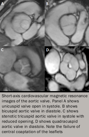
Bicuspid valve disease affects 0.5–0.8% of the population and is often hereditary displaying an autosomal dominant pattern of inheritance and incomplete penetrance (figure 5). It appears to be related to an underlying connective tissue disorder and is frequently associated with abnormalities of the aorta including coarctation and ascending aortic dilatation. The latter predisposes patients to a low risk of aortic dissection such that combined valve and root replacement is often required at the time of surgery. Other congenital abnormalities of the aortic valve include unicuspid and quadricuspid valves that are also associated with calcification, regurgitation and a predisposition to endocarditis (figure 5).
Mitral valve prolapse
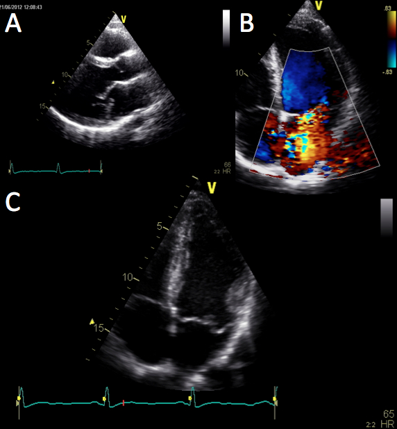
Mitral valve prolapse (MVP) can be defined as the billowing of one or both leaflets of the mitral valve into the left atrium during systole (see figures 6A and 6B). Methods of defining prolapse have been refined with advances in echocardiography, particularly with the realisation that the mitral annulus is saddle-shaped. The prevalence of prolapse in recent studies is around 2% and is associated with mitral regurgitation in about 9% of cases.

Courtesy of John Chambers
