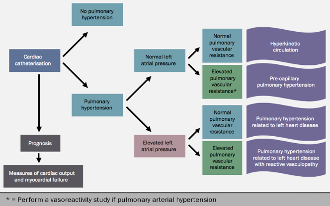
Cardiac catheterisation
Cardiac catheteristion is normally carried out after other, non-invasive imaging has been undertaken and reviewed. Invasive haemodynamic assessment with cardiac catheterisation should be performed in all patients to confirm the diagnosis from non-invasive testing and in selected patients with a high pre-test probability of pulmonary hypertension despite normal echocardiography (such as CTD). Cardiac catheterisation is used to ascertain the haemodynamic mechanism and/or its differential diagnoses (such as restrictive cardiomyopathy and pericardial constriction). It is used in PAH to identify patients who might benefit from long-term treatment with high-dose calcium channel blockers (CCBs). Cardiac catheterisation may also be carried out if the patient deteriorates and to obtain baseline readings before treatment escalation or combination drug therapy, and to guide treatment and transplant decisions. In selected patients, coronary angiography may also be carried out (figure 12).

Hydration needs to be maintained before and during the procedure; renal function may be precarious in these patients, and should be checked before giving contrast media. Warfarin should be discontinued. Adenosine should not be given to patients with asthma. Patients with appearances of pulmonary venous hypertension or veno-occlusive disease should not undergo vasodilator studies.
Venous and arterial sheaths are inserted. Arterial blood gases and oxygen saturation (the latter using a finger probe) are measured at the start of the procedure, and systemic blood pressure and heart rate are measured throughout.
Pressure measurements
Pressure measurements are made from the pulmonary wedge, pulmonary artery, right ventricle, right atrium (and left atrium if entered). All pressure measurements are made at functional residual capacity.
Oximetry is required to measure pulmonary and systemic flow and to identify significant shunts. Blood samples to measure oxygen saturation are taken from: left atrium if entered, pulmonary artery (two or three saturations), right ventricle (body and outflow), high right atrium, mid right atrium, low right atrium, high SVC, low SVC and inferior vena cava.
Cardiac output is measured either by thermodilution or by the Fick principle. The Fick principle with measure oxygen consumption is preferred for patients with low cardiac output (most patients with PH fall into this category). In the differential diagnosis of PH due to left heart disease, accurate pulmonary wedge pressure measurement is needed. Left heart catheterisation should be considered in patients where left heart disease is suspected, or an accurate pulmonary wedge pressure measurement is not successful.
Cardiac catheterisation is safe when performed by experienced operators in well-equipped centres. A recent review of more than 7,000 procedures performed at 20 different medical centres reported 1.1% serious events.16 The most frequent complications were related to venous access, arrhythmias and hypotensive episodes, and most of these were mild to moderate; the overall procedure-related mortality in this study was 0.05%.
Vasodilator testing
Acute vasodilator testing is performed at the same time as cardiac catheterisation in patients with a suspected diagnosis of idiopathic pulmonary arterial hypertension, heritable PAH, and PAH associated with anorexigen use: patients with a positive response have a better prognosis, and additionally the test identifies patients who might benefit from long-term treatment with CCBs. The usefulness of this type of testing in other group 1 patients is uncertain, and this testing is not recommended for patients in clinical classification groups 2 to 5. It is indicated in CTD except scleroderma, and in asymmetric thromboembolic disease. A vasodilator study is contra-indicated in patients with pulmonary venous hypertension, pulmonary veno-occlusive disease or pulmonary capillary haemangiomatosis and severe coronary artery disease.
Acute challenge with a vasodilator should employ only short-acting and safe drugs. The preferred agent is inhaled nitric oxide;17 other possibilities are epoprostenol and adenosine by intravenous infusion. CCBs, sodium nitroprusside and nitrates should not be used for acute vasodilator testing as their safety and efficacy have not been established in this setting (AHA guidelines).Haemodynamic measurements recorded at baseline and during drug administration include pulmonary artery pressure, systemic pressure, pulmonary wedge pressure, right atrial pressure and cardiac output.
The definition of a positive response to acute vasodilator testing is controversial. It is defined by Sitbon17 as a reduction of mean PAP > 10 mmHg to reach an absolute value of mean PAP < 40 mmHg with an increased or unchanged cardiac output. Fewer than 10% of patients with IPAH meet these criteria: they are more likely than other groups to respond to long-term treatment with high doses of CCBs, with roughly half of acute responders also responding in the longer term.
Exercise and fluid loading
Borderline pressure results present difficulties in interpretation of haemodynamic data. If patients become dehydrated, then cardiac filling pressures may become reduced. Fluid loading and exercise are used to detect abnormal haemodynamics in a stable patient, and if pericardial constriction is suspected.
Coronary angiography

Angiography is performed in patients over the age of 40 with thromboembolic disease who are being assessed for PEA, and in patients in whom angina pectoris is a significant symptom.
Definitions and classification
Cardiac catheterisation findings may be used to establish whether pulmonary hypertension is present, and to classify it. Module 1 contains a description of the clinical and haemodynamic classification of pulmonary hypertension.
Genetic testing
Patients with sporadic or familial PAH or PVOD/PCH should be advised in regards to the availability of genetic testing and counselling. BMPR2 mutation screening should be offered by referral centres to patients with IPAH considered to be sporadic, induced by anorexigens and those with a family history. Screening for ACVRL1 and ENG genes may be performed when no BMPR2 mutations are identified in familial PAH patients or in IPAH patients <40 years old, or in patients with a personal or family history of hereditary haemorrhagic telangiectasia. If none of these genes are identified, then screening of rare mutations may be considered. Patients with sporadic or familial PVOD/PCH should be tested for EIF2AK4 mutations.
Prognosis
The prognosis of the individual patient is affected by the cause of disease18 but a number of clinical and investigational findings give prognostic information.
World Health Organization Functional Class (WHO-FC, see table for definitions of functional classes) is a predictor of survival. Registry data19 showed that untreated patients in WHO-FC IV had a median survival of six months, compared with 2.5 years for those in FC III and six years for those in FC I and II. Patients whose functional class improves on treatment have a better prognosis then those whose functional class remains unchanged. Worsening of functional class, rapid worsening of symptoms and the development or increase in frequency and severity of syncope are poor prognostic signs.
Progression of right ventricular failure, with increasing oedema, the need to escalate diuretic therapy or increasing severity and frequency of angina, is indicative of deteriorating and unstable disease.
Performance on the six-minute walk test (6MWT) is indicative of survival: those able to walk 500 metres or more have a better prognosis compared to those able to walk shorter distances. In one epoprostenol trial (see treatments section), distances greater than 380 metres after three months of treatment correlated with better survival. Similarly, maximal oxygen consumption on exercise testing (using cycle ergometry), treadmill exercise time and peak systemic blood pressure greater than 120 mmHg are predictive of survival.
Haemodynamic findings measured at cardiac catheterisation are predictive of prognosis. Based upon available data, it is agreed20 that mean right atrial pressure RAP), cardiac index (CI) and mean pulmonary arterial pressure (mPAP) are predictive of survival, although the mPAP may in fact decrease as right ventricular function worsens. It is important to note that the estimated systolic PAP at rest is usually not prognostic, is not relevant to therapy and nor does an increase or decrease in PAP reflect disease progression or improvement. According to the ESC 2015 guidelines,2 RAP <8 mmHg and CI >2.5 L/min/m2 indicate a better prognosis in the individual patient. In addition, true vasodilator responders have an excellent prognosis.
As right ventricular function is a key determinant of outcome, echocardiographic findings are important in determining the prognosis. A comprehensive assessment of RV function is required, most importantly chamber sizes particularly the right atrium and right ventricle, magnitude of tricuspid regurgitation and an assessment of RV and LV contractility. Contractility can be assessed using several indices, including strain/strain rate, Tei index, RV fractional area change and TAPSE. Speckle tracking can also improve quantification of RV function. An overall impression, however, is more important than a single variable. Furthermore, presence of a pericardial effusion indicates a worse prognosis.
CMR imaging is more accurate than echocardiography for the assessment of the right ventricle and is the current gold standard. Increased RV volume, reduced LV volume, reduced RVEF and reduced RV SV have been shown to be prognostic and there may be value with serial CMR in long term management. However, this requires further investigation.
Several circulating biomarkers are informative in PAH, including BNP, NT-proBNP, serum uric acid and cardiac troponins. Normal or near-normal levels of BNP and NT-proBNP indicate a better prognosis, and levels of these markers may also be used to monitor the effects of treatment.
Determinants of worse prognosis (from ESC/ERS 2015 guidelines)2 are summarised in table 5.
Learning point
- Many markers of prognosis have been established. These are used to assess the patient at baseline, and they may also be used to monitor the progress of disease and the effects of treatment
close window and return to take test
References
1. Black CM. Consensus statement on the management of pulmonary hypertension in clinical practice in the UK and Ireland. Thorax 2008;63 (Suppl II): ii1–ii41. http://dx.doi.org/10.1136/thx.2007.090480
2. Galie N, Hoeper MM, Humbert M et al. The task force for the diagnosis and treatment of pulmonary hypertension of the European Society of Cardiology (ESC) and the European Respiratory Society (ERS), endorsed by the International Society of Heart and Lung Transplantation (ISHLT). Guidelines for the diagnosis and treatment of pulmonary hypertension. Eur Heart J 2015. http://dx.doi.org/10.1093/eurheartj/ehv317
3. Xiao HB, Purcell HJ, Minhas R. ECGs for healthcare professionals. London: Concise Clinical Consulting, 2009.
4. Howard L. Echocardiographic assessment of pulmonary hypertension standard operating procedure. Imperial College Healthcare NHS Trust 2010
5. Yeo TC, Dujardin KS, Tei C et al. Value of a Doppler-derived index combining systolic and diastolic time intervals in predicting outcome in primary pulmonary hypertension. Am J Cardiol1998;81:1157–61. http://dx.doi.org/10.1016/S0002-9149(98)00140-4
6. Fijalkowska A, Kurzyna M, Torbicki A et al. Serum N-terminal brain natriuretic peptide as a prognostic parameter in patients with pulmonary hypertension. Chest 2006;129:1313–21. http://dx.doi.org/10.1378/chest.129.5.1313
7. Nagaya N, Nishikimi T, Uematsu M et al. Plasma brain natriuretic peptide as a prognostic indicator in patients with primary pulmonary hypertension. Circulation 2000;102:865–70. http://dx.doi.org/10.1161/01.CIR.102.8.865
8. American Thoracic Society guidelines for the six minute walk test. Am J Respir Crit Care Med 2002;166:111–7.
9. Miyamoto S, Nagaya N, Satoh T et al. Clinical correlates and prognostic significance of six-minute walk test in patients with primary pulmonary hypertension: comparison with cardiopulmonary exercise testing. Am J Respir Crit Care Med 2000;161:487–92.
10. Humbert M, Sitbon O, Chaouat A et al. Pulmonary arterial hypertension in France: results from a national registry. Am J Respir Crit Care Med 2006;173:1023–30. http://dx.doi.org/10.1164/rccm.200510-1668OC
11. Wensel R, Opitz CF, Anker SD et al. Assessment of survival in patients with primary pulmonary hypertension: importance of cardiopulmonary exercise testing. Circulation2002;106:319–24. http://dx.doi.org/10.1161/01.CIR.0000022687.18568.2A
12. Tunariu N, Gibbs SJ, Win Z et al. Ventilation-perfusion scintigraphy is more sensitive than multidetector CTPA in detecting chronic thromboembolic pulmonary disease as a treatable cause of pulmonary hypertension. J Nucl Med 2007;48:680-4. http://dx.doi.org/10.2967/jnumed.106.039438
13. Grosse C, Grosse A. CT Findings in Diseases Associated with Pulmonary Hypertension: A Current Review. RadioGraphics 2010;30:1753–77. http://dx.doi.org/10.1148/rg.307105710
14. Resten A, Maitre S, Humbert M et al. Pulmonary hypertension: CT of the chest in pulmonary venooclusive disease. Am J Roentgenol 2004;183:65–70. http://dx.doi.org/10.2214/ajr.183.1.1830065
15. Van Wolferen SA, Marcus JT, Boonstra A et al. Prognostic value of right ventricular mass, volume, and function in idiopathic pulmonary arterial hypertension. Eur Heart J 2007;28:1250–7. http://dx.doi.org/10.1093/eurheartj/ehl477
16. Hoeper MM, Lee SH, Voswinckel R et al. Complications of right heart catheterization procedures in patients with pulmonary hypertension in experienced centres. J Am Coll Cardiol2006;48:2546–52. http://dx.doi.org/10.1016/j.jacc.2006.07.061
17. Sitbon O, Humbert M, Jais X et al. Long-term response to calcium channel blockers in idiopathic pulmonary arterial hypertension. Circulation 2005; 111: 3105–11. http://dx.doi.org/10.1161/CIRCULATIONAHA.104.488486
18. McLaughlinVV, Presberg KW, Doyle RL et al. Prognosis of pulmonary arterial hypertension: ACCP evidence-based practice guidelines. Chest 2004;126:78S–92S. http://dx.doi.org/10.1378/chest.126.1_suppl.78S
19. D’Alonzo GE, Barst RJ, Ayres SM et al. Survival in patients with primary pulmonary hypertension. Results from a national prospective registry. Ann Intern Med 1991;115: 343–9.
20. McLaughlin VV, Archer SL, Badesch DB et al. ACCF/AHA 2009 expert consensus document on pulmonary hypertension: a report of the American College of Cardiology Foundation Task Force on expert consensus documents and the American Heart Association developed in collaboration with the American College of Chest Physicians; American Thoracic Society, Inc, and the Pulmonary Hypertension Association. J Am Coll Cardiol 2009;53:1573–619. http://dx.doi.org/10.1016/j.jacc.2009.01.004
Suggested further reading:
Buechel ERV, Mertens LL. Imaging the right heart: the use of integrated multimodality imaging. Eur Heart J 2012;33:949-60. http://dx.doi.org/10.1093/eurheartj/ehr490
close window and return to take test
All rights reserved. No part of this programme may be reproduced, stored in a retrieval system, or transmitted in any form or by any means, electronic, mechanical, photocopying, recording or otherwise, without the prior permission of the publishers, Medinews (Cardiology) Limited.
It shall not, by way of trade or otherwise, be lent, re-sold, hired or otherwise circulated without the publisher’s prior consent.
Medical knowledge is constantly changing. As new information becomes available, changes in treatment, procedures, equipment and the use of drugs becomes necessary. The editors/authors/contributors and the publishers have taken care to ensure that the information given in this text is accurate and up to date. Readers are strongly advised to confirm that the information, especially with regard to drug usage, complies with the latest legislation and standards of practice.
Healthcare professionals should consult up-to-date Prescribing Information and the full Summary of Product Characteristics available from the manufacturers before prescribing any product. Medinews (Cardiology) Limited cannot accept responsibility for any errors in prescribing which may occur.
