“I will give you a new heart and put a new spirit in you; I will remove from you your heart of stone and give you a heart of flesh” – Ezekiel 36:26
A 72-year-old woman was referred to our cardiology service with increasing dyspnoea on exertion. Her background history was notable for haemochromatosis, type 2 diabetes mellitus, chronic kidney disease (stage 3a), treated pulmonary tuberculosis and known pericardial calcification. Echocardiography (figure 1A) demonstrated a calcified structure evident on the apical four-chamber view, which appeared to indent the right ventricle. Computed tomography (CT) of the thorax (figure 1B) demonstrated extensive and circumferential pericardial calcification with a maximal thickness of up to 20 mm in the right atrial pericardial region, 12 mm in the inferior pericardium and 18 mm in the left atrioventricular region, extending along the basal lateral left ventricular (LV) pericardial region. We proceeded to right and left heart catheterisation. A heavily calcified pericardium was evident on coronary angiography (figure 1C). Simultaneous pressure tracings from the right and left ventricles were taken and demonstrated equalisation of the ventricular end diastolic pressures. Mean pulmonary capillary wedge pressure was 25 mmHg, pulmonary artery pressure was 37/16 mmHg, right ventricular (RV) pressure was 51/20 mmHg (mean 24 mmHg) and mean right atrial (RA) pressure was 21 mmHg. LV end diastolic pressure was 25 mmHg. The patient was diagnosed with constrictive pericarditis, likely secondary to previous tuberculosis. Her case was discussed at the cardiothoracic conference and a decision made to proceed to pericardiectomy. However, after discussion with the cardiothoracic surgeon, the patient elected not to proceed to surgery. She is currently being managed medically and at the last outpatient clinic review was clinically stable.
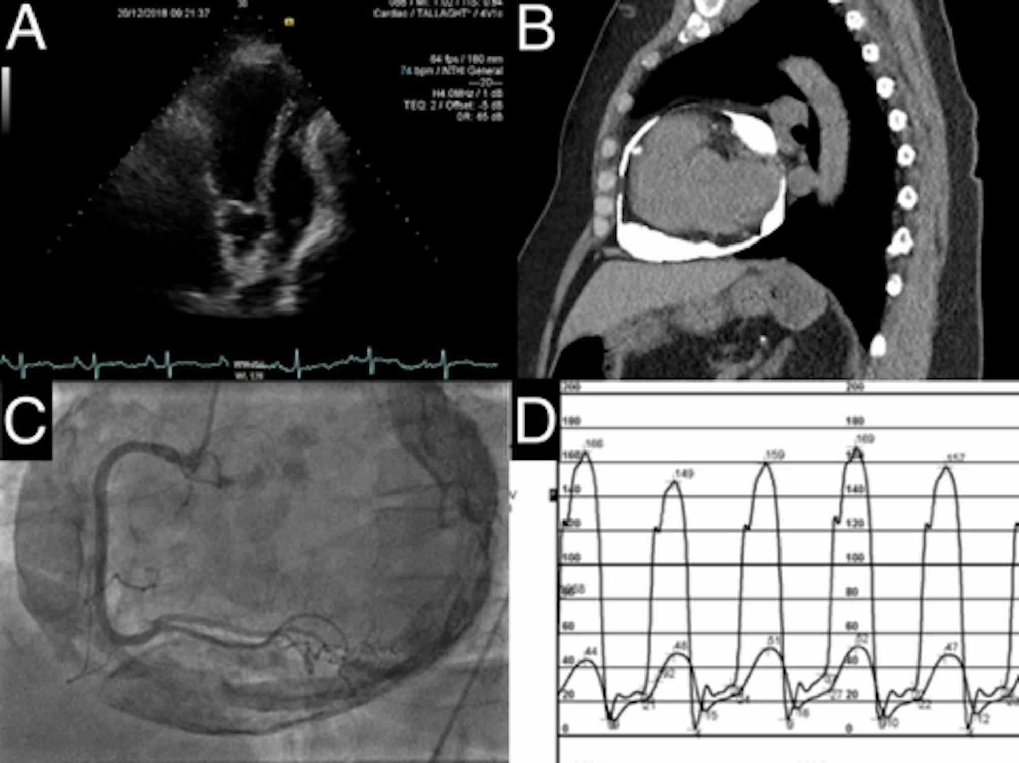
D. Right and left heart catheterisation
Conflicts of interest
None declared.
Funding
None.
Consent
Patient consent for publication was obtained.
