

We present a case of malignancy-induced thromboembolism resulting in global ST-elevation myocardial infarction and complete heart block.
Introduction
Acute myocardial infarction (AMI) is a common presentation in patients, mainly caused by plaque rupture or erosion; however, it is rarely caused by thromboembolism. The incidence of AMI secondary to thromboembolism is 2.9%.1 Several cases of arterial thrombosis resulting in AMI have been reported; however, multiple thrombi affecting all three coronary arteries are rare.2,3 We present a case of malignancy-induced thromboembolism resulting in global ST-elevation myocardial infarction and complete heart block.
Case presentation
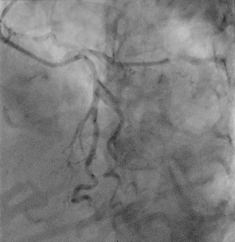
A woman in her mid-80s presented to a specialist eye hospital with right eye visual loss in the morning, and was diagnosed with right-sided giant cell arteritis before being referred to another hospital for high-dose steroids. While waiting in the emergency department, she had an episode of seizure, and an electrocardiogram (ECG) following seizure showed complete heart block with global ST-segment-elevation myocardial infarction (figure 1). Past medical history was significant for hypertension, glaucoma, right-knee replacement, and trigeminal neuralgia. Arterial blood gas post-seizure showed pH 7.23 (reference range 7.35–7.45), and the lactate was 5.0 mmol/L. Initial troponin T was 63 ng/L (reference range 0–14 ng/L), and the repeat troponin T level was 326 ng/L. Bedside echocardiography showed mild inferior wall hypokinesia, and an emergency coronary angiogram showed thromboembolism in all three coronary arteries (figures 2 and 3). Coronary angioplasty with a drug-coated balloon (DCB) was performed on the right coronary artery, resulting in TIMI (thrombolysis in myocardial infarction) 2 flow, and she was given a tirofiban bolus followed by infusion, in view of the heavy thrombus burden. She also underwent computed tomography (CT) of the head and pulmonary angiography, which revealed lytic lesions and oesophageal dilatation with lung nodules.

 |
 |
She was admitted to the coronary care unit (CCU) and commenced on dual antiplatelet therapy, including aspirin and clopidogrel. She was hypotensive and was given intravenous fluids. Resuscitation and ceiling of care were discussed with the family and patient, and, with their agreement, a do-not-attempt-resuscitation form was completed and a ceiling of care established for ward-based care. The patient showed further clinical deterioration and commenced on end-of-life care. She passed away peacefully 24 hours after admission.
Discussion
The ECG shows inferior and antero-lateral ST-segment elevation with complete heart block and ventricular ectopy. The ECG changes are inconsistent with one coronary territory, as confirmed by coronary angiography.1 The differential diagnosis would include large vessel arteritis, given the recent diagnosis of giant cell arteritis, myocardial infarction secondary to coronary artery disease or thromboembolism, or takotsubo cardiomyopathy secondary to the stress of physical illness and loss of vision in her right eye.
This patient had significant thromboembolism, which likely resulted in visual loss in the right eye and occlusion of all three coronary vessels, resulting in global ST-segment-elevation myocardial infarction and complete heart block.2,3 The patient had a DCB to the right coronary artery and a tirofiban bolus followed by infusion.4 Stenting was not the right option given the extent of thromboembolism, with the primary aim being to restore TIMI 3 flow, and a DCB to restore flow was a reasonable option. The right coronary artery was treated in this case due to the patient having complete heart block, although the patient had evidence of thromboembolism in all coronary arteries.
She had a CT of the head and CT pulmonary angiography (CTPA) confirming a lytic lesion in the skull, lung nodules and dilated oesophagus, highly suggestive of underlying malignancy. The CTPA did not show any pulmonary embolism, and there was no clinical evidence of deep vein thrombosis. She was not stable enough to have a CT abdomen and pelvis to confirm the primary malignancy.
After discussion with the patient and her family, a do-not-attempt-resuscitation form was completed, and she passed away the following day. It is important to remember that malignancies can present with thromboembolism, resulting in visual loss, stroke, pulmonary embolism, and acute myocardial infarction. In this case, ECG findings were consistent with occlusion in all three coronary arteries, which is rare.
Key messages
- Malignancy-induced thromboembolism should be considered in patients presenting with widespread ST-segment elevation
- Imaging, including whole body imaging, should be considered in patients suspected of having malignancy
- Drug-coated balloons, rather than stenting, should be considered in patients with significant thromboembolism
Conflicts of interest
None declared.
Funding
None.
Patient consent
Consent could not be obtained from the patient as the patient passed away, unfortunately. However, written consent for publication was obtained from the next of kin.
References
1. Deshpande A. ST-segment elevation: distinguishing ST elevation myocardial infarction from ST elevation secondary to nonischemic etiologies. World J Cardiol 2014;6:1067–79. https://doi.org/10.4330/wjc.v6.i10.1067
2. Farris W, Waymack JR. Central retinal artery occlusion. In: StatPearls [Internet]. Treasure Island (FL): StatPearls Publishing; 2021. Available from: https://www.ncbi.nlm.nih.gov/books/NBK470354/
3. Alameer A, Kanodia AK, Duraikannu C, Nair P. Isolated superior ophthalmic vein thrombosis in a patient with prostate cancer. BMJ Case Rep 2023;16:e253919. https://doi.org/10.1136/bcr-2022-253919
4. Hu H, Shen L. Drug-coated balloons in the treatment of acute myocardial infarction (review). Exp Ther Med 2021;21:464. https://doi.org/10.3892/etm.2021.9895


