There is much debate about the optimal sedation strategy for transoesophageal echocardiography (TEE). Despite previous studies demonstrating the potential benefits of combining opiates and benzodiazepines for conscious sedation, and previous published national surveys and recommendations, sedation practice for TEE in clinical practice varies widely within the UK. All UK centres routinely use midazolam, but only 7% of centres use it in combination with an opiate: 14% of hospitals report no routine use of sedation for TEE. There
is no British Society of Echocardiography (BSE) recommended TEE sedation protocol within the UK and even where guidelines exist locally, 82% of operators report being unaware of their details. Consequently, a wide range of sedative doses are used and many patients are reported to be over-sedated. We developed a new protocol for conscious sedation using intravenous pethidine and midazolam for TEE and have shown it to be safe and effective when implemented within an existing TEE service.
From the days of Virchow and the analysis of post-mortem coronary specimens, an enormous amount of knowledge has been built about coronary pathophysiology. In the 1950s the dream of in vivo coronary imaging became a reality with the invention of coronary arteriography under the guidance of Mason Sones. As we fast forward 50 years, it has become clear that angiography has helped us focus on areas of stenosis and flow limitation, but the main problem of coronary artery disease is much more complex than can appear on a luminal silhouette. The finding of ‘normal coronary arteries’ following angiography is short-sighted and does not take into account the potential of unstable disease lurking within the vessel wall. We begin the series with intravascular ultrasound.
Background
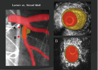
Figure 1. Positive remodelling: arterial expansion to protect luminal size
Coronary luminal narrowing is prevented by a vascular mechanism known as positive remodelling. This is an outwards expansion of the blood vessel to accommodate the build up of plaque within the artery (figure 1). This phenomenon appears within diseased segments of coronary arteries and the build up of these plaques is dependent on multiple, well-known factors from genetics and lifestyle through to coronary anatomy and flow patterns. The complex pathobiology that creates these plaques and leads to plaque structure weakening is beyond the scope of this review. However, contemporary coronary imaging can now focus on the vessel wall and identify high-risk, positively remodelled, plaques and their contents. This series of review articles will focus on what is now possible in the field of coronary artery imaging and how each modality can try to improve both the invasive treatment of unstable coronary syndromes and the prevention of unstable coronary artery disease in the future.
IVUS and virtual histology
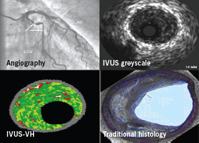
Intravascular ultrasound (IVUS) is performed at the time of coronary angiography and involves a tiny ultrasound probe that emits high frequency signals (20–40 mHz). This wire-based probe can be placed over a coronary guidewire into the artery and withdrawn at a set rate (0.5 mm/sec) to provide segmental tomographic images of the vessel. IVUS has demonstrated discrepancies between the extent of atherosclerosis seen by coronary angiography and the actual extent of atherosclerotic disease.1 Quantitative assessment of the vessel and plaque within a lesion was made possible with the introduction of greyscale IVUS analysis and the further analysis of individual plaque components is now possible with virtual histology (VH).2
Figure 2. Intravascular ultrasound virtual histology (IVUS-VH): producation of a colour-coded map of the atherosclerotic plaque
IVUS-VH uses advanced radiofrequency analysis of ultrasound backscatter signals to overcome the limitations of greyscale IVUS by providing a more detailed analysis of plaque morphology (figure 2).3 In addition, IVUS-VH has the potential to provide in vivo patient-specific plaque analysis to determine the range of characteristics in relation to clinical factors and risk, rather than making assumptions about living plaque from a highly selected autopsy population.
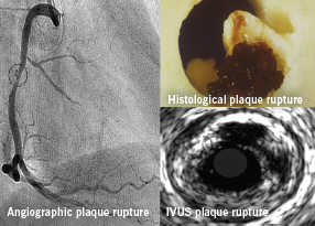
Greyscale IVUS has also demonstrated the multiplicity of plaque ruptures seen in patients with acute coronary syndromes (ACS) (figure 3).4-6 A recent study also highlighted that the number of vulnerable plaques with less than 75% luminal obstruction identified by IVUS had a positive correlation with future cardiovascular events.7 Of note, serial IVUS analysis of a small patient cohort showed that 50% of ruptured coronary plaques detected on first ACS event had spontaneously healed at 22 months’ follow-up.8
Figure 3. Plaque rupture on angiography and IVUS
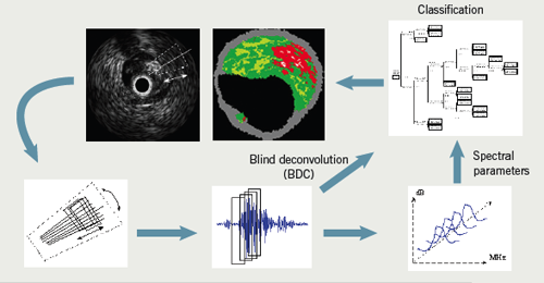
Obviously, many different cell and tissue types are commonly found in atherosclerotic plaques. To simplify image interpretation, and because of the fundamental resolution limitations of the underlying ultrasound signal, plaque components are grouped into four basic tissue types during IVUS-VH imaging. These components are displayed on the image as different colour pixels. This technique is based on advanced radiofrequency analysis of reflected ultrasound signals in a frequency domain analysis and displays the reconstructed colour-coded tissue map of plaque composition superimposed on cross-sectional images of the coronary artery obtained by greyscale IVUS (figure 4).3
Figure 4.The reflected ultrasound signal being converted to its corresponding tissue type
Tissue types
1. Fibrous
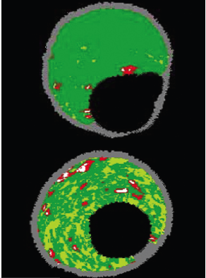
Fibrous tissue is represented by dark-green pixels (figure 5). Histologically, this tissue is collagenous with no lipid.9,10 On greyscale IVUS, these tissues tend to be medium-bright regions.
2. Fibrofatty
Figure 5. Fibrous plaque appearing as dark green (top) and fibrofatty plaque appearing as light green (bottom)
Fibrofatty tissue is denoted in VH by light-green pixels (figure 5). This tissue is loosely packed collagen, but it can have a cellular quality with potential for foam cells to start invading.10 There is usually no necrotic core and even cholesterol products are rare. If thrombus or plaque rupture are included as plaque during analysis, then they are displayed as fibrofatty plaque.11
3. Necrotic core
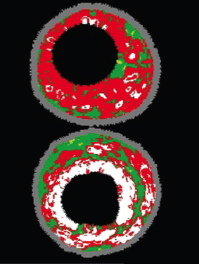
In VH the necrotic core is seen as red (figure 6). This tissue is a mixture of soft, lipid-like dead cells, foam cells and trapped blood cells.9 Most of any real structure is lost and with some areas producing micro-calcification as a by-product (from the dead cells) this leads to a recipe for gross instability and rupture with friable areas next to sharp calcification.
4. Dense calcium
White pixels represent dense calcium (figure 6). These calcified regions can be lost during histology processing but on plain greyscale IVUS, they act as extremely strong reflectors of signal and appear as bright white.
Plaque risk assessment
Figure 6. Necrotic core in a thin-cap fibroatheroma (TCFA) lesion appearing as red (top) and dense calcium in fibrocalcific disease appearing as white (bottom)
In vivo plaque classification with IVUS-VH is based on a histopathological classification system developed by Virmani et al. in 2000.12,13 From this system, coronary lesions can be classified as adaptive intimal thickening, pathologic intimal thickening, fibroatheroma, fibrocalcific and thin-cap fibroatheroma (TCFA) plaques. For the purpose of this review we focus on the highest-risk plaque type described below.
Thin-cap fibroatheroma (TCFA)
TCFA have a confluent necrotic core (>10%) in direct contact with the lumen (no IVUS evidence of a cap) and a minor amount of calcium (<10%). If this is present on three consecutive IVUS-VH cross-sectional frames, this confers an increase in vulnerability. It is currently thought that the higher the extent of surface contact the necrotic core has with the lumen, and the presence of increased amounts of calcium, produces the highest risk of rupture. This appears to be in some way different from the commonly taught theory that non-significant plaques are the ones which are more likely to rupture.14 It may simply be that non-significant plaques are just more frequent along a given coronary segment.
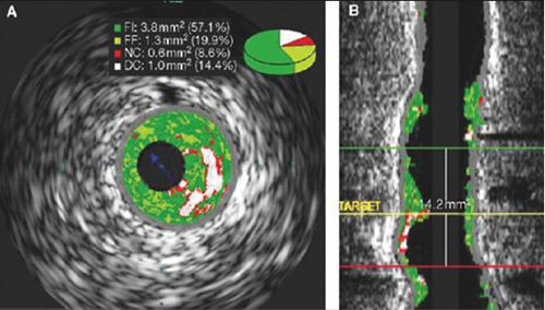
Figure 7. IVUS-VH in longitudinal view showing a specific frame and length/distibution of disease
As the majority of acute coronary events are triggered by plaque rupture,4,5 defining the anatomic features that lead to plaque rupture should be of central importance to lesion imaging. Post-mortem analyses have shown that TCFA is probably the main precursor lesion for plaque rupture.12 According to histological studies, the size of the necrotic core and the thickness of the fibrous cap have a critical influence on plaque stability, along with our previously mentioned positive remodelling. In addition, other characteristics of vulnerable lesions such as micro-calcification within the lesion, its location within the coronary tree, the length of the lesion and the degree of stenosis relative to healthy reference lumen size, are also important parameters for the evaluation of plaque vulnerability. The reconstruction of IVUS-VH images in a longitudinal view enables a comprehensive analysis of the total length of the plaque and its complete orientation to the rest of the coronary tree (figure 7).
Therefore, exact coronary dimensions and elements of plaque composition, such as the presence and amount of necrotic core, plaque burden, the degree of calcification and coronary remodelling, are all anatomic features visualised by IVUS and IVUS-VH, but not by traditional angiography. However, the limited resolution of IVUS-VH (approximately 150 µm) renders it unable to truly claim the ability to visualise thin fibrous caps (<65 µm). In order to improve visualisation of the coronary lumen we need to improve resolution, and this is best seen with a newer invasive tool called optical coherence tomography (OCT). OCT uses spectroscopic light emission in place of the ultrasound waves and will be covered in our next article.
Summary
Angiographic imaging of coronary disease is flawed and cannot be relied on to understand the full extent of plaque development. Imaging tools are now available that can directly image the vessel wall and calculate plaque burden and plaque composition with great accuracy. IVUS remains an under-utilised but invaluable clinical and research tool to improve the understanding of coronary artery disease. It is hoped that in the future it may not only help improve the outcome from vulnerable plaque treatment by percutaneous intervention,
but also help stratify the vulnerable patient to improve risk factor modification and appropriate preventative drug therapy.
Conflict of interest
None declared
Key messages
Angiographic assessment of coronary plaque is limited and does not provide the full story
Intravascular ultrasound (IVUS) can image the entire vessel in tomographic slices around 0.01 mm thick and with 150 µm resolution
Virtual histology (VH) is a new technique that can provide a sensitive guide to plaque components within four groups: fibrous, fibrofatty, necrotic core, and dense calcium
It is hoped that future studies will show that IVUS-VH can improve results and outcomes from coronary intervention
References
1. Nissen SE, Yock P. Intravascular ultrasound: novel pathophysiological insights and current clinical applications. Circulation 2001;103:604–16.
2. Nissen SE. Application of intravascular ultrasound to characterize coronary artery disease and assess the progression or regression of atherosclerosis. Am J Cardiol 2002;89:24B–31B.
3. Nair A. Coronary plaque classification with intravascular radiofrequency data analysis. Circulation 2006;106:2200–06.
4. Tanaka A, Shimada K, Sano T et al. Multiple plaque rupture and C-reactive protein in acute myocardial infarction. J Am Coll Cardiol 2005;45:1594–9.
5. Rioufol G, Finet G, Ginon I et al. Multiple atherosclerotic plaque rupture in acute coronary syndrome: a three-vessel intravascular ultrasound study. Circulation 2002;106:804–08.
6. Schoenhagen P, Stone GW, Nissen SE et al. Coronary plaque morphology and frequency of ulceration distant from culprit lesions in patients with unstable and stable presentation. Arterioscler Thromb Vasc Biol 2003;23:1895–900.
7. Yamagishi M, Terashima M, Awano K et al. Morphology of vulnerable plaque: insights from follow-up of patients examined by intravascular ultrasound before an acute coronary syndrome. J Am Coll Cardiol 2000;35:106–11.
8. Rioufol G, Finet G, Ginon I et al. Evolution of spontaneous atherosclerotic plaque rupture with medical therapy: long-term follow-up with intravascular ultrasound. Circulation 2004;110:2875–80.
9. Burke AP, Kolodgie FD, Farb A et al. Morphological predictors of arterial remodeling in coronary atherosclerosis. Circulation 2002;105:297–303.
10. Nair A, Margolis MP, Kuban VD, Vice DG. Automated coronary plaque characterisation with intravascular ultrasound backscatter: ex vivo validation. Eurointervention 2007;3:113–20.
11. Murray SW, Palmer ND. Intravascular ultrasound and virtual histology interpretation of plaque rupture and thrombus in acute coronary syndromes. Heart 2009;95:1494.
12. Kolodgie FD, Virmani R, Burke AP et al. Pathologic assessment of the vulnerable human coronary plaque. Heart 2004;90:1385–91.
13. Uemura R, Tanabe J, Ohaki M et al. Impact of histological plaque characteristics on intravascular ultrasound parameters at culprit lesions in coronary artery disease. Int Heart J 2006;47:683–92.
14. Kolodgie FD, Gold HK, Burke AP et al. Intraplaque hemorrhage and progression of coronary atheroma. N Engl J Med 2003;349:2316–25.
In UK cardiac rehabilitation programmes, exercise training is often set at a percentage of maximal heart rate or heart rate reserve, either predicted or measured. Problems may arise when using this method for chronic heart failure (CHF) patients who often have chronotropic incompetence and are treated with beta blockers. A safer approach is to use cardiopulmonary exercise testing to prescribe training below the ventilatory threshold, thus ensuring that the exercise is moderate. The aim of this study was to determine whether British Association for Cardiac Rehabilitation (BACR) heart rate guidelines prescribe moderate intensity exercise for CHF patients. The only target heart rate range to prescribe exercise below the ventilatory threshold was 60–80% measured maximum heart rate. Target heart rates calculated from predicted maximum values were higher than those from measured values, and the heart rate reserve method resulted in the highest target heart rates. Cardiac rehabilitation exercise practitioners should be aware that these methods may well result in CHF patients performing heavy rather than moderate exercise.
Diabetes mellitus is caused by an absolute or relative lack of insulin.1 This article covers people with type 2 diabetes, as most people with type 1 diabetes will be under the care of a secondary care team for at least some of their care. Type 2 diabetes is not primarily about sugar, but about moderating the vascular and neurological damage resulting from chronic hyperglycaemia. Many people with type 2 diabetes will also have components of the metabolic syndrome,2 namely hypertension, dyslipidaemia and obesity, all of which need separate and sometimes overlapping interventions.
Introduction
People diagnosed with diabetes have the condition for the rest of their life, and like many other long-term conditions, most of the interventions that effect control and outcomes are in the hands of the patient themselves.3 The key to successful treatment in primary care is to effectively educate and engage the person with diabetes in a self-management collaboration, using the skills and treatment options offered by healthcare professionals.4 Given the possible complications of diabetes, it is important that this support is provided using the combined skills of several different healthcare professional groups, such as nurses, general practitioners (GPs), podiatrists, dietitians and psychologists. Sometimes referral to a specialist team, which may be community or hospital based, will be required. The key ethos in the diabetes “Commissioning diabetes without walls”5 concept to make high-quality care available to everyone who needs it, is that it is the skills of the various healthcare professionals needed at any particular time that matter, and not where they are based.
1. Are you sure of the diagnosis?
This is not as simple as it sounds.6 The advent of the quality outcomes framework (QOF) forced GPs to code people with diabetes as having either type 1 or type 2 diabetes. Very few were coded as having other forms of diabetes, such as genetic diabetes, and still less as diabetes due to other causes, such as Cushing’s syndrome or acromegaly. The result was that there was a huge variation in the prevalence of people with type 1 diabetes from 1% to 25%.7 The implication is that a significant number of people with type 2 diabetes on insulin were misclassified as having type 1 diabetes, as well as people with rarer forms of diabetes, such as one of the variety of gene abnormalities now termed maturity onset diabetes of the young (MODY).8
Differentiating type 2 diabetes from type 1 can sometimes be challenging, these rules help:
type 1 diabetes can present at any age but most cases occur in childhood
patients with type 1 diabetes tend to lose weight but, as with the general population, they still may be overweight
the presence of ketones strongly suggests type 1 diabetes.
If in doubt discuss with an expert, they may check diabetes-specific antibodies (glutamic acid decarboxylase [GAD] antibodies, Islet cell antibodies and insulin antibodies) or initiate a therapeutic trial under close supervision.
2. Is this an unusual presentation of diabetes?
Most people would recognise the classic symptoms of diabetes, i.e. weight loss, thirst and polyuria, but sometimes the symptoms cause diagnostic confusion, particularly in the elderly, and may result in an inappropriate or unnecessary referral to the wrong speciality. Patients may present with confusion, memory loss, paraesthesia, mood changes and recurrent infections, particularly vaginal candidiasis. Persistent diarrhoea or vomiting may be a manifestation of early autonomic neuropathy. Sexual dysfunction in women, as well as men, is an increasingly recognised complication, and may be the presenting symptom of diabetes.9
3. Have they accepted the diagnosis?
As with many long-term conditions, failure of therapeutic effect may be due to lack of compliance with the agreed treatment regimen, and this is particularly true in diabetes.10 It is not unusual for people diagnosed with diabetes to go through a period of denial which is akin to a grief reaction. This traditionally has five components: denial, anger, bargaining, depression and, eventually, acceptance. Primary care teams should suspect stages of denial when medication concordance is erratic, or there is a perceived lack of engagement or interest in controlling diabetes. Depression is about twice as common in people with diabetes,11 and this may impair the individual’s ability to self-care.12 Encouraging the attendance of family members, partners or carers to diabetes clinics may shed light on what is going on.
4. Have we mixed the right medications?
Everyone newly diagnosed with type 2 diabetes should be offered a structured education programme along with locally appropriate weight management and exercise opportunities. If this fails to achieve a lowering of glycosylated haemoglobin (HbA1c) to an individually tailored target, the latest National Institute for Health and Clinical Excellence (NICE) guidance (CG 66 and CG 87)13 supports the use of an increasingly complex cocktail of medications, including insulin. Certainly, triple therapy with metformin, a sulphonylurea and either a glitazone or a gliptin (dipeptidyl peptidase [DPP] IV inhibitor) should be used where appropriate. Many practices are achieving competence in using insulin regimens, which now have licences to be used with pioglitazone or sitagliptin, as well as metformin. The recently licensed injectible incretin mimetics (exanatide and liraglutide) may well become established primary care drugs in the future but for now they are probably best grouped with insulins, i.e. if you have the confidence, skills and knowledge to initiate insulin, you can certainly initiate incretin mimetics. The role of weight should always be borne in mind, and any intervention that lowers weight is likely to have a beneficial impact on diabetes control. Adjusting the energy balance (eat less, do more) is the cornerstone of weight management, but the use of weight-reducing drugs (orlistat is now the sole remaining licenced drug in this area) and bariatric surgery is recommended, the latter achieving normoglycaemia in over 50% of cases.14
5. Have we considered the contraindications of the drugs?
Having a good working knowledge of what works with what and the potential side effects and relative contraindications is important. Metformin, although universally regarded as the drug of first choice, does cause gastrointestinal upset in about 10% of people who take it.15 Gastrointestinal intolerance can be moderated by starting at 500 mg a day and building up slowly, or using the slow-release option, which may also allow less tablets to be taken as it is now available in 1 g tablets. Metformin doses should be halved at estimated glomerular filtration rates (eGFRs) under 45 ml/min/1.73 m2 and metformin stopped at eGFR of less than 30 ml/min/1.73 m2. Further considerations include the relative contraindication for glitazones in people prone to heart failure or in women at risk of fractures. Sulphonylureas alone or in combination with other oral hypoglycaemic agents can precipitate hypoglycaemia and this may be an issue for people who do a lot of driving or who work in hazardous occupations.
6. Have we looked for complications?
It is estimated that people diagnosed with diabetes have had raised blood sugars for between four to seven years, and, at diagnosis, half have detectable complications.16 All people with diabetes should be screened for signs of retinopathy (by digital retinal photography) and nephropathy (by assessment of urinary microalbumin). All members of the primary healthcare team should be competent in basic foot examination and refer appropriately to community podiatry clinics for non-urgent matters, or to local rapid-access foot clinics in the case of ulceration, ischaemia or the suspicion of a Charcot foot with a warm, swollen, subluxed, and often painless, ankle joint.
Half of men with diabetes have erectile dysfunction,17 and although the condition is often easily improved by the use of phosphodiesterase (PDE) inhibitors (sildenafil, tadalafil, vardenafil), it is important to remember that erectile dysfunction may be an early marker of cardiovascular disease.18 The less well-recognised complication of diabetic autonomic neuropathy (DAN) can cause diarrhoea, abdominal bloating, orthostatic hypotension and loss of exercise-induced tachycardia, which may contribute to the increased risk of silent myocardial infarctions in people with diabetes. DAN also contributes to loss of hypoglycaemia awareness, which is a major issue for people on insulin.
7. Have we considered the effect on recreation, work and driving?
Diabetes may impact on the working lives of many people, but it is particularly relevant if the risk of hypoglycaemia might cause harm to the person themselves or others. This may involve driving, operating machinery or working at heights, so knowing someone’s occupation may help to guide treatment options. There are some occupations that people on insulin are not allowed to do, such as flying a plane or driving a train, and there are restrictions on driving passenger-carrying or heavy goods vehicles. The Driver and Vehicle Licensing Agency (DVLA) at-a-glance document (http://www.dft.gov.uk/dvla/medical/ataglance.aspx) gives up-to-date advice. For drivers who need licence reviews, the opinion of a consultant diabetologist must be sought to confirm that the risk of hypoglycaemia is minimal. This is also true for people who wish to take up hazardous sports, including diving which is no longer prohibited. It should be noted that people with diabetes are covered by the Disability Discrimination Act (1995) and as such employers must make appropriate adjustments to allow them to work. GPs may have to advise employers that making time and arrangements for blood sugar testing or injecting at work is a requirement.
8. Have we considered the technical problems?
Diabetes, almost like no other condition, is complicated by technical issues. People who choose, or are advised, to do self-monitoring of blood glucose (SMBG), need the skills to act on the results. Like all technology, these machines may give erroneous readings for a variety of reasons, and GPs and nurses need to have an index of suspicion when SMBG, HbA1c or indeed symptoms, do not tie up. The same applies to insulin pens and needles, which can malfunction and result in the wrong dose of insulin being dialled up. Insulin itself can denature if incorrectly stored. Batches of blocked needles are not unknown. With the advent of long-haul travel, some patients run out of insulin and return with U40 insulin (40 units per ml), which is still made in China and Russia. This is 2.5 times weaker than European or US insulin, which can lead to rapid loss of control, or hypoglycaemia on returning to using the U100 insulin in the UK.
9. Who do we refer to?
For the majority of diabetes-related problems, the advice of a diabetes specialist nurse or nurse consultant will be sufficient. On occasion, referral to a consultant diabetologist will be necessary, but this could be done by virtual referral as long as the consultant has full access to the primary care records.
Many patients, as mentioned, have psychological and adjustment issues, and referral to a diabetes psychologist is recommended.
Eye, vascular and renal problems may need direct referral, but given the huge variety of possible diabetes complications, diabetologists often regard themselves as the last of the true general physicians, and, as such, are a valuable source of information and advice.
10. Have we considered other sources of support?
There are 2.5 million people in the UK with diabetes and most areas will have a local support group usually affiliated and supported by Diabetes UK (www.diabetes.org.uk) who also run a telephone care line for patients and have a focus on supporting diabetes-related research.
In addition, there are the Insulin Dependent Diabetes Trust (who campaign to maintain supplies of animal insulins; www.iddtinternational.org), Diabetes Lifeline (who will contact a family member in case of an emergency; www.diabeteslifeline.co.uk), and Diabetes Aware (who run awareness events with primary care trusts; www.diabetesaware.org.uk). Healthcare professionals can join Diabetes UK as a professional member, or the Primary Care Diabetes Society (www.pcdsociety.org).
Conflict of interest
None declared.
References
1. Butler RN, Rubenstein AH, Gracia AM, Zweig SC. Type 2 diabetes: causes, complications, and new screening recommendations. I. Geriatrics 1998;53:47–50, 53–4.
2. Targher G, Bertolini L, Tessari R, Zenari L, Arcaro G. The International Diabetes Federation definition of the metabolic syndrome independently predicts future cardiovascular events in type 2 diabetic patients. The Valpolicella Heart Diabetes Study. Diabet Med 2006;23:1270–1.
3. Clark M. Diabetes self-management education: a review of published studies. Prim Care Diabetes 2008;2:113–20.
4. Dagogo-Jack S. Preventing diabetes-related morbidity and mortality in the primary care setting. J Natl Med Assoc 2002;94:549–60.
6. Diabetes UK. The classification and coding of diabetes in primary care. London: Diabetes UK, March 2010.
7. Rollason W, Khunti K, de Lusignan S. Variation in the recording of diabetes diagnostic data in primary care computer systems: implications for the quality of care. Inform Prim Care 2009;17:113–19.
8. Winckler W, Weedon MN, Graham RR et al. Evaluation of common variants in the six known maturity-onset diabetes of the young (MODY) genes for association with type 2 diabetes. Diabetes 2007;56:685–93.
9. Muniyappa R, Norton M, Dunn ME, Banerji MA. Diabetes and female sexual dysfunction: moving beyond ‘benign neglect’. Curr Diab Rep 2005;5:230–6.
10. Cramer JA, Benedict A, Muszbek N, Keskinaslan A, Khan ZM. The significance of compliance and persistence in the treatment of diabetes, hypertension and dyslipidaemia: a review. Int J Clin Pract 2008;62:76–87.
11. Anderson RJ, Freedland KE, Clouse RE, Lustman PJ. The prevalence of comorbid depression in adults with diabetes: a meta-analysis. Diabetes Care 2001;24:1069–78.
12. Antai-Otong D. Diabetes and depression: pharmacologic considerations. Perspect Psychiatr Care 2007;43:93–6.
13. National Institute for Health and Clinical Excellence. NICE Guidance on type 2 diabetes. London: NICE, May 2010.
14. Treadwell JR, Turkelson CM. Systematic review of bariatric surgery. JAMA 2005;293:1726.
16. Amos AF, McCarty DJ, Zimmet P. The rising global burden of diabetes and its complications: estimates and projections to the year 2010. Diabet Med 1997;14(suppl 5):S1–S85.
17. Brown JS, Wessells H, Chancellor MB et al. Urologic complications of diabetes. Diabetes Care 2005;28:177–85.
18. Grover SA, Lowensteyn I, Kaouache M et al. The prevalence of erectile dysfunction in the primary care setting: importance of risk factors for diabetes and vascular disease. Arch Intern Med 2006;166:213–19.
Authors: Kyle J Stewart, Pippa Woothipoom, Jonathan N Townend
Kyle J Stewart
Medical Student
Pippa Woothipoom
Medical Student
University of Birmingham, Edgbaston, Birmingham, B15 2TT.
Jonathan N Townend
Consultant and Honorary Senior Lecturer in Cardiology
Department of Cardiology, Queen Elizabeth Hospital, Edgbaston, Birmingham, B15 2TH.
Correspondance to:
Mr K J Stewart
Sundowner Lodge,
Higher Broad Oak Road,
West Hill, Devon, EX11 1XJ.
[email protected]
To establish whether the medication received by patients post-myocardial infarction was prescribed at therapeutic doses, we performed a retrospective audit of discharge summaries. Over three quarters (75.1%) of all patients in the study group were discharged on sub-therapeutic doses of angiotensin-converting enzyme (ACE) inhibitors and beta blockers. In contrast, nearly all (94–97%) patients received a statin at a therapeutic dose. Aspirin and clopidogrel, where prescribed, were also within the therapeutic range in 100% of patients. These findings illustrate the difficulty in optimising the doses of drugs that have a wide range of possible doses during short hospital admissions.
Introduction
This retrospective audit was performed to assess whether patients discharged from the cardiology ward at the Queen Elizabeth Hospital, Birmingham, following ST-segment elevation myocardial infarction (STEMI) or non-ST-segment elevation myocardial infarction (NSTEMI) were prescribed the recommended medication at appropriate doses. The evidence for the prognostic benefit of drugs such as angiotensin-converting enzyme (ACE) inhibitors, beta blockers and statins after a myocardial infarction (MI) is derived from studies in which these drugs were used at high doses, such as Acute Infarction Ramipril Efficacy (AIRE),1 Carvedilol Post-Infarct Survival Control in Left-Ventricular Dysfunction (CAPRICORN),2 and Pravastatin or Atorvastatin Evaluation and Infection Therapy (PROVE IT).3 Lower doses may not confer the same benefit.
Recent studies on prescribing post-MI show that between 60% and 90% of patients are receiving each discharge medication as recommended by the National Institute for Health and Clinical Excellence (NICE),4-7 although this does appear to be increasing.5 This study aims to go further by assessing how many patients are being prescribed these drugs at doses likely to be effective.
Methods
Computerised discharge summaries, which included the discharge medications, were analysed retrospectively for 400 patients discharged between May and September 2008.
For study purposes, the sub-therapeutic doses of NICE recommended drugs for use post-MI were defined as follows:
ACE inhibitors: lisinopril <10 mg per day, perindopril <4 mg per day, ramipril <5 mg per day
beta blockers: atenolol <50 mg per day, bisoprolol <5 mg per day, carvedilol <25 mg per day, metoprolol <50 mg per day
statins: atorvastatin <80 mg per day, rosuvastatin <10 mg per day, simvastatin <40 mg per day
antiplatelets: aspirin <75 mg per day, clopidogrel <75 mg per day.
Results
Of the 173 patients with the diagnosis of STEMI/NSTEMI (65 and 108, respectively) 126 were male. The age range was from 39 to 87 years with a mean age of 65.1 years. Forty were current smokers (23%), 69 were ex-smokers (40%), 35 had never smoked (20%) and smoking status was unknown in 29 (17%). There were 18 patients with diabetes in the group (10%).
Over three quarters (75.1%) of all patients in the study group (72.2% of STEMI patients and 77.2% of NSTEMI patients) were discharged on sub-therapeutic doses of ACE inhibitors and beta blockers.
In contrast, 97% of STEMI and 94% of NSTEMI patients received a statin at a therapeutic dose. Aspirin was prescribed within the therapeutic range in 100% of patients. Clopidogrel was also prescribed within the therapeutic range for all patients, except in a small number of patients in the NSTEMI group where it was not prescribed at all (3%).
In an attempt to improve prescribing practice, posters were displayed above the computers on the ward, in full view of the doctors writing the discharge summaries. These stated the therapeutic doses of ACE inhibitors and beta blockers and also suggested giving instructions to general practitioners (GPs) regarding up-titration.
During the six weeks with the intervention in place, there was no significant improvement in the doses of ACE inhibitors or beta blockers prescribed for patients post-MI. There was also no evidence that the intervention improved the instructions given to GPs for ongoing patient care.
Discussion
These findings illustrate the difficulty in optimising the doses of drugs that have a wide range of possible doses during short hospital admissions. It is also notable that discharge summaries are usually completed by junior doctors with limited experience of prescribing cardio-active drugs. While a few patients may have had absolute contraindications to some drugs (e.g. asthmatics and beta blockers) and a few may have had problems such as hypotension limiting drug dosing, these factors are unlikely to have prevented the prescription of higher doses of drugs in over 75% of cases.
These findings suggest that doctors were either unwilling to change their prescribing habits, or perhaps they disagreed with the information on the posters. This study suggests that posters are not an effective method of influencing prescribing habits.
While short admission times limit scope for up-titration, all discharge summaries should include information for GPs regarding up-titration of ACE inhibitors and beta blockers if this cannot be achieved during the patient’s stay. Instructions to stop clopidogrel after one year should also be included. This could be accomplished by modifying the electronic prescribing systems, such as the one in use in our institution, to alert doctors to the therapeutic doses of the drugs they are prescribing, as well as providing information regarding up-titrations. This system could also be modified to include a space for manual input allowing doctors to explain why they were unable to follow guidelines, for example, in patients with contraindications. This would improve communication between the hospital and primary-care setting and allow more accurate results to be collected in future studies of discharge medication.
By achieving these targets the post-MI mortality rate could be significantly lowered.
Conflict of interest
None declared.
Editors’ note
Kyle Stewart and Pippa Woothipoom are joint first authors of this article.
Key messages
Some patients are receiving sub-therapeutic doses of medication following hospital admission for myocardial infarction
The reasons for prescribing sub-therapeutic doses should be made clear on the patient record
GPs should be provided with information regarding titration of medication, as appropriate
Achieving therapeutic doses of post-myocardial infarction medications could significantly lower the mortality rate
References
1. The AIRE Study Investigators. Effect of ramipril on mortality and morbidity of survivors of acute myocardial infarction with clinical evidence of heart failure. The acute infarction ramipril efficacy (AIRE) study investigators. Lancet 1993;342:821–8.
2. McMurray JJ, Dargie HJ, Ford I et al. Carvedilol reduces supraventricular and ventricular arrhythmias after myocardial infarction: evidence from the CAPRICORN study. Presented at American Heart Association Scientific Sessions in Anaheim, CA; November 11–14, 2001: abstract 3303.
3. Cannon C, Braunwald E, McCabe C et al. Intensive versus moderate lipid lowering with statins after acute coronary syndromes (PROVE IT-TIMI 22). N Engl J Med 2004;350:1495–1504.
4. Birkhead JS, Walker L, Pearson M, Weston C, Cunningham AD, Rickards AF; on behalf of the MINAP Steering Group. Improving care for patients with acute coronary syndromes: initial results from the National Audit of Myocardial Infarction Project (MINAP). Heart 2004;90:1004–09.
5. Cox J, Johnstone D, Nemis-White D, Montague T; for the ICONS Investigators. Optimizing healthcare at the population level: results of the improving cardiovascular outcomes in Nova Scotia partnership. Healthc Q 2008;11(2):28–41.
6. Underwood P, Beck P. Secondary prevention following myocardial infarction: evidence from an audit in South Wales that the National Service Framework for coronary heart disease does not address all the issues. Qual Saf Health Care 2002;11:230–2.
7. Scott IA, Denaro CP, Flores JL, Bennett CJ, Hickey AC, Mudge AM. Quality of care of patients hospitalized with acute coronary syndromes. Intern Med J 2002;32:502–11.
Cardiac magnetic resonance (CMR) has much to offer in the clinical assessment of intra-cardiac space-occupying lesions (SOL). Below we describe the use of CMR as a second-line investigation complementing the use of other imaging modalities, using the example of three patients with atrial SOL. We briefly review the literature and discuss the use of CMR within the context of multi-modality imaging of cardiac SOL.
Case 1
A 49-year-old woman with an unremarkable past medical history presented to her local hospital with irregular palpitations and two syncopal episodes. On both occasions she had regained consciousness without any neurological features, neither as prodrome nor in recovery. Examination revealed a diastolic murmur. Electrocardiogram (ECG) and chest X-ray were normal.
A transthoracic echocardiogram (TTE) revealed a 2–3 cm mass in a non-dilated left atrium. Her transoesophageal echocardiogram showed the mass to be located close to the right, lower pulmonary vein but suggested the point of attachment to be the posterior wall rather than the atrial septum, somewhat atypical for atrial myxoma. Although she had a normal-sized atrium, paroxysmal atrial fibrillation was suspected from her history and, consequently, a differential diagnosis of thrombus was entertained, given that atrial thrombus is per se a more frequent finding than atrial myxoma,1 and tends to arise from the posterior wall, rather than atrial septum.2
A myocardial contrast enhancement echo was performed to demonstrate perfusion. Here, transpulmonary contrast is administered using low-energy ultrasound interleaved with high-energy pulses to destroy the phospholipid-shell bubbles within the field of view and monitoring of tissue bubble replenishment to assess perfusion. This supported a diagnosis of a cardiac tumour over thrombus.
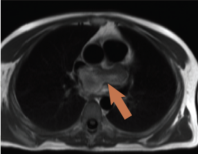
Figure 1. T2 weighted spin echo sequence (CMR) showing high signal intensity of left atrial mass
Cardiac magnetic resonance (CMR) (figure 1) confirmed a 3.7 x 2.3 x 2 cm mass with a broad-based attachment to the inter-atrial septum at the floor of the fossa ovalis, partially obstructing the right, lower pulmonary vein. The mass had high signal intensity on T2-weighted spin echo images (figure 1), persisting on fat suppression imaging, excluding a lipoma or liposarcoma. Signal intensity doubled on first-pass perfusion, excluding thrombus. Delayed enhancement imaging revealed patchy hyperenhancement suggestive of cystic cavitation, features all strongly suggestive of a myxoma. The patient proceeded to an uncomplicated surgical removal of the mass. Histology revealed a gelatinous mass with a chronic inflammatory cell infiltrate at the periphery of the tumour. No necrosis was seen within the mass.
Case 2
A 59-year-old woman presented to a nearby hospital with two days of left iliac fossa pain. On admission she had a low-grade fever, but other observations were normal, as was the remainder of the clinical examination. All blood tests were normal except for raised C-reactive protein (CRP) and white cell count (WCC). ECG and plain X-rays were unremarkable. Diverticulitis was initially suspected.
A contrast-enhanced computed tomography (CT) scan revealed multiple splenic infarcts. Additionally, a filling defect was noticed in a dilated left atrium. A subsequent TTE was performed showing a homogenous left atrial mass prolapsing into the mitral valve plane. The point of attachment was thought to be the inter-atrial septum but this was not clearly seen.
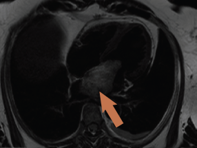
Figure 2. Cardiac magnetic resonance (CMR) four-chamber view showing large left atrial mass prolapsing into left ventricle
CMR was again requested to tissue characterise the mass and delineate the site of attachment in relation to the neighbouring structures. The scan confirmed a 7 x 3.6 x 3.7 cm mass in the left atrium attached to the fossa ovalis via a stalk, with a 1.3 cm footprint (figure 2). The mass had low signal intensity on ‘white blood’ imaging using a steady-state free precession sequence (True-FISP), with a similar isodense signal to myocardium on T1 imaging. The mass was signal intense on T2-weighted images, and remained bright on fat-suppression with no significant contrast uptake on first-pass perfusion. There was no hyperenhancement on delayed enhancement imaging.
Surgical removal and histological assessment confirmed atrial myxoma attached to the superior and posterior aspects of the left atrial side of the fossa ovalis. Histological assessment demonstrated a haemorrhagic mass with haemosiderin-laden macrophages.
Case 3
A 42-year-old man presented with abdominal pain. He was found to have an iron-deficiency anaemia with a family history of bowel cancer. He, therefore, went on to have excision of a polyp by colonoscopy, which also incidentally identified an abnormal anal tag, subsequently found to be a squamous cell carcinoma on histology; this was also excised. A contrast CT scan of the abdomen was arranged due to ongoing pain, and this revealed an area of thickened, inflamed large bowel loop, thought to be due to an embolic event. A filling defect in the left atrium was also described. The appearances of the mass on subsequent echocardiography were suggestive of a myxoma; a small pericardial effusion was also seen.
Figure 3. ‘Black blood’ image showing left atrial mass with extracardiac extension
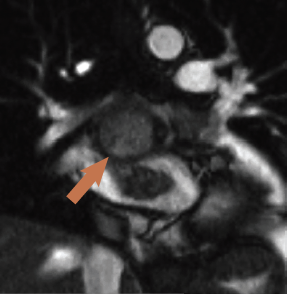
CMR was performed revealing a multi-lobular mass of approximately 3.8 x 4.2 x 3.5 cm (figure 3), thought to arise from the inferior aspect of the fossa ovalis extending with a broad base to the roof of the fossa, one lobe apparently growing into and almost completely filling the left atrial appendage (3 x 3.1 cm). There was partial obstruction to the lower pulmonary veins and compression of the superior vena cava/right atrial (SVC/RA) junction. On additional coronal cine imaging (figure 4), the mass appeared to be superior to the left atrial roof, and it was unclear if the mass had grown through the atrial roof or indeed primarily arisen from the left atrial roof and grown bi-directionally. The mass was mildly hyperintense on T1 and more so on T2, with evidence of cystic cavitation. First-pass perfusion demonstrated very little blood supply and a small pericardial effusion was also noted.
Figure 4. Coronal ‘white blood’ slice showing breach of the left atrial roof
There was debate among cross-sectional imaging physicians about whether this was a myxoma or the anatomic appearance was supporting malignancy. The patient went for surgery where the mass was subtotally excised due to extension into the visceral pericardium and transverse sinus. Such behaviour is atypical for myxomas. Histology revealed myxoid stroma with considerable mitotic features, extensive necrosis and haphazard arrangement within myxoid stroma, features all in keeping with a grade 3 undifferentiated, pleomorphic sarcoma.
Discussion
Epidemiology of myxomas
Atrial myxomas are the most common benign, primary cardiac tumour (followed by lipomas and papillary fibroelastomas) and can vary in size from 1 to 15 cm.3 Approximately 80% are located in the left atrium, 13% right atrium and 1.3% are bi-atrial. Occurrence in the ventricles and on valves is described, but remains very rare.3-6
Although benign in terms of invasive potential, malignant haemodynamic features include valve obstruction, valvular regurgitation and systemic embolic phenomena.5 However, other malignant features include its ability to recur following resection, believed to be due to the multi-centric nature of the disease.7 The Carney syndrome accounts for 5–7% of cardiac myxomas and exhibits an autosomal dominant inheritance pattern, characterised by myxomas in cardiac and extracardiac sites associated with skin pigmentation and endocrine hyperactivity; thyroid and pituitary tumours are also associated.6,8
Modes of presentation
These can be divided into obstructive, embolic and constitutional. In a study of 112 patients,4 approximately 29% had evidence of systemic embolisation and 34% had constitutional symptoms thought to be due to elevated interluekin-6 (IL-6) levels.5 Another retrospective analysis of modes of presentation3 revealed around 15% present with chest pain and 19% present with arrhythmias in association with one of the above; 5% were asymptomatic and discovered on routine echocardiography performed for other reasons. The classic tumour ‘plop’ in diastole was identified in only a minority (15%) of cases.
Comparison of cases
All the cases reflect that ECG and chest X-ray are not helpful in the diagnosis. The ‘malignant’ potential of this benign tumour is emphasised by case 2 as is the poor negative predictive value of the tumour plop, which was clinically not detected on auscultation despite CMR evidence of tumour prolapse through the mitral valve.
Presentation with syncope has generally been considered to result from obstruction of mitral valve inflow. However, case 1 presented with syncope, yet cine imaging did not provide strong support for mitral valve inflow obstruction as the underlying mechanism. Indeed, this tumour was the smallest of the presented cases, with the least imaging evidence of impact on mitral valve inflow.
Case 3 is of particular interest as many of the imaging features, for example location and apparent origin, T1 and T2 signal characteristics, moderate signal increase on first-pass perfusion and diffuse hyperenhancement on late enhancement imaging, appeared in keeping with left atrial myxoma. Indeed, histology confirmed myxoid stroma not dissimilar from that seen in myxomas. However, some features, most notably the expansive and irregular (‘cauliflower’) growth, apparent growth into left atrial appendage (intra-operatively the left atrial appendage was found to be free of tumour mass but instead was completely externally compressed by the mass), compression of the SVC/RA junction, and the apparent invasion and penetration of the left atrial roof, were features pointing towards a possible malignancy. Somewhat ambivalent features were a small pericardial effusion and minimal mediastinal lymphadenopathy.
Multi-modality imaging
A range of imaging modalities were used in our cases. All had a TTE as this is the most widely available and versatile modality. TTE provides rapid anatomical and functional information and the classic pedunculated tumour with a stalk attaching it to the atrial septum is occasionally sufficient for diagnosis. Agostini et al. combined positron emission tomography (PET) imaging to demonstrate metabolic activity of the mass, but this has not found widespread clinical use.9
Contrast-enhanced CT characteristically reveals a lobular contour with lower or equivalent signal intensity than myocardium and allows accurate delineation of the point of attachment in some cases, but this becomes increasingly limited as the size of the tumour increases and obliterates the atrium. The detection of calcium or calcification is the domain of CT, which is occasionally helpful in the differential diagnosis of cardiac tumours from mitral annular calcification that can mimic tumour masses.10,11 Some early work suggested that mean CT signal intensity, as per Hounsfield Units (HU), could help differentiate myxomas from thrombus (mean value 44.5 HU for thrombus versus 30.2 HU for myxomas),10 but this has not been assessed in a larger case series.
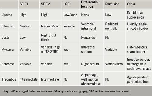
Table 1. Comparing image characteristics for cardiac masses. Adapted from refs 13 and 14
Advantages of CMR are its unparalleled soft-tissue imaging capabilities and, thus, its ability to tissue characterise by virtue of perfusion, late-enhancement imaging, fat-suppression and T1 and T2 characteristics.2,12 These are summarised in table 1.
Due to slice selectivity and reproducibility, first-pass perfusion is more reliably assessed on CMR than echocardiography. In fact, myocardial contrast echocardiography (MCE) can create false-positive results by virtue of the blood pool projecting onto the structure being interrogated (i.e. in front of the mass), giving the erroneous appearance of perfusion of the mass on a two-dimensional image. First-pass perfusion with CMR, if planned meticulously, avoids this problem.
Disadvantages of CMR and CT are the decline in image quality in the setting of frequent ectopic beats and irregular rhythms. Small, mobile, pedunculated myxomas can occasionally be overlooked during steady-state free precession imaging due to their alternating position throughout the cardiac cycle.15-17
Conclusion
From a practical point of view, it is possibly fair to say that the better image quality afforded by CMR imaging of larger cardiac masses outweigh the theoretical advantage, in terms of better temporal and spatial resolution, provided by TTE, in aiding the diagnosis. Morphological appearance and adherence to anatomical boarders are clinically more useful and reproducible features than signal intensity on T1 and T2 weighting. This is likely due to the histological overlap and non-uniformity of myxomas and malignant intra-cardiac space-occupying lesions.18
The above cases illustrate that myxomas display a broad spectrum of presenting clinical signs and symptoms, and a variety of imaging and histological findings. It is for this reason that a multi-modality imaging approach may be justified in selected cases.
While information obtained from such investigations will inform about the need, urgency and possible success of (complete) surgical removal, the ultimate diagnosis, however, must remain a histological one.
Conflict of interest
None declared.
Key messages
Cardiac magnetic resonance is a powerful tool for the assessment of intra-cardiac masses, particularly in patients with sinus rhythm
Morphological appearance of the space-occupying lesions and adherence to anatomical borders are clinically more important and useful features than signal intensity on T1 and T2 weighting
References
1. Matsuoka H, Hamada M, Honda T et al. Morphologic and histologic characterisation of cardiac myxomas by magnetic resonance imaging. Angiology 1996;47:693–8.
2. Sparrow PJ, Kurian JB, Jones TR, Sivananthan MU. MR imaging of cardiac tumours. Radiographics 2005;25:1255–76.
3. Grebenc ML, Rosado-de-Christenson ML, Green CE, Burke AP, Galvin JR. Cardiac myxoma: imaging features in 83 patients. Radiographics 2002;22:673–89.
4. Pinede L, Duhaut P, Loire R. Clinical presentation of left atrial cardiac myxoma. A series of 112 consecutive cases. Medicine (Baltimore) 2001;80:159.
5. McManus B, Lee C. Primary tumours of the heart. In: Braunwald E (ed.). Heart disease. 8th edition. Philadelphia: Elsevier, 2008:1815–23.
6. Sheppard MN. Tumours of the heart. In: Camm J (ed.). The ESC textbook of cardiovascular medicine. Oxford: Oxford University Press, 2006:535–51.
7. Sadeghi N, Sadeghi S, Karimi A. Mitral valve recurrence of a left atrial myxoma. Eur J Cardiothorac Surg 2002;21:568–73.
8. Stratakis CA, Jenkins RB, Pras E et al. Cytogenetic and microsatellite alterations in tumors from patients with the syndrome of myxomas, spotty skin pigmentation, and endocrine overactivity (Carney complex). J Clin Endocrinol Metab 1996;81:3607–14.
9. Agostini D, Babatasi G, Galateau F et al. Detection of cardiac myxoma by F-18 FDG PET. Clin Nucl Med 1999;24:159.
10. Araoz PA, Mulvagh Sl, Tazelaar HD, Julsrud PR, Breen JF. CT and MR imaging of benign cardiac neoplasms with echocardiographic correlation. Radiographics 2000;20:1303–19.
11. Hongo M, Okubo S, Amemiya H et al. Diagnosis of left atrial masses by computed tomography: with special reference to the differentiation between mural thrombi and myxomas. J Cardiogr 1983;13:935–47.
12. Gilkeson RC, Chiles C. MR evaluation of cardiac and pericardial malignancy. Magn Reson Imaging Clin N Am 2003;11:173–86.
13. Lombardi M Frank H. Tumours and masses of the heart and pericardium. In: Schwitter J (ed.). CMR update. Zurich: Juerg Schwitter, 2008:190–206.
15. Constantine G, Shan K, Flamm SD, Sivananthan MU. Role of MRI in clinical cardiology. Lancet 2004;363:2162–71.
16. Kaminaga T, Takeshita T, Kimura I. Role of magnetic resonance imaging for evaluation of tumors in the cardiac region. Eur Radiol 2003;13(suppl 4):L1–L10.
17. Luna A, Ribes R, Caro P, Vida J, Erasmus JJ. Evaluation of cardiac tumours with MRI. Eur Radiol 2005;15:1446–55.
18. Paydarfar D, Krieger D, Dib N et al. In vivo MRI and surgical histopathology of intracardiac masses: distinct features of subacute thrombi. Cardiology 2001;95:40–7.
Welsh School of Pharmacy, Cardiff University, Cardiff, CF10 3NB.
For more than 30 years, the term ‘cardioprotection’ has been applied widely and often without precision to sundry interventions that reduce the incidence and severity of cardiovascular disease. For those of us who spend time occluding and reperfusing coronary arteries to model experimentally the effects of coronary thrombosis, ‘cardioprotection’ has a precise (and arguably the definitive) meaning; i.e. cardioprotection is the limitation or prevention of irreversible cellular injury in heart muscle as a consequence of ischaemia and reperfusion. In this little book, this definition is applied and is extended to consider all aspects of the medical management of acute myocardial infarction. This is a highly successful and justified approach that will maximise the appeal and utility of this text to a wide clinical and scientific audience. Barely 120 pages of text, nevertheless the book provides an up-to-date and remarkably comprehensive series of essays that convey an accurate snapshot of the basic and clinical science relevant to the rational treatment of myocardial infarction.
The editors and contributing authors are distinguished experts in experimental and clinical aspects of acute coronary syndromes. In eleven commendably succinct chapters, the authors provide coverage of the essential information, from the fundamental concepts of cellular pathophysiology and molecular physiology, to their clinical application in the management of acute myocardial infarction. Each chapter is furnished with key references that provide ideal preliminary bibliographies for those unfamiliar with the field. As I indicated, the coverage is very comprehensive. The chapters provide information on basic definitions and descriptions of the pathological features of myocardial infarction; the current status of reperfusion therapies and adjunctive treatments, including antiplatelet and anticoagulant therapies; approaches to myocardial imaging; the longer term management of cardiac remodelling and dysfunction in survivors of myocardial infarction; emerging and experimental therapeutics, including the phenomenon called ‘postconditioning’, and reparative and regenerative therapies based on stem cell delivery.
At £5.99, this book represents excellent value for money. Its appeal should be wide and I would commend Cardioprotection to anyone with an interest, experimental or clinical, in the scientific and medical challenge of myocardial infarction. The obvious audience will include clinical cardiologists and cardiologists in training who require brief but authoritative information on the evidence underpinning practice. But basic scientists and clinicians embarking on research in cardioprotection will also find the book to be a highly accessible starting point. As an indication of the strength of my recommendation, I should add that I have bought multiple copies to present to new postgraduate students and research fellows as essential reading. So far, they have expressed their delight with my present.
My initial reaction on reading this slim, elegant volume was a twinge of professional jealousy which was rapidly replaced by enjoyment. An important feature is that nationalism has been set aside, with authors from Europe, New Zealand, America, Canada and Australia. Perhaps the next edition will embrace the challenges of developing countries as well.
This book is aimed at general practitioners, junior doctors, medical students and nurses. After reading it, these people (and I hope consultant physicians and health planners as well) will be well versed in the principles of total risk assessment and the need to assess the impact of all risk factors in planning the management of the person at risk of cardiovascular disease. The meticulously fair acknowledgment of the major international guidelines in each chapter leads to a certain amount of repetition.
Will the intended audience receive specific guidance from this book as well as a solid grounding in principles? Possibly not, in that all international thresholds and targets are presented but without a very concise summary of the common ground, so that what are often minor differences between guidelines can cause confusion and perhaps frustration – the fault of the guidelines, not of this book.
While behavioural medicine may be regarded as ‘soft’ and qualitative by some, a little more detail on how to assist our patients in lifestyle change would have been welcome. Barriers to guideline implementation are indeed considered but late in the day. Perhaps these issues could helpfully be considered earlier.
I believe that this book is showing us the way of the future – the need for international dialogue to achieve as much common ground between different guidelines as the evidence permits.
I found this book thoroughly enjoyable, and not just because guidelines are ‘what I do’. It can be recommended to all those involved in cardiovascular risk management. If it steers us closer to simplification of risk estimation and management, and in particular to international dialogue, so much the better
There are no guidelines on the practice of antibiotic prophylaxis in pacemaker implantation resulting in wide variation in practice. We sought to investigate this and identify areas for further study and improvement. Using an email questionnaire, followed up with a telephone call if no response, all 121 adult National Health Service hospitals in England implanting pacemakers were asked about use of systemic prophylactic antibiotics at implantation. Data were obtained from 61 hospitals (50.4% of total contacted), covering a wide geographic distribution.
In this issue, Sandler’s paper (see pages 86–8) reinforces the growing body of evidence that should lead to the demise of the routine use of direct current cardioversion (DCCV) for patients with atrial fibrillation. This interesting paper highlights several issues surrounding DCCV within the context of a service re-design within a district general hospital. Despite a state-of-the-art service, the success of DCCV was limited, with sinus rhythm maintained in between the stated 20% (22/110) or even optimistically 40% (22/55) at around one year. I would suggest that this is unacceptable and that we would not allow any other procedure with significant associated morbidity to be undertaken with such a low chance of succeeding.
It may be that these figures are actually very good compared with data from other centres, given that this service was designed specifically to reduce the delay in receiving DCCV. It would be interesting, therefore, to have more data on the types of patients receiving cardioversion, and whether there are any factors that may predict both initial and long-term success. It is clear, for example, from other data that the current National Institute for Health and Clinical Excellence (NICE) recommendations to utilise cardioversion for patients with heart failure need revising.1
Patient selection
If we look at the results in some detail it is clear that the whole issue of patient selection and management prior to the procedure remains to be elucidated. In common with other centres, a high number of procedures were cancelled due to low International Normalised Ratio (INR) measurement. The current recommendation is for the INR to be maintained between 2.5 and 3.0 for six weeks prior to cardioversion. This is very difficult to achieve. Problems arise when the INR, having been stabilised using one monitoring system, is measured using a different monitoring system on admission. Surely the issue is not what the absolute INR value is, which can vary by 0.5 INR units even between reliable systems,2 but rather whether there is clot in the left atrium. If we are to reduce the overall numbers of patients undergoing cardioversion would it not be safer, and more effective, to undertake high-resolution scanning to detect clot, rather than relying on indirect markers such as INR?
Sandler provides no data on outcomes other than sinus rhythm, thus we do not know if any direct harm was attributable to the procedure itself. Even without these data, however, only around half of patients were in sinus rhythm six weeks after the procedure. Thus, even in a state-of-the-art facility designed to minimise delay in receiving the procedure, only half of patients had a successful outcome at six weeks, with possibly only 20% at one year. No mention is made here of management of oral anticoagulation in these patients, and whether the patients lost to follow-up had significant morbidity or not. Given that other studies have demonstrated worse outcomes in terms of morbidity, such as hospital admissions and pulmonary symptoms, attributable to anti-arrhythmic drugs, this may be the cause of loss to follow-up.3
Is it acceptable?
The time has come, therefore, for those of us who manage patients with atrial fibrillation to question whether routine cardioversion is acceptable. Sandler outlines the current NICE criteria for consideration of restoration of sinus rhythm as “symptomatic, younger patients, those presenting for the first time with lone AF, those with AF secondary to a treated/corrected precipitant and those with congestive heart failure”. We have already seen that heart failure is not an indication for cardioversion, and it may be that, with time, the other indications drop off also.
I would argue that cardioversion should currently only be considered for patients with acute onset of atrial fibrillation, probably within 24 hours, or for patients who remain symptomatic despite optimal medical therapy. I would re-iterate that even in these scenarios, successful cardioversion cannot automatically lead to discontinuation of oral anticoagulation therapy. As it stands, I would recommend continuing oral anticoagulation therapy for at least one year following successful cardioversion. If sinus rhythm is maintained at one year, it may be worth a trial without anticoagulants, however, regular review would be absolutely essential.
Conflict of interest
None declaired.
Editors’ note
Please see the article ‘Whatever happens to the cardioverted’ by David A Sandler on pages 86–8 of this issue.
References
Roy D, Talajic M, Nattel S et al. Rhythm control versus rate control for atrial fibrillation and heart failure. N Engl J Med 2008;358:2667–77.
Fitzmaurice DA, Gardiner C, Kitchen S, Mackie I, Murray ET, Machin SJ. An evidence-based review and guidelines for patient self-testing and management of oral anticoagulation. Br J Haematol 2005;131:156–65.
The AFFIRM investigators. Relationships between sinus rhythm, treatment and survival in the atrial fibrillation follow-up investigation of rhythm management (AFFIRM) study. Circulation 2004;109:1509–13.
You are not logged in
You need to be a member to print this page.
Find out more about our membership benefits


 People diagnosed with diabetes have the condition for the rest of their life, and like many other long-term conditions, most of the interventions that effect control and outcomes are in the hands of the patient themselves.3 The key to successful treatment in primary care is to effectively educate and engage the person with diabetes in a self-management collaboration, using the skills and treatment options offered by healthcare professionals.4 Given the possible complications of diabetes, it is important that this support is provided using the combined skills of several different healthcare professional groups, such as nurses, general practitioners (GPs), podiatrists, dietitians and psychologists. Sometimes referral to a specialist team, which may be community or hospital based, will be required. The key ethos in the diabetes “Commissioning diabetes without walls”5 concept to make high-quality care available to everyone who needs it, is that it is the skills of the various healthcare professionals needed at any particular time that matter, and not where they are based.


 This retrospective audit was performed to assess whether patients discharged from the cardiology ward at the Queen Elizabeth Hospital, Birmingham, following ST-segment elevation myocardial infarction (STEMI) or non-ST-segment elevation myocardial infarction (NSTEMI) were prescribed the recommended medication at appropriate doses. The evidence for the prognostic benefit of drugs such as angiotensin-converting enzyme (ACE) inhibitors, beta blockers and statins after a myocardial infarction (MI) is derived from studies in which these drugs were used at high doses, such as Acute Infarction Ramipril Efficacy (AIRE),1 Carvedilol Post-Infarct Survival Control in Left-Ventricular Dysfunction (CAPRICORN),2 and Pravastatin or Atorvastatin Evaluation and Infection Therapy (PROVE IT).3 Lower doses may not confer the same benefit.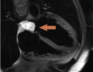
 Editors: Hausenloy DJ, Yellon DM
 Editors: Hobbs R, Arroll B
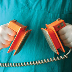 It may be that these figures are actually very good compared with data from other centres, given that this service was designed specifically to reduce the delay in receiving DCCV. It would be interesting, therefore, to have more data on the types of patients receiving cardioversion, and whether there are any factors that may predict both initial and long-term success. It is clear, for example, from other data that the current National Institute for Health and Clinical Excellence (NICE) recommendations to utilise cardioversion for patients with heart failure need revising.1