Pacing in patients with congenital heart disease: part 3

Br J Cardiol 2014;21:38doi:10.5837/bjc.2014.006 1 Comment
Click any image to enlarge
Authors:

W e continue our series looking at pacing in patients with congenital heart disease. In this final article, we discuss the challenge of device implantation in patients with more complex congenital structural cardiac defects.

Introduction

In the first article, we discussed those anomalies that are usually encountered by chance at, or just prior to, implantation; patent foramen ovale/atrial septal defect, Ebstein’s anomaly and ventricular septal defect, and the potential problems that they may provide to the device implanter. In the second article, we discussed the challenge of device implantation in patients with more complex congenital structural cardiac defects, which the operator should be aware of prior to device implantation, including congenitally corrected L-transposition of great arteries, tetralogy of Fallot and tricuspid atresia/univentricular heart. In this final article, we will discuss D-transposition of the great arteries and how to deal with those cases where trans-superior vena cava (SVC) pacing is practically impossible.

D-transposition of great arteries
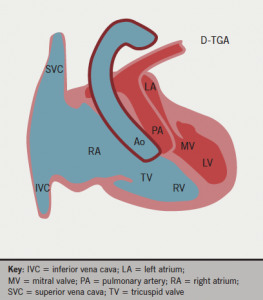
Figure 1a. Schematic representation of transposition of great arteries (D-TGA). The aorta (Ao) originates from the right ventricle (RV) and the pulmonary artery (PA) from the left ventricle (LV)

Complete transposition of the great arteries (D-TGA) is relatively common and accounts for 5% of all congenital heart disease cases. It consists of the origin of the aorta from the right ventricle (RV) and that of the pulmonary artery from the left ventricle (LV) (figure 1a). Without surgery, soon after birth, infants would not survive this situation.1 Usually, a co-existing communication, such as a patent ductus arteriosus, atrial septal defect (ASD) or ventricular septal defect (VSD), allows the infant to survive. Infants may have had a palliative ASD created or a balloon atrial septostomy to allow mixing of venous and arterial blood. Nowadays, D-TGA is corrected with the arterial switch operation.2 This was adopted in the 1980s and, therefore, long-term data on adults are still lacking. In this procedure, the aorta and the pulmonary artery (PA) are transected and the orifices of the coronary arteries are excised and transferred to the PA, which is anastomosed to the proximal aortic stump. The majority of the current adult patients with D-TGA had the atrial switch operation, which was pioneered in the 1950s–1960s.3 In it, blood is redirected at the atrial level using a baffle made of Dacron or pericardium (Mustard operation) or atrial flap (Senning operation). The systemic venous blood is diverted through the mitral valve into the LV while the pulmonary venous blood is rerouted through the tricuspid valve (TV) into the RV.
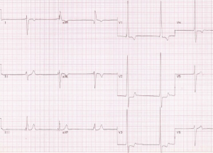
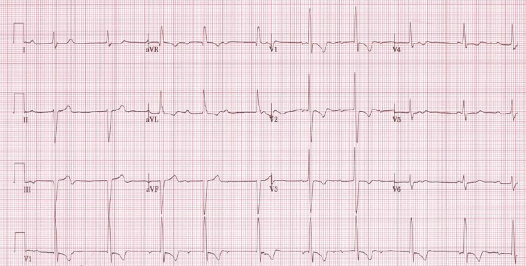
Figure 1b. Electrocardiogram (ECG) from a patient with transposition of great arteries (D-TGA) treated by the Mustard procedure. It shows junctional bradycardia, right axis deviation, and right ventricular hypertrophy

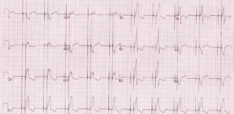
Atrial tachyarrhythmias, junctional rhythm (figure 1b) and complete heart block can occur in these patients. Although pacing in patients with D-TGA may be a daunting task, surprisingly, it may be straightforward. The atrial lead is advanced via the SVC and the stump of the right atrium (RA), behind the baffle and into the left atrium (LA), where it should be actively fixed to the roof of the LA (figure 1c). Lateral screening should show posterior positions of both LA and LV electrodes (figure 1d). Preferably, a curved or steerable stylet should be used to place the lead as medial as possible in order to avoid phrenic nerve stimulation. Steerable catheter delivery systems may be useful for positioning the atrial lead in optimum position. The ventricular lead is advanced along the same route, across the mitral valve and into the LV, where it should be actively fixed. The electrocardiogram (ECG) should confirm satisfactory dual-chamber pacing (figures 1e and 1f).
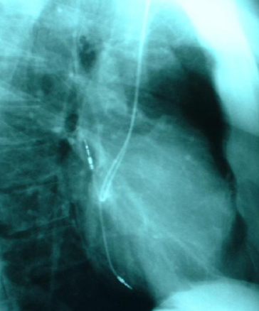
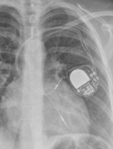
Figure 1c. After a Mustard or Senning procedure, if dual-chamber pacing is required, the atrial lead may be passed behind the baffle into the left atrium (LA) and actively fixed to the roof of the LA. The ventricular lead follows the same route into the LA and then is advanced across the mitral valve into the left ventricle (LV), where it is actively fixed
Figure 1d. This illustration shows a posterior position of both leads in the left atrium and left ventricle in this patient who had undergone a Mustard procedure
Figure 1c. After a Mustard or Senning procedure, if dual-chamber pacing is required, the atrial lead may be passed behind the baffle into the left atrium (LA) and actively fixed to the roof of the LA. The ventricular lead follows the same route into the LA and then is advanced across the mitral valve into the left ventricle (LV), where it is actively fixed
Figure 1e. Complete heart block in a patient after a Mustard procedure
Figure 1f. ECG after dual-chamber pacemaker implantation with right bundle branch block (RBBB) pattern paced ventricular complexes

When the baffle becomes obstructed as the patient grows (>20% of patients), computed tomography (CT), magnetic resonance imaging (MRI) scans or venography might be helpful to show the anatomy better prior to device implantation. Ventricular leads must pass into the LA and into the LV before being anchored actively. Figure 1g shows a more medial position of the atrial lead in order to try and prevent phrenic nerve stimulation. It is worth remembering that chronic atrial arrhythmias are not uncommon, because of the extensive atrial surgery and, if atrial fibrillation is present, then a rate-responsive (VVIR) pacemaker with a single pacing electrode is appropriate (figure 1h).
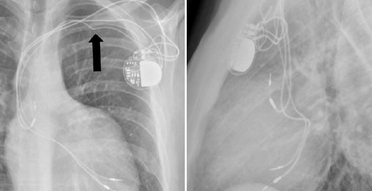
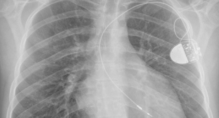
Figure 1g. Following a Mustard procedure at the age of 19 months, a dual-chamber pacemaker was implanted in this patient at the age of 16 years. An active-fixation lead was placed in the LA (venous atrium) but placed more medially to try and avoid phrenic nerve stimulation. An active-fixation lead was placed in the apex of the LV and an Elite DDDR generator (Medtronic) implanted. Ten years later, the generator was replaced with a Clarity DDDR (Vitatron). A subsequent procedure was required four years later to replace the fractured ventricular lead (arrow), which was not extracted. The lateral view shows the atrial lead positioned posteriorly, but pointing anteriorly, and the two activelyfixed ventricular leads posteriorly placed in the LV. The generator was also replaced with a SensiaTM DDDR device (Medtronic)
Figure 1h. After a Mustard procedure, if atrial fibrillation and a slow ventricular rate are present, or if atrial pacing is not possible, a single active-fixation lead can be passed into the left ventricle and attached to a rate-responsive pacemaker. This 19-year-old man had transposition of the great vessels, had an atrial septostomy at day 1 and a Mustard procedure at 15 months of age. At the age of 12 years he developed complete heart block. No acceptable pacing threshold could be obtained in the baffle or LA and so the proposed dual-chamber procedure was changed to a Thera SR rate-responsive pacemaker using a single, actively-fixed LV lead. Seven years later, the generator was replaced by an IdentityTM SR (St. Jude Medical) and six years later by a Symphony SR device (Sorin) – both as a result of battery depletion

Cases when trans-SVC pacing is not possible

If the SVC is severely stenosed or occluded (figure 2a), transvenous pacing can be attempted via the femoral vein.4 Trans-inferior vena cava (IVC) implantation is performed by entering the femoral vein using the standard Seldinger technique. A guidewire and sheath are inserted to enable the delivery of the ventricular lead. Usually, long pacing leads with an active-fixation mechanism are required to reach the target cardiac chamber (RV). The lead can then be tunnelled under the skin into the lower abdominal wall, where an incision can be made and a pocket created for the generator to which the lead can be attached (figure 2b). An atrial lead can be similarly delivered if dual-chamber pacing is desired.
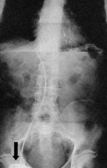
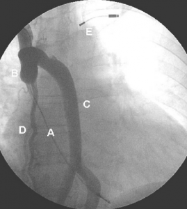
Figure 2a. Right-sided venogram in a 72-year-old woman with multiple previous pacing procedures. She was admitted this time to replace the rightsided ventricular lead (A) because of insulation fracture. The operator started with a right-sided venogram, which revealed occluded superior vena cava (B) with two collaterals (C and D), which drain the venous blood down to the inferior vena cava. Interestingly, this patient did not exhibit clinical signs of superior vena cava (SVC) obstruction. Remnant of a previous left-sided lead can be seen (E). It was then decided to implant an epicardial pacing system
Figure 2b. Dual-chamber KappaTM (Medtronic) pacemaker is implanted in the right inguinal region (arrow) using CapSure FixTM active-fixation leads in the right atrium and ventricle, avoiding traversing the SVC in this patient recently treated by balloon angioplasty for SVC obstruction at the site of multiple previous pacing leads – which were removed in the same sitting
Figure 2a. Right-sided venogram in a 72-year-old woman with multiple previous pacing procedures. She was admitted this time to replace the rightsided ventricular lead (A) because of insulation fracture. The operator started with a right-sided venogram, which revealed occluded superior vena cava (B) with two collaterals (C and D), which drain the venous blood down to the inferior vena cava. Interestingly, this patient did not exhibit clinical signs of superior vena cava (SVC) obstruction. Remnant of a previous left-sided lead can be seen (E). It was then decided to implant an epicardial pacing system
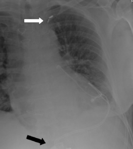
Figure 2c. When several procedures have resulted in difficult or impossible access to the RV transvenously (white arrow shows remnant of old leads from previous extractions), epicardial pacing may be necessary. In this case, new epicardial atrial and ventricular electrodes have been tunnelled to the abdominally-placed generator (black arrow)

Epicardial pacing can always be used as a last resort in any pacing indication, if access to the atrium or ventricle cannot be achieved transvenously (figures 2c and 2d).

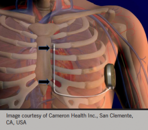
In cases where an implantable cardioverter defibrillator (ICD) implantation is indicated, without any need for cardiac pacing, a subcutaneous implant can always be considered.5 In this approach, the device functions without transvenous leads, overcoming adverse venous access issues. The system is placed, guided by anatomical landmarks, without need for fluoroscopy (figures 3a and 3b). This technology of subcutaneous implantation has a promising future because of its predictability, safety and efficacy. The current ‘hurdle’, that is yet to be overcome, is that the subcutaneous ICD generator remains relatively large (about 70 ml), when compared with a standard transvenous system (about half the size), due to the fact that more energy is needed subcutaneously to defibrillate the heart.
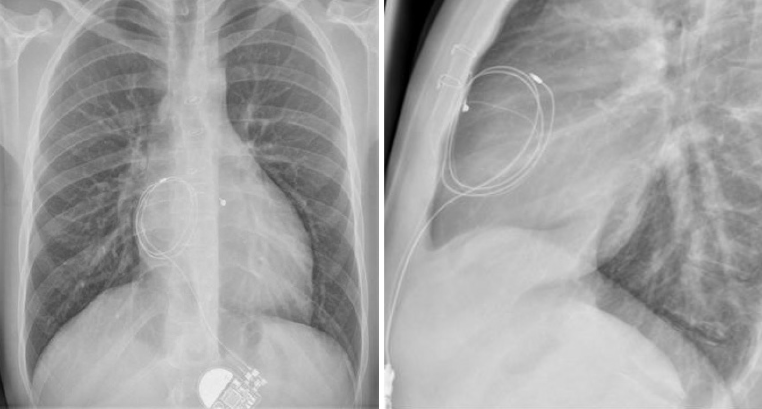
Figure 2d. After closure of a peri-membranous ventricular septal defect with a Dacron patch, atrioventricular block is not uncommon. This patient developed complete heart block five years later and a Legend VVIR pacemaker was implanted using a single ventricular lead. When SVC obstruction developed as a result of lead adhesion/fibrosis accompanied by a very low lead impedance, it was decided to remove the lead and dilate the SVC obstruction and implant a DDDR device instead. However, it proved impossible to either remove the adherent lead or dilate the stenosis. Surgical treatment was, therefore, performed to remove the lead and repair the SVC obstruction, at which time an endocardial lead was fixed in the RA and tunnelled to the epigastrium. An epicardial lead was attached to the surface of the RV and the lead tunnelled to the epigastrium where both leads are attached to a DDDR generator and buried behind the rectus sheath

Conclusions

Figure 3a. Schematic representation of the subcutaneous implantable cardioverter defibrillator (ICD) system manufactured by Cameron Health. It is a 9 Fr lead with two sense electrodes (arrows). Multiple sense vectors are automatically analysed to identify the most robust cardiac signal. The maximum shock energy is 80 J delivered in a biphasic fashion

Congenital cardiac structural abnormalities can provide a serious challenge to device implanting cardiologists who need to be aware of how to recognise the expected, and sometimes unexpected, anatomical anomalies and how to deal with them in order to achieve safe, stable and effective implantation of leads and devices. Besides a careful history and physical examination, ECG and chest X-ray, other tests, such as echocardiography, CT and MRI imaging and cardiac catheterisation, may prove invaluable in helping to answer important anatomical questions prior to device implantation. Every opportunity should be taken to review previous surgical records or previous cardiac catheterisation information, before proceeding to the pacing theatre. Many older patients will have had several previous procedures, and this may add to the complexity of the current task in hand. These complex cases are best done in a tertiary cardiothoracic centre by experienced implanters, where cardiac surgeons are available for their help and advice.
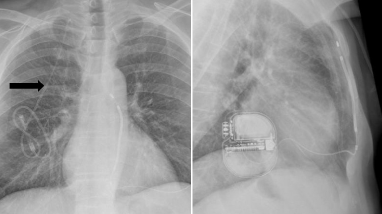
Figure 3b. Postero-anterior and lateral chest X-rays in a patient with Crohn’s disease who had a subcutaneous ICD system implanted. This patient is on long-term total parenteral nutrition (TPN) and had multiple previously infected transvenous ICD systems extracted. TPN feeding is the likely source for the recurrent infection. The black arrow points to the permanent TPN venous line

Conflict of interest

None declared.

Editors’ note

This article concludes our series on pacing in patients with congenital heart disease.  The first two articles can be found online at www.bjcardio.co.uk and in print (Br J Cardiol 2013;20:117–20 and 151–3).

References

1. Williams WG, McCrindle BW, Ashburn DA et al. Outcomes of 829 neonates with complete transposition of the great arteries 12–17 years after repair. Eur J Cardiothorac Surg 2003;24:1–9. http://dx.doi.org/10.1016/S1010-7940(03)00264-1

2. Rehnstrom P, Gilljam T, Sudow G, Berggren H. Excellent survival and low complication rate in medium-term follow-up after arterial switch operation for complete transposition. Scand Cardiovasc J 2003;37:104–06.

3. Wells WJ, Blackstone E. Intermediate outcome after Mustard and Senning procedures: a study by the Congenital Heart Surgeons Society. Semin Thorac Cardiovasc Surg Pediatr Card Surg Annu 2000;3:186–97.

4. Mathur G, Stables RH, Heaven D, Ingram A, Sutton R. Permanent pacemaker implantation via the femoral vein: an alternative in cases with contraindications to the pectoral approach. Europace 2001;3:56–9. http://dx.doi.org/10.1053/eupc.2000.0135

5. Bardy GH, Smith WM, Hood MA et al. An entirely subcutaneous implantable cardioverter-defibrillator. N Engl J Med 2010;363:36–44. http://dx.doi.org/10.1056/NEJMoa0909545

One thought on “Pacing in patients with congenital heart disease: part 3”